Complete genome sequence of bacteriophage P26218 … genome sequence of bacteriophage P26218...
7
SHORT GENOME REPORT Open Access Complete genome sequence of bacteriophage P26218 infecting Rhodoferax sp. strain IMCC26218 Kira Moon 1 , Ilnam Kang 2 , Suhyun Kim 2 , Jang-Cheon Cho 2* and Sang-Jong Kim 1* Abstract Bacteriophage P26218 is a virus that thrives in freshwater and infects Rhodoferax sp. strain IMCC26218, both of which were isolated from Soyang Lake, Korea. The bacterial host, IMCC26218, belongs to the genus Rhodoferax and is closely related to R. saidenbachensis, with 98.7 % 16S rRNA gene sequence similarity. Bacteriophage P26218 has an icosahedral head structure with a diameter of ~52 nm and short tail of ~9 nm, which is a typical morphology of the Podoviridae family. Its complete dsDNA genome was 36,315 bp with 56.7 % G + C content. This is the first genome sequence reported for a lytic phage of the genus Rhodoferax. Keywords: Bacteriophage, Rhodoferax, Freshwater, Podoviridae, Genome Introduction Bacteriophages, which are obligate parasites of bacterial cells, are the most abundant biological entities that can be found in all biospheres [1–3]. Considering the fact that phages heavily influence the bacterial community structure [4] and various biochemical cycles such as the carbon cycle [5], understanding the genetic potential and diversity of phages would be important in the study of microbial community dynamics. Due to the lack of a universal phylogenetic marker gene to help understand phage diversity, several studies have been reported that include a survey of entire phage populations via metage- nomics, from various environments including seawater, hot springs, soil, and freshwater [6–8]. These viral meta- genomic studies demonstrate the extremely diverse na- ture and novel genetic repertoire of viruses, but the limited number of phage genomes poses a challenge for interpretation of virome data. Difficulty in phage isola- tion and genome sequencing is simply due to the lack of available bacterial hosts, since many of bacteria in nat- ural environments are yet to be cultured [2]. Therefore, isolation of phages infecting major groups of bacteria and unveiling their genomic information are required to provide detailed information about each phage and enable meaningful interpretation of virome data. The class Betaproteobacteria is often the most abun- dant group in freshwater environments, though less abundant in marine environments [9, 10]. Metagenomic studies on several freshwater bacteria revealed that the family Comamonadaceae, arbitrarily named betI [9], is the most frequently found family [11] within this class. The genus Rhodoferax [12], belonging to the family Comamonadaceae, is found in diverse habitats including ditch water, activated sludge, Antarctic microbial mats, and water reservoirs [10, 12–14]. Additionally, this is one of the most abundant genera within the 16S rRNA gene database [15]. Therefore, understanding the ecol- ogy of the genus Rhodoferax and its lytic phage will con- tribute to the understanding of freshwater microbial dynamics and help in further freshwater phage genomic studies. To isolate bacteriophages infecting Rhodoferax spp., we successfully isolated phage P26218, which in- fects Rhodoferax sp. IMCC26218 and further details of its genome features and annotations are described below. * Correspondence: [email protected]; [email protected] 2 Department of Biological Sciences, Inha University, Incheon 402-751, Republic of Korea 1 School of Biological Sciences, Seoul National University, Seoul 151-747, Republic of Korea © 2015 Moon et al. Open Access This article is distributed under the terms of the Creative Commons Attribution 4.0 International License (http://creativecommons.org/licenses/by/4.0/), which permits unrestricted use, distribution, and reproduction in any medium, provided you give appropriate credit to the original author(s) and the source, provide a link to the Creative Commons license, and indicate if changes were made. The Creative Commons Public Domain Dedication waiver (http://creativecommons.org/publicdomain/zero/1.0/) applies to the data made available in this article, unless otherwise stated. Moon et al. Standards in Genomic Sciences (2015) 10:111 DOI 10.1186/s40793-015-0090-1
Transcript of Complete genome sequence of bacteriophage P26218 … genome sequence of bacteriophage P26218...

SHORT GENOME REPORT Open Access
Complete genome sequence ofbacteriophage P26218 infectingRhodoferax sp. strain IMCC26218Kira Moon1, Ilnam Kang2, Suhyun Kim2, Jang-Cheon Cho2* and Sang-Jong Kim1*
Abstract
Bacteriophage P26218 is a virus that thrives in freshwater and infects Rhodoferax sp. strain IMCC26218, both ofwhich were isolated from Soyang Lake, Korea. The bacterial host, IMCC26218, belongs to the genus Rhodoferax andis closely related to R. saidenbachensis, with 98.7 % 16S rRNA gene sequence similarity. Bacteriophage P26218 hasan icosahedral head structure with a diameter of ~52 nm and short tail of ~9 nm, which is a typical morphology ofthe Podoviridae family. Its complete dsDNA genome was 36,315 bp with 56.7 % G + C content. This is the firstgenome sequence reported for a lytic phage of the genus Rhodoferax.
Keywords: Bacteriophage, Rhodoferax, Freshwater, Podoviridae, Genome
IntroductionBacteriophages, which are obligate parasites of bacterialcells, are the most abundant biological entities that canbe found in all biospheres [1–3]. Considering the factthat phages heavily influence the bacterial communitystructure [4] and various biochemical cycles such as thecarbon cycle [5], understanding the genetic potentialand diversity of phages would be important in the studyof microbial community dynamics. Due to the lack of auniversal phylogenetic marker gene to help understandphage diversity, several studies have been reported thatinclude a survey of entire phage populations via metage-nomics, from various environments including seawater,hot springs, soil, and freshwater [6–8]. These viral meta-genomic studies demonstrate the extremely diverse na-ture and novel genetic repertoire of viruses, but thelimited number of phage genomes poses a challenge forinterpretation of virome data. Difficulty in phage isola-tion and genome sequencing is simply due to the lack ofavailable bacterial hosts, since many of bacteria in nat-ural environments are yet to be cultured [2]. Therefore,isolation of phages infecting major groups of bacteria
and unveiling their genomic information are required toprovide detailed information about each phage andenable meaningful interpretation of virome data.The class Betaproteobacteria is often the most abun-
dant group in freshwater environments, though lessabundant in marine environments [9, 10]. Metagenomicstudies on several freshwater bacteria revealed that thefamily Comamonadaceae, arbitrarily named betI [9], isthe most frequently found family [11] within this class.The genus Rhodoferax [12], belonging to the familyComamonadaceae, is found in diverse habitats includingditch water, activated sludge, Antarctic microbial mats,and water reservoirs [10, 12–14]. Additionally, this isone of the most abundant genera within the 16S rRNAgene database [15]. Therefore, understanding the ecol-ogy of the genus Rhodoferax and its lytic phage will con-tribute to the understanding of freshwater microbialdynamics and help in further freshwater phage genomicstudies. To isolate bacteriophages infecting Rhodoferaxspp., we successfully isolated phage P26218, which in-fects Rhodoferax sp. IMCC26218 and further details ofits genome features and annotations are describedbelow.
* Correspondence: [email protected]; [email protected] of Biological Sciences, Inha University, Incheon 402-751,Republic of Korea1School of Biological Sciences, Seoul National University, Seoul 151-747,Republic of Korea
© 2015 Moon et al. Open Access This article is distributed under the terms of the Creative Commons Attribution 4.0International License (http://creativecommons.org/licenses/by/4.0/), which permits unrestricted use, distribution, andreproduction in any medium, provided you give appropriate credit to the original author(s) and the source, provide a link tothe Creative Commons license, and indicate if changes were made. The Creative Commons Public Domain Dedication waiver(http://creativecommons.org/publicdomain/zero/1.0/) applies to the data made available in this article, unless otherwise stated.
Moon et al. Standards in Genomic Sciences (2015) 10:111 DOI 10.1186/s40793-015-0090-1

Virus informationClassification and featuresA bacteriophage, designated P26218 that infects the bac-terial strain IMCC26218 was isolated from Soyang Lake,located inland of Gangwon-do, Korea, in October 2014.A bacterial strain, IMCC26218, was also isolated fromthe same site, using standard dilution plating techniqueon R2A agar (Becton, Dickenson and Company, FranklinLakes, NJ, USA) in April 2014. Based on a comparative16S rRNA gene sequence analyses, strain IMCC26218was found to belong to the genus Rhodoferax with98.7 % sequence similarity to R. saidenbachensis ED16T.To screen a representative lytic phage infecting repre-sentatives of the class Betaproteobacteria, Rhodoferax sp.IMCC26218 was used as the bacterial host.Phage P26218 is a lytic phage that forms plaques of 1
to 2 mm in diameter, on Rhodoferax sp. IMCC26218culture plates. Transmission electron microscopy ofpurified phage particles revealed its icosahedral-shapedhead (52.1 nm in diameter) with a 9.4-nm long short tail(Fig. 1). The capsid encapsulates a linear dsDNA withlength of 36,315 bp with 56.7 % G + C content. Themorphology of the viral particle, including a uniform,icosahedral-shaped head with a short tail indicated thatthis phage belonged to the family Podoviridae of theorder Caudovirales [16]. However, when its genomiccharacteristics were considered, no similar genomicarchitecture was found among the known viral genera,leaving P26218 without an assigned genus. The aminoacid sequence of DNA polymerase I (encoded by polA)of P26218, one of the widely used viral phylogeneticmarkers [17, 18], was aligned with that of representativestrains of the families Podoviridae and Siphoviridae andthe aligned sequences were used for phylogenetic ana-lysis. The phylogenetic tree based on DNA polymerase Irevealed that P26218 formed a clade with a marinemetagenome sequence, parted from previously knowntype species, confirming limitations in its assignment to
a known genus (Fig. 2). A summary of the general phylo-genetic features and isolation information are shown inTable 1.
Genome sequencing informationGenome project historyCompared to phage genomics and viromics in marineenvironments, relatively fewer studies with phage isola-tion and viral metagenome have been conducted infreshwater environments. Bacteriophage P26218 is thefirst lytic phage identified that infects the genus Rhodo-ferax, one of the representatives of the class Betaproteo-bacteria in freshwater environments. In this study, bothvirus and host were isolated from Soyang Lake in Korea.This phage was selected for genome sequencing as aninitial approach to understand phages infecting membersof the Betaproteobacteria isolated from surface fresh-water in Korea. Genomic DNA was sequenced by theChunLab Inc. Genome assembly, annotation, and sub-mission to GenBank were performed at the Departmentof Biological Sciences, Inha University. This genomeproject was registered in Genomes Online Database,with accession ID, Gp0111341 as well as GenBank, withan accession ID of KP792623. A summary of the projectinformation is described in Table 2.
Growth conditions and genomic DNA preparationThe bacterial host, Rhodoferax sp. IMCC26218, was rou-tinely cultured and maintained on R2A agar or in R2Abroth (MB Cell, Los Angeles, CA, USA) at 20 °C. Toscreen lytic phages infecting this bacterial host, 10 l ofwater sample was collected from Soyang Lake at a depthof 1 m. The water sample was initially filtered using a 0.2-μm polyvinylidene difluoride membrane filter (MerckMillipore, Darmstadt, Germany) to remove bacterial-sizedparticles. To 400 ml filtrate, 100 ml of 5× R2A broth and20 ml of IMCC26218 culture in the exponential phasewere added, followed by incubation at 20 °C for 2 weeks for
Fig. 1 Transmission electron micrographs of phage P26218 particles infecting Rhodoferax sp. IMCC26218. The TEM images were obtained usingPhilips CM200 electron microscope. Scale bars represent 100 nm in (a) and 20 nm in (b)
Moon et al. Standards in Genomic Sciences (2015) 10:111 Page 2 of 7

enrichment of bacteriophages that specifically infectRhodoferax sp. IMCC26218. During the incubationperiod, 10 ml of the enrichment culture was sub-sampled5 times at a 3-day interval. Each sub-sample was treatedwith approximately 3 ml chloroform to inactivate the bac-terial cells. The treated samples were used for spot-doubleagar layer plaque assay on a Rhodoferax sp. IMCC26218lawn plate for phage screening via appearance of plaques[19], resulting in the isolation of phageP26218.The purification of phage P26218 genomic DNA was
performed as per the ‘Molecular Cloning: A LaboratoryManual’ [20] with minor modifications. To 200 ml ofphage lysate prepared for DNA purification, 1 μg ml−1 ofDNase I and RNase A were added, followed by 11.7 g ofNaCl. The obtained mixture was transferred to centrifugebottles, to which PEG 8000 (Sigma-Aldrich, St. Louis,MO, USA) was added to attain a concentration of 10 %(w/v). After overnight incubation at 4 °C, the mixture wascentrifuged at 11,000 × g for 40 min, supernatant was dis-carded by gentle inversion of the bottle, and the pellet wasresuspended in 3–5 ml of SM buffer (50 mM Tris–HCl,pH 7.5; 100 mM NaCl; 10 mM MgSO4 · 7H2O; 0.01 %
gelatin). PEG was removed from the liquid by treating itwith equal volume of chloroform. The aqueous phase wasthen collected and further concentrated by ultracentrifu-gation at 246,000 × g for 2 h using a Beckman Coulter L-90 K ultracentrifuge with a SW 50 Ti swinging-bucketrotor. The phage pellet was resuspended in 100 μl SMbuffer and used for genomic DNA extraction, usingQiagen DNeasy Blood and Tissue Kit, according to themanufacturer instructions.
Genome sequencing and assemblyThe genome of phage P26218 was sequenced at ChunLabInc. using Illumina MiSeq system with 2 × 300-bp paired-end reads. The Illumina platform produced a total of2 × 798,245 reads. The initial total reads were split by2 × 50,000 reads into 16 sets [21] to facilitate theassembly process. Each set of sequence reads wasindependently assembled using SPAdes-3.1.1 [22],yielding a single contig but with different start points.Gap-closing PCR was performed with primers de-signed within the end region of a contig, which resultedin the circularization of the genome sequence. Circular
Fig. 2 Phylogenetic tree highlighting the relationship of phage P26218 infecting Rhodoferax sp. IMCC26218 with representatives of the familiesPodoviridae and Siphoviridae. Sequences of DNA polymerase I (polA) collected from NCBI were aligned using CLUSTALW software [37], withBacillus phage SPO1 (NC011421.1) as an outgroup. The phylogenetic tree was generated using the neighbor-joining method implemented inMEGA6 [38]. Bootstrap values representing over 60 % in 1,000 replicates are shown in the tree
Moon et al. Standards in Genomic Sciences (2015) 10:111 Page 3 of 7

assembly of the genome sequence suggested that thephage genome is terminally redundant or circularly per-muted. This procedure for genome sequencing and as-sembly finally produced 36,315 bp with approximately2,500× fold-coverage of the genome.
Genome annotationThe ORFs were predicted using 3 gene prediction pro-grams: GeneMark.hmm version 3.25 [23], Rapid Annota-tion using Subsystem Technology server version 2.0[24], and NCBI Gene Locator and Interpolated MarkovModelER version 3.02 [25]. Only the ORFs that wereidentified by 2 of the 3 gene-prediction programs wereincluded in the annotation. Each predicted ORF wastranslated and used to search for its homologous pro-teins and predict its domains using the NCBI BLASTP[17, 26], HHpred server [27] and HMMER [28] uponNCBI non-redundant database [26], the Conserved Do-main Database [29], Pfam database [30], COG [31], PRK[29], and TIGRFam [32]. Then, TMHMM [33] and Sig-nalP [34] were used to predict transmembrane helicesand signal peptides.
Genome propertiesThe properties and statistics of P26218 genome are sum-marized in Tables 3 and 4. The total length of theP26218 dsDNA genome was found to be 36,315 bp with56.7 % G + C content. All 44 predicted ORFs wereprotein-coding sequences. However, only 15 of themwere assigned to putative protein functions, while 29
Table 1 Classification and general features of phage P26218infecting Rhodoferax sp. IMCC26218
MIGS ID Property Term Evidencecodea
Classification Genome group: dsDNAviruses, no RNA stage
IDA
Phylum: unassigned
Class: unassigned
Order: Caudovirales TAS [16]
Family: Podoviridae TAS [16]
Genus: unassigned
Species: unassigned
Strain: P26218
Particle shape Icosahedral head witha short tail
IDA
MIGS-6 Habitat Freshwater lake, surface IDA
MIGS-15 Biotic relationship Intracellular parasite ofRhodoferax sp. IMCC26218
IDA
MIGS-14 Pathogenicity Virulent phage ofRhodoferax sp. IMCC26218
IDA
MIGS-4 Geographic location Soyang Lake, Gangwon-do,South Korea
IDA
MIGS-5 Sample collection Oct. 17, 2014 IDA
MIGS-4.1 Latitude 37°57’11” N IDA
MIGS-4.2 Longitude 127°49’02” E IDA
MIGS-4.3 Depth 1 m IDA
MIGS-4.4 AltitudeaIDA: Inferred from Direct Assay and TAS: Traceable Author Statement. Theevidence codes are from the Gene Ontology project [39]
Table 2 Project information
MIGS ID Property Term
MIGS 31 Finishing quality Finished
Number of contigs 1
MIGS-28 Libraries used One paired-endIllumina library
MIGS 29 Sequencing platforms Illumina Miseq
MIGS 31.2 Fold coverage 2,551×
MIGS 30 Assemblers SPAdes version 3.1.1
MIGS 32 Gene calling method RAST version 2.0,GeneMark.hmm version3.25 and GLIMMER version 3.02
GenBank ID KP792623
GenBank Date of Release April, 2015
GOLD ID Gp0111341
BIOPROJECT NAa
MIGS 13 Source Material Identifier NAa
Project relevance Diversity of freshwaterbacteriophage
aNot available
Table 3 Nucleotide content and gene-count levels of thegenome
Attribute Value % of Totala
Genome size (bp) 36,315 100.00
DNA coding (bp) 33,796 93.06
DNA G + C (bp) 20,589 56.70
DNA scaffolds 1 100.00
Total genes 44 100.00
Protein coding genes 44 100.00
RNA genes 0 0.00
Pseudo genes 0 0.00
Genes in internal clusters 0 0.00
Genes with function prediction 15 34.09
Genes assigned to COGs 11 25.00
Genes with Pfam domains 21 36.36
Genes with signal peptides 1 2.27
Genes with transmembrane helices 0 0.00
CRISPR repeats 0 0.00aThe total percentage is based on the total number of protein coding genes inthe genome
Moon et al. Standards in Genomic Sciences (2015) 10:111 Page 4 of 7
were assigned to hypothetical proteins. One gene with asignal peptide was identified but none were found tohave transmembrane helices.
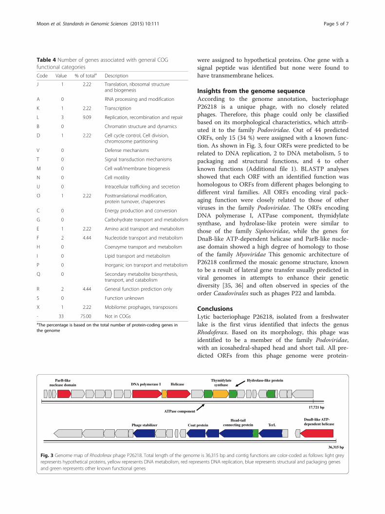
Insights from the genome sequenceAccording to the genome annotation, bacteriophageP26218 is a unique phage, with no closely relatedphages. Therefore, this phage could only be classifiedbased on its morphological characteristics, which attrib-uted it to the family Podoviridae. Out of 44 predictedORFs, only 15 (34 %) were assigned with a known func-tion. As shown in Fig. 3, four ORFs were predicted to berelated to DNA replication, 2 to DNA metabolism, 5 topackaging and structural functions, and 4 to otherknown functions (Additional file 1). BLASTP analysesshowed that each ORF with an identified function washomologous to ORFs from different phages belonging todifferent viral families. All ORFs encoding viral pack-aging function were closely related to those of otherviruses in the family Podoviridae. The ORFs encodingDNA polymerase I, ATPase component, thymidylatesynthase, and hydrolase-like protein were similar tothose of the family Siphoviridae, while the genes forDnaB-like ATP-dependent helicase and ParB-like nucle-ase domain showed a high degree of homology to thoseof the family Myoviridae This genomic architecture ofP26218 confirmed the mosaic genome structure, knownto be a result of lateral gene transfer usually predicted inviral genomes in attempts to enhance their geneticdiversity [35, 36] and often observed in species of theorder Caudovirales such as phages P22 and lambda.
ConclusionsLytic bacteriophage P26218, isolated from a freshwaterlake is the first virus identified that infects the genusRhodoferax. Based on its morphology, this phage wasidentified to be a member of the family Podoviridae,with an icosahedral-shaped head and short tail. All pre-dicted ORFs from this phage genome were protein-
Table 4 Number of genes associated with general COGfunctional categories
Code Value % of totala Description
J 1 2.22 Translation, ribosomal structureand biogenesis
A 0 RNA processing and modification
K 1 2.22 Transcription
L 3 9.09 Replication, recombination and repair
B 0 Chromatin structure and dynamics
D 1 2.22 Cell cycle control, Cell division,chromosome partitioning
V 0 Defense mechanisms
T 0 Signal transduction mechanisms
M 0 Cell wall/membrane biogenesis
N 0 Cell motility
U 0 Intracellular trafficking and secretion
O 1 2.22 Posttranslational modification,protein turnover, chaperones
C 0 Energy production and conversion
G 0 Carbohydrate transport and metabolism
E 1 2.22 Amino acid transport and metabolism
F 2 4.44 Nucleotide transport and metabolism
H 0 Coenzyme transport and metabolism
I 0 Lipid transport and metabolism
P 0 Inorganic ion transport and metabolism
Q 0 Secondary metabolite biosynthesis,transport, and catabolism
R 2 4.44 General function prediction only
S 0 Function unknown
X 1 2.22 Mobilome: prophages, transposons
- 33 75.00 Not in COGsaThe percentage is based on the total number of protein-coding genes inthe genome
Coat proteinHead-tail
connecting protein TerL
HelicaseParB-like
nuclease domain DNA polymerase I
ATPase component
Thymidylate synthase
DnaB-like ATP-dependent helicasePhage stabilizer
Hydrolase-like protein
17,721 bp
36,315 bp
Fig. 3 Genome map of Rhodoferax phage P26218. Total length of the genome is 36,315 bp and contig functions are color-coded as follows: light greyrepresents hypothetical proteins, yellow represents DNA metabolism, red represents DNA replication, blue represents structural and packaging genesand green represents other known functional genes
Moon et al. Standards in Genomic Sciences (2015) 10:111 Page 5 of 7

coding, with 3 specifically coding for DNA replication, 7for DNA metabolism, and 5 for packaging and structuralproteins. The group of ORFs with similar function waspostulated to originate from different groups of viralfamilies (Podoviridae, Siphoviridae, and Myoviridae),which was indicative of the mosaic property of theP26218 genome. It is expected that phage P26218 iso-lated in this research and its genome sequence would befurther used to study bacteria-phage interactions infreshwater environments, to reveal the evolutionary roleof phage lateral gene transfer and to interpret freshwatervirome data.
Additional file
Additional file 1: Table S1. Description of data: Gene annotation tableof bacteriophage P26218. (PDF 183 kb)
AbbreviationsPEG: Polyethylene glycol.
Competing interestsThe authors declare that they have no competing interests.
Authors’ contributionsIK and J-CC designed the study. SK isolated the bacterial host and collectedfreshwater samples. KM performed laboratory experiments, analyzed thedata, and drafted the manuscript. KM, IK, J-CC, and S-JK wrote the manuscripttogether and finalized the study. We all authors have read and approved thefinal manuscript.
AcknowledgementsThis work was supported by Mid-Career Research Program through NationalResearch Foundation (NRF) funded by the Ministry of Science, ICT and FuturePlanning (to J-CC, NRF-2013R1A2A2A01068004) and partially by the GeneralResearch Program through NRF (to S-JK; 2012R1A1B3003609), Korea.
Received: 8 April 2015 Accepted: 29 October 2015
References1. Casas V, Rohwer F. Phage metagenomics. Methods Enzymol.
2007;421:259–68.2. Edwards RA, Rohwer F. Viral metagenomics. Nat Rev Microbiol.
2005;3:504–10.3. Rohwer F. Global phage diversity. Cell. 2003;113:141.4. Rodriguez-Brito B, Li L, Wegley L, Furlan M, Angly F, Breitbart M, et al. Viral
and microbial community dynamics in four aquatic environments. ISME J.2010;4:739–51.
5. Wommack KE, Nasko DJ, Chopyk J, Sakowski EG. Counts and sequences,observations that continue to change our understanding of viruses innature. J Microbiol. 2015;53:181–92.
6. Suttle CA. Viruses in the sea. Nature. 2005;437:356–61.7. Breitbart M, Wegley L, Leeds S, Schoenfeld T, Rohwer F. Phage
community dynamics in hot springs. Appl Environ Microbiol.2004;70:1633–40.
8. Srinivasiah S, Bhavsar J, Thapar K, Liles M, Schoenfeld T, Wommack KE.Phages across the biosphere: contrasts of viruses in soil and aquaticenvironments. Res Microbiol. 2008;159:349–57.
9. Newton RJ, Jones SE, Eiler A, McMahon KD, Bertilsson S. A guide to the naturalhistory of freshwater lake bacteria. Microbiol Mol Biol R. 2011;75:14–49.
10. Zwart G, Crump BC, Kamst-van Agterveld MP, Hagen F, Han S-K. Typicalfreshwater bacteria: an analysis of available 16S rRNA gene sequences fromplankton of lakes and rivers. Aquat Microbiol Ecol. 2002;28:141–55.
11. Cottrell MT, Waidner LA, Yu L, Kirchman DL. Bacterial diversity ofmetagenomic and PCR libraries from the Delaware River. Environ Microbiol.2005;7:1883–95.
12. Kaden R, Spröer C, Beyer D, Krolla-Sidenstein P. Rhodoferax saidenbachensissp. nov., a psychrotolerant, very slowly growing bacterium within the familyComamonadaceae, proposal of appropriate taxonomic position ofAlbidiferax ferrireducens strain T118T in the genus Rhodoferax andemended description of the genus Rhodoferax. Int J Syst Evol Microbiol.2014;64:1186–93.
13. Madigan MT, Jung DO, Woese CR, Achenbach LA. Rhodoferax antarcticus sp.nov., a moderately psychrophilic purple nonsulfur bacterium isolated froman Antarctic microbial mat. Arch Microbiol. 2000;173:269–77.
14. Hiraishi A, Hoshino Y, Satoh T. Rhodoferax fermentans gen. nov., sp. nov., aphototrophic purple nonsulfur bacterium previously referred to as the“Rhodocyclus gelatinosus-like” group. Arch Microbiol. 1991;155:330–6.
15. Salka I, Čuperová Z, Mašín M, Koblížek M, Grossart HP. Rhodoferax‐related pufM gene cluster dominates the aerobic anoxygenicphototrophic communities in German freshwater lakes. EnvironMicrobiol. 2011;13:2865–75.
16. King AM, Adams MJ, Carstens EB, Lefkowitz EJ. The double stranded DNAviruses. In: Virus Taxonomy: Classification and Nomenclature of Viruses;Ninth Report of the International Committee on Taxonomy of Viruses. SanDiego: Elsevier Academic Press; 2012. p. 63–85.
17. Adriaenssens EM, Cowan DA. Using signature genes as tools to assessenvironmental viral ecology and diversity. Appl Environ Microbiol.2014;80:4470–80.
18. Breitbart M, Miyake JH, Rohwer F. Global distribution of nearly identicalphage-encoded DNA sequences. FEMS Microbiol Lett. 2004;236:249–56.
19. Grabow W. Bacteriophages: update on application as models for viruses inwater. Water Sa. 2004;27:251–68.
20. Green MR, Sambrook J. Molecular cloning: a laboratory manual. New York:Cold Spring Harbor Laboratory Press; 2012.
21. Lonardi S, Mirebrahim H, Wanamaker S, Alpert M, Ciardo G, Duma D, et al.When less is more: “slicing” sequencing data improves read decodingaccuracy and de novo assembly quality. bioRxiv. 2015; doi:10.1101/013425
22. Bankevich A, Nurk S, Antipov D, Gurevich AA, Dvorkin M, Kulikov AS, et al.SPAdes: a new genome assembly algorithm and its applications to single-cell sequencing. J Comput Biol. 2012;19:455–77.
23. Lukashin AV, Borodovsky M. GeneMark.hmm: new solutions for genefinding. Nucleic Acids Res. 1998;26:1107–15.
24. Aziz RK, Bartels D, Best AA, DeJongh M, Disz T, Edwards RA, et al. The RASTserver: rapid annotations using subsystems technology. BMC Genomics.2008;9:75.
25. Delcher AL, Harmon D, Kasif S, White O, Salzberg SL. Improved microbialgene identification with GLIMMER. Nucleic Acids Res. 1999;27:4636–41.
26. Lavigne R, Seto D, Mahadevan P, Ackermann H-W, Kropinski AM. Unifyingclassical and molecular taxonomic classification: analysis of the Podoviridaeusing BLASTP-based tools. Res Microbiol. 2008;159:406–14.
27. Söding J. Protein homology detection by HMM–HMM comparison.Bioinformatics. 2005;21:951–60.
28. Eddy SR. Profile hidden Markov models. Bioinformatics. 1998;14:755–63.29. Marchler-Bauer A, Lu S, Anderson JB, Chitsaz F, Derbyshire MK, DeWeese-
Scott C, et al. CDD: a Conserved Domain Database for the functionalannotation of proteins. Nucleic Acids Res. 2011;39:D225–9.
30. Finn RD, Bateman A, Clements J, Coggill P, Eberhardt RY, Eddy SR, et al.Pfam: the protein families database. Nucleic Acids Res.2014;42:D222–30.
31. Tatusov RL, Galperin MY, Natale DA, Koonin EV. The COG database: a toolfor genome-scale analysis of protein functions and evolution. Nucleic AcidsRes. 2000;28:33–6.
32. Haft DH, Selengut JD, White O. The TIGRFAMs database of protein families.Nucleic Acids Res. 2003;31:371–3.
33. Krogh A, Larsson B, Von Heijne G, Sonnhammer EL. Predictingtransmembrane protein topology with a hidden Markov model:application to complete genomes. J Mol Biol. 2001;305:567–80.
34. Petersen TN, Brunak S, von Heijne G, Nielsen H. SignalP 4.0:discriminating signal peptides from transmembrane regions. NatMethods. 2011;8:785–6.
35. Yoshida M, Yoshida-Takashima Y, Nunoura T, Takai K. Genomiccharacterization of a temperate phage of the psychrotolerant deep-seabacterium Aurantimonas sp. Extremophiles. 2015;19:49–58.
Moon et al. Standards in Genomic Sciences (2015) 10:111 Page 6 of 7
36. Swanson MM, Reavy B, Makarova KS, Cock PJ, Hopkins DW, Torrance L, et al.Novel bacteriophages containing a genome of another bacteriophagewithin their genomes. PLoS ONE. 2012;7:e40683.
37. Thompson JD, Gibson T, Higgins DG. Multiple sequence alignment usingClustalW and ClustalX. Curr Protoc Bioinformatics. 2002;Chapter 2:Unit 2.3.
38. Tamura K, Stecher G, Peterson D, Filipski A, Kumar S. MEGA6: molecularevolutionary genetics analysis version 6.0. Mol Biol Evol. 2013;30:2725–9.
39. Ashburner M, Ball CA, Blake JA, Botstein D, Butler H, Cherry JM, et al. Geneontology: tool for the unification of biology. Nat Genet. 2000;25:25–9.
Submit your next manuscript to BioMed Centraland take full advantage of:
• Convenient online submission
• Thorough peer review
• No space constraints or color figure charges
• Immediate publication on acceptance
• Inclusion in PubMed, CAS, Scopus and Google Scholar
• Research which is freely available for redistribution
Submit your manuscript at www.biomedcentral.com/submit
Moon et al. Standards in Genomic Sciences (2015) 10:111 Page 7 of 7