Comparison of radiographic appearances associated and ...
9
British Journal of Industrial Medicine 1984;41:459-467 Comparison of radiographic appearances with associated pathology and lung dust content in a group of coalworkers V ANNE RUCKLEY,' JUNE M FERNIE,' J S CHAPMAN,' PAULA COLLINGS,' J M G DAVIS,' A N DOUGLAS,' D LAMB,2 AND A SEATON' From the Institute of Occupational Medicine, ' Edinburgh, and the Department of Pathology,2 University of Edinburgh Medical School, Edinburgh, UK ABSTRACr The pathology and dust content of lungs from 261 coalminers in relation to the appearances of their chest radiographs taken within four years of death were examined. Radiological opacities of coalworkers' pneumoconiosis were more profuse the more dust was retained in lungs. Among the men who had mined low rank coal-that is, with a relatively high proportion of ash-the increase in profusion was most closely related to the ash component of the dust, whereas in men who had mined high rank coal both coal and ash increased in the lungs in relation to radiological profusion. The fine p type of opacity was found to be associated with more dust and a higher proportion of coal and less ash than the nodular r opacity, and was also more likely to be associated with emphysema. The pathological basis of the different types of opacity found on the radiographs of coalminers related to the number, size, and nodularity of the dust lesions. Larger fibrotic lesions were likely to appear as r opacities, whereas fine reticular dust deposition was most likely to present as p opacities, q opacities showing a mixture of appear- ances. The study has shown that the composition of dust retained in the lung, as well as its amount, makes an important contribution to the radiographic appearances of pneumoconiosis. In particular, the r type of lesion on the radiograph of a low rank coalminer indicates the possibility of a silicotic like lesion. The relation between exposure to coalmine dust and the radiographic profusion of small opacities in sim- ple pneumoconiosis has been well documented.'-3 The primary relation appears to be with exposure to mixed respirable dust, and no generally applicable additional effect of the ash component of the dust has been shown.3 Consequently it seems logical to suppose that radiographic opacities are due largely to an accumulation of dust and, indeed, such an association between retained dust and profusion of opacities has been clearly shownm.45 Most workers, however, describe groups in which the radiological changes appear disproportionate for the amount of dust in the lungs and recognise that factors other than the mass of retained dust contri- bute to the radiographic picture. It has been noted, for example, that the higher the proportion of coal Received 6 June 1983 Accepted 7 November 1983 in the retained dust, the more dust is required to produce a given radiological category,6 and Caplan found that the radiological category was more closely associated with the profusion of fibrotic nodules in the lung than with the profusion of dust foci of all types.7 The principal aim of the present study was to examine further the factors that affect the relation between the profusion of small opacities and the mass of retained lung dust. Additional aims were to determine the pathological basis for the three types of small rounded opacity and to verify the reported association of both the p type of small rounded opac- ity8 and irregular opacities9 with the presence of emphysema. Methods SOURCE AND CLASSIFICATION OF RADIOGRAPHS The study was based on 261 men drawn from a group of 500 coalworkers, all of whom had been 459 AB36-COMM-55-7
Transcript of Comparison of radiographic appearances associated and ...

British Journal of Industrial Medicine 1984;41:459-467
Comparison of radiographic appearances withassociated pathology and lung dust content in a group
of coalworkersV ANNE RUCKLEY,' JUNE M FERNIE,' J S CHAPMAN,' PAULA COLLINGS,'J M G DAVIS,' A N DOUGLAS,' D LAMB,2 AND A SEATON'
From the Institute of Occupational Medicine, ' Edinburgh, and the Department ofPathology,2 University ofEdinburgh Medical School, Edinburgh, UK
ABSTRACr The pathology and dust content of lungs from 261 coalminers in relation to theappearances of their chest radiographs taken within four years of death were examined.Radiological opacities of coalworkers' pneumoconiosis were more profuse the more dust wasretained in lungs. Among the men who had mined low rank coal-that is, with a relatively highproportion of ash-the increase in profusion was most closely related to the ash component of thedust, whereas in men who had mined high rank coal both coal and ash increased in the lungs inrelation to radiological profusion. The fine p type of opacity was found to be associated with moredust and a higher proportion of coal and less ash than the nodular r opacity, and was also morelikely to be associated with emphysema. The pathological basis of the different types of opacityfound on the radiographs of coalminers related to the number, size, and nodularity of the dustlesions. Larger fibrotic lesions were likely to appear as r opacities, whereas fine reticular dustdeposition was most likely to present as p opacities, q opacities showing a mixture of appear-ances. The study has shown that the composition of dust retained in the lung, as well as itsamount, makes an important contribution to the radiographic appearances of pneumoconiosis. Inparticular, the r type of lesion on the radiograph of a low rank coalminer indicates the possibilityof a silicotic like lesion.
The relation between exposure to coalmine dust andthe radiographic profusion of small opacities in sim-ple pneumoconiosis has been well documented.'-3The primary relation appears to be with exposure tomixed respirable dust, and no generally applicableadditional effect of the ash component of the dusthas been shown.3 Consequently it seems logical tosuppose that radiographic opacities are due largelyto an accumulation of dust and, indeed, such anassociation between retained dust and profusion ofopacities has been clearly shownm.45Most workers, however, describe groups in which
the radiological changes appear disproportionate forthe amount of dust in the lungs and recognise thatfactors other than the mass of retained dust contri-bute to the radiographic picture. It has been noted,for example, that the higher the proportion of coal
Received 6 June 1983Accepted 7 November 1983
in the retained dust, the more dust is required toproduce a given radiological category,6 and Caplanfound that the radiological category was moreclosely associated with the profusion of fibroticnodules in the lung than with the profusion of dustfoci of all types.7The principal aim of the present study was to
examine further the factors that affect the relationbetween the profusion of small opacities and themass of retained lung dust. Additional aims were todetermine the pathological basis for the three typesof small rounded opacity and to verify the reportedassociation of both the p type of small rounded opac-ity8 and irregular opacities9 with the presence ofemphysema.
Methods
SOURCE AND CLASSIFICATION OF RADIOGRAPHSThe study was based on 261 men drawn from agroup of 500 coalworkers, all of whom had been
459
AB36-COMM-55-7
Ruckley, Fernie, Chapman, Collings, Davis, Douglas, Lamb, and Seaton
employed at collieries included in thepneumoconiosis field research of the National CoalBoard and whose lungs were collected for a nec-ropsy study. The pneumoconiosis medical panelswere the source of 95% of the lungs. In terms of thepneumoconiosis field research population fromwhich they were drawn, men in the necropsy studywere older and more severely diseased but neverthe-less they covered the full range of dust associateddisease. Selection for this study was dependent onthe availability of a chest radiograph taken withinfour years of death, and men thus selected were rep-resentative of the 500 for distribution over coalrank, age range, and pathological severity. Radio-graphs were obtained from pneumoconiosis medicalpanels or from hospitals, or were taken during theperiodic field research surveys.The radiographs (one for each man) were ran-
domised and read in two batches by experiencedCoal Board medical officers; five readers were pres-ent on the first occasion, four on the second. Read-ings were made according to the 1971 ILO U/Cclassification'0 whereby the profusions of roundedand irregular small opacities are recorded on a12-point incremental scale together with the pre-dominant type of opacity, and large opacities (>1cm) are recorded in three size categories.Average readings, where reported, were based on
the records of four doctors who were present at bothreading sessions and were derived as follows. TheILO 12-point scale for the profusion of smallopacities was converted to a 12-point notional scaleof -1 to + 10. The notional scale scores for eachreader were summed and a mean score was assignedto the radiograph. Opacity types, determined bytheir size, were recorded using two characters-forexample, pr, qq for small rounded opacities. Takingthe first letter of the pair the most frequently occur-ring value of these readings was used as the "aver-age." In cases where an even split in reading occur-red the pneumoconiosis field research identificationnumber was used as a random tie breaker, an oddnumber resulting in the smaller type classificationbeing assigned.The main results of the investigation are shown
for five radiographic readers. Details of dust valuesare shown for a single observer (reader 3) chosen asbest illustrating the trends observed. Data generatedfor all readers are available elsewhere."
LUNG EXAMINATIONFor 50 radiographs, at least three of the four readerswere in agreement that the small rounded opacitytype was pp, qq, or rr. From this group a consultantchest physician (AS) selected zones on the chestradiographs which displayed opacities typical of the
three types and for which lung tissue was availablefor examination. Twenty four subjects were selectedin this way, to which were added seven with normal(0/0) radiographs giving 53 zones for examination.Four observers, working independently,
examined the same sagittal lung slice from each ofthe 31 subjects without knowledge of the radiologi-cal classification. Lung slices were orientated asthough within the upright chest, and the distancebetween the apex and the base was divided horizon-tally into three equal parts to correspond with theradiographic zones. Thread was attached to the edgeof the lung at either end of the demarcation of thezone, being long enough on one side to be stretchedacross to delineate zones in a comparable mannerfor each observer. Three observers undertook lungexaminations on two occasions separated by severalweeks, the fourth was present on only one occasion.
Within the preselected zones all foci of dustdeposition were counted and the number of nodules,defined as having a solid centre of 1 mm or more,was recorded using three size ranges < 1P5 mm, 1P5- 3 mm, and > 3 mm. Three shapes of nodule wererecognised, those with a small central region sur-rounded by elongated strands of emphysematoustissue (type a), those with a larger central regionsurrounded by shorter peripheral strands (type b),and rounded lesions (type c); the predominant typefor each zone was recorded. The extent ofemphysema on each lung slice was estimated using amethod" based on that of Heard.'2One or more histological sections were available
for each zone examined. Sections were stained withhaematoxylin and eosin, and by the methods of VanGieson and Gordon and Sweet for collagen andreticulin respectively, and were reviewed indepen-dently by two of the four observers.
DETERMINATION OF LUNG DUST CONTENTOne lung of each pair was used for measurement ofdust content. Dust was recovered from representa-tive samples of dried lung by the method of Guest.'3Recovered dust was ashed to constant weight, coalcontent being measured by weight loss. Residual ashwas analysed *for quartz, kaolin, and mica byinfrared spectrophotometry.'4 A figure for the dustcontent of both lungs was calculated by multiplyingthe measured weight in a single lung by 2 16 whenthe left lung was analysed, and by 1-86 for the rightlung. The corrective factors were determined usingthe average weights of dust in left and right lungsfrom 490 subjects.At times in this paper there is reference to the
" rank" of coal, a descriptive term for the position onthe geological "coalification" scale (from lignite toanthracite), increasing rank being associated with
460

Comparison of radiographic appearances with associated pathology and lung dust content in a group ofcoalworkers 461
diminishing "volatiles" and increasing carbon con-tent. Subjects were divided into "high" and "low"rank groups depending on the rank of coal mined atthe pit where each man had worked, the carbon con-tent of the coal being above and below 88% respec-tively.
Results
ASSOCIATIONS BETWEEN RETAINED DUST ANDTHE PROFUSION OF SMALL ROUNDED OPACITIESIN SIMPLE PNEUMOCONIOSISSubjects categorised as having large radiographicopacities (>1 cm) by the individual reader wereexcluded and in the remainder the profusion scoresfor small rounded opacities (SRO) were reduced toa four point scale giving categories 0, 1, 2, and 3.The mean weight of retained dust and its meas-
ured components coal, ash, quartz, and kaolin plusmica increased over the profusion range. The differ-ences were statistically significant (F-test of equalityof means) for each component and for all readers.Figure 1 illustrates the association between lungdust and profusion of small rounded opacities forfive readers. The composition of retained dust, inpercentage terms, did not vary consistently with pro-fusion category.When subjects were divided into high and low
rank groups the observed increase in mean lung dustcontent with rising profusion category was evidentfor both but was more striking in the high rankgroup (table 1). At category 0, the dust content ofthe lung was similar for both groups but at category3, lungs in the high rank group contained, on aver-age, twice as much dust as those from the low rankgroup.
In the high rank group both coal and ash contri-buted to the overall increase in lung dust contentwhereas in low rank subjects the increase waslargely accounted for by an increase in the ash con-tent of the dust (table 2). Depending on the readerthis increase seemed either to be distributed over
(D(DOQQ)(D
Fo
0u0
a#1 20 3
CL
E
0
0
10 20 30
Lung dust content (g)Fig 1 Mean lung dust content for each category ofsimplepneumoconiosis shown for five* readers identified bynumber.*Reader S classified 118 of261 cases.
the whole category range or to occur betweencategories 0 and 1.
ASSOCIATIONS BETWEEN RETAINED DUST ANDTYPE OF SMALL ROUNDED OPACITYOn average, 40% of the 261 radiographs showed nosmall rounded opacities (category 0). Of theremainder, q opacities were the most commonlyrecorded accounting for 61 %, whereas r opacitieswere least common being found in 11% of subjects.About half the radiographs showing predominantlyp or q opacities also showed progressive massivefibrosis (PMF). Between 77% and 100% of theradiographs with predominantly r opacities showedPMF (depending on the reader).
Within the study group only four specimens wereregarded pathologically as showing Caplan' s syn-drome. These were almost exclusively described asshowing r opacities and were removed beforefurther data analysis on the grounds that they might
Table 1 Mean weight ofretained dust (and standard deviation) for each profusion category ofsmall rounded opacitydivided by coal rank
Coal Radiographic Reader I Reader 2 Reader 3 Reader 4rank category No of Mean lung dust No of Mean lung dust No of Mean lung dust No of Mean lung dust
men (g) men (g) men (g) men (g)
High 0 86 13-4 (10.2) 61 1067 (8 9) 52 10.4 (85) 70 11-8 (963)1 12 20-6 (8-1) 20 20-6 (90) 12 17-1 (10.3) 24 19-4 (8.6)2 9 26.0 12-5) 9 24-0 (14-1) 24 203 (9-8) 4 264 (13-5)3 1 37-7 - 3 41-3 (7.1) 2 44-6 (5.9)
Low 0 67 12-3 (9.2) 51 11-0 8.5) 28 9-3 (8-6) 47 11-2 (8-0)1 16 20.0 7.1) 15 18-6 11-4) 19 12-7 (7-3) 32 16 6 (10.1)2 6 17-2 8-1) 4 19.5 5.4) 17 15-7 (8.3) 8 175 (4-0)3 1 15-8 5 18-2 8-0) 8 19.8 (68) 2 21-4 (14-6)
*Reader 5 had only six men at category 2 or above and was excluded.
Ruckley, Fernie, Chapman, Collings, Davis, Douglas, Lamb, and Seaton
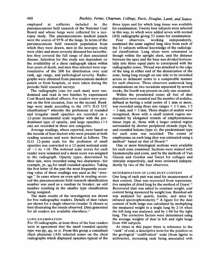
Table 2 Mean retained lung dust by weight and percentage composition for each profusion category ofsmall roundedopacity divided by coal rank (differences examined by F test)-reader 3
Radiographic No of Total Total Total Total Total kaolin % Coal % Ash % Quartz % Kaolin +category men dust (g) coal (g) ash (g) quartz (g) + mica (g) mica
High rank0 52 10-3 7-8 2-6 04 1-8 75-1 24-9 3-8 17-51 12 17-1 14-0 3-1 0.5 2-2 79-6 20-4 3-1 14-52 24 20-3 16-2 4-1 06 2-9 74-3 25-7 3-5 15-73 3 41-3 35-6 5-7 0 7 4-5 85-5 14-5 1-8 11-6Standard deviation 11.1 9-6 2-1 03 1-5 12-1 12-1 1-7 6-8F value 15-9 17-2 5-3 4-8 6-5 1-2 1-2 1-5 1-4p value <0-001 <0-001 <0-01 <0-01 <0-001 NS NS NS NS
Low rank0 28 9 3 5-8 3-5 0-6 2-1 56-9 43-1 7-6 25-91 19 12-7 7 0 5-7 09 3.7 56-7 43-3 7-1 28-52 17 15-7 7-3 8-4 1-5 5-4 48-2 51-8 9-2 32-33 8 19-8 8-9 10-9 2-0 7-3 44-1 55-9 10-2 37.4Standard deviation 8-6 6-0 4-7 0-8 3-2 16-3 16-3 3-7 11-0F value 4 5 0-6 9-8 10-4 105 2-2 2-2 2-0 30p value <0-01 NS <0-001 <0-001 <0-001 NS NS NS <0 05
Table 3 Mean weight ofretained lung dust and coal, and mean percentage composition ofretained dust (with standarddeviations) for men presenting without small rounded opacities or with type p, q, or r-reader 3
Opacity No ofmen Dust (g) Coal (g) % Coal % Ash % Quartz % Kaolin + mica
0/0 105 130(120) 9-3 (96) 683(150) 317 (150) 52 (31) 211 (97)p 23 24-9 (11 0 184 (10.8) 70:S5(17-2) 295(172) 5-0 (39) 20-1 (11-1)q 102 187 (11.0) 117 (10.0) 59.9 (20.0) 40-1 (20-0) 72 (4.6) 26-5 (134r 27 18-2 (7.7) 10-3 (8-2) 53.7 (233) 46-3 (233) 80 (48) 31-7 (153)
obscure any specific associations between r opacitiesand retained dust.For all readers p opacities were associated with a
higher mass of retained lung dust which generallyshowed a higher proportion of coal and lower pro-portion of non-coal mineral than was seen for theother types of opacity (table 3). The proportion ofnon-coal mineral (ash) was greatest in lungs showingr opacities. This observation that both the mass andcomposition of lung dust might afford some distinc-tion between types of opacity was confirmed usingdiscriminant analysis whereby the linear combina-tion of dust variables which best characterised thedifferences between the types was found. Radio-graphs showing p opacities were associated with thehighest mean mass of retained dust in both coal rankgroups (fig 2). In general r opacities were associatedwith a igher proportion of ash in retained dust thanother opacity types in each rank group. Neverthe-less, the differences between ranks were greaterthan those between types of opacity, all opacitytypes in the low rank group having a higher propor-tion of ash in lung dust than r opacities in the highrank group (fig 3).
ASSOCIATION BETWEEN TYPE OF OPACITY ANDTHE PRESENCE OF EMPHYSEMAEmphysema data were available for 256 of the 261subjects. Using the average radiographicclassification they were divided into four groups;
those not showing small opacities, those with smallrounded opacities only, those with small irregularopacities only, and those showing both types. Halfthe men without radiographic opacities showedsome emphysema. When small irregular opacitieswere present about 90% of the lungs showedemphysema (table 4). If small rounded opacitiesalone were present 60% of the lungs on averageshowed emphysema.
35-
- 30-
Ch
u 20-U)
,15-
c 10-a2 5-
0
i
!I
0
I0
S
I9
III0
OJ . .% p q r % p q r
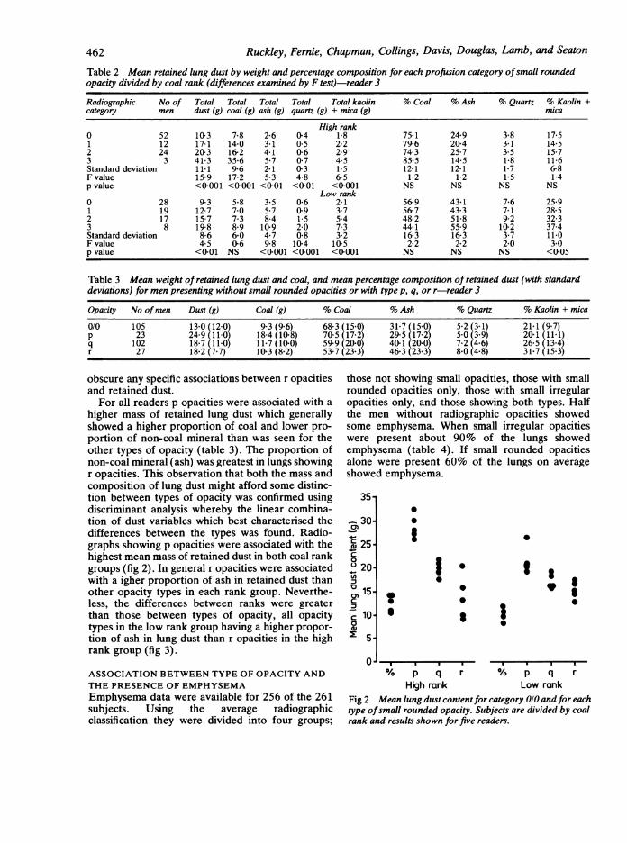
High rank Low rankFig 2 Mean lung dust content for category 0/0 and for eachtype ofsmall rounded opacity. Subjects are divided by coalrank and results shown for five readers.
462

Comparison of radiographic appearances with associated pathology and lung dust content in a group ofcoalworkers 463
group with p type small rounded opacities whereas60% of those presenting with q or r opacities
* * showed emphysema (table 5).
% p q r % p q rHigh rank Low rank
Fig 3 Mean percentage ash in retained dust for category0/0 and for each type ofsmall rounded opacity. Subjects aredivided by coal rank and results shown for five readers.
An "average" opacity type was produced for 131radiographs showing small rounded opacities.Emphysema was present in the lungs of 90% of the
Table 4 Proportion ofspecimens showing emphysemaconsidered by type ofradiographic opacity present.(Number ofmen in each group shown in parentheses)
No of SRO only SRO + SIO onlyopacities S10
All men 49% (57) 64% (96) 89% (79) 92% (24)Men showing simplepneumoconiosis only45% (53) 57% (53) 87% (23) 92% (13)
'K SRO = Small rounded opacity.SIO = Small irregular opacity.
Table 5 Proportion ofspecimens showing emphysemaconsidered by the size ofsmall rounded opacity presentingradiographically. (Number ofmen in each group shown inparentheses)
p q r
All men 92% (37) 66% (80) 57% (14)Men with simplepneumocomosis only 89% (18) 61% (41) *
*Only two men with r opacities were classified as simplepneumoconiosis; one showed emphysema.
PATHOLOGICAL BASIS OF SMALL ROUNDEDOPACITIESIn the group of 31 subjects studied the mean profu-sion scores for each opacity type (p, q, and r) weresimilar. The results of the pathological examinationare summarised in table 6. The figures given are themean of seven values produced by the four obser-vers. While intraobserver variation was small,interobserver variation was evident, particularly forcounts of dust foci. These differences were a matterof interpretation of lesions, regarded by someobservers as single and by others as composite.
Table 6 shows that although dust foci were pres-ent in subjects without radiographic opacities, thelungs showed fewer foci than were found for menwho had opacities, and these foci tended to be smalland were rarely palpable. For half the subjectscategorised as showing p type opacities thepathological appearances consisted predominantlynot of discrete dust foci but rather of an extensivelattice work of tissue made rigid by the presence ofdust.Where dust foci could be counted the mean
number per zone when opacities were of the p or qtype was similar, being 81 and 95 respectively (table6). Fewer dust foci per zone were associated with ropacities, the mean value being 41. There werestriking differences in the proportion of foci whichwere palpable as nodules depending on the type ofopacity. When p opacities were observed, on aver-age only 16% of dust foci in the corresponding lungswere palpable, whereas for r opacities more thanhalf the foci were palpable.For all three types of opacity the greatest propor-
tion of the total dust foci measured between 1-5 and3 mm but examination of the size distributions ofpalpable nodules showed differences which relatedto opacity type. For subjects showing p typeopacities 75% of nodules were evenly divided bet-ween the <1.5 mm and the 1.5 - 3-0 mm sizeranges. When q opacities were recorded half the
Table 6 Mean counts (and standard deviations) ofall dust lesions and ofnodules in lung zones showing specific types ofsmall roundedopacity
Radiographic Mean No of Percentage distribution Mean No of Mean % of Percentage distribution Mean % ofclassification dust focilzone of all foci by size palpable nodules' dust foci ofnodules by size lung affected by(No ofsubjects) <1-5 mm 1-5-3 mm >3 mm zone palpable (range) <1-5 mm 1-5-3 mm >3 mm emphysema
0/0 (7) 28 (21) 47 36 17 <1 0 - - - 9*p (10) 81 59) 28 44 28 13 (15 16 5-48) 37 38 23 18*q (9) 95 (48) 26 62 12 41 t31 43 11-75) 31 54 15 13r (5) 41 (21) 23 49 28 23 (18 56 (32-94) 10 48 41 8
;For five p cases and one q case total dust foci could not be counted although it was possible to count palpable nodules.
70*
, 60
C 50Q
l*O0
0 30c
5b x 20
I
*0.
09:0
310-

Ruckley, Fernie, Chapman, Collings, Davis, Douglas, Lamb, and Seaton
palpable lesions were in the 1-5 - 3*0 mm range andfor r opacities, 48% of palpable lesions were in the1-5 - 3 0 mm size range and a further 41% weremore than 3 mm in size (table 6).The proportion of lung showing emphysema was
also related to type of opacity. Lungs presentingwith p opacities showed the most emphysemawhereas r opacities were associated with the least,being similar to lungs without radiographic evidenceof disease (table 6).Three shapes of dust deposit were observed (table
7), and there was good agreement between obser-vers about the predominant shape in each subject.Type a (stellate) was usually associated with a cen-tral palpable lesion of less than 3 mm in size; mostwere considerably smaller than this. Circumscribednodules (type c) were commonly larger than 3 mmand were characteristic of subjects presenting with ropacities. The b lesion, showing a crenated border,predominated in subjects with q type opacities.More than half the subjects with p opacities showedthe classic stellate lung lesions (type a). Where thepredominant lesion was of type b the differencebetween p and q subjects was one of size, p opacitiesbeing associated with smaller lesions than qopacities.
Examination of histological material confirmedthe differences in lesion size and shape observedmacroscopically. In lungs categorised as 0/0 dust
Table 7 Twenty four lungs presenting as typical smallrounded opacities divided by radiographic type and dustlesion shape
Opacity type a b c
p 6 4 -
q 2 7 -
r - - 5
macules were present but individual deposits in rela-tion to respiratory bronchioles were generally rathersmall, reaching a maximum thickness of 0-75 mm.Lungs presenting p type opacities contained largernumbers of dust macules. In addition a proportionof the dust foci showed a measurable solid centre of1-0 to 1.5 mm and were associated with focalemphysema, with dust accumulation accentuatingthe peripheral extensions of the stellate lesion (fig4). The peripheral extensions of the larger lesionsassociated with q opacities appeared shorter (fig 5)and some showed central fibrosis with relatively lessdust in this area than at the periphery. The largestlesions often showed some evidence of centraldegeneration. In subjects showing r opacities palp-able nodules predominated and these ranged in sizefrom 2mm to 10 mm, commonly showing distinctperipheral and central zones. In the five examinedhere the picture was either that induced by a highquartz content of the dust or that seen in Caplan's
t.*4t s w ~ '
Fig 4 Classic centriacinar stellate dust lesions presenting radiographically as p type opacities (x 11 5.)
464
Comparison of radiographic appearances with associated pathology and lung dust content in a group ofcoalworkers 465
A
*
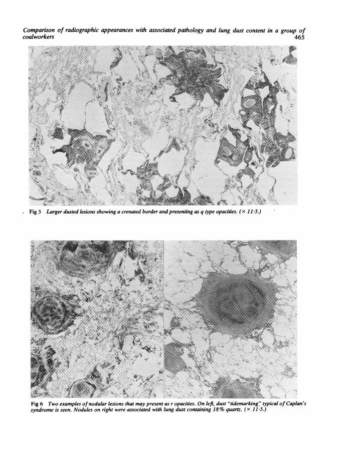
Fig 5 Larger dusted lesions showing a crenated border and presenting as q type opacities. (x II S)
.S.. ....
..v-
Fig 6 Two exampkes ofnodular lesions that may present as r opacities. On left, dust "tidemarking" typical of Caplan'ssyndrome is seen. Nodules on right were associated with lung dust containing 18%o quartz. (x 11 5S.)

Ruckley, Femnie, Chapman, Collings, Davis, Douglas, Lamb, and Seaton
syndrome and associated with the rheumatoiddiathesis (fig 6). In the former the central regions ofnodules showed a characteristic whorling pattern ofcollagen with little visible dust and seldom any evi-dence of tissue breakdown. Caplan's lesions, on theother hand, frequently displayed necrosis of col-lagen, in addition to the classic peripheral"tidemarking" by dust.
Discussion
Although opacity type has been less closelyexamined than opacity profusion, there is evidencefor an association between both type p smallrounded opacities8 and small irregular opacities9 andthe presence of emphysema. These associations areconfirmed in the present study. The positive associa-tion reported previously4 5 between the mass ofretained dust and the profusion of opacities in sim-ple pneumoconiosis is also clearly shown. Both Ros-siter and Caplan have emphasised factors that con-tribute to the complexity of this relationship.6'
This study adds further information concerningthe nature and extent of tissue reaction to retaineddust. Several points are of special interest. One isthat, for men who worked low rank coal, there is aless distinct association between increasing profu-sion of small rounded opacities and an increase inthe amount of retained dust. A second is that,depending on the predominant type of smallrounded opacity present, the amount of dust in thelungs differs. The latter finding might be explainedby an uneven distribution of men showing highercategories of simple pneumoconiosis or progressivemassive fibrosis, which are known to be associatedwith a higher lung dust content. In fact, proportion-ately fewer radiographs with p opacities were readas category 2 or higher compared with those show-ing q or r, and most of the r cases showed PMF. Theview that the composition of retained dust mayinfluence the degree of radiographic change is, tosome extent, reflected in the association between popacities and dust of high coal content. In this con-text the rank of coal mined may be important sincehigh rank coal contains more coal and less non-coalmineral (ash) than low rank coal. Although, how-ever, there were proportionately more radiographsshowing p opacities and fewer showing q opacities inhigh rank areas when compared with low, the differ-ences were minor. The general conclusion that,other factors such as category of pneumoconiosisand rank of coal mined being equal, p opacities arespecifically associated with a higher mass of retaineddust is supported.The study of the pathological counterparts of
small rounded opacities goes some way to offer a
rationale for these differences. Lungs presentingwith p opacities had foci of dust deposition that weresmaller, less often palpable, and more numerousthan those associated with r opacities. Indeed, aprominent feature of some p specimens was a rathergeneralised dusting of tissue not clearly separatedinto dust foci. The production of radiographicopacities in such subjects may depend on the super-imposition of lattices of dusted tissue, a possibilityconfirmed experimentally by Carstairs.'5 A corollaryof this is that more dust may have to be retained toproduce a given effect. Certainly individual maculeswill not be seen on a radiograph. It has been sug-gested that solitary lesions will not be visualised ifthey are less than 3 mm in diameter,'617 althoughthis refers to a non-dusted lesion. Even when welldefined lesions are present, as in silicosis, it has beenshown that the number of silicotic nodules seen atnecropsy exceeds that predicted by radiography.'8 19Where macules and very small fibrotic nodules arethe only response to dust, as in subjects with popacities, then clearly larger numbers will berequired to produce radiographic opacities, theseprobably being the result of summation. Ruckley etal found a positive association between counts ofany dust foci in the lung and the profusion ofopacities (r = 0-43)." When the number of foci wasrecorded for specific size ranges, opacity profusionwas more closely associated with nodules than withmacules (for nodules >3 mm r = 0 5 - 0 8 for fiveradiographic readers).
Radiographs showing r opacities were the leastcommon. The study of a small group of pure exam-ples suggested that this group will always include aconsiderable proportion of subjects with nodularconditions such as silicosis or Caplan' s syndrome. Asmost of the lesions present were palpable and oftengreater than 3 mm in diameter they were more likelyto produce a radiographic image and, overall, lessdust would be required for a given effect.5 Neverthe-less, the general association with a dust of high ashcontent was not striking in men showing r opacitiesdrawn from high rank mining areas. The differencebetween men showing r opacities from high and lowrank areas therefore requires further clarification.The q opacity is generally regarded as the com-
monest expression of coalworkers' pneumoconiosis.Fletcher and Oldham, describing opacities between1 5 and 3 mm, said they were of two types, thosemade up of aggregations of minute opacities, andthose that appear homogeneous.20 These types mayrepresent different tissue reactions to dust within theq group which depend on the composition of thedust to which men were exposed.The absence of observations over time precludes
any judgment about changes from one opacity type
466

Comparison of radiographic appearances with associated pathology and lung dust content in a group ofcoalworkers 467
to another for an individual. Lange et al suggestedthat this occurs only in a small proportion of men,
although they noted that p opacities were more
common in younger men.2' In the present studygroup, for whom the average age was 65, one mightassume that the opacity type was a characteristic of aman's response to dust. Whereas a study of "pure"opacity type cases is of interest, for most men theradiograph will show opacities covering a range ofsizes that reflect the age of dust lesions and perhapsexposure to dusts of different composition.Although effective control of mass exposure
remains a primary aim it becomes increasingly clearthat the composition of the dust that a man breathesmay be of significance. We have shown that theincrease in opacity profusion for subjects drawnfrom low rank areas is not simply associated with an
increase in the mass of retained dust but closelyrelates to the ash component of that dust. Theresults suggest that, in terms of dust mass, less maybe required for a given radiographic effect when theash content of the dust is high. This conclusion is inagreement with Casswell et al5 who found that,weight for weight, the contribution of the mineralcomponent of dust to the relationship with radiolog-ical appearances was greater than that of coal. Otherwork in this necropsy study" suggests that the clear-ance of coal and ash components of dust may differ,and it is possible that some increase in radiographicchange is attributable to a continuing tissue reactionto dust held in the lung.
We are grateful to the staff of the pneumoconiosismedical panels and several hospitals for supplyingnecropsy material and some chest radiographs.Dr J G Bennett, Dr J Burns, Dr J A Dick, Dr D J
Thomas, and Dr J S Washington of the NationalCoal Board gave valuable time to provide radiog-raphic readings.Many staff of the Institute of Occupational
Medicine gave skilled help in this study, which was
jointly funded by the National Coal Board and theCommission of the European Communities.
Requests for reprints to: V Anne Ruckley, Instituteof Occupational Medicine, 8 Roxburgh Place, Edin-burgh EH8 9SU.
References
'Jacobsen M, Rae S, Walton WH, Rogan JM. The relation bet-ween pneumoconiosis and dust exposure in British coal mines.
In: Walton WH, ed. Inhaled particles I1I. Old Woking, Surrey:Unwin Bros, 1971:903-17.
2 Reisner MTR. Results of epidemiological studies ofpneumoconiosis in West German coal mines. In: Walton WH,ed. Inhaled pariicle1' III. Old Woking, Surrey: Unwin Bros,1971:921-9.
3Hurley JF, Burns J, Copland L, Dodgson J, Jacobsen M. Coal-worker's simple pneumoconiosis and exposure to dust at 10British coalmines. Br J Ind Med 1982;39: 120-7.
Rivers D, Wise ME, King EJ, Nagelschmidt G. Dust content,radiology, and pathology in simple pneumoconiosis of coal-workers. Br J Ind Med 1960; 17:87-108.
Casswell C, Bergman I, Rossiter CE. The relation of radiologicalappearance in simple pneumoconiosis of coal workers to thecontent and composition of the lung. In: Walton WH, ed.Inhaled particles III. Old Woking, Surrey: Unwin Bros,1971:713-24.
6 Rossiter CE. Relation of lung dust content to radiologicalchanges in coal workers. Ann NY Acad Sci 1972;200:465-77.
7Caplan A. Correlation of radiological category with lung pathol-ogy in coal-workers' pneumoconiosis. Br J Ind Med1962; 19:171-9.
Hankinson JL, Palmes ED, Lapp NL. Pulmonary air space size incoal miners. Am Rev Respir Dis 1979; 119: 391-7.
9 Lyons JP, Ryder RC, Cambell H, Clarke WG, Gough J.Significance of irregular opacities in the radiology of coalwor-kers' pneumoconiosis. Br J Ind Med 1974;31:36-44.
'° International Labour Office. International classification of radio-graphs ofpneumoconioses, 1971. Geneva: ILO, 1972. (Occu-pational Safety and Health series No 22.)
"Ruckley VA, Chapman JS, Collings PL, et al. Autopsy studies ofcoalminers' lungs-phase II. Edinburgh: Institute of Occupa-tional Medicine, 1981. (IOM report TM/81/18.)
12 Heard BE. Pathology of chronic bronchitis and emphysema.London: J and A Churchill, 1969.
13 Guest L. The recovery of dust from formalin-fixed pneumoconio-tic lungs: a comparison of the methods used at SMRE. AnnOccup Hyg 1976; 19:.37-47.
4 Dodgson J, Whittaker W. The determination of quartz in respir-able dust samples by infrared spectrophotometry-I. Thepotassium bromide disc method. Ann Occup Hyg1973; 16: 373-87.
'Carstairs LS. The interpretation of shadows in a restricted area ofa lung field on the chest radiograph. Proceedings of the RoyalSociety ofMedicine 1961;54:978-80.
16 Greening RR, Pendergrass EP. Post mortem roentgenographywith particular emphasis on the lung. Radiology1954;62:720-5.
7 Spratt JS, Ter-Pogossian M, Long RTL. The detection andgrowth of intra-thoracic neoplasms: the lower limits of radiog-raphic distinction of the antemortem size, the duration and thepattern of growth as determined by direct mensuration oftumour diameters from random thoracic roentgenograms.Arch Surg 1963;86:283-8.
18 Theron GP, Walters LG, Webster J. The internationalclassification ofradiographs in the pneumoconioses. MedicalProceedings (Johannesburg) 1964; 10. 352-4.
9 Heitznan ER. The lung. Radiologic-pathologic correlatons. StLouis: CV Mosby Co, 1973.
20 Fletcher CM, Oldham PD. The problem of consistent radiologi-cal diagnosis in coalminers' pneumoconiosis. An experimentalstudy. Br J Ind Med 1949; 6: 168-83.
21 Lange R, Worth G, Smidt U, Stahlmann W. Longitudinal studyof the radiology of coal workers' pneumoconiosis. I. Small andlarge opacities. IntArch Occup Environ Health 1980;45: 1-13.