
Comparison of Parallel High-Throughput RNA Sequencing Between
14
INVESTIGATION Comparison of Parallel High-Throughput RNA Sequencing Between Knockout of TDP-43 and Its Overexpression Reveals Primarily Nonreciprocal and Nonoverlapping Gene Expression Changes in the Central Nervous System of Drosophila Dennis J. Hazelett, Jer-Cherng Chang, Daniel L. Lakeland, and David B. Morton 1 Department of Integrative Biosciences, Oregon Health & Science University, Portland, Oregon 97239 ABSTRACT The human Tar-DNA binding protein, TDP-43, is associated with amyotrophic lateral sclerosis (ALS) and other neurodegenerative disorders. TDP-43 contains two conserved RNA-binding motifs and has documented roles in RNA metabolism, including pre-mRNA splicing and repression of transcription. Here, using Drosophila melanogaster as a model, we generated loss-of-function and overexpression genotypes of Tar-DNA binding protein homolog (TBPH) to study their effect on the transcriptome of the central nervous system (CNS). By using massively parallel sequencing methods (RNA-seq) to profile the CNS, we find that loss of TBPH results in widespread gene activation and altered splicing, much of which are reversed by rescue of TBPH expression. Conversely, TBPH overexpression results in decreased gene expression. Al- though previous studies implicated both absence and mis-expression of TDP-43 in ALS, our data exhibit little overlap in the gene expression between them, suggesting that the bulk of genes affected by TBPH loss-of-function and overexpression are different. In combination with computational approaches to identify likely TBPH targets and orthologs of previously identified vertebrate TDP-43 targets, we provide a compre- hensive analysis of enriched gene ontologies. Our data suggest that TDP-43 plays a role in synaptic trans- mission, synaptic release, and endocytosis. We also uncovered a potential novel regulation of the Wnt and BMP pathways, many of whose targets appear to be conserved. KEYWORDS TARDBP neuro- degeneration neuropathy invertebrate models of human disease RNA binding protein genomics Amyotrophic lateral sclerosis (ALS) is a devastating neurological dis- ease affecting about 2 in 100,000 of the population per year (Beghi et al. 2006). This progressive and irreversible disease is characterized by the asymmetric paralysis of the lower limbs and extremities. The prognosis for patients suffering from ALS is grim; 90% will die within 10 years of diagnosis (Beghi et al. 2006). In post-mortem samples, ALS patients have intraneuronal aggregates or inclusions composed of hyperphosphorylated, hyperubiquitinated protein (Mitsuyama 1984; Leigh et al. 1988; Lowe et al. 1988, Migheli et al. 1990; Leigh et al. 1991; Neumann et al. 2006). Six years ago, TDP-43 was identified as one of the primary constituents of these inclusions in ALS and a re- lated neurodegenerative disorder, Fronto-temporal lobar dementia with ubiquitin-positive inclusions (FTLD-U) (Neumann et al. 2006), thus spurring a new age of research into the etiology of this devastat- ing family of diseases. TDP-43 was previously described as a splicing factor (Buratti et al. 2001) and a transcriptional repressor (Ou et al. 1995; Abhyankar et al. 2007). Since then, numerous researchers have sought to assign cellular roles to TDP-43 as a regulator of splicing (Ayala et al. 2006, Bose et al. 2008), transcription (Ayala et al. 2008b), and microRNA biogenesis (Buratti et al. 2010), and as a factor that binds and stabilizes neuro- filament RNA in the cytoplasm (Strong et al. 2007). Consistent with its major function in RNA metabolism, TDP-43 contains two highly conserved RNA binding domains (Ou et al. 1995). It also has nuclear Copyright © 2012 Hazelett et al. doi: 10.1534/g3.112.002998 Manuscript received April 10, 2012; accepted for publication May 10, 2012 This is an open-access article distributed under the terms of the Creative Commons Attribution Unported License (http://creativecommons.org/licenses/ by/3.0/), which permits unrestricted use, distribution, and reproduction in any medium, provided the original work is properly cited. Supporting information is available online at http://www.g3journal.org/lookup/ suppl/doi:10.1534/g3.112.002998/-/DC1 Sequence data from this article have been deposited with the EMBL/GenBank Data Libraries under accession no. GSE31194. 1 Corresponding author: Oregon Health & Science University, 611 SW Campus Drive, SD 719C, Portland, OR 97239. E-mail: [email protected] Volume 2 | July 2012 | 789 Downloaded from https://academic.oup.com/g3journal/article/2/7/789/5986886 by guest on 01 January 2022
Transcript of Comparison of Parallel High-Throughput RNA Sequencing Between

INVESTIGATION
Comparison of Parallel High-Throughput RNASequencing Between Knockout of TDP-43 and ItsOverexpression Reveals Primarily Nonreciprocaland Nonoverlapping Gene Expression Changesin the Central Nervous System of DrosophilaDennis J. Hazelett, Jer-Cherng Chang, Daniel L. Lakeland, and David B. Morton1
Department of Integrative Biosciences, Oregon Health & Science University, Portland, Oregon 97239
ABSTRACT The human Tar-DNA binding protein, TDP-43, is associated with amyotrophic lateral sclerosis(ALS) and other neurodegenerative disorders. TDP-43 contains two conserved RNA-binding motifs and hasdocumented roles in RNA metabolism, including pre-mRNA splicing and repression of transcription. Here,using Drosophila melanogaster as a model, we generated loss-of-function and overexpression genotypes ofTar-DNA binding protein homolog (TBPH) to study their effect on the transcriptome of the central nervoussystem (CNS). By using massively parallel sequencing methods (RNA-seq) to profile the CNS, we find thatloss of TBPH results in widespread gene activation and altered splicing, much of which are reversed byrescue of TBPH expression. Conversely, TBPH overexpression results in decreased gene expression. Al-though previous studies implicated both absence and mis-expression of TDP-43 in ALS, our data exhibitlittle overlap in the gene expression between them, suggesting that the bulk of genes affected by TBPHloss-of-function and overexpression are different. In combination with computational approaches to identifylikely TBPH targets and orthologs of previously identified vertebrate TDP-43 targets, we provide a compre-hensive analysis of enriched gene ontologies. Our data suggest that TDP-43 plays a role in synaptic trans-mission, synaptic release, and endocytosis. We also uncovered a potential novel regulation of the Wnt andBMP pathways, many of whose targets appear to be conserved.
KEYWORDS
TARDBPneuro-degeneration
neuropathyinvertebratemodels ofhumandisease
RNA bindingprotein
genomics
Amyotrophic lateral sclerosis (ALS) is a devastating neurological dis-ease affecting about 2 in 100,000 of the population per year (Beghiet al. 2006). This progressive and irreversible disease is characterizedby the asymmetric paralysis of the lower limbs and extremities. Theprognosis for patients suffering from ALS is grim; 90% will die within10 years of diagnosis (Beghi et al. 2006). In post-mortem samples,
ALS patients have intraneuronal aggregates or inclusions composed ofhyperphosphorylated, hyperubiquitinated protein (Mitsuyama 1984;Leigh et al. 1988; Lowe et al. 1988, Migheli et al. 1990; Leigh et al.1991; Neumann et al. 2006). Six years ago, TDP-43 was identified asone of the primary constituents of these inclusions in ALS and a re-lated neurodegenerative disorder, Fronto-temporal lobar dementiawith ubiquitin-positive inclusions (FTLD-U) (Neumann et al. 2006),thus spurring a new age of research into the etiology of this devastat-ing family of diseases.
TDP-43 was previously described as a splicing factor (Buratti et al.2001) and a transcriptional repressor (Ou et al. 1995; Abhyankar et al.2007). Since then, numerous researchers have sought to assign cellularroles to TDP-43 as a regulator of splicing (Ayala et al. 2006, Bose et al.2008), transcription (Ayala et al. 2008b), and microRNA biogenesis(Buratti et al. 2010), and as a factor that binds and stabilizes neuro-filament RNA in the cytoplasm (Strong et al. 2007). Consistent withits major function in RNA metabolism, TDP-43 contains two highlyconserved RNA binding domains (Ou et al. 1995). It also has nuclear
Copyright © 2012 Hazelett et al.doi: 10.1534/g3.112.002998Manuscript received April 10, 2012; accepted for publication May 10, 2012This is an open-access article distributed under the terms of the CreativeCommons Attribution Unported License (http://creativecommons.org/licenses/by/3.0/), which permits unrestricted use, distribution, and reproduction in anymedium, provided the original work is properly cited.Supporting information is available online at http://www.g3journal.org/lookup/suppl/doi:10.1534/g3.112.002998/-/DC1Sequence data from this article have been deposited with the EMBL/GenBankData Libraries under accession no. GSE31194.1Corresponding author: Oregon Health & Science University, 611 SW CampusDrive, SD 719C, Portland, OR 97239. E-mail: [email protected]
Volume 2 | July 2012 | 789
Dow
nloaded from https://academ
ic.oup.com/g3journal/article/2/7/789/5986886 by guest on 01 January 2022

import and export motifs and is actively shuttled between the nucleusand cytoplasm in a transcription-dependent manner, consistent withthese reported activities (Ayala et al. 2008a).
Recent studies have identified potential targets for TDP-43(Polymenidou et al. 2011; Sephton et al. 2011; Tollervey et al. 2011)and provided important insights into the general role that TDP-43 fillsin the nervous system. But there remain some deeply perplexingquestions about the basic etiology of TDP-43-opathies. Presently, itremains unclear whether aggregation of TDP-43 into inclusions isdetrimental (see Hanson et al. 2010), or whether cellular distress iscaused by cytoplasmic expression of TDP-43 or by loss of TDP-43from the nucleus (Lee et al. 2012). Overexpression of TDP-43 ina variety of contexts from yeast to mice is certainly detrimental tocells and can cause ALS-like phenotypes at the cellular and organismallevel (Johnson et al. 2009, Wegorzewska et al. 2009; Li et al. 2010).The major model that our article is attempting to examine is theproposition that TDP-43 pathology is associated with loss-of-functionin the nucleus. Many papers have now been published using over-expression of wild-type and mutant versions of TDP-43 and its variousorthologs in a variety of cell-culture and animal models. Generallyspeaking, overexpression is reported to result in cytoplasmic mislocal-ization. It remains unclear, however, what the effect of such manipu-lations are, and what their implications for disease are. For example,does cytoplasmic mislocalization of TDP-43 in an overexpression ex-periment have the same consequence as loss-of-function in the nu-cleus? We have attempted to clarify this situation by examining theeffect on the transcriptome directly. Here we describe experiments inwhich we have altered TDP-43 levels by means of genetic manipula-tions to make a direct comparison of loss-of-function with overexpres-sion. We examined the effect on gene expression and splicing in thenervous system of Drosophila melanogaster. Our findings show thatoverexpression and loss-of-function of TDP-43 have very differentconsequences with respect to gene expression and splicing, both interms of the types of genes and the way they are regulated.
MATERIALS AND METHODS
Fly stocksAll fly strains were obtained from the Bloomington stock center (http://flystocks.bio.indiana.edu/) unless otherwise indicated. All animals and gen-otypes were reared at 25� using standard procedures (Greenspan 2004).The D42-GAL4 motor neuron driver was obtained from the Bloomingtonstock center and the TBPH loss of function and overexpression and theTPH-GAL4 driver lines were generated in this study as described below.
Generation of TBPH null mutationsInsertion line KG08578 (Bloomington stock center) contains a p-element upstream of TBPH and is lethal, but it complements lethalityof an overlapping deficiency, Df[2R]or-BR11. Therefore we out-crossedKG08578 with the w1118 wild-type strain to remove the lethality untilwe obtained a new homozygous viable strain. The p-element insertionwithin this strain was mobilized using standard procedures (Greenspan2004). In this fashion 2932 chromosomes were screened, of which 161(5%) lost the white+ marker. Of these 5 (0.1% of total) had lethalmutations, and 3 strains, including TBPH[PxG2] (G2), failed to com-plement the lethality of Df(2R)or-BR11. These three strains also failedto complement D142 and D23, two previously described alleles ofTBPH (Feiguin et al. 2009). Additional strains, including TBPH[PxA1] (A1), were also retained from among the nonlethal white-eyedstrains as potential controls. Polymerase chain reaction (PCR) primersflanking the adjacent upstream gene (CG4585) and the first large intron
of TBPH were used to amplify the deletion. The resulting PCR productswere then rapid-ligated into a TA-cloning vector (pCR4, Invitrogen)and sequenced. The deleted region in G2 was determined to be G2:(2R)19749305.0.19751447. There was residual transcription detectable byRT-PCR (not shown) and deep sequencing, but no protein was detectedby Western blot of larval CNS tissue (Figure 1A). The predicted tran-script should “read through” from the immediate upstream gene,CG4585, which is truncated, thus producing transcripts containing mul-tiple stop codons in all three frames within 54 bp of the breakpoints.Although it is formally possible for TBPH to be spliced into a CG4585transcript, no such events were detected in our RNA-seq data.
Generation of GAL4 and UAS lines for rescueand overexpressionFor rescue and overexpression of TBPH we used the GAL4/UASheterologous expression system (Brand and Perrimon 1993). On thebasis of the distribution of chromosomal proteins and histone marksavailable at modENCODE (Kharchenko et al. 2010; Hoskins et al.2011) we predicted the likely promoter region of TBPH to be con-tained within the first large intron, 59 UTR and upstream sequencesbetween TBPH and the adjacent gene CG4585. We designed primersflanking this region (GGTACCAAGGCTGCTAGAAACGAGGA, CCGCGGCATTTCCATGGCGAGCTAAT) and subcloned the resultingproduct of PCR amplification from a preparation of Canton-S wild-typegenomic DNA into a TA cloning vector (Invitrogen). These primerscontained ectopic KpnI and SacII restriction sites, allowing for direc-tional cloning of the insert into the pPTGAL vector. The resultingTBPH-GAL4 plasmid was injected into embryos (Best Gene Inc, ChinoHills, CA), and the resulting transformants were mapped and balanced.To generate rescue constructs of the endogenously expressed TBPHgene, we designed oligonucleotides complementary to regions (GAGCGTGGAACGTACAGTGA, GGTACCACATCATTGGGTGACA) flank-ing the 59 and 39 ends of the coding region, with an introduced KpnIsite at the 59 end of the C-terminal primer. Total RNA was isolatedfrom wild-type adult flies homogenized in Trizol (Invitrogen) and sin-gle-stranded cDNA prepared using the superscript II kit (Invitrogen).PCR resulted in three bands of approximate size 1.2 kb, 1.6 kb, and1.8 kb, which were then TA cloned into pCR4. The inserts were se-quenced with vector primers and determined to be consistent withFlyBase transcript annotations “RA” (1.2 kb form) and “RB” through“RF” (1.8 kb form). The 1.8 kb full-length wild-type isoform of TBPHwas chosen for rescue and overexpression studies and was subclonedinto the pPUAST vector at the EcoRI and KpnI sites and used to trans-form embryos (BestGene). To obtain rescue of TBPH loss-of-functionmutants, strains of genotype G2/CyO[GFP]; UAS-TBPH and G2/CyO[GFP]; TBPH-GAL4 were crossed, and the resulting larvae werescreened for lack of GFP, 100% of which express TBPH under thecontrol of the proximal TBPH promoter region.
Locomotion assaysThird instar larvae were removed from food, rinsed briefly in water, andplaced on a 90 mm petri plate with 2% heat-sterilized agarose. The pathof travel for 3–5 larvae was traced for 5 min. Plates were photographedand the distance traveled analyzed using ImageJ software (http://imagej.nih.gov/ij/). Adult locomotion assays were carried out as described byS. Benzer (1967).
TBPH antisera and immunofluoresenceFull-length TBPH was amplified by PCR and TA-cloned into aninducible system with His-tag, pET 200D (Invitrogen). The resulting
790 | D. J. Hazelett et al.
Dow
nloaded from https://academ
ic.oup.com/g3journal/article/2/7/789/5986886 by guest on 01 January 2022
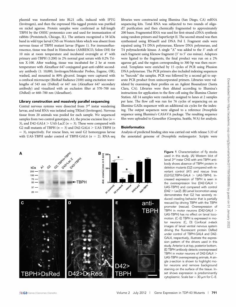
plasmid was transformed into BL21 cells, induced with IPTG(Invitrogen), and then the expressed His-tagged protein was purifiedon nickel agarose. Protein samples were confirmed as full-lengthTBPH by the OHSU proteomics core and used for immunization ofrabbits (Proteintech, Chicago, IL). The antisera recognized a 58 kDaband in wild type larval CNS on Western blots which was absent fromnervous tissue of TBPH mutant larvae (Figure 1). For immunofluo-rescence, tissue was fixed in Histochoice (AMRESCO, Solon OH) for10 min at room temperature and incubated overnight at 4� withprimary anti-TBPH (1:200) in 2% normal goat serum with 0.2% Tri-ton X-100. After washing, tissue was incubated for 2 hr at roomtemperature with Alexafluor 647-conjugated goat-anti-rabbit second-ary antibody (1: 10,000, Invitrogen/Molecular Probes, Eugene, OR),washed, and mounted in 80% glycerol. Images were captured witha confocal microscope (BioRad Radiance 2100) using excitation wave-lengths of 543 nm (DsRed) or 647 nm (Alexafluor 647 secondaryantibody) and visualized with an exclusion filter at 570–700 nm(DsRed) or 660–700 nm (Alexafluor).
Library construction and massively parallel sequencingCentral nervous systems were dissected from 3rd instar wanderinglarvae, and total RNA was isolated using TRIzol (Invitrogen). Nervoustissue from 20 animals was pooled for each sample. We sequencedsamples from two control genotypes, A1, the precise excision line (n¼3), and D42-GAL4 . UAS-LacZ (n ¼ 3). These were compared withG2 null mutants of TBPH (n ¼ 3) and D42-GAL4 . UAS-TBPH (n¼ 3), respectively. For rescue lines, we used G2 homozygous larvaewith UAS-TBPH under control of TBPH-GAL4 (n ¼ 2). RNA-seq
libraries were constructed using Illumina (San Diego, CA) mRNAsequencing kits. Total RNA was subjected to two rounds of oligo-dT purification and then chemically fragmented to approximately200 bases. Fragmented RNA was used for first-strand cDNA synthesisusing random primers and SuperScript II. The second strand was thensynthesized using RNaseH and DNA Pol I. Fragment ends wererepaired using T4 DNA polymerase, Klenow DNA polymerase, andT4 polynucleotide kinase. A single “A” was added to the 39 ends ofeach fragment using Klenow fragment (39 to 59 exo minus). Adaptorswere ligated to the fragments, the final product was run on a 2%agarose gel, and the region corresponding to 300 bp was then recov-ered. Templates were enriched by 15 cycles of PCR using PhusionDNA polymerase. The PCR primers also included indexing sequencesto “barcode” the samples. PCR was followed by a second gel to sep-arate PCR product from unincorporated primers. Libraries were val-idated by examining their profiles on an Agilent Bioanalyzer (SantaClara, CA). Libraries were then diluted according to Illumina’sinstructions for application to the flow cell using the Illumina ClusterStation. All 14 samples were randomly assigned to lanes at 2 samplesper lane. The flow cell was run for 76 cycles of sequencing on anIllumina GAIIx sequencer with an additional six cycles for the index-ing. The output sequences were aligned to a reference Drosophilasequence using Illumina’s CASAVA package. The resulting sequencefiles were uploaded to Genesifter (Geospiza, Seattle, WA) for analysis.
BioinformaticsAnalysis of predicted binding sites was carried out with release 5.33 ofthe annotated genome of Drosophila melanogaster. Scripts were
Figure 1 Characterization of fly stocksused in this study. (A) Western blot oflarval 3rd instar CNS with anti-TBPH anti-body shows absence of TBPH protein indeletion mutants (G2) compared with re-vertant control (A1) and rescue lines(G2/G2;TBPH-GAL4 . UAS-TBPH). In-creased expression of TBPH is seen inthe overexpression line (D42-GAL4 .UAS-TBPH) and compared with control(D42. LacZ). (B) Larval locomotion assaydemonstrates that G2 has severely re-duced crawling behavior that is partiallyrescued by driving TBPH with the TBPHpromoter (rescue). Overexpression ofTBPH in motor neurons (D42-GAL4 .UAS-TBPH) has no effect on larval loco-motion. (C–E) TBPH is expressed in mo-tor neurons: (C, D) Confocal z-stackimages of larval ventral nervous systemdriving the fluorescent protein DsRedunder control of TBPH-GAL4 and D42-GAL4, respectively, illustrate the expres-sion pattern of the drivers used in thisstudy. Anterior is at top, posterior bottom.(E) TBPH antibody detects overexpressedTBPH in motor neurons of D42-GAL4 .UAS-TBPH overexpressing animals. A sin-gle z-section is shown to highlight mo-tor neurons and remove backgroundstaining on the surface of the tissue. In-set shows expression is predominantlycytoplasmic. Scale bar¼ 30 mm for C–E.
Volume 2 July 2012 | Gene Expression in TDP-43 Mutants | 791
Dow
nloaded from https://academ
ic.oup.com/g3journal/article/2/7/789/5986886 by guest on 01 January 2022

composed in python (http://www.python.org). Gene regions or othersequence features (e.g., introns or exons) were queried using one ormore of three different regular expressions corresponding to publishedTDP-43 binding sites in mouse or human. The regular expressionswere 1:(GT){3,}T{3,}, 2:(GT){2,}(GTA){1}(TG){3,}, and 3:(GT){4,}[^T{3,}], and their reverse complements, corresponding to the canonicalTDP-43 motifs (UG)mUn, (UG)n, and the novel (UG)mUA(UG)n(Buratti and Baralle 2001; Sephton et al. 2011). For searches of singlestranded molecules (introns or mRNAs), only the forward regularexpressions were used. Differentially expressed genes were comparedwith predicted targets of TDP-43 binding in R and SQLite.
Statistical analysesGenetic rescue of TBPH mutant in genotype G22/2; TBPH-GAL4.UAS-TBPH was calculated using 105 samples from a Bayesian modelwith the JAGS software package (Plummer 2003) to estimate the ratioof survival probabilities between rescue and mutant phenotype. Themodel was based on multinomial/Dirichlet and binomial/beta conju-gate distributions (Gelman and Hill 2006). The genetic frequencieswere constrained by an informative prior distribution to be withina few percent of the theoretical Mendelian frequencies. The totalprogeny (including unobserved embryonic and larval lethality) wereestimated from a normal approximation to the binomial distributionusing the number of observed adults expressing the dominant visiblebalancer marker (viable nonmutants). Estimation of gene expressionwas carried out using Genesifter (Geospiza, Seattle, WA). Gene ex-pression values were normalized for each sample by indexing numberof reads per kilobase per mapped million reads of each biologicalsample (Mortazavi et al. 2008; Pepke et al. 2009; Wilhelm and Landry2009) and reported as log2 values. There were three biological repli-cates for each genotype (null G2, control A1, D42 . LacZ, D42 .TBPH) except for rescue (G2; TBPH . TBPH), for which there weretwo replicates. For pair-wise comparisons of gene expression, t-testswere performed on genes with a minimum average quality of 10 readsin at least one genotype. We also report the edgeR P values adjustedfor multiple pair-wise comparisons (Benjamini and Hochberg 1995).For pathway and ontology analysis, we used DAVID bioinformaticssuite at http://david.abcc.ncifcrf.gov/ (Huang et al. 2009a, Huang et al.2009b). We used the 7897 genes derived from control gene expressiondescribed in the text as our background. Annotation clustering wasperformed with stringency set to “high.” Enrichment scores representthe negative log of the average P value in each cluster. We reportenrichment scores of 1.3 or greater (corresponding to log2 ¼ 0.05cutoff value) for categories with at least 5 genes in the result set.
Analysis of splicingFor analysis of splicing in mutants vs. control, both genotypes hada minimum quality of 200 reads and were indexed according toGenesifter’s “correlation coefficient”method (Eisen et al. 1998). Genesthat had an index less than 0.7 and that also contained predicted TDP-43 binding sites were selected. For specific exon-exon junctions ofbiological interest, we used a beta binomial conjugate pair to estimatethe P value for a binomial event having a particular splice pattern(Gelman and Hill 2006). Successes were defined as counts of a specificexon-exon junction of interest and failures as any exon junctions in-volving the first exon and an alternate second one. Alternatively, fail-ures were defined as all other exon-exon junctions from the samegene. We chose a beta (1/2, 1/2) prior (Jeffreys 1946) to reflect thebiologically reasonable assumption that reads across specific exon-exon junctions tend to be either very uncommon or common.
RESULTS
Generation of TBPH mutantThe fly ortholog of human TDP-43 is the Tar DNA-binding proteinhomolog (TBPH). We used imprecise p-element excision to generateTBPH deletion mutants. From this we recovered a balanced lethalline, TBPH[xG2], referred to hereafter as “G2,” that deletes the pro-moter region, 59 UTR and most of the first intron. G2 fails to com-plement a deletion covering the same region as TBPH, and it also failsto complement previously isolated alleles of TBPH (Feiguin et al.2009). Antibodies raised against Drosophila TBPH fail to recognizebands of the expected size on Western blots of CNS isolated fromhomozygous G2 mutant compared with control (Figure 1A). As pre-viously reported for loss-of-function mutations in TBPH (Feiguinet al. 2009), G2 homozygous larvae also exhibited severely reducedlocomotion in larval crawling assays (Figure 1B). Consistent with ourobservation that ubiquitous expression of TBPH RNAi is lethal in thelate pupa (unpublished data), the G2 allele is 100% pupal lethal. Also,consistent with G2 being a loss-of-function mutation in TBPH, ex-pression of a UAS-TBPH transgene either in motor neurons with theD42-GAL4 transgene (Figure 1E) or under the control of the endog-enous promoter region (TBPH-GAL4, Figure 1C) was sufficient topartially rescue lethality (10.7% survival to adult, .9.6:1 ratio of res-cue:mutant, 95% confidence, n ¼ 393) and the crawling phenotype inG2 homozygous larvae (Figure 1B). Thus, TBPH-GAL4 likely partiallyreplicates the endogenous expression pattern of TBPH and is anappropriate driver to use as a rescue transgene in our expressionprofile study. Together these data indicate that the G2 allele retainsthe coding sequence but does not express protein in the CNS of larvaewhere it is normally found. Therefore, G2 is a null allele or at leasta severe hypomorph of the TBPH gene.
Overexpression of TBPH is toxic and causeslocomotion defectsMany studies have used overexpression of human TDP-43 (hTDP-43)to model ALS. In particular, expression of hTDP-43 in motor neuronscause climbing deficits in adult flies (Li et al. 2010). To compare theeffects of loss-of-function with overexpression, we drove expression ofTBPH pan-neuronally or in more restricted sets of cells. TBPH mis-expression had broadly deleterious effects, causing early lethality witha variety of different cell-type specific drivers (data not shown). Pan-neuronal expression of TBPH was highly toxic, leading to 100% lethalitywith 90% lethality as first instar larvae (unpublished observations). Bycontrast, overexpression of TBPH in motor neurons (D42 . TBPH)had no effect in larval stages (Figure 1B), and adult flies exhibitedsignificant climbing deficits relative to controls within a week of eclosion(supporting information, Figure S1). By the end of the second week ofadult life, D42 . TBPH flies were unable to scale the sides of a testtube in the same assay (Figure S1). Thus overexpression of TBPH,the fly ortholog of TDP-43, is toxic to neurons. Immunocytochem-istry of larval nervous tissue overexpressing TBPH using the D42-GAL4 driver showed extensive expression in the cytoplasm of motorneurons (Figure 1E). Although the staining shows clear enrichmentof expression in the cytoplasm, we were unable to detect endogenousTBPH to determine whether overexpression leads to loss of endog-enous TBPH from the nucleus.
Loss of TBPH results in increased transcript abundancein the CNSTo determine the effects of loss of TBPH function on transcriptabundance in the larval nervous system, we performed RNA-seq on
792 | D. J. Hazelett et al.
Dow
nloaded from https://academ
ic.oup.com/g3journal/article/2/7/789/5986886 by guest on 01 January 2022

genotypes in which we manipulated endogenous TBPH expressionlevels, first by removing and then restoring expression in loss-of-function animals (rescue). We obtained approximately 49 millionreads with an average of 3.5 million reads per sample. Seventy-fivepercent of the reads mapped to exons, as expected for a sampleconsisting of poly-A(+) selected RNA. Two percent of the reads weremapped to exon/exon boundaries indicative of splicing. rRNA/snRNAaccounts for only 2% of the reads, suggesting a very efficient selectionprocess, as these are the most abundant species in total RNA. Thesestatistics are summarized in Figure 2A. We estimated wild-type geneexpression by combining the six control samples (two genotypes),resulting in a count of 7897 genes, about half of the Drosophilagenome. We pooled six samples in this way only to simulate thedepth of coverage in other pair-wise comparisons in the study thatinvolve five or six samples. Two different genotypes were used ascontrols in this study to reduce genetic background differences be-tween the loss-of-function and overexpression comparisons. The A1line, a p-element revertant (see Materials and Methods) has the samegenetic background as G2, and we used it for the loss-of-functioncontrol genotype. GAL4 expression itself has been reported to pro-mote a transcriptional response in Drosophila (Liu and Lehmann2008), so some fraction of the genes may result from irrelevantGAL4 activity. Therefore, we used D42-GAL4 . UAS-LacZ as a con-trol for ectopic GAL4 transcriptional activation and overexpression offoreign protein in the overexpression experiment.
In TBPH loss-of-function CNS, we detected 910 differentiallyexpressed (DE) genes, of which 112 exhibited a 2-fold or greater
change (Table 1 and expression data in Table S1). Of these 910 DEgenes, the majority, 681 genes (75%), were upregulated compared withcontrol with 229 genes (25%) downregulated. Likewise, the majority of2-fold differentially expressed genes were upregulated (85 genes). Todetermine which of these 910 differentially expressed genes were af-fected due to the loss of TBPH expression, we replaced TBPH in theG2 mutants by expressing TBPH under the control of its endogenousproximal promoter region. We then compared the CNS expressionprofile of these rescue larvae with A1 controls, and identified all thegenes in this set that were not differentially expressed in the rescueexperiment but that were differentially expressed in the mutants. Ofthe 910 differentially expressed genes identified in mutant larvae, 398(44%) were completely rescued, and an additional 76 genes (8%) werepartially rescued. These 474 genes likely represent the best-candidategenes whose expression depends on TBPH. The proportion of upre-gulated and downregulated genes in this group was similar to alldifferentially regulated genes in mutant animals: 346 (73%) were upre-gulated and 128 (27%) were downregulated in mutants. We interpretthese data to mean that our rescue experiment results in a partialrestoration of endogenous TBPH activity. This interpretation is sup-ported by the partial rescue of larval crawling behavior (Figure 1C)and lethality.
When TBPH was overexpressed in motor neurons under thecontrol of the D42-GAL4 transgene, we observed 623 differentiallyexpressed genes, with 51 genes showing changes that were 2-fold orgreater (Table 1, Table S2). In contrast to the loss-of-function data, themajority (464) of differentially expressed genes were downregulated
Figure 2 Summary of readcounts and read types fromRNA-seq. (A) The proportion ofreads mapping to each class ofRNA. Exon-intron includesreads that mapped to exonregions but may overlapintrons. (B) Total number ofreads mapping to exons (exon-intron category as described inpanel A) for each biologicalsample. Horizontal bars indicatethe mean.
n Table 1 Summary of RNA-seq results
Condition Upregulated Downregulated Total
TBPH null (G2 vs. A1) 681 229 910TBPH null . 2-fold change 85 27 112Rescued and partially rescueda
(G2;TBPH . TBPH) 346 128 474Null with TBPH binding site 308 92 400Null, rescued with TBPH binding site 134 47 181Overexpression (D42 . TBPH vs. .LacZ) 159 464 623Overexpression . 2-fold change 23 28 51Overexpression with TBPH binding site 33 237 270Null + overexpressiona,b 48 9 79
In control CNS, 7897 genes were expressed. Genes in row 9 do not sum to total because some genes in this set changed in the samedirection between null and overexpressed.aThe direction of change refers to the original null-mutant gene expression data set.
bThe mathematical intersection of null and overexpression datasets.
Volume 2 July 2012 | Gene Expression in TDP-43 Mutants | 793
Dow
nloaded from https://academ
ic.oup.com/g3journal/article/2/7/789/5986886 by guest on 01 January 2022

(74%), and only 159 (25%) were upregulated compared with control.These statistics are summarized in Table 1. These data suggest thatmodulating TBPH expression in the nervous system dramaticallyalters gene expression, and that TBPH plays a direct or indirect rolein transcription or message stability. To our surprise however, only 79genes were shared between the mutant loss-of-function and overex-pression datasets. Of these 79 genes, 57 (72%) were regulated inopposite directions—48 (84% of 57) were increased in mutants anddecreased in overexpression CNS.
Identification of genes with putative TBPH binding sitesTDP-43 is an RNA-binding protein with two conserved RNA-bindingmotifs. The high degree of homology between the two RNA-bindingmotifs of Drosophila TBPH and human TDP-43 and the finding thatthey can functionally substitute for each other (Feiguin et al. 2009; Luet al. 2009; Li et al. 2010) suggest that the target binding sequence isalso conserved. Therefore, to predict direct mRNA binding targets ofTBPH and relate these predictions to our expression data, we surveyedthe Drosophila genome for potential TBPH binding sites. Using pre-viously identified TDP-43 binding site sequence motifs (see Materialsand Methods), we identified 3742 putative target genes, about 24% of15,065 annotated genes. Of the 3742 predicted targets, 3018 (81%)were expressed in control CNS (7897 genes), constituting a significant(P , 1e2369) enrichment of putative TBPH target genes in thenervous system over genomic background. We compared these geneswith the list of TDP-43 targets in mammalian nervous tissue as pre-viously identified by CLIP-seq (Sephton et al. 2011). Using one-wayBLAST against Drosophila proteins, we identified 2612 unique Dro-sophila orthologs (17% of genome) that mapped to 3647 of the 4338genes from the Sephton et al. (2011) study (using expect cutoff1e27). Of these 2612 Drosophila orthologs, only 1053 also hadpredicted TBPH binding sites. Of these, 1007 (96% of 1053, P ¼4.48e2172) were expressed in controls, suggesting a very highenrichment for orthologs of mammalian TDP-43 targets in thelarval nervous system. The majority of predicted binding sites inDrosophila were found in introns rather than exons, 59 UTRs, and39 UTRs (Figure 3A). We did not detect any bias within theseclasses of RNA for different motifs.
Next, we compared our predicted RNA targets to the expressiondata. We identified 400 genes (44% of 910) that were differentiallyexpressed in TBPH loss of function. This number constitutessignificant enrichment (P ¼ 5.44e25) compared with 38% of genesexpressed in nervous tissue with binding sites. Similarly, 44% of upre-gulated genes in loss-of-function mutants contain TBPH binding sites(P ¼ 7.7e25). For the genes that were differentially expressed inoverexpression samples, although there was no overall enrichmentfor binding sites, there were 200 genes (43%) that were downregulated
and had binding sites, representing a significant enrichment (P ¼1.16e22) compared with all CNS genes with binding sites. Twenty-six of the genes that contained putative TBPH binding sites wereidentified in both loss-of-function and overexpression data sets.The expression levels of 20 out of the 26 genes changed in oppositedirections comparing knockout vs. overexpression (Table S3) (i.e.,upregulated in mutants and downregulated in overexpression orvice versa).
We also identified 181 differentially expressed genes in loss-of-function samples that contained binding sites and were either fullyrescued or partially rescued (Table S4A) by TBPH expression. Ofthese 181 genes, we identified a subset of 52 genes that were orthol-ogous to one or more previously identified TDP-43 binding targets inCLIP-seq studies (Sephton et al. 2011). These genes are listed with themammalian orthologs in Table 2.
TBPH target genes are enriched for synaptictransmission, neurotransmitter secretion,and endocytosisTo determine the biological role of TBPH function in the centralnervous system, we focused on the set of genes whose differentialexpression could be rescued or partially rescued in mutant genotypesby expression of transgenic TBPH. To look for enrichment in geneontology annotations, we used the publicly available bioinformaticstools at DAVID (http://david.abcc.ncifcrf.gov/). To eliminate inherentbias from using nervous tissue, we used the 7897 genes (Table S4C)from control genotypes as our background instead of the whole ge-nome. Specific biological themes were evident in the enriched ontol-ogy terms; therefore, we used annotation clustering to organizeenriched terms and identify the sets of genes relevant to each cluster.This analysis revealed enrichment of synaptic transmission, neuro-transmitter secretion, and endocytosis (Figure 4 and Figure S2). Asignificant number of ion channels were also differentially expressedin G2 mutants (Figure S3, cluster 3, enrichment score: 2.29), ligand- orneurotransmitter-gated ion-channels (Figure S3, cluster 6, enrichmentscore 2.00), and neuropeptide receptors (Figure S3, cluster 13 enrich-ment score 1.55). Clustered annotation data for G2 mutants are dis-played in Figure S3. We also performed this analysis on the list ofrescued genes that were orthologous to previously identified orthologsof mammalian TDP-43 targets (Sephton et al. 2011; see Table 2).The enrichment results were similar to the set of all rescued genes(data not shown), consistent with the hypothesis that the orthol-ogous genes identified in our study are conserved TDP-43 targetgenes. Therefore, instead of presenting this analysis separately, theorthologous targets are highlighted red in Figure S2 and Figure S3.We did not detect any pathway enrichment by KEGG analysis, butthree important BMP pathway genes, Smad on X (Smox), skpA,
Figure 3 Genomic distribution ofTBPH binding sites. Genes with morethan one TBPH target binding se-quence are counted only once. (A)Distribution of three reported TBPHbinding motifs (described in Materialsand Methods) among different RNAstructural subdomains. (B) Distributionof TBPH binding sites among upregu-lated and downregulated genes inRNA-seq of TBPH knockout.
794 | D. J. Hazelett et al.
Dow
nloaded from https://academ
ic.oup.com/g3journal/article/2/7/789/5986886 by guest on 01 January 2022

n Table 2 Conserved TDP targets whose differential expression in G2 mutants was restored in rescue experiment
Flybase_IDa Mouse TDP Targetb Molecular Functionc Biological Processd
Rescued expression, downregulated in mutantAdar NM_001111055, NM_001111056,
NM_001111057, NM_031006,NM_012894
RNA-binding protein RNA editing
baz NM_031235 Phosphatidylinositol binding Asymmetric division, planar polarity,synapse assembly, other
CG15822 NM_001134514 No dataCG17754 NM_001047093 No data PhagocytosisCG32226 NM_001037191 Membrane sugar bindingCG33214 NM_017211 Golgi membrane proteinCG5214 NM_001006981 Succinyl transferase Tricarboxylic acid cyclechrb NM_080906 No data Cell death, signal transductiondlg1 NM_031639, NM_019621,
NM_022282, NM_022599,NM_022940, NM_012788
Guanylate kinase, egfr binding Synaptic transmission, basal proteinlocalization, other
MED1 NM_001134361 Mediator complex RNA Pol II cofactorpgant2 NM_001100863, NM_001012109 N-acetylgalactoseaminotransferase Golgi, posttranslational modificationPk61C NM_031081 Kinase Receptor mediated signaling, cell-growthSema-1b NM_001108526 Receptor Axon guidanceTBPH NM_001011979 RNA-binding protein Regulation of splicing
Rescued expression, upregulated in mutantAmph NM_022217,NM_053959 Synaptic vesicle protein Exocytocis; neurotransmitter secretionAP-1g NM_134460 Clathrin adaptor complex Neurotransmitter secretion; Notch signalingatt-ORFA NM_001100860 No datacact NM_030867 Transcription factor binding Nervous system development,
environmental insult, otherCG3308 NM_001109252 EndodeoxyribonucleaseCG33181 NM_001108742 No dataCG34127 NM_134336, NM_053868 Neurexin family protein
bindingNeurogenesis, phagocytosis
CG34353 NM_001163168, NM_001163169 No dataCG4293 NM_001024984 ArginaseCG4400 NM_001106731, NM_001009605 SIN3-type complex Assembly of HDAC complexesCG6287 NM_031620 Phosphoglycerate
dehydrogenaseSerine biosynthesis
CG8223 NM_001005543 No dataCG9705 NM_152790, NM_001170542 Transcription factor UnknownClic NM_031818 Chloride intracellular channel Adult lifespan, oxidative stressdve NM_001109306 Transcription factor Copper import, morphogenesisExn NM_001136241 Rho-GTP exchange factor Regulation of neurotransmitter secretionform3 NM_001106437 Actin binding Tracheal developmentHph NM_001004083 Oxidoreductase Response to DNA damage and hypoxial(1)G0289 NM_001108422 No dataMAPk-Ak2 NM_001025761, NM_001164043,
NM_178102Kinase Cell adhesion
Nap1 NM_133402, NM_001024789,NM_053561, NM_001012170
Histone binding Nucleosome assembly, regulation oftranscription
nvy NM_001108657 Transcription factor Axon guidance, dendrite morphogenesis,muscle
opa NM_001108391, NM_001108392 Transcription factor dpp/BMP signaling, eye development,germ cell migration, midgut
Rab35 NM_001013046 GTPase Signaling, cytokinesisRala NM_053821 GTPase Cell morphogenesis, Notch regulation, otherrb NM_001107532, NM_001107646 Regulates ubiquitination Endocytosis, synaptic vesicle coating,
pigment granule biogenesis, lysosomalorganization
RpS15Aa NM_053982 Small ribosomal subunit Translation, mitotic spindle organizationSap-r NM_013013 Lysosomal protein, activator
of lysosomal enzymesdsRNA transport, sphingolipid metabolism
sax NM_024486 dpp/BMP type I receptor Morphogenesis, NMJ morphogenesis, otherSh NM_012971, NM_023954 Voltage-gated K+ channel axon potentialskpA NM_001007608 SCF ubiquitin ligase complex Centrosome dup., neurogenesis, cell cycle
(continued)
Volume 2 July 2012 | Gene Expression in TDP-43 Mutants | 795
Dow
nloaded from https://academ
ic.oup.com/g3journal/article/2/7/789/5986886 by guest on 01 January 2022

and saxophone, were upregulated in mutants and rescued in G2;TBPH . TBPH animals.
Overexpressed TBPH leads to novel changes in geneexpression with some functional overlap with mutantsSimilar to our analysis of loss-of-function altered genes, we alsofocused on 270 genes with TBPH binding sites (Table S4B) whoseexpression was altered when TBPH was overexpressed in motor neu-rons. As with the prior analysis, we clustered annotations to identifyoverlapping sets of functionally related genes. Once again, we identi-fied enrichment of genes involved in tissue and epithelial sheet mor-phogenesis (cluster 3, enrichment score 2.76). Another cluster ofterms related to larval metamorphosis and pupal or imaginal discdevelopment (enrichment score 3.63) included many of the samegenes (Figure 5 and Figure S4). More than a few of these genes werealso shared with loss of function (e.g., u-shaped, steamer duck, anddomeless). Most shared genes—including these three—were upregu-lated in mutants and repressed in overexpression (Table S3). Therewere additional categories present in the overexpression and absent inmutants (Figure 5 and Figure S4): imaginal disc development (enrich-ment score 3.63), proximal-distal patterning (enrichment score 2.79),oogenesis and gamete production (enrichment score 2.32), stem celldifferentiation and asymmetric division (enrichment score: 1.56), andLeucine-rich repeat proteins (enrichment score: 1.81). Immunoglob-ulin domain proteins were enriched in mutants (Figure 4 and FigureS2, cluster 8) and overexpression (Figure 5 and Figure S4, cluster 4).We also performed the KEGG pathway analysis for the differentiallyexpressed genes in overexpression D42 . TBPH samples. The Wntpathway was enriched (P ¼ 0.0014, corrected P ¼ 0.05 [Benjaminiand Hochberg 1995]), including the Wnt receptor arrow/LRP-5,CG7913/PP2A, Rho-kinase or rok/ROCK2, supernumerary limbs orslmb/beta-TrCP, armadillo (beta-catenin), cAMP-dependant proteinkinase 1 or Pka-C1 (PKA), and Smad on X or Smox (SMAD3). Whenwe considered only conserved TDP-43 targets, WNT pathway geneswere clearly enriched (P ¼ 3.4e24, corrected P ¼ 7.2e23 [Benjaminiand Hochberg 1995]). One of the targets, Smox, is an effector of dpp/BMP signaling. Other dpp pathway genes decapentaplegic and saxo-phone were affected in loss of function or overexpression of TBPH,although the pathway as a whole was not enriched for. Wnt/BMPpathway genes were upregulated in mutants and downregulated inoverexpression samples, suggesting that TDP-43 modulates thesepathways by altering expression levels.
Analysis of altered splicingTDP-43 is described as a splicing factor (Buratti et al. 2001; Burattiand Baralle 2001), so we also identified genes with altered splicing. Inan attempt to identify splicing changes that were directly regulated byTBPH, we restricted our analyses to the set of genes with TBPHbinding sites. There were 30 genes in mutants and 50 genes in theoverexpression samples (Table 3) that met these criteria. All but 7 ofthe genes with altered splicing in mutants returned a higher spliceindex (reduced altered splicing) in the rescue samples, suggesting thatthe changes in splicing were due to loss of TBPH. The overall expres-sion levels of the majority of all 80 genes with altered splicing inmutants and overexpression samples were unchanged, although
n Table 2, continued
Flybase_IDa Mouse TDP Targetb Molecular Functionc Biological Processd
Smox NM_019191 Transcription factor TGF-beta signaling, axon guidance, dendritemorphogenesis, neuron development,cell cycle
spin NM_001144991, NM_001039208 No data PCD, dpp signaling, CNS remodel,glial migration/differentiation,NMJ remodeling
Su(var)3–9 NM_001100542 H3-K9 methyltransferase Gene silencingSynd NM_017294, NM_001009966 Vesicle endocytosis, neurotransmitter
secretionTimp NM_001109393, NM_012886 Metalloprotease inhibitor Basement membrane organizationtwin NM_001108355 CCR4-NOT complex mRNA polyA shortening, transcript
stabilityUGP NM_001024743 Transferase Carbohydrate metabolism
aGenes whose expression was rescued in G2 mutants when driving TBPH with the TBPH promoter region under the GAL4 system.
bIdentity of mouse orthologous mRNAs found to be bound to TDP-43 in CLIP-seq experiment (Sephton et al. 2011).
cSummary of most informative gene ontology terms for molecular function.
dSummary of most informative biological process annotations.
Figure 4 Clusters of related annotation terms enriched in rescuedgenes with binding sites. Frequency of different categories of clustersis represented by a single descriptive term as a function of the numberof genes in each cluster. Percentages indicate proportion of enrichedterms, not all genes. For details on the identities of the genes and theindividual terms and significance, see Figure S2 and Figure S3.
796 | D. J. Hazelett et al.
Dow
nloaded from https://academ
ic.oup.com/g3journal/article/2/7/789/5986886 by guest on 01 January 2022

expression of individual exons varied. Eight genes were misspliced inboth loss-of-function and overexpression samples. These genes werecacophony (cac), CG17341, strawberry notch (sno), CG3744, toucan(toc), Rho kinase (rok), division abnormally delayed (dally), and sticksand stones (sns). We also clustered annotations of misspliced genesand discovered some functional overlap with differentially expressedgenes. One cluster of enriched terms was related to nervous systemdevelopment and cell projection morphogenesis (enrichment score2.23). Six misspliced genes associated with cell or projection morpho-genesis terms are Rho kinase, Netrin-B (NetB), Cadherin-N (CadN),hamlet (ham), Insulin-like receptor (InR), division abnormally delayed,and the ATP-dependant chromatin assembly factor large subunit Acf-1.Three genes annotated with regulation of dendrite morphogenesiswere warts (wts), hamlet, and Cadherin N. In Kegg pathway analysis,the Wnt pathway was again enriched (P ¼ 0.041, corrected P ¼ 0.42),which includes the genes Casein kinase II beta subunit (Ckiibeta),division abnormally delayed, thus reinforcing the importance of thispathway as a potential target of regulation by TBPH/TDP-43 activity.
We tabulated the frequency of individual species of exon-exonjunctions as additional evidence for different splice forms in differentgenotypes (Table S5). We calculated the expected frequency of anexon junction and the likelihood of the observed junction reads rel-ative either to alternative junctions or relative to all other mappedexon junctions for that gene. In some instances, a gene that had a lowsplicing index in G2 mutants but not in overexpression samples mightnonetheless have significant exon junctions when examined this way.One such gene is Apaf-1-related-killer (Ark), which was only detectedin the overexpression splicing index but yielded significant findings inboth the mutant and overexpression for a pair of mutually exclusivealternative exon junctions (see Table S5). The most common splicingevents that we detected by comparison of junction reads were those inwhich multiple exons were spliced out of the middle of genes, such thatthe first one or two exons were joined to the last or next-to-last exons,producing a transcript with little or no coding sequence. Exon junctionsof this type were frequently observed in the controls but absent frommutant nervous tissue. Interestingly, three such genes, strawberry notch,
rho kinase, and Pkn, were found to be either upregulated in mutants ordownregulated in overexpression, suggesting a potential novel mecha-nism by which TBPH regulates these transcripts. None of these putativeshort transcripts is currently annotated in public databases.
TDP-43 regulates the expression and splicing of genesassociated with neurological disordersWe compared our lists of differential expression with human diseasedatabases via “Homophila” (Chien et al. 2002). The results of thiscomparison are presented in Table S6. A large number of genes, in-cluding 211 genes from G2 mutant (plus 8 misspliced) and 142 genes(plus 21 misspliced) from D42 . TBPH overexpression were associ-ated with human diseases. To narrow our list to the most interestingcandidates, we selected the rescued genes, overexpression set genes,and misspliced genes associated with neurological disorders, and thenclassified them as developmental, neuropathy, movement disorder,sensory or “other” (e.g., epilepsy), with some genes falling into morethan one category. Twenty-three (23) direct homologs or paralogs ofsuch genes were associated with human neurological disorders.
Among the 23 genes with Drosophila orthologs, 10 are linked todevelopmental disorders, the majority associated with some type ofmental retardation or microcephaly. There were 9 genes associatedwith degenerative neuropathies including, notably, tau, which is bothupregulated in G2 mutants and downregulated in overexpressionsamples. tau is the Drosophila homolog of the well-known neuropathycausing microtubule-associated protein MAPT (Heidary and Fortini2001). Overexpression of tau is sufficient to cause neurodegenerationin a variety of contexts, including Drosophila (Jackson et al. 2002; Cheeet al. 2006; Chen et al. 2007), and it interferes with Wnt pathwayactivity (Jackson et al. 2002). Another microtubule-associated proteinfutsch, the MAP1B homolog, was recently shown to be regulated byTBPH in flies (Godena et al. 2011). Consistent with the tau result,futsch was downregulated in our overexpression samples, whereas nochange was detected in G2 mutants.
Six genes were associated with movement disorders, includingCharcot-Marie-Tooth disease and Parkinson’s disease (PD). One ofthe largest effects we observed in TBPH mutants was the downregu-lation of L-dopa decarboxylase (Ddc; expression ¼23.52), an enzymewhose activity is significantly reduced in the substantia nigra of PDpatients (Lloyd and Hornykiewicz 1970; Gjedde et al. 2006). TDP-43is reported to be deposited in Parkinson’s in 7% of cases and 19% ofcases of PD with dementia (Nakashima-Yasuda et al. 2007), so it isplausible that TDP-43 pathology affects PD-related disease processes.TDP-43 overexpression also enhances toxicity of a-synuclein in do-paminergic neurons in mice (Tian et al. 2011), but TDP-43 has not yetbeen implicated as a requirement for dopamine production. Thestrong downregulation of Ddc in our mutants suggests that TDP-43dysregulation or mislocalization could directly impact this pathwayand thus contribute to the development of Parkinson’s. Thus, ourobservations of human-disease genes in TBPH knockout and over-expression indicate that some TBPH targets are orthologs of genesassociated with developmental disorders of the nervous system, age-related neuropathies, and motor neuropathies. This list of genes com-prises attractive candidates for follow-up studies.
DISCUSSION
Implications for loss-of-function vs. overexpressionof TDP-43 as models of ALSThere are two prevailing models for how TDP-43 dysfunction mightbe involved in pathogenesis of ALS/FTD-U. In one, cytoplasmic
Figure 5 Clusters of related annotation terms enriched in TBPHoverexpression DE genes with binding sites. Frequency of differentcategories of clusters is represented by a single descriptive term asa function of the number of genes in each cluster. Percentagesindicate proportion of enriched terms, not all genes. For details on theidentities of the genes and the individual terms and significance, seeFigure S2 and Figure S3.
Volume 2 July 2012 | Gene Expression in TDP-43 Mutants | 797
Dow
nloaded from https://academ
ic.oup.com/g3journal/article/2/7/789/5986886 by guest on 01 January 2022

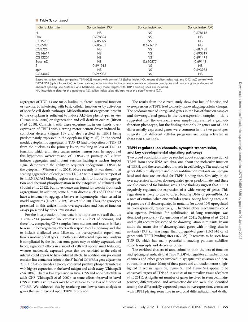
n Table 3 Candidate splicing targets of TBPH
Gene_Identifier Splice_Index_KO Splice_Index_rsc Splice_Index_OX
CG15628 NS NS 0.012231cac 0.035131 0.096865 0.610466CG17341 0.374411 NS 0.120439CG2747 NS 0.440132 0.150027sno 0.559002 NS 0.246462CG3744 0.623733 NS 0.283057Rab8 NS NS 0.294291Pde9 0.337865 0.584513 NACG8179 NS NS 0.3732NetB NS NS 0.381535toc 0.382134 0.399532 0.499912CG31352 0.40346 0.616599 NSCG1607 NS NS 0.42337InR NS NS 0.437603CG6051 0.44685 NS NSaxo NS 0.653959 0.461114CG34318 NS 0.539601 0.463593CG10353 NA NS 0.485735CG42404 0.489271 0.282117 NSssp3 NA 0.623646 0.489643CG32372 NS 0.692738 0.49013pgant5 0.493896 NS NSCG32479 NS NS 0.503011CG14322 NA 0.341229 0.50317tai NS NS 0.510347CG9318 NS 0.660374 0.51649wts NS NS 0.527354CG34394 0.530439 0.666341 NSsmi35A 0.531367 0.67484 NSCG11399 0.537233 NS NSCG10011 NS 0.561528 0.539412CG14764 NS NS 0.547603CG15356 0.320978 0.548626CG9919 0.550806 0.431502 NSmAcR-60C 0.560996 0.511291 NSCadN NS NS 0.564077Su(dx) 0.573514 NS NSSurf4 NA NS 0.574626CG4080 0.586081 0.558437 NSCG31150 NS NS 0.604517CG31739 NS NS 0.605173Ark NS NS 0.615061CkIIbeta NS 0.671316 0.61684Trn NS NS 0.621424CG6966 0.62369 NS NSrok 0.626402 0.406334 0.656452NaCP60E 0.636625 NS NSham NS NS 0.639029CG3078 NS NS 0.645845CRMP 0.646375 0.612456 NSCG7028 NA NS 0.651044htt 0.652084 NS NSCG34113 NA NS 0.658926dally 0.661246 0.669615 0.677181l(3)05822 0.661522 NS NSsns 0.683637 NS 0.662555CG12084 NA NS 0.670823Acf1 NS 0.500329 0.670858dbo NS NS 0.6765Nak NS NS 0.676703CG11880 NS NS 0.676724ppa NS NS 0.677093
(continued)
798 | D. J. Hazelett et al.
Dow
nloaded from https://academ
ic.oup.com/g3journal/article/2/7/789/5986886 by guest on 01 January 2022
aggregates of TDP-43 are toxic, leading to altered neuronal functionor survival by interfering with basic cellular function or by activationof specific cell-death pathways. Mislocalization of exogenous proteinto the cytoplasm is sufficient to induce ALS-like phenotypes in vivo(Ritson et al. 2010) or degeneration and cell death in culture (Ritsonet al. 2010). Consistent with these experiments, in our hands, over-expression of TBPH with a strong motor neuron driver induced lo-comotion defects (Figure 1B) and also resulted in TBPH beingpredominantly expressed in the cytoplasm (Figure 1E). In the secondmodel, cytoplasmic aggregates of TDP-43 lead to depletion of TDP-43from the nucleus as the primary lesion, resulting in loss of TDP-43function, which ultimately causes motor neuron loss. In support ofthis hypothesis, overexpression of TDP-43 in primary cell cultureinduces aggregates, and mutant versions lacking a nuclear importsignal demonstrate the ability to sequester endogenous TDP-43 tothe cytoplasm (Winton et al. 2008). More recently, it was shown thatseeding aggregation of endogenous TDP-43 with a multimer repeat ofits hnRNPA1/A2 binding region was sufficient to induce its aggrega-tion and aberrant phosphorylation in the cytoplasm of cultured cells(Budini et al. 2012), but no evidence was found for toxicity from suchaggregations. In addition, some human disease alleles of TDP-43 thathave a tendency to aggregate behave as hypomorphs when tested inmodel organisms (Lu et al. 2009; Estes et al. 2010). Thus, the genotypespresented in this article mimic overexpression and loss-of-functionassays presented by other investigators.
For the interpretation of our data, it is important to recall that theTBPH-GAL4 promoter line expresses in a subset of neurons, andtherefore, comparing CNS samples from mutants and rescues is likelyto result in heterogeneous effects with respect to cell autonomy and alsoto include unaffected cells. Likewise, the overexpression experimentshave a mixture of cell types. In both cases, differential expression analysisis complicated by the fact that some genes may be widely expressed, andhence, significant effects in a subset of cells will appear small (dilution),whereas moderately expressed genes that are restricted to the cells ofinterest could appear to have outsized effects. In addition, our p-elementexcision line contains a lesion in the 39 half of CG4585, a gene adjacent toTBPH. CG4585 encodes a poorly conserved putative phosphotransferasewith highest expression in the larval midgut and adult ovary (Chintapalliet al. 2007). There is low expression in larval CNS and none detectable inadult CNS (Chintapalli et al. 2007), so some effects observed in larvalCNS in TBPH G2 mutants may be attributable to the loss of function ofCG4585. We addressed this by restricting our downstream analysis togenes that were rescued upon TBPH expression.
The results from the current study show that loss of function andoverexpression of TBPH lead to mostly nonoverlapping cellular changes.The predominance of upregulated genes in the loss-of-function samplesand downregulated genes in the overexpression samples initiallysuggested that the overexpression simply represented a gain-of-function phenotype, but the finding that only 79 genes out of 1533differentially expressed genes were common in the two genotypessuggests that different cellular programs are being activated inthese two situations.
TBPH regulates ion channels, synaptic transmission,and key developmental signaling pathwaysTwo broad conclusions may be reached about endogenous function ofTBPH from these RNA-seq data, one about the molecular functionof TBPH, and the second about its role in cell biology. The majority ofgenes differentially expressed in loss-of-function mutants are upregu-lated and these are enriched for TBPH binding sites. Similarly, in theoverexpression samples, downregulated genes predominate, and theseare also enriched for binding sites. These findings suggest that TBPHnegatively regulates the expression of a wide variety of genes. Thisregulation is likely to due to direct binding DNA or pre-mRNA. Asa note of caution, when one excludes genes lacking binding sites, 20%of genes are still downregulated in mutants (or about 10% upregulatedin overexpression, respectively). Therefore other mechanisms couldalso operate. Evidence for stabilization of long transcripts wasdescribed previously (Polymenidou et al. 2011; Sephton et al. 2011)and might account for some of the downregulation in mutants. In ourstudy the mean size of downregulated genes with binding sites inmutants (19.7 kb) was larger than upregulated genes (16.2 kb) or allgenes with TBPH binding sites (16.7 kb). It remains to be seen howTDP-43, which has many potential interacting partners, stabilizessome transcripts and decreases others.
The enriched clusters of annotations in both the loss-of-functionand splicing set indicate that TBPH/TDP-43 regulates a number of ionchannels and other genes involved in synaptic transmission and neu-rotransmitter release. Many of these genes and annotation terms (high-lighted in red in Figure S2, Figure S3, and Figure S4) appear to beconserved targets of TDP-43 in studies of mammalian tissue (Sephtonet al. 2011). A significant number of genes involved in stem cell main-tenance, differentiation, and asymmetric division were also identifiedamong the differentially expressed genes in overexpression, consistentwith TBPH playing some role in neuronal differentiation and death.
n Table 3, continued
Gene_Identifier Splice_Index_KO Splice_Index_rsc Splice_Index_OX
H NS NS 0.678118Pkn 0.678824 NS NSCG15735 0.685029 NS NSCG6509 0.685753 0.671619 NSCG8726 NS NS 0.687488CG14614 NS NS 0.690319CG13204 NS NS 0.691471Socs16D NS 0.610877 0.69148S 0.691913 NS NSspir NS NS 0.693973CG34449 0.699088 NS NS
Based on splice index comparing TBPH[G2] mutant with control A1 (Splice Index KO), rescue (Splice Index rsc), and D42-lacZ control withD42-TBPH (Splice Index OX). A lower splicing index number indicates less correlation between genotypes and hence a greater degree ofaberrant splicing (see Materials and Methods). Only those targets with TBPH binding sites are included.NA, insufficient data for the genotype; NS, splice index value did not meet the cutoff criteria (0.7).
Volume 2 July 2012 | Gene Expression in TDP-43 Mutants | 799
Dow
nloaded from https://academ
ic.oup.com/g3journal/article/2/7/789/5986886 by guest on 01 January 2022

Elements of the TGF-beta and Wnt signaling pathways, includingSmad on X, arrow, armadillo, and the bmp receptor homolog saxo-phone, were upregulated in mutants and downregulated in our rescuesamples. TGF-beta signaling pathways regulate growth of the neuro-muscular endplate via retrograde signal from the muscle to the neuron[reviewed by Collins and Diantonio (2007)]. These genes are strongcandidates for TDP-43 effectors as both Smad and sax have TBPHbinding sites in their pre-mRNAs and were identified targets ofTDP-43 in vertebrate cells as well (Sephton et al. 2011). If this findingis validated in subsequent genetic and molecular studies, it wouldsuggest that TDP-43 plays a role in the modulation of synaptic ho-meostasis via the dpp/BMP signaling cascade. In support of such a role,the endplate of the larval neuromuscular junction is reduced in com-plexity and number of synapses in TBPH mutants (Feiguin et al.2009). Another interesting candidate in the Wnt group is rho kinase(rok). Wnt3-induced neurite retraction is mediated by Rho-kinase(Kishida et al. 2004), suggesting that Rho-kinase/ROCK may regulatearborization downstream of Wnts. Sensory neurons of the larval epi-dermis show severely reduced complexity and length in the dendriticarbor with loss of TBPH, and both human and Drosophila TDP-43expression drive overgrowth of this structure in the same cells, suggest-ing that TDP-43 levels play an important role in regulating the growthof the dendritic arbor (Li et al. 2010). Although this process couldpotentially be effected by dpp/Smad, we also identified other strongcandidates for effector genes in TDP-mediated dendritic arborization.Two genes identified in the endocytosis/membrane group, Syndapin andamphiphysin, are localized to the postsynapse and regulate aspects ofpost-synaptic development and function ( Mathew et al. 2003; Kumaret al. 2009). In addition, we identified dendritic arborization genes warts,hamlet, and Cadherin N as putative splicing targets of TBPH.
Thus, our findings are consistent with a model in which TDP-43regulates nerve transmission, process outgrowth, and synaptic ho-meostasis by regulation of transcript abundance and alternativesplicing of key genes in important signaling pathways, includingWnt and TGF-beta. Moreover our results indicate that TDP-43 maybe important for post-synaptic refinement and maintenance as well asfor axon terminal differentiation on the basis of functional annota-tions, and they help explain why phenotypes may be observed in bothcompartments. One or more of our identified candidates associatedwith these terms may ultimately be shown to be critical for mediatingTDP-43 activity in these cell types and compartments.
Altered splicing in TBPH mutantsTDP-43 is a splicing factor with roles in splicing of the cystic fibrosistransmembrane conductance regulator (CFTR) gene (Buratti et al.2001; Ayala et al. 2006). It was demonstrated to affect the splicingof the spinal motor neuron (SMN) gene, (Bose et al. 2008), and thehuman apoAII gene (Mercado et al. 2005). Our analysis identifieda number of potentially important target genes, including the Dro-sophila ortholog of the Cav2.1 channel cacophony (cac) and the mus-carinic acetyl choline receptor (mAchR-60C). Mutants of cac inDrosophila have been reported to have reduced endplate growth atthe neuromuscular junction (Xing et al. 2005) similar to TBPHmutants (Feiguin et al. 2009), and cac is likely to play an importantrole in the regulation of presynaptic vesicle fusion, thus making it anattractive candidate for follow-up studies on the perturbation of neu-romuscular junction function in TBPH mutants. Orthologs of cacoph-ony have also been identified as targets of TDP-43 binding in RIP-seqdata (Sephton et al. 2011) and TDP-43 loss-of-function studies (Poly-menidou et al. 2011).
Comparison with other expression-profiling studiesSeveral massively parallel sequencing or microarray studies have beencarried out previously by others. Sephton et al. (2011) identified RNAtargets bound by TDP-43 in vivo by RIP-seq. We found that ourdifferentially expressed genes were statistically enriched in orthologsof many of the same targets identified in that study, which supportsa conserved role for TDP-43 in the CNS. Also, Polymenidou et al.(2011), using RNA-seq of TDP-43 knockdown in mouse CNS, showeda 3:2 proportion of upregulated genes to downregulated genes, a sim-ilar ratio to that seen with our data. This observation suggests thatbiological effects we observed were similar despite large evolutionarydistances. In fact, one interesting aspect of TDP-43 biology is thatmany of the effects of human TDP-43 expression in model systemsrecapitulate the effects of overexpression of endogenous orthologs,despite the low degree of conservation of the C-terminus from theperspective of an Altschul alignment (Altschul et al. 1990). Here wehave provided evidence that Drosophila TBPH regulates processessimilar to those reported previously for mammalian TDP-43 (Poly-menidou et al. 2011; Sephton et al. 2011), especially with respect toregulators of synaptic release. Moreover, a significant fraction of thegenes regulated by TBPH in synaptic transmission and tissue mor-phogenesis was found to be conserved targets of TDP-43, most strik-ingly a set of genes in the wingless/Wnt and decapentaplegic/BMPpathways. Thus, our data provide support for functional conservationof TBPH/TDP-43 and are consistent with the hypothesis that TDP-43evolved early in animal evolution to play a fundamental role in thedifferentiation and cellular function of complex nervous systems. It isimportant to note, however, that TDP-43 is widely expressed in bothmammals and insects; therefore, our findings represent a fractionalview of its role from an organismal perspective. Also, as our experi-ments address only changes in messenger RNA, other potential rolesin the metabolism of noncoding RNA are excluded from this analysis.
In conclusion, we have identified a relatively small group of genesthat are probably directly regulated by the action of TBPH. Consistentwith this hypothesis and published phenotypes, a number of thesetargets are well-characterized promoters of motor neuron differenti-ation, function, and survival. Surprisingly, a large majority of genesaffected in knockout were unaffected in overexpression and vice versa,supporting different mechanisms underlying behavioral and cellulardefects of these two genetic models. Further studies using a combina-tion of genetics, behavior, and electrophysiology will be necessary tovalidate these candidates and to elucidate more precisely how theymediate the biological effects of TDP-43 expression or loss of functionin motor neurons. Such studies in flies and other model systems areneeded to determine the causes of motor neuron vulnerability to theseperturbations and to provide clues to future therapies.
ACKNOWLEDGMENTSWe thank Sudeshna Dutta for critical comments on this manuscriptduring the course of this work. We also thank the Bloomington StockCenter and FlyBase for providing vital resources. This work wassupported by grants from the National Institutes of Health(NS071186), the ALS Association, and the Muscular DystrophyAssociation to D.B.M.
LITERATURE CITEDAbhyankar, M. M., C. Urekar, and P. P. Reddi, 2007 A novel CpG-free
vertebrate insulator silences the testis-specific SP-10 gene in somatictissues. J. Biol. Chem. 282: 36143–36154.
Altschul, S. F., W. Gish, W. Miller, E. W. Myers, and D. J. Lipman,1990 Basic local alignment search tool. J. Mol. Biol. 215: 403–410.
800 | D. J. Hazelett et al.
Dow
nloaded from https://academ
ic.oup.com/g3journal/article/2/7/789/5986886 by guest on 01 January 2022

Ayala, Y. M., F. Pagani, and F. E. Baralle, 2006 TDP43 depletion rescuesaberrant CFTR exon 9 skipping. FEBS Lett. 580: 1339–1344.
Ayala, Y. M., P. Zago, A. D’Ambrogio, Y.-F. Xu, L. Petrucelli et al.,2008a Structural determinants of the cellular localization and shuttlingof TDP-43. J. Cell Sci. 121: 3778–3785.
Ayala, Y. M., T. Misteli, and F. E. Baralle, 2008b TDP-43 regulates retinoblas-toma protein phosphorylation through the repression of cyclin-dependentkinase 6 expression. Proc. Natl. Acad. Sci. USA 105: 3785–3789.
Beghi, E., G. Logroscino, A. Chiò, O. Hardiman, D. Mitchell et al., 2006 Theepidemiology of ALS and the role of population-based registries. Biochim.Biophys. Acta 1762: 1150–1157.
Benjamini, Y., and Y. Hochberg, 1995 Controlling the false discovery rate:a practical and powerful approach to multiple testing. J. R. Stat. Soc., B 57(1): 289–300.
Benzer, S., 1967 Behavioral mutants of Drosophila isolated by counter-current distribution. Proc. Natl. Acad. Sci. USA 58: 1112–1119.
Bose, J. K., I.-F. Wang, L. Hung, W.-Y Tarn, and C.-K. James Shen,2008 TDP-43 overexpression enhances exon 7 inclusion during thesurvival of motor neuron pre-mRNA splicing. J. Biol. Chem. 283: 28852–28859.
Brand, A. H., and N. Perrimon, 1993 Targeted gene expression as a meansof altering cell fates and generating dominant phenotypes. Development118: 401–415.
Budini, M., E. Buratti, C. Stuani, C. Garnaccia, V. Romano et al., 2012 Cel-lular model of TAR DNA-binding protein 43 (TDP-43) aggregation basedon its C-terminal Gln/Asn-rich region. J. Biol. Chem. 287: 7512–7525.
Buratti, E., and F. E. Baralle, 2001 Characterization and functional impli-cations of the RNA binding properties of nuclear factor TDP-43, novelsplicing regulator of CFTR exon 9. J. Biol. Chem. 276: 36337–36343.
Buratti, E., T. Dork, E. Zuccato, F. Pagani, M. Romano et al., 2001 Nuclearfactor TDP-43 and SR proteins promote in vitro and in vivo CFTR exon 9skipping. EMBO J. 20: 1774–1784.
Buratti, E., L. De Conti, C. Stuani, M. Romano, M. Baralle et al.,2010 Nuclear factor TDP-43 can affect selected microRNA levels. FEBSJ. 277: 2268–2281.
Chee, F., A. Mudher, T. A. Newman, M. Cuttle, S. Lovestone et al., 2006 Over-expression of tau results in defective synaptic transmission in Drosophilaneuromuscular junctions. Biochem. Soc. Trans. 34: 88–90.
Chen, X., Y. Li, J. Huang, D. Cao, G. Yang et al., 2007 Study of tauopathiesby comparing Drosophila and human tau in Drosophila. Cell Tissue Res.329: 169–178.
Chien, S., L. T. Reiter, E. Bier, and M. Gribskov, 2002 Homophila: humandisease gene cognates in Drosophila. Nuc Ac Res 30: 149–151.
Chintapalli, V. R., J. Wang, and J. A. T. Dow, 2007 Using FlyAtlas toidentify better Drosophila melanogaster models of human disease. Nat.Genet. 39: 715–720.
Collins, C. A., and A. DiAntonio, 2007 Synaptic development: insights fromDrosophila. Curr. Opin. Neurobiol. 17: 35–42.
Eisen, M. B., P. T. Spellman, P. O. Brown, and D. Botstein, 1998 Clusteranalysis of genome-wide expression patterns. Proc Natl Acad USA 95:14863–14868.
Estes, P. S., A. Boehringer, R. Zwick, J. E. Tang, B. Grigsby et al.,2010 Wild-type and A315T mutant TDP-43 exert differential neuro-toxicity in a Drosophila model of ALS. Hum. Mol. Genet. 20: 2308–2321.
Feiguin, F., V. K. Godena, G. Romano, A. D’Ambrogio, R. Klima et al.,2009 Depletion of TDP-43 affects Drosophila motoneurons terminalsynapsis and locomotive behavior. FEBS Lett. 583: 1586–1592.
Gelman, A., and J. Hill, 2006 Data Analysis Using Regression and Multi-level/Hierarchical Models. Cambridge University Press, Cambridge,England.
Gjedde, A., G. C. Léger, P. Cumming, Y. Yasuhara, A. C. Evans et al.,2006 Striatal L-DOPA decarboxylase activity in Parkinson’s diseasein vivo: implications for the regulation of dopamine synthesis. J. Neu-rochem. 61: 1538–1541.
Godena, V. K., G. Romano, M. Romano, C. Appocher, R. Klima et al.,2011 TDP-43 regulates Drosophila neuromuscular junctions growth by
modulating futsch/MAP1B levels and synaptic microtubules organiza-tion. PLoS ONE 6: e17808.
Greenspan, R. J., 2004 Fly Pushing, Ed. 2. Cold Spring Harbor LaboratoryPress, Cold Spring Harbor, NY.
Hanson, K. A., S. H. Kim, D. A. Wassarman, and R. S. Tibbets, 2010 Ubiquilinmodifies TDP-43 toxicity in a Drosophila model of amyotrophic lateralsclerosis (ALS). J. Biol. Chem. 285: 11068–11072.
Heidary, G., and M. E. Fortini, 2001 Identification and characterization ofthe Drosophila tau homolog. Mech. Dev. 108: 171–178.
Hoskins, R. A., J. M. Landolin, J. B. Brown, J. E. Sandler, H. Takahashi et al.,2011 Genome-wide analysis of promoter architecture in Drosophilamelanogaster. Genome Res. 21: 182–192.
Huang, D. W., B. T. Sherman, and R. A. Lempicki, 2009a Systematic andintegrative analysis of large gene lists using DAVID Bioinformatics Re-sources. Nature Protoc. 4: 44–57.
Huang, D. W., B. T. Sherman, and R. A. Lempicki, 2009b Bioinformaticsenrichment tools: paths toward the comprehensive functional analysis oflarge gene lists. Nucleic Acids Res. 37: 1–13.
Jackson, G. R., M. Wiedau-Pazos, T. K. Sang, N. Wagle, C. A. Brown et al.,2002 Human wild-type tau interacts with wingless pathway compo-nents and produces neurofibrillary pathology in Drosophila. Neuron 34:509–519.
Jeffreys, H., 1946 An invariant form for the prior probability in estimationproblems. Proc. R. Soc. Lond. 186: 453–461.
Johnson, B. S., D. Snead, J. J. Lee, J. M. McCaffery, J. Shorter et al.,2009 TDP-43 is intrinsically aggregation-prone, and amyotrophic lat-eral sclerosis-linked mutations accelerate aggregation and increase tox-icity. J. Biol. Chem. 284: 20329–20339.
Kharchenko, P. V., A. A. Alekseyenko, Y. B. Schwartz, A. Minoda, N. C.Riddle et al., 2010 Comprehensive analysis of the chromatin landscapein Drosophila melanogaster. Nature 471: 480–485.
Kishida, S., H. Yamamoto, and A. Kikuchi, 2004 Wnt-3a and Dvl induceneurite retraction by activating rho-associated kinase. Mol. Cell. Biol. 24:4487–4501.
Kumar, V., S. R. Alla, K. S. Krishnan, and M. Ramaswami, 2009 Syndapin isdispensable for synaptic vesicle endocytosis at the Drosophila larvalneuromuscular junction. Mol. Cell. Neurosci. 40: 234–241.
Lee, E. B., V. M.-Y. Lee, and J. Q. Trojanowski, 2012 Gains or losses:molecular mechanisms of TDP43-mediated neurodegeneration. NatNeurosci Rev 13: 38–50.
Leigh, P. N., B. H. Anderton, A. Dodson, J. M. Gallo, and M. Swash,1988 Ubiquitin deposits in anterior horn cells in motor neurone dis-ease. Neurosci. Lett. 93: 197–203.
Leigh, P. N., H. Whitwell, O. Garofolo, J. Buller, M. Swash et al.,1991 Ubiquitin-immunoreactive intraneuronal inclusions in amyotro-phic lateral sclerosis. Morphology, distribution and specificity. Brain 114:775–788.
Li, Y., P. Ray, E. J. Rao, C. Shi, W. Guo et al., 2010 A Drosophila model forTDP-43 proteinopathy. Proc. Natl. Acad. Sci. USA 107: 3169–3174.
Liu, Y., and M. Lehmann, 2008 A genomic response to the yeast tran-scription factor GAL4 in Drosophila. Fly (Austin) 2: 92–98.
Lloyd, K., and O. Hornykiewicz, 1970 Parkinson’s disease: activity ofL-Dopa decarboxylase in discrete brain regions. Science 170: 1212–1213.
Lowe, J., G. Lennox, D. Jefferson, K. Morrell, D. McQuire et al., 1988 Afilamentous inclusion body within anterior horn neurones in motorneurone disease defined by immunocytochemical localisation of ubiqui-tin. Neurosci. Lett. 94: 203–210.
Lu, Y., J. Ferris, and F.-B. Gao, 2009 Frontotemporal dementia andamyotrophic lateral sclerosis-associated disease protein TDP-43 promotesdendritic branching. Mol. Brain 2: 30.
Mathew, D., A. Popescu, and V. Budnik, 2003 Drosophila amphiphysinfunctions during synaptic Fasciclin II membrane cycling. J. Neurosci. 23:10710–10716.
Mercado, P. A., Y. M. Ayala, M. Romano, E. Buratti, and F. E. Baralle,2005 Depletion of TDP43 overrides the need for exonic and intronicsplicing enhancers in the human apoA-II gene. Nuc Ac Res 33: 6000–6010.
Volume 2 July 2012 | Gene Expression in TDP-43 Mutants | 801
Dow
nloaded from https://academ
ic.oup.com/g3journal/article/2/7/789/5986886 by guest on 01 January 2022

Migheli, A., L. Autilio-Gambetti, P. Gambetti, C. Mocellini, M. C. Viglianiet al., 1990 Ubiquitiniated filamentous inclusions in spinal cord of pa-tients with motor neuron disease. Neurosci. Lett. 114: 5–10.
Mortazavi, A., B. A. Williams, K. McCue, L. Schaeffer, and B. Wold,2008 Mapping and quantifying mammalian transcriptomes by RNA-Seq. Nat. Methods 5: 621–628.
Nakashima-Yasuda, H., K. Uryu, J. Robinson, S. X. Xie, H. Hurtig et al.,2007 Co-morbidity of TDP-43 proteinopathy in Lewy body relateddiseases. Acta Neuropathol. 114: 221–229.
Neumann, M., D. M. Sampathu, L. K. Kwong, A. C. Truax, M. C. Micsenyiet al., 2006 Ubiquitinated TDP-43 in frontotemporal lobar degenerationand amyotrophic lateral sclerosis. Science 314: 130–133.
Ou, S. H., F. Wu, D. Harrich, L. F. Garcia-Martinez, and R. B. Gaynor,1995 Cloning and characterization of a novel cellular protein, TDP-43,that binds to human immunodeficiency virus type 1 TAR DNA sequencemotifs. J. Virol. 69: 3584–3596.
Pepke, S., B. Wold, and A. Mortazavi, 2009 Computation for ChIP-seq andRNA-seq studies. Nat. Methods 6: S22–S32.
Plummer, M., 2003 JAGS: a program for analysis of Bayesian graphicalmodels using Gibbs sampling. Proceedings of the 3rd InternationalWorkshop on Distributed Statistical Computing. Vienna, Austria.
Polymenidou, M., C. Lagieer-Tourenne, K. Hutt, S. C. Huelga, J. Moran et al.,2011 Long pre-mRNA depletion and RNA missplicing contribute toneuronal vulnerability from loss of TDP-43. Nat. Neurosci. 14: 459–468.
Ritson, G. P., S. K. Custer, B. D. Freibaum, J. B. Guinto, D. Geffel et al.,2010 TDP-43 mediates degeneration in a novel Drosophila model ofdisease caused by mutations in VCP/p97. J. Neurosci. 30: 7729–7739.
Sephton, C. F., C. Cenik, A. Kucukural, E. B. Dammer, B. Cenik et al.,2011 Identification of neuronal RNA targets of TDP-43-containingribonucleoprotein complexes. J. Biol. Chem. 286: 1204–1215.
Strong, M. J., K. Volkening, R. Hammond, W. Yang, W. Strong et al.,2007 TDP43 is a human low molecular weight neurofilament (hNFL)mRNA-binding protein. Mol. Cell. Neurosci. 35: 320–327.
Tian, T., C. Huang, J. Tong, M. Yang, H. Zhou et al., 2011 TDP-43 po-tentiates alpha-synuclein toxicity to dopaminergic neurons in transgenicmice. Int. J. Biol. Sci. 7: 234–243.
Tollervey, J. R., T. Curk, B. Rogelj, M. Briese, M. Cereda et al., 2011 Char-acterizing the RNA targets and position-dependent splicing regulation byTDP-43. Nat. Neurosci. 14: 452–458.
Wegorzewska, I., S. Bell, N. J. Cairns, T. M. Miller, and R. H. Baloh,2009 TDP-43 mutant transgenic mice develop features of ALS andfrontotemporal lobar degeneration. Proc. Natl. Acad. Sci. USA 106:18809–18814.
Wilhelm, B. T., and J.-R. Landry, 2009 RNA-Seq—quantitative measure-ment of expression through massively parallel RNA-sequencing. Methods48: 249–257.
Winton, M. J., L. M. Igaz, M. M. Wong, L. K. Kwong, J. Q. Trojanowski et al.,2008 Disturbance of nuclear and cytoplasmic TAR DNA-binding pro-tein (TDP-43) induces disease-like redistribution, sequestration, and ag-gregate formation. J. Biol. Chem. 283: 13302–13309.
Xing, B., A. Long, D. A. Harrison, and R. L. Cooper, 2005 Developmentalconsequences of neuromuscular junctions with reduced presynaptic cal-cium channel function. Synapse 57: 132–147.
Communicating editor: D. S. Fay
802 | D. J. Hazelett et al.
Dow
nloaded from https://academ
ic.oup.com/g3journal/article/2/7/789/5986886 by guest on 01 January 2022