Comparison between integrated backscatter intravascular
8
RESEARCH Open Access Comparison between integrated backscatter intravascular ultrasound and 64-slice multi-detector row computed tomography for tissue characterization and volumetric assessment of coronary plaques Takahiko Yamaki 1 , Masanori Kawasaki 1* , Ik-Kyung Jang 2 , Owen Christopher Raffel 2 , Yoshiyuki Ishihara 1 , Munenori Okubo 1 , Tomoki Kubota 1 , Arihiro Hattori 1 , Kazuhiko Nishigaki 1 , Genzou Takemura 1 , Hisayoshi Fujiwara 1 and Shinya Minatoguchi 1 Abstract Background: The purpose of this study was to determine the cut-off values of Hounsfield units (HU) for the discrimination of plaque components and to evaluate the feasibility of measurement of the volume of plaque components using multi-detector row computed tomography (MDCT). Methods: Coronary lesions (125 lesions in 125 patients) were visualized by both integrated backscatter intravascular ultrasound (IB-IVUS) and 64-slice MDCT at the same site. The IB values were used as a gold standard to determine the cut off values of HU for the discrimination of plaque components. Results: Plaques were classified as lipid pool (n =50), fibrosis (n =65) or calcification (n =35) by IB-IVUS. The HU of lipid pool, fibrosis and calcification were 18 ± 18 HU (-19 to 58 HU), 95 ± 24 HU (46 to 154 HU) and 378 ± 99 HU (188 to 605 HU), respectively. Using receiver operating characteristic curve analysis, a threshold of 50 HU was the optimal cutoff values to discriminate lipid pool from fibrosis. Lipid volume measured by MDCT was correlated with that measured by IB-IVUS (r =0.66, p <0.001), whereas fibrous volume was not (r =0.21, p =0.059). Conclusion: Lipid volume measured by MDCT was moderately correlated with that measured by IB-IVUS. MDCT may be useful for volumetric assessment of the lipid volume of coronary plaques, whereas the assessment of fibrosis volume was unstable. Keywords: Computed tomography, Integrated backscatter, Intravascular ultrasound, Coronary plaque Introduction Enhanced multi-detector row computed tomography (MDCT) is a promising minimally-invasive method for detecting coronary artery disease. This method uses low radiation and requires the intravenous injection of con- trast medium. The accuracy of MDCT for evaluating the degree of stenosis in coronary arteries was established in previous studies by direct comparison with angiography [1-6]. However, the ability of MDCT to characterize the tissue components of coronary plaques has been contro- versial, with some studies showing that MDCT produced results that were similar to conventional intravascular ultrasound (IVUS) [7,8], whereas other studies found that MDCT was not as accurate as IVUS [9,10]. Although MDCT has the potential for discriminating plaque components, the validity of this method in the clinical setting will depend upon development of object- ive and quantitative methods to analyze MDCT images. Recently, many techniques for the tissue characteri- zation of plaque composition have been developed using * Correspondence: [email protected] 1 Department of Cardiology, Gifu University Graduate School of Medicine, 1-1 Yanagido, Gifu 501-1194, Japan Full list of author information is available at the end of the article CARDIOVASCULAR ULTRASOUND © 2012 Yamaki et al.; licensee BioMed Central Ltd. This is an Open Access article distributed under the terms of the Creative Commons Attribution License (http://creativecommons.org/licenses/by/2.0), which permits unrestricted use, distribution, and reproduction in any medium, provided the original work is properly cited. Yamaki et al. Cardiovascular Ultrasound 2012, 10:33 http://www.cardiovascularultrasound.com/content/10/1/33
Transcript of Comparison between integrated backscatter intravascular
CARDIOVASCULAR ULTRASOUND
Yamaki et al. Cardiovascular Ultrasound 2012, 10:33http://www.cardiovascularultrasound.com/content/10/1/33
RESEARCH Open Access
Comparison between integrated backscatterintravascular ultrasound and 64-slicemulti-detector row computed tomographyfor tissue characterization and volumetricassessment of coronary plaquesTakahiko Yamaki1, Masanori Kawasaki1*, Ik-Kyung Jang2, Owen Christopher Raffel2, Yoshiyuki Ishihara1,Munenori Okubo1, Tomoki Kubota1, Arihiro Hattori1, Kazuhiko Nishigaki1, Genzou Takemura1,Hisayoshi Fujiwara1 and Shinya Minatoguchi1
Abstract
Background: The purpose of this study was to determine the cut-off values of Hounsfield units (HU) for thediscrimination of plaque components and to evaluate the feasibility of measurement of the volume of plaquecomponents using multi-detector row computed tomography (MDCT).
Methods: Coronary lesions (125 lesions in 125 patients) were visualized by both integrated backscatter intravascularultrasound (IB-IVUS) and 64-slice MDCT at the same site. The IB values were used as a gold standard to determinethe cut off values of HU for the discrimination of plaque components.
Results: Plaques were classified as lipid pool (n =50), fibrosis (n =65) or calcification (n =35) by IB-IVUS. The HU oflipid pool, fibrosis and calcification were 18 ± 18 HU (−19 to 58 HU), 95 ± 24 HU (46 to 154 HU) and 378 ± 99 HU(188 to 605 HU), respectively. Using receiver operating characteristic curve analysis, a threshold of 50 HU was theoptimal cutoff values to discriminate lipid pool from fibrosis. Lipid volume measured by MDCT was correlated withthat measured by IB-IVUS (r =0.66, p <0.001), whereas fibrous volume was not (r =0.21, p =0.059).
Conclusion: Lipid volume measured by MDCT was moderately correlated with that measured by IB-IVUS. MDCTmay be useful for volumetric assessment of the lipid volume of coronary plaques, whereas the assessment offibrosis volume was unstable.
Keywords: Computed tomography, Integrated backscatter, Intravascular ultrasound, Coronary plaque
IntroductionEnhanced multi-detector row computed tomography(MDCT) is a promising minimally-invasive method fordetecting coronary artery disease. This method uses lowradiation and requires the intravenous injection of con-trast medium. The accuracy of MDCT for evaluating thedegree of stenosis in coronary arteries was established inprevious studies by direct comparison with angiography
* Correspondence: [email protected] of Cardiology, Gifu University Graduate School of Medicine, 1-1Yanagido, Gifu 501-1194, JapanFull list of author information is available at the end of the article
© 2012 Yamaki et al.; licensee BioMed CentralCommons Attribution License (http://creativecreproduction in any medium, provided the or
[1-6]. However, the ability of MDCT to characterize thetissue components of coronary plaques has been contro-versial, with some studies showing that MDCT producedresults that were similar to conventional intravascularultrasound (IVUS) [7,8], whereas other studies foundthat MDCT was not as accurate as IVUS [9,10].Although MDCT has the potential for discriminatingplaque components, the validity of this method in theclinical setting will depend upon development of object-ive and quantitative methods to analyze MDCT images.Recently, many techniques for the tissue characteri-
zation of plaque composition have been developed using
Ltd. This is an Open Access article distributed under the terms of the Creativeommons.org/licenses/by/2.0), which permits unrestricted use, distribution, andiginal work is properly cited.
Yamaki et al. Cardiovascular Ultrasound 2012, 10:33 Page 2 of 8http://www.cardiovascularultrasound.com/content/10/1/33
IVUS [11,12]. We previously reported that integratedbackscatter (IB)-IVUS had with high sensitivity and spe-cificity (90-95 %) for the characterization of plaque tis-sue components using histology as a gold standard[13,14]. The reliability and the usefulness of IB-IVUShave been established in many reports [13-17].The purpose of the present study was [1] to determine
the cut-off values of Hounsfield units (HU) for thediscrimination of plaque components using IB values asa gold standard and [2] to evaluate the feasibility ofmeasurement of the volume of lipid pool and fibrosisusing MDCT.
MethodsStudy protocolsWe enrolled 150 consecutive patients. Inclusion criteriawere patients with stable angina pectoris, who wereundergoing percutaneous coronary intervention (PCI),angina-unrelated lesions with moderate stenosis inwhich calcification did not preclude quantitative assess-ment by IVUS or MDCT and absence of side branchesbetween the proximal and distal portions of the lesion.The plaques analyzed in this study had to be more than20 mm from the lesion that was targeted for interven-tion. Patients with unstable angina or myocardial infarc-tion within the previous three months were excluded.The final enrollment included 125 patients (testing
1
4
1
2
3
4
-20 50 17
B
A
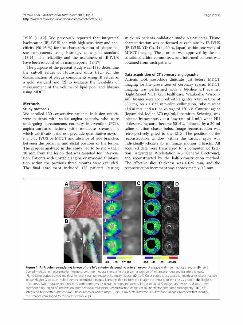
Figure 1 (A) A volume-rendering image of the left anterior descendinCurved multiplaner reconstruction image shows intermediate stenosis in th(Right) Color-coded curved multiplaner reconstruction image of coronary pimage. (Right) Gray-scale multiplaner reconstruction images. Numbers thatof interests (white square, 0.5 x 0.5 mm) with homogenous tissue componcorresponding region of interests on cross-sectional multiplaner reconstrucIntegrated backscatter intravascular ultrasound color-coded maps. (Right) Gthe images correspond to the cross-section in (B).
study: 45 patients, validation study: 80 patients). Tissuecharacterization was performed at each site by IB-IVUS(IB-IVUS, YD Co., Ltd., Nara, Japan) within one week ofMDCT imaging. The protocol was approved by the in-stitutional ethics committees, and informed consent wasobtained from each patient.
Data acquisition of CT coronary angiographyPatients took isosorbide dinitrate just before MDCTimaging for the prevention of coronary spasm. MDCTimaging was performed with a 64-slice CT scanner(Light Speed VCT, GE Healthcare, Waukesha, Wiscon-sin). Images were acquired with a gantry rotation time of350 ms, 64 x 0.625 mm-slice collimation, tube currentof 430 mA, and a tube voltage of 120 kV. Contrast agent(Iopamidol, Iodine 370 mg/ml, Iopamiron, Schering) wasinjected intravenously at a flow rate of 4 ml/s when HUof descending aorta became 50 HU, followed by a 30 mlsaline solution chaser bolus. Image reconstruction wasretrospectively gated to the ECG. The position of thereconstruction window within the cardiac cycle wasindividually chosen to minimize motion artifacts. Allacquired data were transferred to a computer worksta-tion (Advantage Workstation 4.3, General Electronic),and reconstructed by the half-reconstruction method.The effective slice thickness was 0.625 mm, and thereconstruction increment was approximately 0.5 mm.
-130 -49 -35 -29 dB
1
2
3
4
0 HU
C D
g artery (arrow). A plaque with intermediate stenosis. (B) (Left)e proximal portion of left anterior descending artery (arrow).laque. (C) (Left) Color-coded cross-sectional multiplaner reconstructionidentify the images correspond to the cross-section in (B). Regionsents were selected on IB-IVUS images, and were used to set thetion images of multidetector computed tomography. (D) (Left)ray-scale intravascular ultrasound images. Numbers that identify

Table 1 Patient Characteristics
Testing study(n = 45)
Validation study(n = 80)
Sex, n (%)
Men 36 (80) 68 (87)
Age, y 67 ± 8 69 ± 9
Body mass index, (kg/m2) 23.1 ± 3.5 23.4 ± 3.9
Heart rate, (beats/minute) 71 ± 11 68 ± 13
Clinical history, n (%)
Prior myocardial infarction 6 (13) 18 (14)
Hypertension 32 (71) 63 (80)
Dyslipidemia 16 (36) 35 (44)
Current smoker 5 (11) 16 (20)
Diabetes mellitus type 2 7 (16) 19 (24)
Medications, n (%)
Antiplatelet medication 45 (100) 80 (100)
Statin 15 (33) 33 (41)
Nirates 25 (56) 53 (66)
Calcium channel blockers 33 (73) 61 (76)
Beta-blockers 16 (36) 30 (38)
Insulin 3 (7) 11 (14)
ACE inhibitors or ARB 34 (76) 53 (66)
Laboratory parameters (mg/dl)
Total cholesterol 201 ± 29 193± 34
Triglycerides 147 ± 67 165± 99
HDL cholesterol 49 ± 12 45 ± 11
LDL cholesterol 125 ± 26 116± 25
HbA1c 6.2 ± 1.0 6.3 ± 1.1
Lesions, n (%)
Left anterior descending branch 21 (44) 32 (40)
Left circumflex branch 10 (22) 18 (23)
Right coronary artery 14 (31) 30 (37)
Values are mean ± SD. Numbers in parenthesis are percentage. SA: stableangina. ACS: acute coronary syndrome. ACE: Angiotensin converting enzyme.ARB: Angiotensin II receptor blockers. LDL: Low-density lipoprotein.HDL: High-density lipoprotein.
Yamaki et al. Cardiovascular Ultrasound 2012, 10:33 Page 3 of 8http://www.cardiovascularultrasound.com/content/10/1/33
Comparison between MDCT images and IB-IVUSimagesConventional IVUS images and ultrasound signals wereacquired using an IVUS imaging system (Clear View,Boston Scientific, MA) with a 40 MHz intravascularcatheter. During IVUS imaging, we administered anintra-coronary optimal dose of isosorbide dinitratebefore the measurements to prevent of coronary spasm.IB-IVUS images were captured at an interval of 0.5 mmusing a motorized pull-back system in each plaque. IBvalues were calculated as previously described and ourdefinition of IB values for each histological category wasdetermined by comparison with histological images asreported in our previous study [16]. In our previousreport, an IB value of ≤ −49 dB was the most reliablecutoff point for discriminating lipid pool (90 % sensitiv-ity, 92 % specificity) and fibrosis (94 % sensitivity, 93 %specificity) and an IB value of >−29 dB was the mostreliable cutoff point for discriminating calcification(95 % sensitivity, 99 % specificity) and fibrosis [16].To ensure that the same coronary sections were always
compared by IB-IVUS and MDCT, we used the distancefrom side branches and bifurcation points as referencemarkers based on the IVUS pullback rate of 0.5 mm/sec.Longitudinal reconstruction of IVUS and MDCT imageswas used to identify the same corresponding coronarysections (Figure 1). In the testing set (n = 45 lesions),we set a region of interest (ROI) (0.5 x 0.5 mm) ina homogenous tissue component in the reconstructedIB-IVUS images after determining the correspondingcross-sections (Figure 1). The region of interests (ROIs)selected in the reconstructed MDCT images were thesame as those selected in the IB-IVUS images, and theHU were compared with the IB values. The one observerindependently set ROIs on MDCT images that weresame lesions as IB-IVUS images, and another observerindependently measured HU of the ROIs.In the validation set (n = 80 lesions), lipid volume was
calculated using the cutoff value that was determined inthe testing study for discriminating lipid pool from fibro-sis by the following formula: lipid volume (mm3) =[plaque (intima + media) area 1 (mm2) x relativelipid area 1 (%) + plaque area 2 (mm2) x relative lipidarea 2 (%) + . . .. . .. + plaque area n (mm2) x relative lipidarea n (%)] / 100 x 0.5, where n is number of lesioncross-sections analyzed. Fibrous volume was calculatedin a similar manner. Calcification volume was also calcu-lated in a similar manner in 49 lesions.
Reproducibility and reliability of HU measurementWe determined interobserver variability of HU using 20randomly-selected cross-sections that were measured bytwo observers in a blinded way. The two observers inde-pendently set ROIs on MDCT images that were same
cross-sections as IB-IVUS images, and measured HU ofthe ROIs. Likewise, we determined intraobserver vari-ability of HU using 20 randomly-selected cross-sectionsthat were measured twice by one observer with a 7-daysinterval between the two measurements.
Statistical analysesContinuous values were expressed as the mean ± onestandard deviation. Categorical data were summarized aspercentages. Receiver operating characteristic (ROC)curves were used to determine the cutoff points for dif-ferentiating each tissue component by MDCT. Linear
Yamaki et al. Cardiovascular Ultrasound 2012, 10:33 Page 4 of 8http://www.cardiovascularultrasound.com/content/10/1/33
regression analysis was performed to determine the rela-tionship between the volume of lipid pool or fibrosismeasured by IB-IVUS and MDCT, and Bland-Altmanplots were constructed. Reclassification analysis was con-ducted by examining the net reclassification improve-ment (NRI) statistics [18]. A p-value of <0.05 wasconsidered statistically significant. Statistical analyseswere performed using Stat View version 5.0 (SAS Institu-tion Inc, Cary, NC, USA).
ResultsPatient characteristicsA total of 125 patients with stable angina underwent IB-IVUS analysis in non-target lesions without any compli-cations. All patients completed MDCT imaging usingcontrast medium (67 ± 4 mL) without any associatedmajor events or complications. There were no clinicalevents within the one week between IB-IVUS andMDCT. The patients’ characteristics in the testing andvalidation studies are shown in Table 1.
Reproducibility and reliability of HU measurementThe interobserver correlation coefficient and mean dif-ferences in HU were 0.97 and 1.3 ± 2.6 HU, respectively.The intraobserver correlation coefficient and mean dif-ferences in HU were 0.96 and 1.0 ± 2.0 HU, respectively.The inter-observer agreement of plaque components(lipid pool, fibrosis and calcification) by MDCT withoutcutoff values was 0.66.
Cut-off values for the discrimination of tissuecomponentsIn the testing set, a total of 150 homogeneous lesions(0.5 x 0.5 mm) in 45 coronary arteries were diagnosedfrom the IB-IVUS images as lipid pool (n = 50), fibrosis(n = 65) or calcification (n = 35) according to IB values.The HU in the corresponding ROIs (0.5 x 0.5 mm) for
80
0
40
120
-40
160
Lipid pool(n = 50)
18 ± 18 HU
Fibrosis(n = 65)
95 ± 24 HU
Ho
un
sfie
ld u
nit
(HU
)
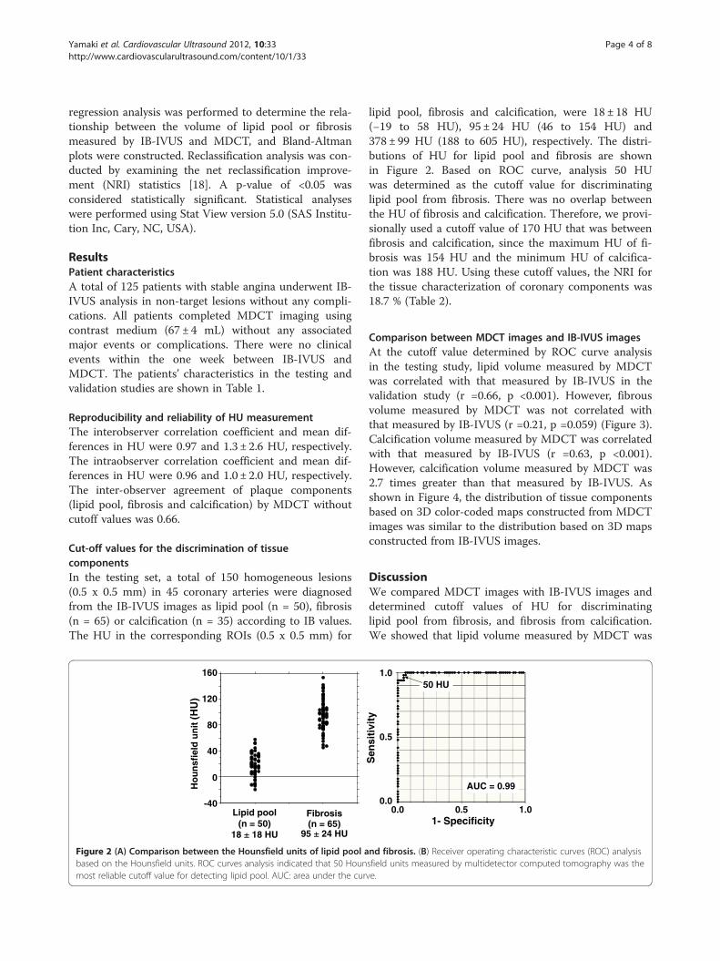
Figure 2 (A) Comparison between the Hounsfield units of lipid pool abased on the Hounsfield units. ROC curves analysis indicated that 50 Hounmost reliable cutoff value for detecting lipid pool. AUC: area under the cur
lipid pool, fibrosis and calcification, were 18 ± 18 HU(−19 to 58 HU), 95 ± 24 HU (46 to 154 HU) and378 ± 99 HU (188 to 605 HU), respectively. The distri-butions of HU for lipid pool and fibrosis are shownin Figure 2. Based on ROC curve, analysis 50 HUwas determined as the cutoff value for discriminatinglipid pool from fibrosis. There was no overlap betweenthe HU of fibrosis and calcification. Therefore, we provi-sionally used a cutoff value of 170 HU that was betweenfibrosis and calcification, since the maximum HU of fi-brosis was 154 HU and the minimum HU of calcifica-tion was 188 HU. Using these cutoff values, the NRI forthe tissue characterization of coronary components was18.7 % (Table 2).
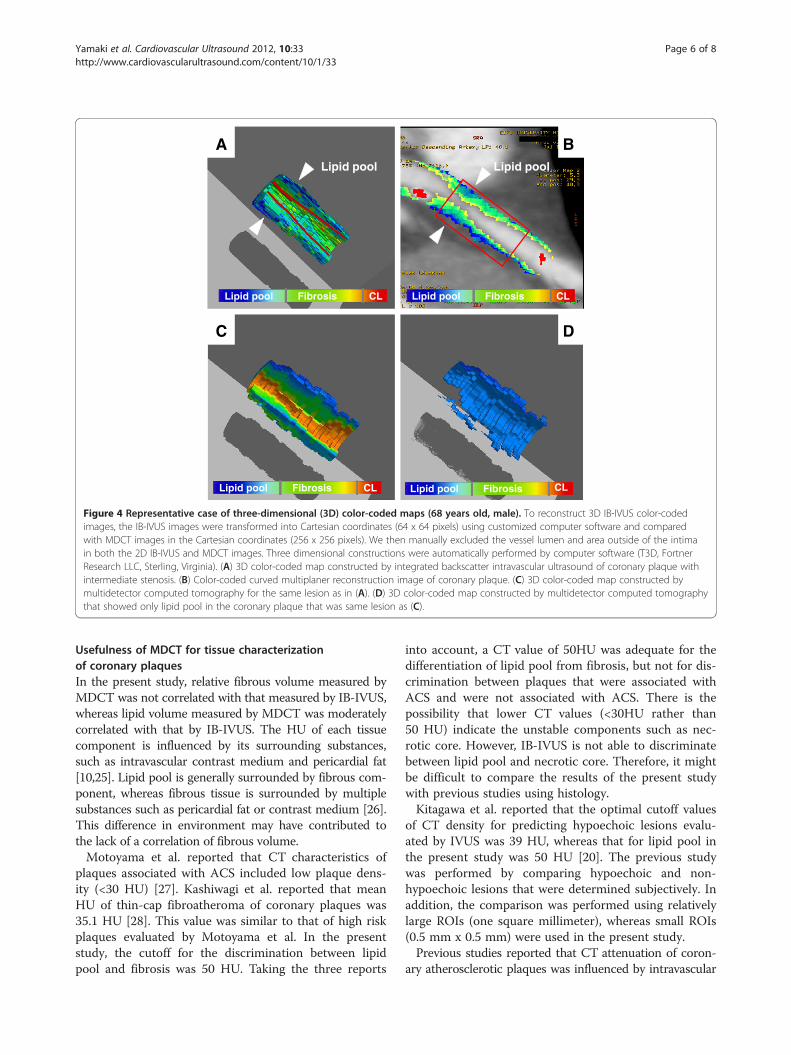
Comparison between MDCT images and IB-IVUS imagesAt the cutoff value determined by ROC curve analysisin the testing study, lipid volume measured by MDCTwas correlated with that measured by IB-IVUS in thevalidation study (r =0.66, p <0.001). However, fibrousvolume measured by MDCT was not correlated withthat measured by IB-IVUS (r =0.21, p =0.059) (Figure 3).Calcification volume measured by MDCT was correlatedwith that measured by IB-IVUS (r =0.63, p <0.001).However, calcification volume measured by MDCT was2.7 times greater than that measured by IB-IVUS. Asshown in Figure 4, the distribution of tissue componentsbased on 3D color-coded maps constructed from MDCTimages was similar to the distribution based on 3D mapsconstructed from IB-IVUS images.
DiscussionWe compared MDCT images with IB-IVUS images anddetermined cutoff values of HU for discriminatinglipid pool from fibrosis, and fibrosis from calcification.We showed that lipid volume measured by MDCT was
Sen
siti
vity
1- Specificity0.5 1.0
1.0
0.5
0.00.0
50 HU
AUC = 0.99
nd fibrosis. (B) Receiver operating characteristic curves (ROC) analysissfield units measured by multidetector computed tomography was theve.
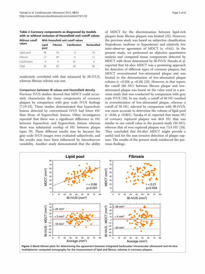
Table 2 Coronary components as diagnosed by modelswith or without inclusion of Hounsfield unit cutoff values
Without cutoffvalues
With Hounsfield unit cutoff values
Lipidpool
Fibrosis Calcification Reclassified
Lipid pool 38 16 0 16
Fibrosis 12 49 0 12
Calcification 0 0 35 0
Total 50 65 35 28
Yamaki et al. Cardiovascular Ultrasound 2012, 10:33 Page 5 of 8http://www.cardiovascularultrasound.com/content/10/1/33
moderately correlated with that measured by IB-IVUS,whereas fibrous volume was not.
Comparison between IB values and Hounsfield densityPrevious IVUS studies showed that MDCT could accur-ately characterize the tissue components of coronaryplaques by comparison with gray scale IVUS findings[7,19-23]. These studies demonstrated that hypoechoiclesions detected by conventional IVUS had lower HUthan those of hyperechoic lesions. Other investigatorsreported that there was a significant difference in HUbetween hypoechoic and hyperechoic lesions whereasthere was substantial overlap of HU between plaquetypes [9]. These different results may be because thegray scale IVUS images were evaluated subjectively, andthe results may have been influenced by interobservervariability. Another study demonstrated that the ability
0
10
20
30
40
50
60
70
80
0 10 20 30 40 50 60 70 80
loop dipiL
-50
-40
-30
-20
-10
0
10
20
30
40
50
0 10 20 30 40 50 60 70 80
+ 3 mm3
- 19 mm3
+ 25 mm3
r = 0.66p<0.001
IB-I
VU
S -
MD
CT
(m
m3 )
Average (mm3)
MD
CT
(m
m3 )
IB-IVUS (mm3)
Figure 3 Bland-Altman plots for determining the agreement betweenmultidetector computed tomography for the measurement of lipid an
of MDCT for the discrimination between lipid-richplaques from fibrous plaques was limited [22]. However,the previous study was based on subjective classification(hypodense, isodense or hyperdense) and relatively lowinter-observer agreement of MDCT (κ =0.61). In thepresent study, we performed an objective quantitativeanalysis and compared tissue components detected byMDCT with those determined by IB-IVUS. Harada et al.reported that 64-slice MDCT was a promising approachfor detection of different types of coronary plaques, butMDCT overestimated low-attenuated plaque and waslimited to the determination of low-attenuated plaquevolume (r =0.328, p =0.18) [23]. However, in that report,the cutoff (60 HU) between fibrous plaque and low-attenuated plaque was based on the value used in a pre-vious study that was conducted by comparison with grayscale IVUS [20]. In our study, a cutoff of 60 HU resultedin overestimation of low-attenuated plaque, whereas acutoff of 50 HU, selected by comparison with IB-IVUS,was more accurate to determine the volume of lipid pool(r =0.66, p <0.001). Tanaka et al. reported that mean HUof coronary ruptured plaques was 46.8 HU that wassimilar to our cutoff value in the present study (50 HU),whereas that of non-ruptured plaques was 73.4 HU [24].They concluded that 64-slice MDCT might provide auseful tool for the non-invasive detection of plaque rup-ture. The results of the present study reinforced the pre-vious findings.
sisorbiF
0
10
20
30
40
50
60
70
80
0 10 20 30 40 50 60 70 80
-50
-40
-30
-20
-10
0
10
20
30
40
50
0 10 20 30 40 50 60 70 80
+ 4 mm3
- 30 mm3
+ 38 mm3
r = 0.21p=0.059
IB-I
VU
S -
MD
CT
(m
m3 )
Average (mm3)
MD
CT
(m
m3 )
IB-IVUS (mm3)
integrated backscatter intravascular ultrasound and 64-sliced fibrous volumes in coronary plaques.
CLFibrosisLipid pool
Lipid pool
CLFibrosisLipid pool
Lipid pool
CLFibrosisLipid poolCLFibrosisLipid pool
A B
C D
Figure 4 Representative case of three-dimensional (3D) color-coded maps (68 years old, male). To reconstruct 3D IB-IVUS color-codedimages, the IB-IVUS images were transformed into Cartesian coordinates (64 x 64 pixels) using customized computer software and comparedwith MDCT images in the Cartesian coordinates (256 x 256 pixels). We then manually excluded the vessel lumen and area outside of the intimain both the 2D IB-IVUS and MDCT images. Three dimensional constructions were automatically performed by computer software (T3D, FortnerResearch LLC, Sterling, Virginia). (A) 3D color-coded map constructed by integrated backscatter intravascular ultrasound of coronary plaque withintermediate stenosis. (B) Color-coded curved multiplaner reconstruction image of coronary plaque. (C) 3D color-coded map constructed bymultidetector computed tomography for the same lesion as in (A). (D) 3D color-coded map constructed by multidetector computed tomographythat showed only lipid pool in the coronary plaque that was same lesion as (C).
Yamaki et al. Cardiovascular Ultrasound 2012, 10:33 Page 6 of 8http://www.cardiovascularultrasound.com/content/10/1/33
Usefulness of MDCT for tissue characterizationof coronary plaquesIn the present study, relative fibrous volume measured byMDCT was not correlated with that measured by IB-IVUS,whereas lipid volume measured by MDCT was moderatelycorrelated with that by IB-IVUS. The HU of each tissuecomponent is influenced by its surrounding substances,such as intravascular contrast medium and pericardial fat[10,25]. Lipid pool is generally surrounded by fibrous com-ponent, whereas fibrous tissue is surrounded by multiplesubstances such as pericardial fat or contrast medium [26].This difference in environment may have contributed tothe lack of a correlation of fibrous volume.Motoyama et al. reported that CT characteristics of
plaques associated with ACS included low plaque dens-ity (<30 HU) [27]. Kashiwagi et al. reported that meanHU of thin-cap fibroatheroma of coronary plaques was35.1 HU [28]. This value was similar to that of high riskplaques evaluated by Motoyama et al. In the presentstudy, the cutoff for the discrimination between lipidpool and fibrosis was 50 HU. Taking the three reports
into account, a CT value of 50HU was adequate for thedifferentiation of lipid pool from fibrosis, but not for dis-crimination between plaques that were associated withACS and were not associated with ACS. There is thepossibility that lower CT values (<30HU rather than50 HU) indicate the unstable components such as nec-rotic core. However, IB-IVUS is not able to discriminatebetween lipid pool and necrotic core. Therefore, it mightbe difficult to compare the results of the present studywith previous studies using histology.Kitagawa et al. reported that the optimal cutoff values
of CT density for predicting hypoechoic lesions evalu-ated by IVUS was 39 HU, whereas that for lipid pool inthe present study was 50 HU [20]. The previous studywas performed by comparing hypoechoic and non-hypoechoic lesions that were determined subjectively. Inaddition, the comparison was performed using relativelylarge ROIs (one square millimeter), whereas small ROIs(0.5 mm x 0.5 mm) were used in the present study.Previous studies reported that CT attenuation of coron-
ary atherosclerotic plaques was influenced by intravascular
Yamaki et al. Cardiovascular Ultrasound 2012, 10:33 Page 7 of 8http://www.cardiovascularultrasound.com/content/10/1/33
contrast medium [29,30]. CT values of coronary athero-sclerotic plaques increased in proportion to the incrementof CT values of the coronary lumen up to 250 HU. In con-trast, the CT values of coronary atherosclerotic plaqueswere relatively stable when the values of the coronarylumen were 250–400 HU [29]. The HU of the vessellumen in the present study was stable (326± 55 HU).However, the CT values of the coronary lumen areimportant when HU is used to characterize tissue compo-nents of coronary plaques.
Clinical implicationsWe previously reported that the relative volume of lipidpool could be determined using IB-IVUS imaging [13].However, IB-IVUS is invasive and can only be performedduring coronary catheterization, whereas MDCT is min-imally invasive and can be applied to the patients whowere suspected as angina pectoris. The volume of lipidpool in coronary atherosclerotic lesions can be calcu-lated using 3D color-coded maps constructed fromMDCT images. MDCT volumetric analysis of lipid poolmay be useful for clinical risk assessment and to provideincremental information on the effectiveness of medica-tions. It was reported that prediction of sudden cardiacdeath using measurement of coronary calcification byMDCT are distinct methods of assessing risk for suddencardiac death [31]. Measurement of lipid pool by MDCTmay be promising methods of assessing risk for coronaryartery disease.
Study limitationsThere are several limitations of the present study. First,calcification is a perfect reflector of ultrasound, causingacoustic shadowing that is typical in IVUS images. Theultrasound signals that are not able to penetrate or passthrough the calcified layer are reflected back towards thetransducer. Therefore, an accurate calculation of calci-fied area and volume is not possible using ultrasound.Likewise, a beam-hardening action by calcification thatis called “partial volume effect”, hinders rigorous evalu-ation of the calcified volume by MDCT. Second, weexcluded the area of the artifact due to the guidewirefrom the IB-IVUS analyses. Therefore, the guidewireartifact and calcification interfere with a rigorous calcu-lation of the area and volume of each component. Com-parison between IB values and HU of unstable plaquesincluding thin cap fibroatheroma in patients with acutecoronary syndrome is required. Third, square shapedROI (0.5 x 0.5 mm) might be too large to enclosehomogenous plaque components in human coronaryplaques. Improvement in resolution of MDCT would beexpected in the future. Finally, the IB-IVUS remains aresearch tool which does not have clinical utility. Theuse of IB values that was surrogate as the gold standard
did not necessarily translate into an accurate analysis ofplaque components.
ConclusionsUsing the IB values as a gold standard, lipid volume mea-sured by MDCT was moderately correlated with thatmeasured by IB-IVUS. MDCT may be useful for volume-tric assessment of the lipid volume of coronary plaques,whereas the assessment of fibrosis volume was unstable.
AbbreviationsACS: Acute coronary syndrome; MDCT: Multi-detector raw computedtomography; IVUS: Intravascular ultrasound; IB: Integrated backscatter;2D: Two-dimensional; HU: Hounsfield units; PCI: Percutaneous coronaryintervention; ROI: Region of interest; ROC: Receiver operating characteristic;3D: Three-dimensional.
Competing interestsThe authors declare that they have no competing interests.
Authors’ contributionsTY, OCR and IKJ carried out subject recruitment and analyzed data. MKanalyzed data and wrote the manuscript. YI, MO, and TK performedintegrated backscatter ultrasound analysis. HF revised manuscript. AH, KN, GTand SM analyzed data. All authors read and approved the final manuscript.We have no financial or other relations that could lead to conflict of interest.
AcknowledgementsThis study was supported, in part, by a Grant-in-Aid for Scientific Research(C), Ministry of Education, Culture, Sports, Science and Technology of Japan(20590822) (2008–2010).
Author details1Department of Cardiology, Gifu University Graduate School of Medicine, 1-1Yanagido, Gifu 501-1194, Japan. 2Cardiology Division, Massachusetts GeneralHospital and Harvard Medical School, Boston, MA, USA.
Received: 1 July 2012 Accepted: 29 July 2012Published: 6 August 2012
References1. Ehara M, Surmely JF, Kawai M, Katoh O, Matsubara T, Terashima M,
Tsuchikane E, Kinoshita Y, Suzuki T, Ito T, Takeda Y, Nasu K, Tanaka N,Murata A, Suzuki Y, Sato K, Suzuki T: Diagnostic accuracy of 64-slicecomputed tomography for detecting angiographically significantcoronary artery stenosis in an unselected consecutive patientpopulation: comparison with conventional invasive angiography. Circ J2006, 70:564–571.
2. Hoffmann U, Moselewski F, Cury RC, Ferencik M, Jang IK, Diaz LJ, Abbara S,Brady TJ, Achenbach S: Predictive value of 16-slice multidetector spiralcomputed tomography to detect significant obstructive coronary arterydisease in patients at high risk for coronary artery disease: patient-versus segment-based analysis. Circulation 2004, 110:2638–2643.
3. Moselewski F, Ropers D, Pohle K, Hoffmann U, Ferencik M, Chan RC,Cury RC, Abbara S, Jang IK, Brady TJ, Daniel WG, Achenbach S:Measurement of cross-sectional coronary atherosclerotic plaques andvessel area by 16-slice multi-detector CT: Comparison to IVUS. Am JCardiol 2004, 94:1294–1297.
4. Hoffmann U, Moselewski F, Nieman K, Jang IK, Ferencik M, Rahman AM,Cury RC, Abbara S, Joneidi-Jafari H, Achenbach S, Brady TJ: Noninvasiveassessment of plaque morphology and composition in culprit and stablelesions in acute coronary syndrome and stable lesions in stable anginaby multidetector computed tomography. J Am Coll Cardiol 2006,47:1655–1662.
5. Motoyama S, Anno H, Sarai M, Sato T, Sanda Y, Ozaki Y, Mochizuki T,Katada K, Hishida H: Noninvasive coronary angiography with a prototype256-row area detector computed tomography system: comparison withconventional invasive coronary angiography. J Am Coll Cardiol 2008,51:773–775.
Yamaki et al. Cardiovascular Ultrasound 2012, 10:33 Page 8 of 8http://www.cardiovascularultrasound.com/content/10/1/33
6. Toepker M, Schlett CL, Irlbeck T, Mahabadi AA, Bamberg F, Leidecker C,Donnelly P, Hoffmann U: Accuracy of dual-source computed tomographyin quantitative assessment of low density coronary stenosis. a motionphantom study. Eur Radiol 2010, 20:542–548.
7. Leber AW, Knez A, Becker A, Becker C, von Ziegler F, Nikolaou K, Rist C,Reiser M, White C, Steinbeck G, Boekstegers P: Accuracy of multidetectorspiral computed tomography in idetifing and differentiating thecomposition of coronary atherosclerotic plaques. A comparative studywith intracoronary ultrasound. J Am Coll Cardiol 2004, 43:1241–1247.
8. Komatsu S, Imai A, Kodama K: Multidetector row computed tomographymay accurately estimate plaque vulnerability. Circ J 2011, 74:1515–1521.
9. Pohle K, Achenbach S, MacNeill B, Ropers D, Ferencik M, Moselewski F,Hoffmann U, Brady TJ, Jang IK, Daniel WG: Characterization of non-calcified coronary atherosclerotic plaque by multi-detector row CT:Comparison to IVUS. Atherosclerosis 2006, 190:174–180.
10. Higashi M: Noninvasive assessment of coronary plaque usingmultidetector row computed tomography. Does MDCT accuratelyestimate plaque vulnerability? (Con). Circ J 2011, 75:1522–1528.
11. Schaar JA, De Korte CL, Mastik F, Strijder C, Pasterkamp G, Boersma E,Serruys PW, Van Der Steen AF: Characterizing vulnerable plaque featureswith intravascular elastography. Circulation 2003, 108:2636–2641.
12. Nair A, Kuban BD, Tuzcu EM, Schoenhagen P, Nissen SE, Vince DG: Coronaryplaque classification with intravascular ultrasound radiofrequency dataanalysis. Circulation 2002, 106:2200–2206.
13. Kawasaki M, Sano K, Okubo M, Yokoyama H, Ito Y, Murata I, Tsuchiya K,Minatoguchi S, Zhou X, Fujita H, Fujiwara H: Volumetric quantitativeanalysis of tissue characteristics of coronary plaques after statin therapyusing three dimensional integrated backscatter intravascular ultrasound.J Am Coll Cardiol 2005, 45:1946–1953.
14. Kawasaki M, Takatsu H, Noda T, Sano K, Ito Y, Hayakawa K, Tsuchiya K,Arai M, Nishigaki K, Takemura G, Minatoguchi S, Fujiwara T, Fujiwara H:In vivo quantitative tissue characterization of human coronary arterialplaques by use of integrated backscatter intravascular ultrasound andcomparison with angioscopic findings. Circulation 2002, 105:2487–2492.
15. Sano K, Kawasaki M, Ishihara Y, Okubo M, Tsuchiya K, Nishigaki K, Zhou X,Minatoguchi S, Fujita H, Fujiwara H: Assessment of vulnerable plaquescausing acute coronary syndrome using integrated backscatterintravascular ultrasound. J Am Coll Cardiol 2006, 47:734–741.
16. Okubo M, Kawasaki M, Ishihara Y, Takeyama U, Kubota T, Yamaki T, Ojio S,Nishigaki K, Takemura G, Saio M, Takami T, Minatoguchi S, Fujiwara H:Development of integrated backscatter intravascular ultrasound fortissue characterization of coronary plaques. Ultrasound Med Biol 2008,34:655–663.
17. Okubo M, Kawasaki M, Ishihara Y, Takeyama U, Yasuda S, Kubota T, Tanaka S,Yamaki T, Ojio S, Nishigaki K, Takemura G, Saio M, Takami T, Fujiwara H:Tissue characterization of coronary plaques. Comparison of integratedbackscatter intravascular ultrasound with Virtual Histology intravascularultrasound. Circ J 2008, 72:1631–1639.
18. Pencina MJ, D'Agostino RB Sr, D'Agostino RB Jr, Vasan RS: Evaluating theadded predictive ability of a new marker: from area under the ROCcurve to reclassification and beyond. Stat Med 2008, 27:157–172.
19. Motoyama S, Kondo T, Anno H, Sugiura A, Ito Y, Mori K, Ishii J, Sato T, InoueK, Sarai M, Hishida H, Narula J: Atherosclerotic plaque characterization by0.5-mm-slice multislice computed tomographic imaging. Circ J 2007,71:363–366.
20. Schroeder S, Kopp AF, Baumbach A, Meisner C, Kuettner A, Georg C,Ohnesorge B, Herdeg C, Claussen CD, Karsch KR: Noninvasive detectionand evaluation of atherosclerotic coronary plaques with multislicecomputed tomography. J Am Coll Cardiol 2001, 37:1430–1435.
21. Kitagawa T, Yamamoto H, Ohhashi N, Okimoto T, Horiguchi J, Hirai N, Ito K,Kohno N: Comprehensive evaluation of noncalcified coronary plaquecharacteristics detected using 64-slice computed tomography inpatients with proven or suspected coronary artery disease. Am Heart J2007, 154:1191–1198.
22. Chopard R, Boussel L, Motreff P, Rioufol G, Tabib A, Douek P, Meyronet D,Revel D, Finet G: How reliable are 40 MHz IVUS and 64-slice MDCT incharacterizing coronary plaque composition? An ex vivo study withhistopathological comparison. Int J Cardiovasc Imaging 2010, 26:373–383.
23. Harada K, Amano T, Uetani T, Funahashi H, Arai K, Okada K, Hirashiki A,Hayashi M, Oshima S, Ishii H, Izawa H, Matsubara T, Murohara T: Accuracy of64-slice multidetector computed tomography for classification and
quantitation of coronary plaque: Comparison with integratedbackscatter intravascular ultrasound. Int J Cardiol 2011, 149:95–101.
24. Tanaka A, Shimada K, Yoshida K, Jissyo S, Tanaka H, Sakamoto M, Matsuba K,Imanishi T, Akasaka T, Yoshikawa J: Non-invasive assessment of plaquerupture by 64-slice multidetector computed tomography-comparisonwith intravascular ultrasound. Circ J 2008, 72:1276–1281.
25. Cademartiri F, Runza G, Mollet NR, Luccichenti G, Belgrano M, Somers P,Knaapen M, Verheye S, Bruining N, Hamers R, Midiri M, De Feyter PJ,Krestin GP: Influence of increasing convolution kernel filtering on plaqueimaging with multislice CT using an ex-vivo model of coronaryangiography. Radiol Med 2005, 110:234–240.
26. Stary HC, Chandler AB, Dinsmore RE, Fuster V, Glagov S, Insull W Jr,Rosenfeld ME, Schwartz CJ, Wagner WD, Wissler RW: A definition ofadvanced types of atherosclerotic lesions and a histological classificationof atherosclerosis. A report from the Committee on Vascular Lesions ofthe Council on Arteriosclerosis, American Heart Association. Circulation1995, 92:1355–1374.
27. Motoyama S, Kondo T, Sarai M, Sugiura A, Harigaya H, Sato T, Inoue K,Okumura M, Ishii J, Anno H, Virmani R, Ozaki Y, Hishida H, Narula J:Multislice computed tomographic characteristics of coronary lesions inacute coronary syndromes. J Am Coll Cardiol 2007, 50:319–326.
28. Kashiwagi M, Tanaka A, Kitabata H, Tsujioka H, Kataiwa H, Komukai K,Tanimoto T, Takemoto K, Takarada S, Kubo T, Hirata K, Nakamura N,Mizukoshi M, Imanishi T, Akasaka T: Feasibility of non-invasive assessmentof thin-cap fibroatheroma by multidetector computed tomography.JACC Cardiovasc Imaging 2009, 12:1412–1419.
29. Cademartiri F, Mollet NR, Runza G, Bruining N, Hamers R, Somers P,Knaapen M, Verheye S, Midiri M, Krestin GP, de Feyter PJ: Influence ofintracoronary attenuation on coronary plaque measurements usingmultislice computed tomography: Observations in an ex vivo model ofcoronary computed tomography angiography. Eur Radiol 2005,15:1426–1431.
30. Halliburton SS, Schoenhagen P, Nair A, Stillman A, Lieber M: Murat Tuzcu E,Geoffrey Vince D, White RD: Contrast enhancement of coronaryatherosclerotic plaque: a high-resolution, multidetector-row computedtomography study of pressure-perfused, human ex-vivo coronaryarteries. Coron Artery Dis 2006, 176:553–560.
31. Taylor AJ, Burke AP, O'Malley PG, Farb A, Malcom GT, Smialek J, Virmani R: AComparison of the Framingham Risk Index, Coronary Artery Calcification,and Culprit Plaque Morphology in Sudden Cardiac Death. Circulation2000, 101:1243–1248.
doi:10.1186/1476-7120-10-33Cite this article as: Yamaki et al.: Comparison between integratedbackscatter intravascular ultrasound and 64-slicemulti-detector row computed tomography for tissue characterizationand volumetric assessment of coronary plaques. CardiovascularUltrasound 2012 10:33.
Submit your next manuscript to BioMed Centraland take full advantage of:
• Convenient online submission
• Thorough peer review
• No space constraints or color figure charges
• Immediate publication on acceptance
• Inclusion in PubMed, CAS, Scopus and Google Scholar
• Research which is freely available for redistribution
Submit your manuscript at www.biomedcentral.com/submit