Comparative utility of diagnostic bone-marrow components: A 10-year study
5
Comparative Utility of Diagnostic Bone-Marrow Components: A 10-Year Study Carol L. Barekman, 1 * Kevaghn P. Fair, 1 and James D. Cotelingam 2 1 Section of Hematology, Brooke Army Medical Center, San Antonio, Texas 2 Hematopathology Branch, National Naval Medical Center, Bethesda, Maryland Ten years of cumulative experience represented by 4,902 consecutive diagnostic bone- marrow examinations at a tertiary care and referral center were reviewed to assess the value of specific components. While it has been shown previously that the information obtained from each component is generally complementary, the inclusion of some or all components may vary between institutions. The components studied included aspirate smears, clot sections, biopsy cores, and touch imprints of biopsy and clot sections. Three clinical presentations accounted for the majority of cases: staging for carcinoma or lymphoma, cytopenias, and acute leukemia. We conclude that bilateral aspirates with biopsies are required for diagnosis in staging of neoplasms and that a unilateral aspirate with biopsy is sufficient to assess patients with cytopenia or leukemia. Only rarely were touch imprints of biopsy cores necessary to establish a diagnosis; however, their early availability prior to examining sections of the clot and core did provide immediate infor- mation, when positive, in the staging of patients with carcinoma. In a small percentage of staging and leukemia cases the diagnosis rested with the clot section alone. The findings in this study address common assumptions associated with routine diagnostic hematol- ogy and oncology procedures, and are important to both clinicians and pathologists concerned with accuracy, quality assurance, turnaround time, and cost containment. Am. J. Hematol. 56:37–41, 1997. © 1997 Wiley-Liss, Inc. Key words: bone marrow; diagnosis; components; utility INTRODUCTION It is the generally-accepted standard of practice to evaluate several components in the process of bone- marrow examination, although among different facilities the elements which are specifically included or excluded may vary. At our institution, aspirate particle smears, touch imprints of the aspirate clot and biopsy cores, and paraffin sections of the clot and biopsy are routinely examined in each instance. While such a thorough ap- proach has been advocated by some [1], other emphasize specific components for selected diagnoses [2,3]. We performed a retrospective review of 4,902 consecutive cases evaluated over a 10-year period to determine which components were useful or necessary to establish a di- agnosis. MATERIALS AND METHODS Four thousand, nine hundred and two bone-marrow procedures were performed between January 1, 1984– December 31, 1993 at the National Naval Medical Center (Bethesda, MD). The procedures were done under local anesthesia using an 8- or 11-gauge Jamshidi like needle (Medical Procedures Inc., Baltimore, MD) after obtain- ing informed consent. A portion of the aspirate was spread onto glass slides for staining with May-Gru ¨nwald- Giemsa and Prussian blue stains, and the remainder was saved for clot section. Touch imprints of the clot and biopsy (both sides if performed bilaterally) were made prior to processing. The core biopsies, which on average each measured 1.5 cm in length, were fixed in 10% for- malin, postfixed in B-5, decalcified in 10% aqueous ni- The opinions or assertions contained herein are the views of the au- thors and should not be construed as the official views of the Depart- ment of the Army, Department of the Navy, or Department of Defense. *Correspondence to: Maj. Carol L. Barekman, D.P.A.L.S., Brooke Army Medical Center, Ft. Sam Houston, TX 78234-6200. Received for publication 5 May 1997; Accepted 7 May 1997. American Journal of Hematology 56:37–41 (1997) © 1997 Wiley-Liss, Inc.
Transcript of Comparative utility of diagnostic bone-marrow components: A 10-year study

Comparative Utility of Diagnostic Bone-MarrowComponents: A 10-Year Study
Carol L. Barekman, 1* Kevaghn P. Fair, 1 and James D. Cotelingam 2
1Section of Hematology, Brooke Army Medical Center, San Antonio, Texas2Hematopathology Branch, National Naval Medical Center, Bethesda, Maryland
Ten years of cumulative experience represented by 4,902 consecutive diagnostic bone-marrow examinations at a tertiary care and referral center were reviewed to assess thevalue of specific components. While it has been shown previously that the informationobtained from each component is generally complementary, the inclusion of some or allcomponents may vary between institutions. The components studied included aspiratesmears, clot sections, biopsy cores, and touch imprints of biopsy and clot sections.Three clinical presentations accounted for the majority of cases: staging for carcinoma orlymphoma, cytopenias, and acute leukemia. We conclude that bilateral aspirates withbiopsies are required for diagnosis in staging of neoplasms and that a unilateral aspiratewith biopsy is sufficient to assess patients with cytopenia or leukemia. Only rarely weretouch imprints of biopsy cores necessary to establish a diagnosis; however, their earlyavailability prior to examining sections of the clot and core did provide immediate infor-mation, when positive, in the staging of patients with carcinoma. In a small percentage ofstaging and leukemia cases the diagnosis rested with the clot section alone. The findingsin this study address common assumptions associated with routine diagnostic hematol-ogy and oncology procedures, and are important to both clinicians and pathologistsconcerned with accuracy, quality assurance, turnaround time, and cost containment. Am.J. Hematol. 56:37–41, 1997. © 1997 Wiley-Liss, Inc.
Key words: bone marrow; diagnosis; components; utility
INTRODUCTION
It is the generally-accepted standard of practice toevaluate several components in the process of bone-marrow examination, although among different facilitiesthe elements which are specifically included or excludedmay vary. At our institution, aspirate particle smears,touch imprints of the aspirate clot and biopsy cores, andparaffin sections of the clot and biopsy are routinelyexamined in each instance. While such a thorough ap-proach has been advocated by some [1], other emphasizespecific components for selected diagnoses [2,3]. Weperformed a retrospective review of 4,902 consecutivecases evaluated over a 10-year period to determine whichcomponents were useful or necessary to establish a di-agnosis.
MATERIALS AND METHODS
Four thousand, nine hundred and two bone-marrowprocedures were performed between January 1, 1984–
December 31, 1993 at the National Naval Medical Center(Bethesda, MD). The procedures were done under localanesthesia using an 8- or 11-gauge Jamshidi like needle(Medical Procedures Inc., Baltimore, MD) after obtain-ing informed consent. A portion of the aspirate wasspread onto glass slides for staining with May-Gru¨nwald-Giemsa and Prussian blue stains, and the remainder wassaved for clot section. Touch imprints of the clot andbiopsy (both sides if performed bilaterally) were madeprior to processing. The core biopsies, which on averageeach measured 1.5 cm in length, were fixed in 10% for-malin, postfixed in B-5, decalcified in 10% aqueous ni-
The opinions or assertions contained herein are the views of the au-thors and should not be construed as the official views of the Depart-ment of the Army, Department of the Navy, or Department of Defense.
*Correspondence to: Maj. Carol L. Barekman, D.P.A.L.S., BrookeArmy Medical Center, Ft. Sam Houston, TX 78234-6200.
Received for publication 5 May 1997; Accepted 7 May 1997.
American Journal of Hematology 56:37–41 (1997)
© 1997 Wiley-Liss, Inc.
tric acid, embedded in paraffin, sectioned, and stainedwith hematoxylin and eosin. Additional special, cyto-chemical, and immunohistochemical stains on aspirate orbiopsy material were performed as indicated. Althoughaspirate material was occasionally submitted for specialstudies such as tissue culture, cytogenetics, or flow cy-tometry, these results were not considered in this study.The aspirate and biopsy touch imprints were examinedon the first day of the procedure and a diagnosis wasrendered, if possible, to expedite patient care. The clotsection, biopsy core, and iron stain of the aspirate andclot section were examined on day 2.
RESULTS
The total number of cases reviewed was 4,902, includ-ing 4,235 HIV (−) adults, 160 HIV (+) adults, and 512pediatric patients. Of the HIV (−) adults, 3,118 (74%)accounted for three presentations: staging for carcinomaor lymphoma, posttreatment follow-up for acute leuke-mia, and cytopenia. The remaining 1,117 (26%) caseswere a heterogeneous group, including 739 evaluated forclinically suspected myelodysplasia, acute or chronicleukemia, myeloproliferative disease, or immunoprolif-erative disease, and 378 for follow-up of such disorders(excluding acute leukemia). The pediatric cases were ex-cluded from this review, since biopsies in addition toaspirate were not consistently performed.
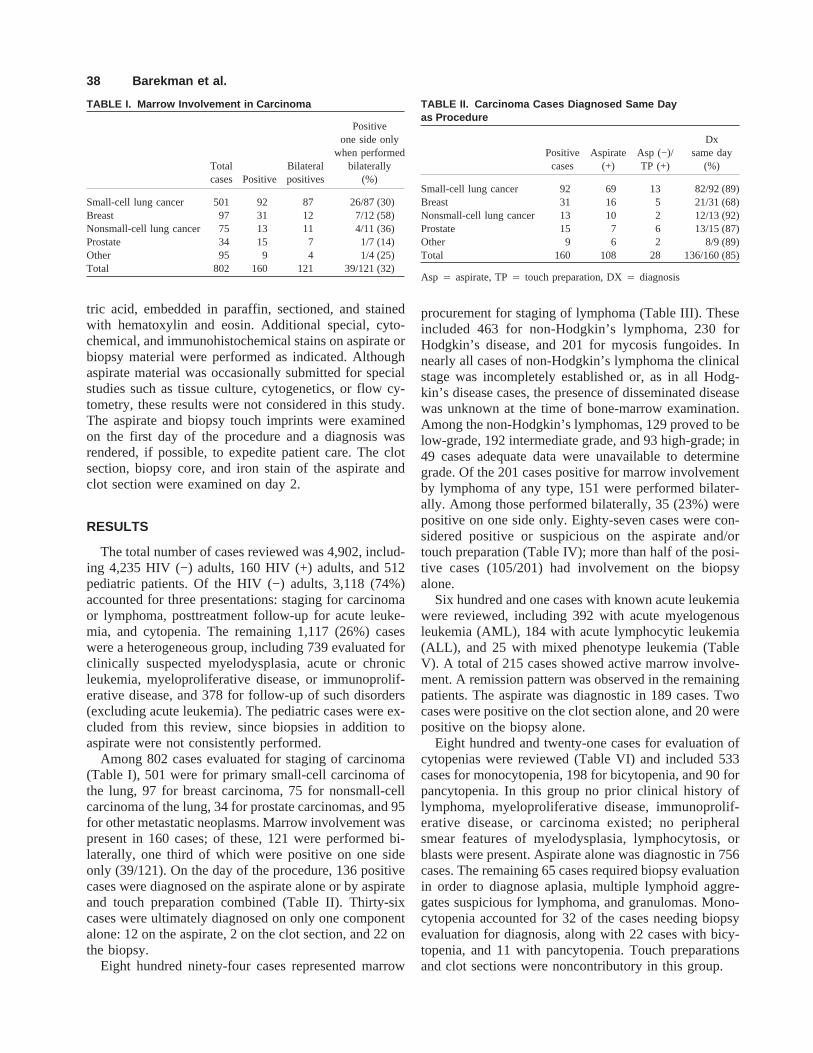
Among 802 cases evaluated for staging of carcinoma(Table I), 501 were for primary small-cell carcinoma ofthe lung, 97 for breast carcinoma, 75 for nonsmall-cellcarcinoma of the lung, 34 for prostate carcinomas, and 95for other metastatic neoplasms. Marrow involvement waspresent in 160 cases; of these, 121 were performed bi-laterally, one third of which were positive on one sideonly (39/121). On the day of the procedure, 136 positivecases were diagnosed on the aspirate alone or by aspirateand touch preparation combined (Table II). Thirty-sixcases were ultimately diagnosed on only one componentalone: 12 on the aspirate, 2 on the clot section, and 22 onthe biopsy.
Eight hundred ninety-four cases represented marrow
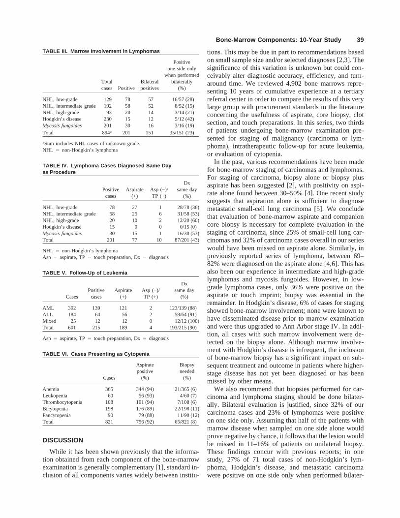
procurement for staging of lymphoma (Table III). Theseincluded 463 for non-Hodgkin’s lymphoma, 230 forHodgkin’s disease, and 201 for mycosis fungoides. Innearly all cases of non-Hodgkin’s lymphoma the clinicalstage was incompletely established or, as in all Hodg-kin’s disease cases, the presence of disseminated diseasewas unknown at the time of bone-marrow examination.Among the non-Hodgkin’s lymphomas, 129 proved to below-grade, 192 intermediate grade, and 93 high-grade; in49 cases adequate data were unavailable to determinegrade. Of the 201 cases positive for marrow involvementby lymphoma of any type, 151 were performed bilater-ally. Among those performed bilaterally, 35 (23%) werepositive on one side only. Eighty-seven cases were con-sidered positive or suspicious on the aspirate and/ortouch preparation (Table IV); more than half of the posi-tive cases (105/201) had involvement on the biopsyalone.
Six hundred and one cases with known acute leukemiawere reviewed, including 392 with acute myelogenousleukemia (AML), 184 with acute lymphocytic leukemia(ALL), and 25 with mixed phenotype leukemia (TableV). A total of 215 cases showed active marrow involve-ment. A remission pattern was observed in the remainingpatients. The aspirate was diagnostic in 189 cases. Twocases were positive on the clot section alone, and 20 werepositive on the biopsy alone.
Eight hundred and twenty-one cases for evaluation ofcytopenias were reviewed (Table VI) and included 533cases for monocytopenia, 198 for bicytopenia, and 90 forpancytopenia. In this group no prior clinical history oflymphoma, myeloproliferative disease, immunoprolif-erative disease, or carcinoma existed; no peripheralsmear features of myelodysplasia, lymphocytosis, orblasts were present. Aspirate alone was diagnostic in 756cases. The remaining 65 cases required biopsy evaluationin order to diagnose aplasia, multiple lymphoid aggre-gates suspicious for lymphoma, and granulomas. Mono-cytopenia accounted for 32 of the cases needing biopsyevaluation for diagnosis, along with 22 cases with bicy-topenia, and 11 with pancytopenia. Touch preparationsand clot sections were noncontributory in this group.
TABLE I. Marrow Involvement in Carcinoma
Totalcases Positive
Bilateralpositives
Positiveone side only
when performedbilaterally
(%)
Small-cell lung cancer 501 92 87 26/87 (30)Breast 97 31 12 7/12 (58)Nonsmall-cell lung cancer 75 13 11 4/11 (36)Prostate 34 15 7 1/7 (14)Other 95 9 4 1/4 (25)Total 802 160 121 39/121 (32)
TABLE II. Carcinoma Cases Diagnosed Same Dayas Procedure
Positivecases
Aspirate(+)
Asp (−)/TP (+)
Dxsame day
(%)
Small-cell lung cancer 92 69 13 82/92 (89)Breast 31 16 5 21/31 (68)Nonsmall-cell lung cancer 13 10 2 12/13 (92)Prostate 15 7 6 13/15 (87)Other 9 6 2 8/9 (89)Total 160 108 28 136/160 (85)
Asp 4 aspirate, TP4 touch preparation, DX4 diagnosis
38 Barekman et al.
DISCUSSION
While it has been shown previously that the informa-tion obtained from each component of the bone-marrowexamination is generally complementary [1], standard in-clusion of all components varies widely between institu-
tions. This may be due in part to recommendations basedon small sample size and/or selected diagnoses [2,3]. Thesignificance of this variation is unknown but could con-ceivably alter diagnostic accuracy, efficiency, and turn-around time. We reviewed 4,902 bone marrows repre-senting 10 years of cumulative experience at a tertiaryreferral center in order to compare the results of this verylarge group with procurement standards in the literatureconcerning the usefulness of aspirate, core biopsy, clotsection, and touch preparations. In this series, two thirdsof patients undergoing bone-marrow examination pre-sented for staging of malignancy (carcinoma or lym-phoma), intratherapeutic follow-up for acute leukemia,or evaluation of cytopenia.
In the past, various recommendations have been madefor bone-marrow staging of carcinomas and lymphomas.For staging of carcinoma, biopsy alone or biopsy plusaspirate has been suggested [2], with positivity on aspi-rate alone found between 30–50% [4]. One recent studysuggests that aspiration alone is sufficient to diagnosemetastatic small-cell lung carcinoma [5]. We concludethat evaluation of bone-marrow aspirate and companioncore biopsy is necessary for complete evaluation in thestaging of carcinoma, since 25% of small-cell lung car-cinomas and 32% of carcinoma cases overall in our serieswould have been missed on aspirate alone. Similarly, inpreviously reported series of lymphoma, between 69–82% were diagnosed on the aspirate alone [4,6]. This hasalso been our experience in intermediate and high-gradelymphomas and mycosis fungoides. However, in low-grade lymphoma cases, only 36% were positive on theaspirate or touch imprint; biopsy was essential in theremainder. In Hodgkin’s disease, 6% of cases for stagingshowed bone-marrow involvement; none were known tohave disseminated disease prior to marrow examinationand were thus upgraded to Ann Arbor stage IV. In addi-tion, all cases with such marrow involvement were de-tected on the biopsy alone. Although marrow involve-ment with Hodgkin’s disease is infrequent, the inclusionof bone-marrow biopsy has a significant impact on sub-sequent treatment and outcome in patients where higher-stage disease has not yet been diagnosed or has beenmissed by other means.
We also recommend that biopsies performed for car-cinoma and lymphoma staging should be done bilater-ally. Bilateral evaluation is justified, since 32% of ourcarcinoma cases and 23% of lymphomas were positiveon one side only. Assuming that half of the patients withmarrow disease when sampled on one side alone wouldprove negative by chance, it follows that the lesion wouldbe missed in 11–16% of patients on unilateral biopsy.These findings concur with previous reports; in onestudy, 27% of 71 total cases of non-Hodgkin’s lym-phoma, Hodgkin’s disease, and metastatic carcinomawere positive on one side only when performed bilater-
TABLE III. Marrow Involvement in Lymphomas
Totalcases Positive
Bilateralpositives
Positiveone side only
when performedbilaterally
(%)
NHL, low-grade 129 78 57 16/57 (28)NHL, intermediate grade 192 58 52 8/52 (15)NHL, high-grade 93 20 14 3/14 (21)Hodgkin’s disease 230 15 12 5/12 (42)Mycosis fungoides 201 30 16 3/16 (19)Total 894a 201 151 35/151 (23)
aSum includes NHL cases of unknown grade.NHL 4 non-Hodgkin’s lymphoma
TABLE IV. Lymphoma Cases Diagnosed Same Dayas Procedure
Positivecases
Aspirate(+)
Asp (−)/TP (+)
Dxsame day
(%)
NHL, low-grade 78 27 1 28/78 (36)NHL, intermediate grade 58 25 6 31/58 (53)NHL, high-grade 20 10 2 12/20 (60)Hodgkin’s disease 15 0 0 0/15 (0)Mycosis fungoides 30 15 1 16/30 (53)Total 201 77 10 87/201 (43)
NHL 4 non-Hodgkin’s lymphomaAsp 4 aspirate, TP4 touch preparation, Dx4 diagnosis
TABLE V. Follow-Up of Leukemia
CasesPositive
casesAspirate
(+)Asp (−)/TP (+)
Dxsame day
(%)
AML 392 139 121 2 123/139 (88)ALL 184 64 56 2 58/64 (91)Mixed 25 12 12 0 12/12 (100)Total 601 215 189 4 193/215 (90)
Asp 4 aspirate, TP4 touch preparation, Dx4 diagnosis
TABLE VI. Cases Presenting as Cytopenia
Cases
Aspiratepositive
(%)
Biopsyneeded
(%)
Anemia 365 344 (94) 21/365 (6)Leukopenia 60 56 (93) 4/60 (7)Thrombocytopenia 108 101 (94) 7/108 (6)Bicytopenia 198 176 (89) 22/198 (11)Pancytopenia 90 79 (88) 11/90 (12)Total 821 756 (92) 65/821 (8)
Bone-Marrow Components: 10-Year Study 39
ally [7], while in another report 30% of 99 cases ofnon-Hodgkin’s lymphoma were positive unilaterally [8].The relative ease of performing an additional biopsy, aswell as the potential savings in time and expense overother staging procedures, renders the effort justifiable.
Although we observed that aspirates and touch prepa-rations were not entirely complementary in positivecases, their earlier availability allowed confirmation ofbone-marrow involvement 1 day sooner in 85% of car-cinomas. The importance of obtaining an aspirate is fur-ther supported in that 9% of cases which were positivefor metastatic disease were observed in the aspiratealone. We also found that the inclusion of touch imprintsincreased the positive yield in carcinoma cases over as-pirate alone by 17%, although our data do not allow us todraw any conclusions concerning clot section vs. biopsycore. In contrast to previous reports concerning lym-phoma [4,6], aspirates and touch preparations were di-agnostic in less than half (43%) of our cases, highlightingthe limited utility of these components without a comple-mentary biopsy. The latter allowed detection of lymphoidaggregates, and their number and location, as well asserving as a source for immunohistochemistry, featurespreviously recognized in definitive diagnosis [8,9–11].No gain in turnaround time was afforded by the aspirateor touch preparations such as was seen in carcinomastaging, and only 2 cases of lymphoma were diagnosedon the aspirate alone. Finally, we recognize the utility ofthe clot section, since 1–2% of all the carcinoma andlymphoma cases we reviewed were detected only in thiscomponent.
Other observers have recommended unilateral biopsyand aspirate as adequate for evaluation following treat-ment of AML to estimate cellularity, stage of marrowreconstitution, and detection of early relapse [12–14].These parameters are less established for ALL, and cur-rent protocols allow for the management of residual dis-ease based on aspirate findings only. We recommendunilateral aspirate and biopsy to monitor therapy in AMLand ALL. In our review, 10% of AML and ALL caseswere positive on the biopsy but not the aspirate; however,we do not suggest that biopsy should replace the aspirate,since 12% of AML and 8% of ALL cases were positiveon the aspirate alone. Touch preparations did not signifi-cantly improve diagnostic accuracy or turnaround times.Similar to our experience in carcinoma and lymphomacases, 1% of the leukemias were diagnosed on the clotsection alone.
There is a relative paucity of information in the litera-ture specifically addressing or comparing the compo-nents of bone-marrow examination in the evaluation ofcytopenias. Standard texts recommend performing a bi-opsy in addition to an aspirate for evaluation of bicyto-penias and pancytopenias [15]. We agree with this ap-proach, since 11% of bicytopenia cases and 12% of pan-cytopenia cases in our series (generally divided between
aplasia, evolving lymphoproliferative disorders, andgranulomas) were diagnosed solely on the biopsy. Incontrast, the aspirate alone has usually been consideredsufficient for monocytopenias [15]. We feel that inclu-sion of the biopsy is warranted in all monocytopeniapresentations as well, since 6% of our monocytopeniacases required biopsy for diagnosis. An additional 8% ofcytopenia cases were diagnosed with myelodysplasticsyndrome, immunoproliferative disorder or carcinoma,leukemia, or myeloproliferative disease. Although inthese instances we found that biopsy was not necessaryfor diagnosis, the role of biopsy in each of these condi-tions has been previously established [3,16–18]. Ourfindings complement these studies and further highlightthe potential value of the biopsy in evaluating cytopenias.
In summary, we reviewed 4,902 consecutive bone-marrow examinations performed over a 10-year period ata tertiary care facility to determine the usefulness of vari-ous components. We conclude that bilateral biopsies arenecessary to stage carcinoma and lymphoma. Aspiratesand touch preparations hasten the diagnosis in a signifi-cant number of carcinoma cases. Unilateral aspirate andbiopsy are considered appropriate for follow-up of acuteleukemia and for evaluation of cytopenias. The clot sec-tion retains its importance as the only diagnostic prepa-ration in a small number of cases, as well as its corrobo-rative usefulness in many instances.
REFERENCES
1. Brynes RK, McKenna RW, Sundberg RD: Bone marrow aspirationand trephine biopsy. An approach to thorough study. Am J Clin Pathol70:753–759, 1978.
2. Savage RA, Hoffman GC, Shaker K: Diagnostic problems involved indetection of metastatic neoplasms by bone marrow aspirate comparedwith needle biopsy. Am J Clin Pathol 70:623–627, 1978.
3. Winfield DA, Polacarz SV: Bone marrow histology 3: Value of bonemarrow core biopsy in acute leukemia, myelodysplastic syndrome, andchronic myeloid leukemia. J Clin Pathol 45:855–859, 1992.
4. Pasquale D, Chikkappa G: Comparative evaluation of bone marrowaspirate particle smears, biopsy imprints and biopsy sections. Am JHematol 22:381–389, 1986.
5. Horlyck A, Henriques Y, Jakobsen A: The value of bone marrowexamination in small cell carcinoma of the lung. Acta Oncol 33:909–911, 1994.
6. Foucar K, McKenna RW, Frizzera G, Brunning R: Bone marrow andblood involvement by lymphoma in relation to Lukes-Collins classi-fication. Cancer 49:888–897, 1982.
7. Brunning RD, Bloomfield CD, McKenna RW, Peterson L: Bilateraltrephine bone marrow in lymphoma and other neoplastic diseases. AnnIntern Med 82:365–366, 1975.
8. Juneja SK, Wolf MM, Cooper IA: Value of bilateral bone marrowbiopsy specimens in non-Hodgkin’s lymphoma. J Clin Pathol 43:630–632, 1990.
9. Rozman C, Montserrat E, Rodriguez-Fernandez JM: Bone marrowhistologic pattern—The best single prognostic parameter in chroniclymphocytic leukemia: A multivariate survival analysis of 329 cases.Blood 64:642–648, 1984.
10. Bartl R, Frisch B, Burkhardt E, Jäger K, Pappenberger R, Hoffman-Fezer G: Lymphoproliferations in the bone marrow: Identification andevolution, classification and staging. J Clin Pathol 37:233–254, 1984.
11. Bluth RF, Casey TT, McCurley TL: Differentiation of reactive from
40 Barekman et al.

neoplastic small cell lymphoid aggregates in paraffin-embedded mar-row particle preparations using L-26 (CD20) and UCHL-1 (CD45RO)monoclonal antibodies. Am J Pathol 99:150–156, 1993.
12. Wittels B: Bone marrow biopsy changes following chemotherapy foracute leukemia. Am J Surg Pathol 4:135–142, 1980.
13. Cassileth PA, Gerson SL, Bonner H: Identification of early relapsingpatients with adult acute nonlymphocytic leukemia by bone marrowbiopsy after initial induction chemotherapy. J Clin Oncol 2:107–111,1984.
14. Cheson BD, Cassileth PA, Head DR, Schiffer CA, Bennett JM,Bloomfield CD, Brunning R, Gale RP, Grever MR, Keating MJ, Sa-witsky A, Stass S, Weinstein H, Woods WG: Report of the NationalCancer Institute-sponsored workshop on definitions of diagnosis andresponse in acute myeloid leukemia. J Clin Oncol 8:813–819, 1990.
15. Lee GR, Bithell TC, Foerster J, Athens JW, Lukens JN (eds): ‘‘Win-trobe’s Clinical Hematology,’’ 9th ed. Lea and Febiger, Philadelphia,1993, pp 74–77.
16. Wolf-Peeters CDE: Bone marrow trephine interpretation: Diagnosticutility and potential pitfalls. Histopathology 18:489–493, 1991.
17. Bartl R, Frisch B, Fatch-Mogadam A, Kettner G, Jaeger K, Sommer-feld W: Histologic classification and staging of multiple myeloma. Aretrospective and prospective study of 674 cases. Am J Clin Pathol87:342–355, 1987.
18. Peterson LC, Brown BA, Crosson JT, Mladenovic J: Application ofthe immunoperoxidase technic to bone marrow trephine biopsies in theclassification of patients with monoclonal gammopathies. Am J ClinPathol 85:688–693, 1986.
Bone-Marrow Components: 10-Year Study 41