Comparative Study of the Cytotoxic Effect of Resilon ... · Cytotoxic effect of Resilon 291...
5
Braz Dent J 19(4) 2008 Correspondence: C. Gogos, Vamvaka 1, Thessaloniki, 546 31, Greece. Tel:/Fax +30-23-1026-5168. e-mail: [email protected] ISSN 0103-6440 Comparative Study of the Cytotoxic Effect of Resilon Against Two Cell Lines Nickolaos ECONOMIDES 1 Elisabeth A. KOULAOUZIDOU 2 Christos GOGOS 1 Ioannis KOLOKOURIS 1 Panagiotis BELTES 1 Demetrios ANTONIADES 3 1 Department of Endodontology; 2 Department of Operative Dentistry; and 3 Department of Oral Medicine and Pathology, Dental School, Aristotle University, Thessaloniki, Greece Resilon is a new material that is a candidate to replace gutta-percha as a root filling material. This study evaluated the antiproliferative effect of Resilon and two commercially available gutta-percha points (Roeko, Dentsply). Two established cell lines (L929 and RPC- C2A) were used for the experiment. Cell survival fraction was estimated by the sulforhodamine-B assay, in reference to controls after 48-h exposure. Non-parametric tests (Kruskal-Wallis followed by Dunn’s multiple comparisons) were used to evaluate the statistical significance of the results (.05). Cytotoxicity in a descending order was: Resilon > Roeko gutta-percha > Dentsply gutta-percha. At 24-h exposure, no statistically significant differences (p>0.05) were observed between tested materials in both cell lines. At 48-h exposure, statistically significant differences (p<0.05) were found between Resilon and the other materials in the L929 cell line. In the RPC-C2A cell line Resilon was significantly more cytotoxic than Dentsply gutta-percha (p<0.05), but no statistically significant differences (p>0.05) were found between Resilon and Roeko gutta-percha. The cytotoxicity of Resilon increased significantly from 24 h to 48 h in both cell lines. Resilon points were more cytotoxic than gutta-percha points. The cytotoxicity was time dependent and increased after 48 h. Key Words: cytotoxic effect, Resilon, gutta-percha. INTRODUCTION Gutta-percha is the most commonly used solid filling material in endodontic therapy. Commercially available endodontic gutta-percha points contain or- ganic (gutta-percha polymer, resins, wax) and inor- ganic (zinc oxide, metal sulphates) substances. It is also possible to detect small quantities of colouring and antioxidant agents. These additives improve the strength, plasticity and radiopacity of the material (1,2). Resilon (Resilon Research LLC, Madison, CT, USA) is a new material that is a candidate to replace gutta-percha as a core root filling material. Its thermo- plastic properties are due to the incorporation of polycaprolactone and it contains methacrylate-based resins. Resilon material behaves similarly to gutta- percha, has the same handling properties and can be heat-softened or dissolved by solvents, such as chloro- form. The overall filler content is approximately 65% by weight. It is used together with Epiphany self-etching primer and sealer (Pentron Clinical Technologies, LLC Wallingford, CT, USA), creating, according to the manufacturers, a “monoblock” root canal filling (3,4). The biocompatibility of endodontic filling mate- rials is a critical factor for their efficacy. A root model has been suggested for cytotoxicity evaluation of endo- dontic filling materials in order to simulate clinical conditions (5,6). However, this model does not replicate all clinical situations. Endodontic filling materials should, in theory, remain inside the root canal, but occasionally they are extruded to the periapical tissues through the apical constriction or via an iatrogenic perforation. Therefore, evaluating the biocompatibility of endodon- tic filling materials based on the biological response of Braz Dent J (2008) 19(4): 291-295
Transcript of Comparative Study of the Cytotoxic Effect of Resilon ... · Cytotoxic effect of Resilon 291...

Braz Dent J 19(4) 2008
Cytotoxic effect of Resilon 291
Correspondence: C. Gogos, Vamvaka 1, Thessaloniki, 546 31, Greece. Tel:/Fax +30-23-1026-5168. e-mail: [email protected]
ISSN 0103-6440
Comparative Study of the Cytotoxic Effectof Resilon Against Two Cell Lines
Nickolaos ECONOMIDES1
Elisabeth A. KOULAOUZIDOU2
Christos GOGOS1
Ioannis KOLOKOURIS1
Panagiotis BELTES1
Demetrios ANTONIADES3
1Department of Endodontology; 2Department of Operative Dentistry; and 3Department of Oral Medicine and Pathology,Dental School, Aristotle University, Thessaloniki, Greece
Resilon is a new material that is a candidate to replace gutta-percha as a root filling material. This study evaluated the antiproliferativeeffect of Resilon and two commercially available gutta-percha points (Roeko, Dentsply). Two established cell lines (L929 and RPC-C2A) were used for the experiment. Cell survival fraction was estimated by the sulforhodamine-B assay, in reference to controls after48-h exposure. Non-parametric tests (Kruskal-Wallis followed by Dunn’s multiple comparisons) were used to evaluate the statisticalsignificance of the results (.05). Cytotoxicity in a descending order was: Resilon > Roeko gutta-percha > Dentsply gutta-percha. At24-h exposure, no statistically significant differences (p>0.05) were observed between tested materials in both cell lines. At 48-h exposure,statistically significant differences (p<0.05) were found between Resilon and the other materials in the L929 cell line. In the RPC-C2A cellline Resilon was significantly more cytotoxic than Dentsply gutta-percha (p<0.05), but no statistically significant differences (p>0.05) werefound between Resilon and Roeko gutta-percha. The cytotoxicity of Resilon increased significantly from 24 h to 48 h in both cell lines.Resilon points were more cytotoxic than gutta-percha points. The cytotoxicity was time dependent and increased after 48 h.
Key Words: cytotoxic effect, Resilon, gutta-percha.
INTRODUCTION
Gutta-percha is the most commonly used solidfilling material in endodontic therapy. Commerciallyavailable endodontic gutta-percha points contain or-ganic (gutta-percha polymer, resins, wax) and inor-ganic (zinc oxide, metal sulphates) substances. It is alsopossible to detect small quantities of colouring andantioxidant agents. These additives improve the strength,plasticity and radiopacity of the material (1,2).
Resilon (Resilon Research LLC, Madison, CT,USA) is a new material that is a candidate to replacegutta-percha as a core root filling material. Its thermo-plastic properties are due to the incorporation ofpolycaprolactone and it contains methacrylate-basedresins. Resilon material behaves similarly to gutta-percha, has the same handling properties and can be
heat-softened or dissolved by solvents, such as chloro-form. The overall filler content is approximately 65% byweight. It is used together with Epiphany self-etchingprimer and sealer (Pentron Clinical Technologies, LLCWallingford, CT, USA), creating, according to themanufacturers, a “monoblock” root canal filling (3,4).
The biocompatibility of endodontic filling mate-rials is a critical factor for their efficacy. A root modelhas been suggested for cytotoxicity evaluation of endo-dontic filling materials in order to simulate clinicalconditions (5,6). However, this model does not replicateall clinical situations. Endodontic filling materials should,in theory, remain inside the root canal, but occasionallythey are extruded to the periapical tissues through theapical constriction or via an iatrogenic perforation.Therefore, evaluating the biocompatibility of endodon-tic filling materials based on the biological response of
Braz Dent J (2008) 19(4): 291-295
1205.pmd 17/12/2008, 00:33291

Braz Dent J 19(4) 2008
292 N. Economides et al.
cell lines is not only useful, but also essential.Gutta-percha points, generally regarded as an
inert material, have shown slight cytotoxicity in somestudies (7,8). The cytotoxicity of Resilon has beenreported to be similar to that of gutta-percha and lowerthan that of Epiphany sealer (9). Other authors (10)evaluating the biological response to contemporaryendodontic sealers, found similar biological response offibroblast cell lines to Epiphany sealer/Resilon. The factthat Resilon is a core filling material raises questionsabout its cytotoxicity compared to gutta-percha pointsand alpha-phase gutta-percha based systems.
This study evaluated the antiproliferative effectof Resilon, and two commercially available gutta-perchapoints on two established cell lines (L929, RPC-C2A) bymeans of the sulforhodamine-B (SRB) assay. The nullhypothesis was that there was no difference in cytotox-icity between Resilon and gutta-percha samples.
MATERIAL AND METHODS
The tested materials were: Resilon points, Roekogutta-percha points (Coltene-Whaledent, Switzerland),Dentsply gutta-percha points (Dentsply Maillefer,Ballaigues, Switzerland).
Cell Lines and Culture Conditions
Two fibroblast cell lines, L929 (mouse skinfibroblasts) and RPC-C2A (rat pulp cells) were used forcytotoxicity evaluation. L929 cells were obtained fromICRF, London, UK, and RPC-C2A cells were kindlydonated by Prof. S. Kasugai (Department of Pharma-cology, Faculty of Dentistry, Tokyo Medical and DentalUniversity, Japan). Cells were grown as monolayercultures in T-75 flasks (Costar/Corning, Cambridge,UK), were subcultured twice a week at 37ºC in anatmosphere containing 5% CO2 in air and 100% relativehumidity and maintained at low passage number (5-20).Cells were cultured in Dulbecco’s modified Eagle me-dium (DMEM; Gibco, Glasgow, UK), supplementedwith 10% fetal bovine serum (FBS, Gibco), 100 µg/mLstreptomycin and 100 IU/mL penicillin.
Cell Inoculation
Adhered cells at a logarithmic growth phase,were detached by the addition of 2-3 mL of a 0.05%
trypsin (Gibco, 1:250) and 0.02% EDTA mixture andincubated for 2-5 min at 37ºC. Cells were plated in 96-well plates (Costar-Corning) at a density of 4,000 cellsper well in 100 µL culture medium, and were maintainedfor 24 h in an incubator to resume exponential growth.
Preparation of Test Materials
The following three materials were tested: Resilonpoints, Dentsply gutta-percha points and Roeko gutta-percha points. As much as 0.2 g of each point wereplaced in sterile vials containing 6 mL DMEM andincubated at 37oC for 24 h. One hundred microliter ofthe extract medium sterile filtrated by a 0.22-µm syringefilter was added to the cells (final volume of 200 µL) andincubated for either 24 h or 48 h. Control wells weretreated with 100 µL DMEM. Six replicate wells for eachmaterial were prepared. At the established time points,cell numbers were estimated by means of thesulforhodamine-B (SRB) assay.
Sulforhodamine-B (SRB) Colorimetric Assay
The SRB assay was undertaken as described bySkehan et al. (11) and modified by Papazisis et al. (12).Briefly, the culture medium was aspirated prior tofixation and 75 µL of 10% cold (4oC) trichloroaceticacid were gently added to the wells. Microplates wereleft for 30 min at 4oC, washed five times with deionisedwater and left drying at room temperature for at least 24h. Subsequently, 70 µL 0.4% (w/v) sulforhodamine B(Sigma Aldrich Corp., St. Louis, MO, USA) in 1%acetic acid solution were added to each well and left atroom temperature for 20 min. The SRB was removedand the plates were washed five times with 1% aceticacid before air-drying. Bound SRB was dissolved in 200µL 10 mM unbuffered tris-base solution (Merck,Darmstadt, Germany) and the plates were taken to aplate shaker for at least 10 min. Absorbance was read at492 nm by subtracting the background measurement of620 nm. The test optical density (OD) was defined asthe mean absorbance of each individual well minus theblank value (‘blank’ is the mean OD of the backgroundcontrol wells). Mean values and coefficient of variation(CV) were calculated. Experiments were performed insixplicates for each material and incubation period andeach experiment was carried out at least twice. Theresults were expressed as “survival fraction”, which
1205.pmd 17/12/2008, 00:33292

Braz Dent J 19(4) 2008
Cytotoxic effect of Resilon 293
was calculated as the percentage of test OD to controlOD (plain medium was added to the control wells).
Non-parametric tests (Kruskal-Wallis followedby Dunn’s multiple comparisons) were used to evaluatethe statistical significance of the results (.05).
RESULTS
The results are presented in Figure 1. Represen-tative photographs of cells are shown in Figure 2.
At 24-h exposure, Resilon exhibited a highercytotoxic effect to both cell lines than the other testedmaterials, though without statistical significance (p>0.05).At 48-h exposure, statistically significant differences(p<0.05) were found between Resilon and the othermaterials in the L929 cell line. In the RPC-C2A cell lineResilon was significantly more cytotoxic than Dentsplygutta-percha (p<0.05), but no statistically significant
Figure 1. Survival fraction means and SD of Resilon, Dentsplygutta-percha and Roeko gutta-percha in L929 and RPC-C2A cells.
Figure 2. Representative photographs of cells (original magnification 100). A= Control L929 cells at 24 h; B= L929 exposed toDentsply gutta-percha for 24 h; C= Control RPC-C2A cells at 48 h; D= RPC-C2A exposed to Resilon for 48 h.
differences (p>0.05) were found between Resilon andRoeko gutta-percha. The cytotoxicity of Resilon
1205.pmd 17/12/2008, 00:33293

Braz Dent J 19(4) 2008
294 N. Economides et al.
increased significantly from 24 h to 48 h in both cell lines.The cytotoxicity of Roeko gutta-percha increased
after 48 h, with a statistically significant differencecompared to the 24-h exposure period, only in the RPC-C2A cell line. There was no statistically significantdifference between the 24- and 48-h values for Dentsplygutta-percha. The null hypothesis was rejected.
DISCUSSION
A number of in vitro and in vivo methodologieshave been used to evaluate the biocompatibility of dentalmaterials. In vitro methods are simple, rapid, reproduc-ible and inexpensive. The established cell lines mostwidely used are L929, BHK21/C13, HeLa, Raji, KB, andNCTC-2544. The experimental settings include thosethat expose the cells to the test materials and measurealterations in cell counts, changes in membrane perme-ability, metabolic alterations and cytopathogenic changes.The cell density used in this study (4,000 cells/well) wassufficient to ensure that cells would be on the exponen-tial phase of the growth curve throughout the experi-ment. The recorded data were cell numbers, estimatedoriginally with the SRB assay, which is both accurateand linear in the estimation of cell populations (12).
Gutta-percha filling points, the most frequentlyused solid core root filling material, only contain about20% organic material (gutta-percha and wax), with 60to 75% of the remainder being zinc oxide filler (1,2).Although regarded as an inert material, gutta-perchaproduces some degree of tissue irritation, probably dueto its zinc oxide content. Pascon and Spangberg (7)evaluated the toxicity of commercially available gutta-percha using the radiochromium release assay. Allgutta-percha points were toxic at longer observationperiods and the researchers attributed this to leakage ofzinc ions into the culture medium. Szep et al. (8)compared the cytotoxicity of medicated (calcium hy-droxide or chlorexidine) and non-medicated gutta-perchapoints using primary human gingival fibroblasts. Com-parison of the non-medicated points (DeTrey and Roeko)to each other and to the control cultures showed anincrease in the number of dead cells which, however,was not statistically significant. The DeTrey culturesshowed a lower, but once again not statistically signifi-cant, mitotic rate than the Roeko cultures.
In the present study, the cytotoxicity of Dentsplyand Roeko gutta-percha to L929 cells was minimal.
After 48-h exposure, Roeko gutta-percha showed higherbut not statistically significant cytotoxic effect to RPC-C2A cells than that of Dentsply gutta-percha. Thedifference between Roeko gutta-percha and the controlin RPC-C2A cells (48-h exposure) was statisticallysignificant.The differences between commercially avail-able gutta-percha points could be attributed to differ-ences in zinc oxide content. Unfortunately, manufactur-ers usually do not inform the relevant quantities of gutta-percha components, nor is it known whether the com-position of the points is consistent over the years,making it impossible to confirm this hypothesis.
Resilon bondable material is made from polyesterpolymers and contains fillers and radiopacifiers in a softresin matrix. The thermoplasticity of Resilon stemsfrom polycaprolactone, a biodegradable polyester witha moderately low melting point, while its bondabilityresults from the inclusion of resins containingmethacryloxy groups. Polycaprolactone is a suitableand biocompatible material that can be used in devices,such as absorbable sutures (13), scaffold for vasculargraft development (14) and experimental epidermalsubstrates for skin regeneration (15). It seems reason-able to attribute the higher cytotoxicity of Resiloncompared to gutta-percha primarily to its resin content.With Resilon, the cytotoxic effect to both cell lines wasmore pronounced after 48 h than after 24 h of exposure,indicating a prolonged and time-dependent effect for theperiods evaluated in this study. A possible explanationwould be the biodegradability of Resilon. Tay et al.(16,17) found that Resilon was susceptible to alkalineand enzymatic hydrolysis and exhibited extensive sur-face thinning and weight loss after incubation in hydro-lytic enzymes. The biodegradation of polycaprolactoneprobably exposes the polymer matrix, a phenomenonthat increases over time. In gutta-percha specimens,only superficial pores were created by enzymes, with nofurther degradation changes (16).
Using a root model, Susini et al. (6) compared thecytotoxicity of fillings composed of Epiphany andResilon with both fillings of Roekoseal and gutta-percha, and fillings of Sealite and gutta-percha. Theseauthors found that Resilon/Epiphany combination wasmore cytotoxic than the other combinations at 1 and 2days. After 7 and 30 days, there were no differences incytotoxicity. In order to determine which material(Resilon or Epiphany) was responsible for the cytotoxiceffect, the authors made a comparison according to ISO
1205.pmd 17/12/2008, 00:33294
Braz Dent J 19(4) 2008
Cytotoxic effect of Resilon 295
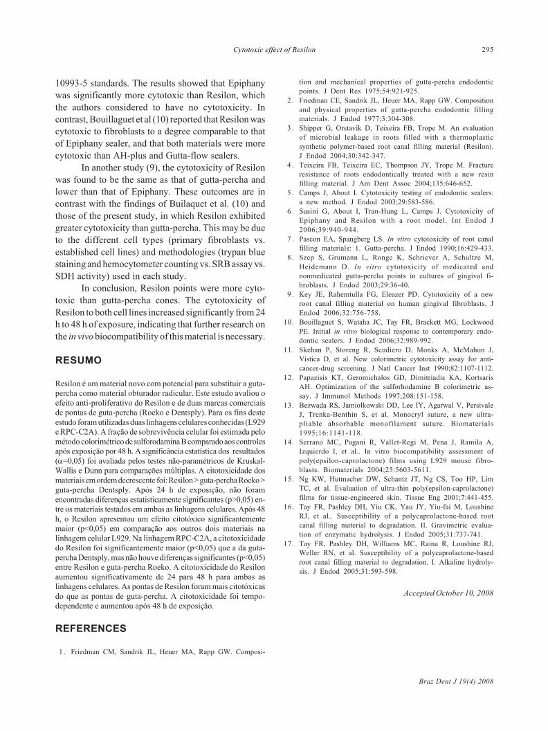
10993-5 standards. The results showed that Epiphanywas significantly more cytotoxic than Resilon, whichthe authors considered to have no cytotoxicity. Incontrast, Bouillaguet et al (10) reported that Resilon wascytotoxic to fibroblasts to a degree comparable to thatof Epiphany sealer, and that both materials were morecytotoxic than AH-plus and Gutta-flow sealers.
In another study (9), the cytotoxicity of Resilonwas found to be the same as that of gutta-percha andlower than that of Epiphany. These outcomes are incontrast with the findings of Builaquet et al. (10) andthose of the present study, in which Resilon exhibitedgreater cytotoxicity than gutta-percha. This may be dueto the different cell types (primary fibroblasts vs.established cell lines) and methodologies (trypan bluestaining and hemocytometer counting vs. SRB assay vs.SDH activity) used in each study.
In conclusion, Resilon points were more cyto-toxic than gutta-percha cones. The cytotoxicity ofResilon to both cell lines increased significantly from 24h to 48 h of exposure, indicating that further research onthe in vivo biocompatibility of this material is necessary.
RESUMO
Resilon é um material novo com potencial para substituir a guta-percha como material obturador radicular. Este estudo avaliou oefeito anti-proliferativo do Resilon e de duas marcas comerciaisde pontas de guta-percha (Roeko e Dentsply). Para os fins desteestudo foram utilizadas duas linhagens celulares conhecidas (L929e RPC-C2A). A fração de sobrevivência celular foi estimada pelométodo colorimétrico de sulforodamina B comparado aos controlesapós exposição por 48 h. A significância estatística dos resultados(=0,05) foi avaliada pelos testes não-paramétricos de Kruskal-Wallis e Dunn para comparações múltiplas. A citotoxicidade dosmateriais em ordem decrescente foi: Resilon > guta-percha Roeko >guta-percha Dentsply. Após 24 h de exposição, não foramencontradas diferenças estatisticamente significantes (p>0,05) en-tre os materiais testados em ambas as linhagens celulares. Após 48h, o Resilon apresentou um efeito citotóxico significantementemaior (p<0,05) em comparação aos outros dois materiais nalinhagem celular L929. Na linhagem RPC-C2A, a citotoxicidadedo Resilon foi significantemente maior (p<0,05) que a da guta-percha Dentsply, mas não houve diferenças significantes (p<0,05)entre Resilon e guta-percha Roeko. A citotoxicidade do Resilonaumentou significativamente de 24 para 48 h para ambas aslinhagens celulares. As pontas de Resilon foram mais citotóxicasdo que as pontas de guta-percha. A citotoxicidade foi tempo-dependente e aumentou após 48 h de exposição.
REFERENCES
1 . Friedman CM, Sandrik JL, Heuer MA, Rapp GW. Composi-
tion and mechanical properties of gutta-percha endodonticpoints. J Dent Res 1975;54:921-925.
2 . Friedman CE, Sandrik JL, Heuer MA, Rapp GW. Compositionand physical properties of gutta-percha endodontic fillingmaterials. J Endod 1977;3:304-308.
3 . Shipper G, Orstavik D, Teixeira FB, Trope M. An evaluationof microbial leakage in roots filled with a thermoplasticsynthetic polymer-based root canal filling material (Resilon).J Endod 2004;30:342-347.
4 . Teixeira FB, Teixeira EC, Thompson JY, Trope M. Fractureresistance of roots endodontically treated with a new resinfilling material. J Am Dent Assoc 2004;135:646-652.
5 . Camps J, About I. Cytotoxicity testing of endodontic sealers:a new method. J Endod 2003;29:583-586.
6 . Susini G, About I, Tran-Hung L, Camps J. Cytotoxicity ofEpiphany and Resilon with a root model. Int Endod J2006;39:940-944.
7 . Pascon EA, Spangberg LS. In vitro cytotoxicity of root canalfilling materials: 1. Gutta-percha. J Endod 1990;16:429-433.
8 . Szep S, Grumann L, Ronge K, Schriever A, Schultze M,Heidemann D. In vitro cytotoxicity of medicated andnonmedicated gutta-percha points in cultures of gingival fi-broblasts. J Endod 2003;29:36-40.
9 . Key JE, Rahemtulla FG, Eleazer PD. Cytotoxicity of a newroot canal filling material on human gingival fibroblasts. JEndod 2006;32:756-758.
10. Bouillaguet S, Wataha JC, Tay FR, Brackett MG, LockwoodPE. Initial in vitro biological response to contemporary endo-dontic sealers. J Endod 2006;32:989-992.
11. Skehan P, Storeng R, Scudiero D, Monks A, McMahon J,Vistica D, et al. New colorimetric cytotoxicity assay for anti-cancer-drug screening. J Natl Cancer Inst 1990;82:1107-1112.
12. Papazisis KT, Geromichalos GD, Dimitriadis KA, KortsarisAH. Optimization of the sulforhodamine B colorimetric as-say. J Immunol Methods 1997;208:151-158.
13. Bezwada RS, Jamiolkowski DD, Lee IY, Agarwal V, PersivaleJ, Trenka-Benthin S, et al. Monocryl suture, a new ultra-pliable absorbable monofilament suture. Biomaterials1995;16:1141-118.
14. Serrano MC, Pagani R, Vallet-Regi M, Pena J, Ramila A,Izquierdo I, et al.. In vitro biocompatibility assessment ofpoly(epsilon-caprolactone) films using L929 mouse fibro-blasts. Biomaterials 2004;25:5603-5611.
15. Ng KW, Hutmacher DW, Schantz JT, Ng CS, Too HP, LimTC, et al. Evaluation of ultra-thin poly(epsilon-caprolactone)films for tissue-engineered skin. Tissue Eng 2001;7:441-455.
16. Tay FR, Pashley DH, Yiu CK, Yau JY, Yiu-fai M, LoushineRJ, et al.. Susceptibility of a polycaprolactone-based rootcanal filling material to degradation. II. Gravimetric evalua-tion of enzymatic hydrolysis. J Endod 2005;31:737-741.
17. Tay FR, Pashley DH, Williams MC, Raina R, Loushine RJ,Weller RN, et al. Susceptibility of a polycaprolactone-basedroot canal filling material to degradation. I. Alkaline hydroly-sis. J Endod 2005;31:593-598.
Accepted October 10, 2008
1205.pmd 17/12/2008, 00:33295