COMPARATIVE ANALYSIS OF MEDICAL EQUIPMENT USED IN ...
9
/ J of IMAB. 2016, vol. 22, issue 3/ http://www.journal-imab-bg.org 1189 COMPARATIVE ANALYSIS OF MEDICAL EQUIPMENT USED IN DIAGNOSTIC IMAGING IN BULGARIA AND COUNTRIES WORLDWIDE AND OPTIMIZATION AIMED AT IMPROVING THE HEALTH CARE SYSTEM MANAGEMENT. Svetoslav Garov 1 , Tsvetelina Mihaylova 1 , Galina Makedonska 2 1) Department of Health Policy and Management, Faculty of Public Health, MU - Sofia, Bulgaria 2) NCRRP (National Center of Radiobiology and Radiation Protection), Ministry of Health, Bulgaria. Journal of IMAB - Annual Proceeding (Scientific Papers) 2016, vol. 22, issue 3 Journal of IMAB ISSN: 1312-773X http://www.journal-imab-bg.org ABSTRACT Purpose: to reveal the current condition of medical equipment in Bulgaria related to those major groups of so- cially significant diseases and to make an attempt to define guidelines for its optimization in view of improving the func- tioning and management of the healthcare system in this field. Material and methods: The following research meth- ods have been applied: 1. Document review method – research, processing and analysis of medical statistical information taken from data from WHO and annual reports of NRA. The study in- cludes data from 2009 - 2015. 2. Graphical method – summarizing data in relevant tables and diagram presentations. Results: The article analyzes the condition of medi- cal equipment in the field of oncologic and cardiologic medical aid in Bulgaria based on data taken from WHO (World Health Organization) and annual reports of NRA (Nu- clear Regulatory Agency). Six types of diagnostic imaging and radiation therapy devices have been studied: Magnetic Resonance Imaging units (MRI); Computed Tomography Scanners (CT), Positron Emission Tomography Scanners, Mammographs, Linear accelerators and Telecobalt units (Co- balt-60). The condition of medical equipment since 2009 has been analyzed, results have been reported and trends – studied. Conclusion: The oncologic and cardiologic medical equipment in Bulgaria has been gradually improving in the last seven years, but quantitative indicators regarding the devices studied are still far away from the figures recom- mended by WHO with one single exception, i.e. Computed Tomography Scanners Key words: Oncologic medical aid, cardiologic medi- cal aid, Magnetic Resonance Imaging units (MRI), Com- puted Tomography Scanners (CT), Positron Emission Tom- ography Scanners, Mammographs, Linear accelerators, Telecobalt units (Cobalt-60), INTRODUCTION The greatest challenge that medicine and healthcare face in XXI century is the increasing costs for socially sig- nificant diseases. The most specific feature of those diseases is their large scale distribution. They are the result of a number of reasons, endogenic andexogenic forces acting throughout one’s life. Those diseases mostly affect middle- aged people, which threatens the social and economic wel- fare of individuals, their families and on a more general level – the welfare of the entire population of a country. A posi- tive affect regarding those groups of diseases can be achieved through: limiting major risk factors and taking advantage of the advance in medical science and practice [1]. RESULTS AND DISCUSSION: The analysis includes data taken from 58 countries regarding 6 types of medical devices: Magnetic Resonance Imaging units (MRI); Computed Tomography Scanners (CT), Positron Emission Tomography Scanners, Mammographs, Linear accelerators and Telecobalt units (Co- balt-60). The source of information is WHO (World Health Organization) and part of the data for Bulgaria is provided by NCRRP (National Center of Radiobiology and Radiation Protection) and taken from annual reports of NRA [2, 3]. Countries from five continents take part in the research as follows: Europe – 25 countries; Asia - 13 countries; North and South America – 10 countries; Africa- 10 countries. The costs of medical equipment are related to 1 million inhabit- ants with the purpose of possible comparison and therefore most of them are not whole numbers. The research presents data from 2009 and in the graphs Bulgaria is marked in red so that it could be quickly and easily seen. Finally, average values are presented in tables and graphs for the six types of devices. 1. Magnetic Resonance Imaging units (MRI) The first Magnetic Resonance Imaging unit in Bul- garia was delivered back in 1966 – MRI spectrometer (JEOL – Japan 60 MHz) [4]. As seen from the graph, Japan is lead- ing in terms of quantitative indicators with 45.94 MRI units per 1 million people, the difference with the other countries being significant. (Fig.1.) Japan also leads in early diagno- http://dx.doi.org/10.5272/jimab.2016223.1189
Transcript of COMPARATIVE ANALYSIS OF MEDICAL EQUIPMENT USED IN ...

/ J of IMAB. 2016, vol. 22, issue 3/ http://www.journal-imab-bg.org 1189
COMPARATIVE ANALYSIS OF MEDICALEQUIPMENT USED IN DIAGNOSTIC IMAGING INBULGARIA AND COUNTRIES WORLDWIDE ANDOPTIMIZATION AIMED AT IMPROVING THEHEALTH CARE SYSTEM MANAGEMENT.
Svetoslav Garov1, Tsvetelina Mihaylova1, Galina Makedonska2
1) Department of Health Policy and Management, Faculty of Public Health, MU- Sofia, Bulgaria2) NCRRP (National Center of Radiobiology and Radiation Protection), Ministryof Health, Bulgaria.
Journal of IMAB - Annual Proceeding (Scientific Papers) 2016, vol. 22, issue 3Journal of IMABISSN: 1312-773Xhttp://www.journal-imab-bg.org
ABSTRACTPurpose: to reveal the current condition of medical
equipment in Bulgaria related to those major groups of so-cially significant diseases and to make an attempt to defineguidelines for its optimization in view of improving the func-tioning and management of the healthcare system in thisfield.
Material and methods: The following research meth-ods have been applied:
1. Document review method – research, processingand analysis of medical statistical information taken fromdata from WHO and annual reports of NRA. The study in-cludes data from 2009 - 2015.
2. Graphical method – summarizing data in relevanttables and diagram presentations.
Results: The article analyzes the condition of medi-cal equipment in the field of oncologic and cardiologicmedical aid in Bulgaria based on data taken from WHO(World Health Organization) and annual reports of NRA (Nu-clear Regulatory Agency). Six types of diagnostic imagingand radiation therapy devices have been studied: MagneticResonance Imaging units (MRI); Computed TomographyScanners (CT), Positron Emission Tomography Scanners,Mammographs, Linear accelerators and Telecobalt units (Co-balt-60). The condition of medical equipment since 2009has been analyzed, results have been reported and trends –studied.
Conclusion: The oncologic and cardiologic medicalequipment in Bulgaria has been gradually improving in thelast seven years, but quantitative indicators regarding thedevices studied are still far away from the figures recom-mended by WHO with one single exception, i.e. ComputedTomography Scanners
Key words: Oncologic medical aid, cardiologic medi-cal aid, Magnetic Resonance Imaging units (MRI), Com-puted Tomography Scanners (CT), Positron Emission Tom-ography Scanners, Mammographs, Linear accelerators,Telecobalt units (Cobalt-60),
INTRODUCTIONThe greatest challenge that medicine and healthcare
face in XXI century is the increasing costs for socially sig-nificant diseases. The most specific feature of those diseasesis their large scale distribution. They are the result of anumber of reasons, endogenic andexogenic forces actingthroughout one’s life. Those diseases mostly affect middle-aged people, which threatens the social and economic wel-fare of individuals, their families and on a more general level– the welfare of the entire population of a country. A posi-tive affect regarding those groups of diseases can be achievedthrough: limiting major risk factors and taking advantageof the advance in medical science and practice [1].
RESULTS AND DISCUSSION:The analysis includes data taken from 58 countries
regarding 6 types of medical devices: Magnetic ResonanceImaging units (MRI); Computed Tomography Scanners(CT), Positron Emission Tomography Scanners,Mammographs, Linear accelerators and Telecobalt units (Co-balt-60). The source of information is WHO (World HealthOrganization) and part of the data for Bulgaria is providedby NCRRP (National Center of Radiobiology and RadiationProtection) and taken from annual reports of NRA [2, 3].Countries from five continents take part in the research asfollows: Europe – 25 countries; Asia - 13 countries; Northand South America – 10 countries; Africa- 10 countries. Thecosts of medical equipment are related to 1 million inhabit-ants with the purpose of possible comparison and thereforemost of them are not whole numbers. The research presentsdata from 2009 and in the graphs Bulgaria is marked in redso that it could be quickly and easily seen. Finally, averagevalues are presented in tables and graphs for the six typesof devices.
1. Magnetic Resonance Imaging units (MRI)The first Magnetic Resonance Imaging unit in Bul-
garia was delivered back in 1966 – MRI spectrometer (JEOL– Japan 60 MHz) [4]. As seen from the graph, Japan is lead-ing in terms of quantitative indicators with 45.94 MRI unitsper 1 million people, the difference with the other countriesbeing significant. (Fig.1.) Japan also leads in early diagno-
http://dx.doi.org/10.5272/jimab.2016223.1189

1190 http://www.journal-imab-bg.org / J of IMAB. 2016, vol. 22, issue 3/
sis and effective treatment of oncology diseases. Despite us-ing cutting edge technologies, treatment in Japan is less ex-pensive than, for example, Germany or Greece. Latest trendsin Japanese medicine, serving as standard for other coun-tries, are as follows: minimal invasiveness, efficiency and in-dividual approach to patients. An evidence of the highly ef-ficient Japanese healthcare system is their record-breakinglife expectancy, directly related to the promotion of healthylifestyle, use of cutting edge technical and scientific achieve-ments and the availability of competent medical experts. In2015 Japan manufactured and commissioned the most pow-erful so far MRI unit with operating frequency of 1020 MHz.Before that the most powerful and ensuring the most pre-cise diagnostics device was in Germany, with a frequencyof 1000 MHz and functioning since 2009. Second amongthe countries studied is Belgium, having 24.89 MRI unitsper 1 million people, followed by Iceland with 21.69 MRIunits per 1 million people, etc. Bulgaria in 2009 had 4.02
devices per 1 million people. This means that Bulgaria, withits population of 7.25 million, had in 2009 30 MRI unitsregistered and functioning according to data. The majorityof them are not brand new, but second hand. The main rea-son is the high price of brand new equipment and that iswhy second hand devices are most often purchased in a rela-tively good condition at prices about 800 000 - 1 000 000BGN. Other reasons include high cost of maintenance, highoperating costs and low return on investment. Profitabilityis ensured by a patient flow depending on the regulatorystandards set by NHIF (National Health Insurance Fund) andon the patients’ solvency. NHIF covers 180 BGN, and therest is paid by patients. Financial estimates show that at suchprices it takes 140-145 patients per month so that such kindof apparatus can be profitable for the medical institution.The average price of an MRI in Bulgaria in 2015 was 550BGN with contrast agents and 350 BGN w/o contrast agents.
Fig. 1. MRI units worldwidein 2009

/ J of IMAB. 2016, vol. 22, issue 3/ http://www.journal-imab-bg.org 1191
After 1998 the world of oncologic surgery welcomed atechnical revolution thanks to robot-assisted surgery and pro-ton therapy. Gamma knife was also introduced in 1998 toperform safe and minimal invasive operations throughgamma ray radiation and with no cuts and bleeding. The de-vice operates with the expensive radioactive cobalt and ismainly used in cases of malignant tumors in the head [5, 6, 7].
Cybeknife is a newer generation of equipment and is arobot-assisted radiosurgery set. It combines a radiation de-vice attached to a mobile arm of a super precise robot and aspecial navigation system. The first European Cyberknife wasmade in Germany. Cyberknife operations are used in muchmore types and locations of malignant tumors and the appa-ratus also successfully treats some kinds of metastasis. Theaverage duration of a Cyberknife surgery is approximately 1hour. Cyberknife is famous for its absolute precision and thetherapy itself spares healthy tissues to the maximum possibleextent [8, 9, 10].
Even newer generation of equipment used for non-operative treatment of cancer is the so called Trilogy linearaccelerator (Trilogy linac). The apparatus was introducedby a hi-tech American company and, in contrast to the two
devices described above, operates directly through the elec-tricity network so the cost of surgery is many times lower andthe treatment become much less expensive [11, 12].
In Bulgaria, before 2016, Cyberknives were commis-sioned for radiosurgery and radiation treatment in Sv. IvanRilski Hospital in Sofia, Tokuda Hospital in Sofia and Sv.Georgi University Hospital in Plovdiv.
2. Computed Tomography Scanners (CT)Bulgaria has a quite high number of CT devices –
27.19 per 1 million people. (Fig. 2) Such figures are closeto the ones reported for well-developed countries such asAustria, Portugal, etc. Japan is the world leader, having101.75 devices per 1 million people. The total number ofCT sets functioning in Bulgaria in 2009 amounted to 198.According to their type they are single-slice and multi-slice.The latter can be subdivided further into 2-slice, 4-slice, 16-slice and 64-slice. In Bulgaria the majority of CT devicesare manufactured by Siemens. Tests are usually made in fourareas: head, chest, abdomen and pelvis [13, 14]. Theirnumber has been gradually increasing: 235 in 2010; 237 in2011; 241 in 2012; 266 in 2013 and 287 in 2014.
Fig. 2. Computed Tomography Scanners world-wide in 2009.

1192 http://www.journal-imab-bg.org / J of IMAB. 2016, vol. 22, issue 3/
3. Positron Emission Tomography Scanners (PET scan-ners)
Positron Emission Tomography Scanners (or positronemission tomography) are diagnostic imaging devices thatdisplay biochemical and physiological changes and diag-nose a disease at a very early stage before any morphologi-cal changes have occurred. This makes them different fromMRI and CT devices that register structural anatomicalchanges that have already appeared in organs [15, 16]. Inaddition, they are used to monitor the effects of antitumortherapy. A disadvantage is their high price when comparedto other diagnostic imaging tests: 2800 BGN, now entirelycovered by NHIF, registration being subject to approval bya special medical committee. The first PET scanner was in-troduced in Bulgaria in 2009 at Sv. Marina University Hos-pital in Varna. Gradually their number increased and by 2016there were 3 hospitals operating PET scanners: Sv. MarinaUniversity Hospital in Varna, Sv. Georgi University Hospi-tal in Plovdiv and Aleksandrovska University Hospital inSofia.
As seen from the graph the countries covered by thestudy that have the greatest number of PET scanners per
capita are Belgium – 26.76 devices per 1 million people,Denmark – 6.03, Japan – 4.34, etc. (Fig.3.)
To operate, PET scanners need a specific radioactiveisotope consumable called radionuclide fluorine-18. In 2016Bulgaria had 2 cyclotron sets for their manufacturing: in Sv.Marina University Hospital in Varna and AleksandrovskaUniversity Hospital in Sofia. A third set is to be constructedin INRNE (Institute for Nuclear Research and Nuclear En-ergy) at BAS (Bulgarian Academy of Sciences) – Sofia by theend of 2017. A cyclotron set consists of a mini cyclotron de-vice producing individual doses of radioactive isotopes forabout 180–200 patients a month. It guarantees constant avail-ability of the PET scanner consumable. Until then isotopesfor PET scanners will be delivered by air due to their shortperiod of decay [17, 18].
The radiopharmaceutical most applied is 18FFludeoxyglucose (F18 FDG). It is a substance analogous toglucose used in PET and suitable for testing metabolism ofglucose in the heart, lungs and brain. It is also applied whenmonitoring treatment of Hodgkin’s disease, non-Hodgkin’slymphoma, colorectal cancer, breast cancer, melanoma, lungcancer, etc. [19, 20].
Fig. 3. PET scanners worldwide in2009.
/ J of IMAB. 2016, vol. 22, issue 3/ http://www.journal-imab-bg.org 1193
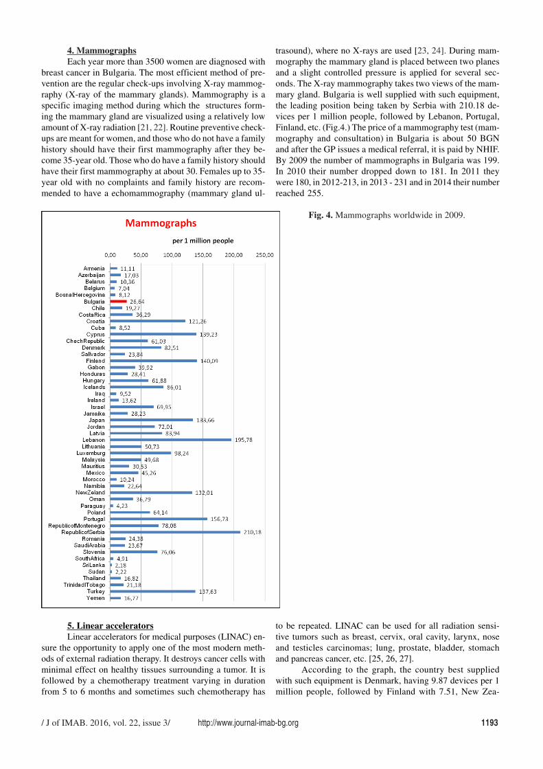
4. MammographsEach year more than 3500 women are diagnosed with
breast cancer in Bulgaria. The most efficient method of pre-vention are the regular check-ups involving X-ray mammog-raphy (X-ray of the mammary glands). Mammography is aspecific imaging method during which the structures form-ing the mammary gland are visualized using a relatively lowamount of X-ray radiation [21, 22]. Routine preventive check-ups are meant for women, and those who do not have a familyhistory should have their first mammography after they be-come 35-year old. Those who do have a family history shouldhave their first mammography at about 30. Females up to 35-year old with no complaints and family history are recom-mended to have a echomammography (mammary gland ul-
trasound), where no X-rays are used [23, 24]. During mam-mography the mammary gland is placed between two planesand a slight controlled pressure is applied for several sec-onds. The X-ray mammography takes two views of the mam-mary gland. Bulgaria is well supplied with such equipment,the leading position being taken by Serbia with 210.18 de-vices per 1 million people, followed by Lebanon, Portugal,Finland, etc. (Fig.4.) The price of a mammography test (mam-mography and consultation) in Bulgaria is about 50 BGNand after the GP issues a medical referral, it is paid by NHIF.By 2009 the number of mammographs in Bulgaria was 199.In 2010 their number dropped down to 181. In 2011 theywere 180, in 2012-213, in 2013 - 231 and in 2014 their numberreached 255.
5. Linear acceleratorsLinear accelerators for medical purposes (LINAC) en-
sure the opportunity to apply one of the most modern meth-ods of external radiation therapy. It destroys cancer cells withminimal effect on healthy tissues surrounding a tumor. It isfollowed by a chemotherapy treatment varying in durationfrom 5 to 6 months and sometimes such chemotherapy has
Fig. 4. Mammographs worldwide in 2009.
to be repeated. LINAC can be used for all radiation sensi-tive tumors such as breast, cervix, oral cavity, larynx, noseand testicles carcinomas; lung, prostate, bladder, stomachand pancreas cancer, etc. [25, 26, 27].
According to the graph, the country best suppliedwith such equipment is Denmark, having 9.87 devices per 1million people, followed by Finland with 7.51, New Zea-

1194 http://www.journal-imab-bg.org / J of IMAB. 2016, vol. 22, issue 3/
land with 6.56, etc. (Fig.5.)By the end of 2009 Bulgaria had 3 linear accelera-
tors used for radiation treatment of oncology diseases in Sv.Georgi University Hospital in Plovdiv, the Specialized Hos-pital for Active Treatment in Oncology in Sofia and the newone located in Tokuda Hospital in Sofia.
In 2014 the total number of linear accelerators usedfor medical purposes was 10: 3 in Sofia, 2 in Plovdiv and 2in the town of Shumen, while 6 were being installed andtested: two new ones in Sofia, 2 in Varna, 1 in Plovdiv and1 in the town of Vratsa.
6. 6. Telecobalt units (Cobalt-60)Cobalt therapy is the use for medical purposes of
gamma rays from cobalt-60 isotope in cases of malignanttumors. After World War II, cobalt radiation therapy equip-ment revolutionized oncology and such devices were wide-spread in the 1950s and 1960s. After the introduction of themedical linear accelerator in the 1970s their role was par-tially replaced by linear accelerators. Advantages: medically
efficient, having simple design, less expensive in terms ofpurchase price and maintenance costs as compared to mod-ern linear accelerators. Disadvantages: radioactive waste re-lated problems; cobalt-60 radioisotope has a half-life of 5.3years and so it has to be periodically replaced [28, 29].
The modern cobalt therapy is also known as GammaKnife therapy and can be applied in oncology for all bodyparts and organs. It is extremely precise and so it is used for
Fig. 5. Linear accelerators worldwide in 2009.

/ J of IMAB. 2016, vol. 22, issue 3/ http://www.journal-imab-bg.org 1195
patients having brain tumors [30, 31, 32].As seen from the graph from the countries covered by
the study Malta has 2.44 devices, Belarus has 2.33, Mauri-tania – 1.55, etc. It is obvious that in countries with well-developed economies linear accelerators prevail instead ofTelecobalt units. ( Fig. 6)
In 2009 the old devices were replaced by new gammaradiation therapy equipment in the Specialized Hospital for
Active Treatment in Oncology in Sofia and the district on-cology clinics in the towns of Stara Zagora and Ruse. NRAissued installation and testing permits for the new devices,approval committees were set up and operation licenses weregranted. In 2010 the medical gamma radiation equipmentin the Oncology Center in the town of Plovdiv wasuninstalled and decommissioned.
Comparative data of the 6 types of studied diagnos-tic imaging and radiation therapy devices used in Bulgariais presented, comparing the average figures on the continentstaken into account with the purpose of displaying the gen-eral condition of the infrastructure studied in different coun-tries worldwide. For better data illustration North and SouthAmerica are presented together under a single name, i.e.America. (Table 1 and Fig.7)
Fig. 6. Telecobalt units (Cobalt-60)worldwide in 2009.

1196 http://www.journal-imab-bg.org / J of IMAB. 2016, vol. 22, issue 3/
Table 1. Average number of the 6 types of medical equipment (per 1 million people) per continents and Bulgaria in2009.
MRI units CT PET scanners Mammographs Linear Telecobalt
accelerators units
EUROPE 8.78 15.84 2.09 81.49 3.13 0.61
ASIA 5.99 13.71 0.58 50.38 0.89 0.3
AMERICA 1.93 4.9 0.116 23.91 0.61 0.69
AFRICA 2.29 4.17 0.08 34.6 0.92 0.397
Bulgaria 4.02 27.19 0.13 26.64 0.4 0.4
CONCLUSIONS:1. As seen from the study, the oncologic and cardio-
logic medical equipment in Bulgaria has been gradually im-proving in the last seven years, but quantitative indicatorsregarding the devices studied are still far away from the fig-ures recommended by WHO with one single exception, i.e.CT. For example, WHO recommends a standard of 1 linearaccelerator per 250 000-300 000 people, which means thatour country by 2016 has approximately half of the numbersrequired for achieving this standard.
2. There is a considerable difference in those indica-
Fig. 7. Average number of the 6 typesof medical equipment (per 1 millionpeople) per continents and Bulgaria in2009.
Key:I. MRI units;II. CTIII. PET scannersIV. MammographsV. Linear acceleratorsVI. Telecobalt units
tors between Bulgaria and the countries with well-developedeconomies. However, it has to be emphasized that the resultsdo show a significant modernization and fast reduction ofthose differences, especially during the last three years.
3. The use of modern technologies by medical expertsis directly related to improving the results in terms of diag-nostics and treatment. Therefore, the recent year trend of in-creasing material resource has to be continued together witha steady improvement of medical specialists’ qualificationwith the purpose of their adequate adjustment to new tech-nologies.
REFERENCES:KD, Wu HM, Shiau CY, et al. GammaKnife Radiosurgery for Atypical andAnaplastic Meningiomas; World Neuro-surgery 2016 Mar;87:557–564.[PubMed]
8. Oppenlander ME, Porter RW. Ra-diosurgery - Gamma Knife andCyberknife. Encyclopedia of the Neuro-logical Sciences (Second Edition)2014; p1050-1051. [CrossRef]
9. Al Kafi MA, Mwidu U, Moftah B.Continuous versus step-by-step scan-ning mode of a novel 3D scanner for
1. Mutafova M, Vodenicharov Ts,Pesheva P, Hristov N, Shipkovenska E,Georgieva L, et al. World populationhealth. Gorexpress – Sofia 2015; page108 [in Bulgarian]
2. http://www.who.int/gho/health_technologies/medical_devices/en/
3. http://ncrrp.org/new/bg/4. http://www.orgchm.bas.bg/~nmr/
History.htm5. Yamanaka K, Iwai Y, Shuto T, Kida
Y, Sato M, Hayashi M, et al. Treatment
results of gamma knife radiosurgery forcentral neurocytoma: report of a Japa-nese multi-institutional co-operativestudy. World Neurosurgery. 2016Jun;90:300-5. [PubMed]
6. Kapitza S, Pangalu A, HorstmannGA, van Eck AT, Regli L, Tarnutzer AA.Acute necrosis after Gamma Knife sur-gery in vestibular schwannoma leadingto multiple cranial nerve palsies. J ClinNeurosci 2016 Mar 3; pii: S0967-5868(16)00068-0 [PubMed]
7. Wang WH, Lee CC, Yang HC, Liu
/ J of IMAB. 2016, vol. 22, issue 3/ http://www.journal-imab-bg.org 1197
Address for correspondence:Svetoslav Garov, MD, PhD;Faculty of Public Health, Medical University-Sofia;1527, Sofia, 8, Bialo more Str., BulgariaÅ-mail: [email protected]
CyberKnife measurements. Appl RadiatIsot. 2015 Nov;105:88-91. [PubMed]
10. Liu SH, Murovic J, Wallach J,Cui G, Soltys SG, Gibbs IC, et al. CyberKnife radiosurgery for brainstem meta-stases: Management and outcomes anda review of the literature. J Clin Neuro-sci. 2016 Mar;25:105-110. [PubMed][CrossRef]
11. Vaccara E, Agostinelli S,Bevegni M, Taccini G. New experiencewith breast volumetric modulation ARCtherapy (VMAT) realized with Trilogylinac: Comparison with tomotherapy,pros and cons. Physica Medica. 2016Feb;32(1):67; [CrossRef]
12. Huntzinger C, Friedman W,Bova F, Fox T, Boushet L, Boeh L. Tril-ogy Image-Guided Stereotactic Radio-surgery. Med Dosim. 2007 Summer;32(2):121-133. [CrossRef]
13. Nieman K, Coenen A,Dijkshoorn M. 5 - Computed tomogra-phy. Advanced Cardiac Imaging. 2015;97-125. [CrossRef]
14. Panetta D. 2.03 – Computed To-mography. Volume 2: X-Ray and Ultra-sound Imaging. Comprehensive Bio-medical Physics. 2014 Sep;2:65-88.[CrossRef]
15. Hess S. FDG-PET/CT: Quovadis? PET Clinics. 2014 Oct;9(4):xi-xiii; [CrossRef]
16. Basu S, Hess S, Braad NielsenPE, Olsen BB, Inglev S, Høilund-Carlsen PF. The basic principles ofFDG-PET/CT imaging. PET Clin-ics. 2014; 9:355-370. [PubMed] [CrossRef]
17. Smirnov V, Vorozhtsov S, Vin-cent J. Design study of an ultra-compactsuperconducting cyclotron for isotopeproduction. Nuclear Instruments andMethods in Physics Research Section A:Accelerators, Spectrometers, Detectorsand Associated Equipment. 2014 Nov1;763:6–12. [CrossRef]
18. Shankar M, Rao J. PET- CTimaging (Positron Emission Tomogra-phy - Computerized Tomography): Abrief overview. Apollo Medicine. 2014Dec;11(4):311–314. [CrossRef]
19. Buvat I. [Quantification in on-cologic FDG-PET: A scientific over-view.] [in French] Médecine Nucléaire.2011 May;35(5):320–321. [CrossRef]
20. Tomasi G, Rosso L. PETimaging: implications for the future oftherapy monitoring with PET/CT in on-cology. Curr Opin Pharmacol. 2012Oct;12(5):569-575. [PubMed]
21. Lee H, Chen Y. Image basedcomputer aided diagnosis system forcancer detection. Expert Systems withApplications. 2015 July 15;42(12):5356-5365. [CrossRef]
22. Griff S, Dershaw D. Chapter16 – Breast Cancer. Oncologic Imaging(Second Edition). 2002; 265–294.[CrossRef]
23. Steyerova P, Frybova J, Sko-vajsova M. 719 Screening and diagnos-tic use of breast ultrasound in youngwomen. European Journal of Cancer.2015 Sep;51(3):S133. [CrossRef]
24. An Y, Kim S, Kang BJ, ParkCS, Jung NY, Kim JY. Breast cancer invery young women (<30 years): Corre-
lation of imaging features with clinico-pathological features and immunohis-tochemical subtypes. Eur J Radiol .2015 Oct;84(10):1894-1902. [PubMed]
25. Owen H, Lomax A, Jolly S. Cur-rent and future accelerator technologiesfor charged particle therapy. NuclInstrum Methods Phys Res A. 2016 Feb;809:96-104. [CrossRef]
26. Ali A, Curran W. Linear Accel-erator-Based Fractionated Radio-therapy. Craniopharyngiomas. 2015;Chapter 21:335-346. [CrossRef]
27. Uesaka M, Tanabe E. 8.01 - Electron Linear Accelerators. Refer-ence Module in Biomedical Sciences.2014; Sep;8:1-16 [CrossRef]
28. Yamashita T, Abe M. Historyand development of radiation oncol-ogy. Int J Radiation Oncology BiolPhys. 1996 Dec;36(5):1263-1265.[PubMed]
29. Henk J. A brief history of Brit-ish radiotherapy. Int J of Radiation On-cology Biol Phys. 1996 Aug; 36(1): 213-218. [CrossRef]
30. Matsunaga S, Shuto T, Sato M.Gamma Knife Surgery for MetastaticBrain Tumors from Gynecologic Can-cer. World Neurosurgery. 2016 May;89:455-463. [PubMed]
31. Ganz J. The journey from pro-ton to gamma knife. Prog Brain Res.2014; 215: 67-75. [PubMed] [CrossRef]
32. Ganz J. The earliest gamma unitpatients; Prog Brain Res. 2014; 215:77-84. [PubMed] [CrossRef]
Please cite this article as: Garov S, Mihaylova T, Makedonska G. Comparative analysis of medical equipment used indiagnostic imaging in Bulgaria and countries worldwide and optimization aimed at improving the health care systemmanagement. J of IMAB. 2016 Jul-Sep;22(3):1189-1197. DOI: http://dx.doi.org/10.5272/jimab.2016223.1189
Received: 03/05/2016; Published online: 01/07/2016