Community Eye Health · PDF file · 2013-08-23At presentation, he had a large...
16
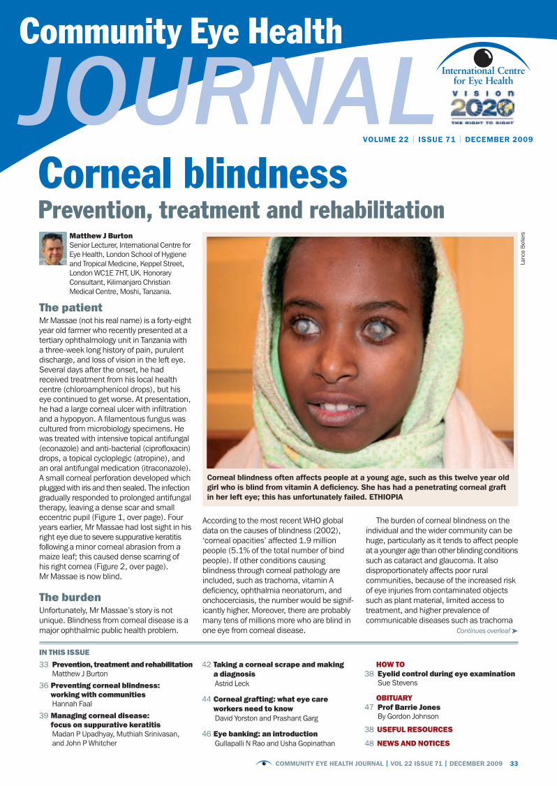
The patient Mr Massae (not his real name) is a forty-eight year old farmer who recently presented at a tertiary ophthalmology unit in Tanzania with a three-week long history of pain, purulent discharge, and loss of vision in the left eye. Several days after the onset, he had received treatment from his local health centre (chloroamphenicol drops), but his eye continued to get worse. At presentation, he had a large corneal ulcer with infiltration and a hypopyon. A filamentous fungus was cultured from microbiology specimens. He was treated with intensive topical antifungal (econazole) and anti-bacterial (ciprofloxacin) drops, a topical cycloplegic (atropine), and an oral antifungal medication (itraconazole). A small corneal perforation developed which plugged with iris and then sealed. The infection gradually responded to prolonged antifungal therapy, leaving a dense scar and small eccentric pupil (Figure 1, over page). Four years earlier, Mr Massae had lost sight in his right eye due to severe suppurative keratitis following a minor corneal abrasion from a maize leaf; this caused dense scarring of his right cornea (Figure 2, over page). Mr Massae is now blind. The burden Unfortunately, Mr Massae’s story is not unique. Blindness from corneal disease is a major ophthalmic public health problem. According to the most recent WHO global data on the causes of blindness (2002), ‘corneal opacities’ affected 1.9 million people (5.1% of the total number of bind people). If other conditions causing blindness through corneal pathology are included, such as trachoma, vitamin A deficiency, ophthalmia neonatorum, and onchocerciasis, the number would be signif- icantly higher. Moreover, there are probably many tens of millions more who are blind in one eye from corneal disease. The burden of corneal blindness on the individual and the wider community can be huge, particularly as it tends to affect people at a younger age than other blinding conditions such as cataract and glaucoma. It also disproportionately affects poor rural communities, because of the increased risk of eye injuries from contaminated objects such as plant material, limited access to treatment, and higher prevalence of communicable diseases such as trachoma Corneal blindness VOLUME 22 | ISSUE 71 | DECEMBER 2009 Community Eye Health JOURNAL Continues overleaf ➤ Prevention, treatment and rehabilitation 33 Prevention, treatment and rehabilitation Matthew J Burton 36 Preventing corneal blindness: working with communities Hannah Faal 39 Managing corneal disease: focus on suppurative keratitis Madan P Upadhyay, Muthiah Srinivasan, and John P Whitcher 42 Taking a corneal scrape and making a diagnosis Astrid Leck 44 Corneal grafting: what eye care workers need to know David Yorston and Prashant Garg 46 Eye banking: an introduction Gullapalli N Rao and Usha Gopinathan HOW TO 38 Eyelid control during eye examination Sue Stevens OBITUARY 47 Prof Barrie Jones By Gordon Johnson 38 USEFUL RESOURCES 48 NEWS AND NOTICES 33 COMMUNITY EYE HEALTH JOURNAL | VOL 22 ISSUE 71 | DECEMBER 2009 Matthew J Burton Senior Lecturer, International Centre for Eye Health, London School of Hygiene and Tropical Medicine, Keppel Street, London WC1E 7HT, UK. Honorary Consultant, Kilimanjaro Christian Medical Centre, Moshi, Tanzania. IN THIS ISSUE Lance Bellers Corneal blindness often affects people at a young age, such as this twelve year old girl who is blind from vitamin A deficiency. She has had a penetrating corneal graft in her left eye; this has unfortunately failed. ETHIOPIA
-
Upload
nguyenthien -
Category
Documents
-
view
218 -
download
2
Transcript of Community Eye Health · PDF file · 2013-08-23At presentation, he had a large...
The patientMr Massae (not his real name) is a forty-eight year old farmer who recently presented at a tertiary ophthalmology unit in Tanzania with a three-week long history of pain, purulent discharge, and loss of vision in the left eye. Several days after the onset, he had received treatment from his local health centre (chloroamphenicol drops), but his eye continued to get worse. At presentation, he had a large corneal ulcer with infiltration and a hypopyon. A filamentous fungus was cultured from microbiology specimens. He was treated with intensive topical antifungal (econazole) and anti-bacterial (ciprofloxacin) drops, a topical cycloplegic (atropine), and an oral antifungal medication (itraconazole). A small corneal perforation developed which plugged with iris and then sealed. The infection gradually responded to prolonged antifungal therapy, leaving a dense scar and small eccentric pupil (Figure 1, over page). Four years earlier, Mr Massae had lost sight in his right eye due to severe suppurative keratitis following a minor corneal abrasion from a maize leaf; this caused dense scarring of his right cornea (Figure 2, over page). Mr Massae is now blind.
The burdenUnfortunately, Mr Massae’s story is not unique. Blindness from corneal disease is a major ophthalmic public health problem.
According to the most recent WHO global data on the causes of blindness (2002), ‘corneal opacities’ affected 1.9 million people (5.1% of the total number of bind people). If other conditions causing blindness through corneal pathology are included, such as trachoma, vitamin A deficiency, ophthalmia neonatorum, and onchocerciasis, the number would be signif-icantly higher. Moreover, there are probably many tens of millions more who are blind in one eye from corneal disease.
The burden of corneal blindness on the individual and the wider community can be huge, particularly as it tends to affect people at a younger age than other blinding conditions such as cataract and glaucoma. It also disproportionately affects poor rural communities, because of the increased risk of eye injuries from contaminated objects such as plant material, limited access to treatment, and higher prevalence of communicable diseases such as trachoma
Corneal blindnessVOLUME 22 | ISSUE 71 | DECEMBER 2009
Community Eye Health
Journal
Continues overleaf ➤
Prevention, treatment and rehabilitation
33 Prevention, treatment and rehabilitation Matthew J Burton
36 Preventing corneal blindness:working with communities
Hannah Faal
39 Managing corneal disease:focus on suppurative keratitis
Madan P Upadhyay, Muthiah Srinivasan, and John P Whitcher
42 Taking a corneal scrape and makinga diagnosis
Astrid Leck
44 Corneal grafting: what eye careworkers need to know
David Yorston and Prashant Garg
46 Eye banking: an introduction Gullapalli N Rao and Usha Gopinathan
HOW TO38 Eyelid control during eye examination
Sue Stevens
OBITUARY47 Prof Barrie Jones
By Gordon Johnson
38 USEFUL RESOURCES
48 NEWS AND NOTICES
33COMMUnIty EyE HEaLtH JOURnaL | VOL 22 ISSUE 71 | DECEMBER 2009
Matthew J BurtonSenior Lecturer, International Centre for Eye Health, London School of Hygiene and Tropical Medicine, Keppel Street, London WC1E 7HT, UK. Honorary Consultant, Kilimanjaro Christian Medical Centre, Moshi, Tanzania.
IN THIS ISSUE
Lanc
e B
elle
rs
Corneal blindness often affects people at a young age, such as this twelve year old girl who is blind from vitamin a deficiency. She has had a penetrating corneal graft in her left eye; this has unfortunately failed. EtHIOPIa
CEHJ71_OA.indd 33 13/01/2010 12:13
Volume 22 | Issue 71 | December 2009
EditorElmien Wolvaardt Ellison [email protected]
Editorial committeeNick AstburyAllen FosterClare GilbertMurray McGavinIan MurdochGVS MurthyDaksha PatelRichard WormaldDavid Yorston
Special advisor for Issue 71David Yorston
Regional consultantsSergey Branchevski (Russia)Miriam Cano (Paraguay)Professor Gordon Johnson (UK)Susan Lewallen (Tanzania)Wanjiku Mathenge (Kenya)Joseph Enyegue Oye (Francophone Africa)Babar Qureshi (Pakistan)BR Shamanna (India)Professor Hugh Taylor (Australia)Min Wu (China)Andrea Zin (Brazil)
AdvisorsLiz Barnett (Teaching and Learning) Catherine Cross (Infrastructure and Technology) Pak Sang Lee (Ophthalmic Equipment)Dianne Pickering (Ophthalmic Nursing)
Editorial assistant Anita ShahDesign Lance BellersPrinting Newman Thomson
Online edition Sally ParsleyEmail [email protected]
Exchange articlesAnita Shah [email protected]
Website Back issues are available at:
www.cehjournal.orgSubscriptions and back issuesCommunity Eye Health Journal, International Centre for Eye Health, London School of Hygiene and Tropical Medicine, Keppel Street, London WC1E 7HT, UK.Tel +44 207 612 7964/72Fax +44 207 958 8317 Email [email protected]
The Community Eye Health Journal is sent freeto applicants from low- and middle-income countries. French, Spanish, Portuguese, and Chinese translations are available and a special supplement is produced for India (in English). Please send details of your name, occupation, and postal address to the Community Eye Health Journal, at the address above. Subscription rates for applicants elsewhere: one year UK £50; three years UK £100. Send credit card details or an international cheque/banker’s order made payable to London School of Hygiene and Tropical Medicine to the address above.
© International Centre for Eye Health, LondonArticles may be photocopied, reproduced or translated provided these are not used for commercial or personal profit. Acknowledgementsshould be made to the author(s) and to Community Eye Health Journal. Woodcut-style graphics by Victoria Francis.
ISSN 0953-6833
The journal is produced in collaboration with the World Health Organization. Signed articles are the responsibility of the named authors alone and do not necessarily reflect the policies of the World Health Organization. The World Health Organization does not warrant that the information contained in this publication is complete and correct and shall not be liable for any damages incurred as a result of its use. The mention of specific companies or of certain manufacturers’ products does not imply that they are endorsed or recommended by the World Health Organization in preference to others of a similar nature that are not mentioned.
Supporting VISION 2020: The Right to Sight
The journal is produced in collaboration with the
World Health Organization
PREVENTION, TREATMENT AND REHABILITATION Continued Community Eye Health
Journal and onchocerciasis. Mr Massae illustrates the burden from corneal disease: he is currently unable to farm his land and provide food for his family.
The causesThere are many different conditions which can damage the structure and shape of the cornea leading to visual impairment and blindness. These include infectious, nutri-tional, inflammatory, inherited, iatrogenic (doctor-caused), and degenerative conditions (see box opposite). Disease patterns vary in different environments. Overall, in low- and middle-income countries, infectious keratitis tends to be the most common problem. However, other conditions, such as trachoma or onchocerciasis, may dominate in some areas.
Controlling corneal blindnessThere are three important elements to addressing corneal blindness: prevention, treatment, and rehabilitation. In this issue of the Community Eye Health Journal, you will find articles addressing aspects of each of these. In Mr Massae’s case, we can see how all three elements are needed, as well as some of the challenges in their implementation.
PreventionSome blinding corneal conditions are very difficult to treat once established; however, they can be prevented by specific public health interventions (see page 36).
• Xerophthalmia, which is caused by vitamin A deficiency and sometimes precipitated by measles, accounts for more than half the new cases of childhood blindness each year. In addition to blindness, these young children are at increased risk of death. Prevention is key: vitamin A supplementation, measles vaccination, and nutritional advice have led to a marked reduction in this condition.
• trachoma, caused by recurrent infection with Chlamydia trachomatis, causes blinding corneal opacification through the
traumatic effect of entropion/trichiasis and possibly secondary bacterial infection. Once established, trachomatous corneal opacification is difficult to treat: the results of corneal grafting are often disappointing, in part due to a dry and damaged ocular surface. Blinding trachoma can be prevented through the full implementation of the SAFE Strategy (Surgery for trichiasis, Antibiotics for infection, Facial cleanliness and Environmental improvement to control transmission).
• Onchocerciasis (river blindness) leads to blindness through an inflammatory response to the microfilaria of Onchocerca volvulus in the retina and the cornea. Control programmes have been very effective in preventing blindness through the mass distribution of ivermectin and measures to control the Simulium fly.
• traumatic corneal abrasion is a common event and is the major risk factor for microbial keratitis in low- and middle-income countries. Simple topical antibiotic prophylaxis for a few days while the epithelium heals can protect the eye from developing potentially blinding infection. Mr Massae lost the sight in his right eye after
‘The results of corneal grafting after blinding trachoma can be disappointing’
34 COMMUnIty EyE HEaLtH JOURnaL | VOL 22 ISSUE 71 | DECEMBER 2009
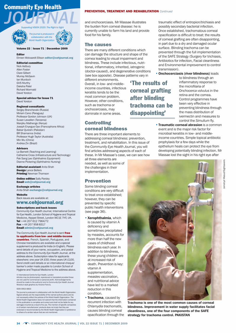
trachoma is one of the most common causes of corneal blindness. Improvement in water supply facilitates facial cleanliness, one of the four components of the SaFE strategy for trachoma control. PaKIStan
Jam
shyd
Mas
ud/S
ight
save
rs
CEHJ71_OA.indd 34 13/01/2010 12:13

an abrasion caused by vegetable matter. It is possible that early antibiotic prophylaxis could have prevented this.
TreatmentIn most low- and middle-income countries, microbial keratitis is the most common acute blinding corneal problem requiring treatment. There is often a history of minor trauma. If appropriate antibiotic prophylaxis is not started soon after the injury, infection can become established. In temperate climates, most infections are bacterial. In contrast, in tropical regions, fungal keratitis is more frequent and may account for about half the cases.
The treatment of microbial keratitis is discussed in detail in the articles on pages 39–41.
Several problems make it difficult to deliver effective treatment for microbial keratitis in a low- and middle-income country setting. These problems need to be addressed by eye care programmes in order to reduce the risk of blindness from microbial keratitis and include:
• Delayed presentation. There may be many days or even weeks between the onset of symptoms and the presentation of the patient at an appropriate health facility. This delay is often catastrophic, allowing time for deep-seated infection to develop and extensive corneal damage to occur. Timely presentation may be promoted through health education and training of staff at primary health facilities to recognise and refer patients with established microbial keratitis.
• traditional medication. This may sometimes be used by the patient before presentation and can make the problem more severe through the harmful effect of toxic compounds and infection with additional microorganisms.
• Microbiology. It may not be possible to obtain a microbiological diagnosis or information about the sensitivity of the organism. This can lead to the use of ineffective treatment and is particularly a
problem when fungal keratitis is missed. The development of a basic microbiological service with gram staining of slides can help to identify some cases of fungal infection. Blindness control programmes need to know which organisms commonly cause microbial keratitis in their population as well as their pattern of antibiotic resistance so that appropriate drugs can be supplied to health facilities.
• Inadequate treatment. The patient may not receive effective treatment. This can occur for several reasons: appropriate antibacterial or antifungal drops may not be available, the microorganism may be resistant to the medication, or the drops may not be given with sufficient intensity. Some of these problems can be overcome with the development of locally appropriate treatment protocols.
Mr Massae’s case illustrates some of these issues. It was several weeks before he reached an ophthalmology unit and received treatment, which contributed to the severity of the case. It was very helpful in his management to have a microbiological diagnosis as it guided the choice and duration of treatment.
RehabilitationMr Massae is now blind. However, his left eye has perception of light and has the potential to see better; to be rehabilitated. It may be possible to offer him some improvement in vision with a pupilloplasty. In addition, as the left corneal scar does not involve the superior cornea, a rotational auto-graft, in which an eccentric corneal button is cut and rotated to bring the clear superior cornea into the centre, may help. However, a penetrating corneal graft (trans-plant) would probably offer him the best quality of vision.
In many low- and middle-income countries, options for visual rehabilitation from corneal disease are limited as it usually requires the services of an ophthal-mologist with sub-specialty training in corneal surgery, equipped to perform the
surgery and with access to donated corneas from an eye bank.
In this issue, there is an article on corneal grafting (page 44) which discusses the indications for corneal grafting, the outcomes for different conditions, and some of the potential complications. The following article focuses on eye banking (page 46) and addresses some of the specific challenges involved in running an eye bank service and finding donors.
Without rehabilitation services, Mr Massae and several million people like him are destined to a life without sight. Without the implementation of the public health and treatment interventions outlined above, many more will be at risk of joining them.
Infectious• Bacterial keratitis• Fungal keratitis• Viral keratitis • Trachoma• Onchocerciasis• Leprosy• Ophthalmia neonatorum
Nutritional• Vitamin A deficiency (xerophthalmia)
Inflammatory• Mooren’s ulcer• Steven’s Johnson Syndrome
Inherited• Corneal stromal dystophies• Fuch’s endothelial dystrophy
Degenerative• Keratoconus
Trauma• Corneal abrasion predisposing to
microbial keratitis• Penetrating trauma• Chemical injury
Doctor-caused (iatrogenic)• Pseudophakic bullous keratopathy
Causes of corneal blindness
Mat
thew
Bur
ton
Mat
thew
Bur
ton
Figure 1. Mr Massae’s left eye, after several weeks of treatment Figure 2. Mr Massae’s right eye
Copyright © 2009 Matthew J Burton. This is an open access article distributed under the Creative Commons Attribution License, which permits unrestricted use, distribution, and reproduction in any medium, provided the original work is properly cited.
CEHJ71_OA.indd 35 13/01/2010 12:14

36 COMMUnIty EyE HEaLtH JOURnaL | VOL 22 ISSUE 71 | DECEMBER 2009
PREVENTION
Preventing corneal blindness: working with communities
Preventing corneal blindness in the community involves action by the community itself, as well as actions by government and non-governmental organisations in the form of health and development services. In order to be effective, eye health workers need to understand how all of the above can work together.
Prevention of corneal blindness takes place at three levels:
1 Primary prevention: Actions or interven-tions taken to prevent the onset of disease
2 Secondary prevention: Actions taken to prevent complications and/or the devel-opment of visual disability due to an existing disease
3 tertiary prevention: After the immediate problem has been addressed by surgery or other treatment, actions to restore function or reduce existing disability from disease complications, i.e. corneal trans-plantation (see page 44).
Right up to the point when someone is seen by an eye care worker or admitted to hospital, the community will influence what happens.
Consider ophthalmia neonatorum as an example. As an eye care worker, you may have little control over the following:
• the risk factors and immediate medical causes, e.g. parents’ sexual behaviour and the presence of Neisseria gonorrhoea
• the contributory and social factors, e.g. the lack of antibiotic drops in the labour ward, poverty associated with
dangerous work, poor access to water and sanitation, or a community’s preference for traditional medicines.
However, the community has the potential to influence most of these factors, either through change in the behaviour of individuals, or by lobbying for improvements at the community level.
The eye care worker’s role, in particular when designing or participating in programmes to reduce corneal blindness, should be to inform and assist the community to address whichever of the above factors are relevant. This will allow the community to become an active partner in the prevention of corneal blindness.
First stepsA successful corneal blindness prevention programme does not:• focus solely on individual diseases• ignore the perceptions, knowledge, and
abilities of the community• work in isolation from other services in the
health system.
A successful corneal blindness prevention programme does:
• address the overall causes of corneal blindness in the community
• aim to understand the community, build on their existing knowledge, and encourage and support them to campaign for better services
• understand the health and development services available in the community with a view to supporting them and making the best possible use of them.
Whether you are planning to improve a prevention programme or designing a new one, it is helpful to learn as much as possible about what the community needs and how they may be able to support your programme.
Doing so ensures that as many people as possible are involved right from the start.
It is a good idea to do a situation analysis, which will help to clarify what you know about the community and identify any gaps in your knowledge (which you will then need to fill).
Here are some questions to get you started:• What are the community’s knowledge and
perceptions regarding the causes and treatment of corneal blindness?
• What are the existing and traditional methods of communication within the community? How can these be used to transmit new health messages?
• How will the community’s knowledge and perceptions influence the content of health messages and how they are presented?
• What skills exist within the community that may be used to support the programme?
• What are the first points of contact for care: homes, schools, traditional healers, or pharmacists/chemist shops? What first aid is usually practiced?
Primary preventionPrimary prevention of corneal blindness is particularly relevant for the following causes:
• vitamin A deficiency and measles• ophthalmia neonatorum• trachoma• eye injuries.
Hannah FaalProgramme Development Adviser: Health Systems, Sightsavers International, 21 Nii Nortei Ababio Road, PO Box KIA 18190, Airport, Accra, Ghana.
traditional remedies, such as the one around this boy’s neck, contribute to delays in seeking medical treatment. tHE GaMBIa
Sig
htsa
vers
Inte
rnat
iona
l
Lanc
e B
elle
rs/S
ight
save
rs In
tern
atio
nal
Community development projects help to reduce the most common causes of corneal blindness. tanZanIa
CEHJ71_OA.indd 36 13/01/2010 12:14

There are many social factors associated with corneal disease, such as poverty, inadequate water supply and sanitation, poor nutrition, and dangerous agricultural practices. Other contributing factors may include the high cost or unavailability of medicines or safety goggles. A good programme should support the community to obtain the health care and other services it needs, either by mobilising the community’s own resources or by lobbying government for help.
To address the immediate medical causes and risk factors, the programme should provide health education about risk factors and how to avoid them, as well as information about what to do and where to go for help if an eye problem develops.
Support for these activities may be possible by closely collaborating with the health promotion unit of the local or national health system.
Good communication is essential. Use what you have learnt from the situation analysis to plan communication activities, for example by using existing and community-friendly methods. In urban areas, use the media and billboards. In rural areas, a meeting of the village elders may be more effective. Integration of eye health messages into the school curriculum is another possibility.
Secondary preventionThe cornea is transparent and sensitive to pain. As a result, patients with corneal disease or injuries are usually in pain and may suffer from photophobia; their eyes may water and they may have blurred vision. These all prompt the patient or carer (in the case of a child) to take action early.
Because of the pain, people may self-medicate, either with harmful medicines obtained from family members or from nearby care providers such as traditional healers or local pharmacists/chemist shops.
These early attempts at seeking care may be harmful, but also delay the process of obtaining correct treatment from the nearest medical facility. Both factors – wrong management and delay – may contribute more to corneal opacity and visual loss than the original cause.
Fighting corneal blindness by strengthening health systemsUnderstanding the health systems which already serve the community will help to ensure that new programmes make the best use of what is available and don’t overload existing services. With careful thought, it may even be possible for a programme to contribute to the existing health systems, leaving them stronger and better able to serve the community in future.
Many health and community devel-opment programmes already in existence, such as measles immunisation, perinatal care, nutrition, water supply, and sanitation, make a significant contribution
to reducing the most common causes of corneal blindness. It is important to support these programmes by informing policy makers and funding agencies of their impact on the prevention of blindness, as this will increase the motivation of those involved and may improve the prospects for
continued political and financial support.The following aspects of existing health
systems can be strengthened with your help.
The health work force• Work with the community to identify
individuals who can provide home-based care and training. These could include retired professional people such as health workers or teachers who have returned to live in their communities. Support them either directly or through existing primary health care structures.
• Collaborate with existing health workers, traditional or not. For example, ensure that they are aware of the dangers of steroid eye drops and that they understand why it is important to instil antibiotic or antiseptic drops in newborn babies’ eyes.
• Teach all health workers to diagnose and refer corneal pathology early.
Medical products and vaccines• Ensure that basic items, such as torches
and antibiotic/antiseptic drops, are available at points of need, e.g. in labour
wards, with traditional birth attendants, or in schools.
• Support existing efforts to provide vaccines and ensure cold chains.
Health information • Gather information about the impact of
corneal disease and trauma in the community. For example, ensure that ophthalmia neonatorum is a notifiable disease and record the number of children with measles or xerophthalmia.
• Gather evidence about the effectiveness of community-focused intervention measures, such as water and sanitation programmes, immunisation campaigns, or free health care for children.
• Use your evidence to improve programme design and service delivery, and to lobby the authorities to maintain and strengthen these programmes.
Health financing• Work with the community to ensure that
emergency eye care for corneal infections or trauma, particularly in children, is free and that cost does not restrict access to treatment.
Leadership and governance• Work with the decision-making bodies
responsible for the local community’s development and health. Encourage them to allocate resources to measures such as latrine construction and home-based care.
• Encourage communities to take the lead on health matters, for example by working with community development groups. Ordinary people can take responsibility for a range of interventions, from household- led health activities such as face washing to demanding better services.
• Support and encourage communication between the community and decision makers within the health system, as well as between different groups or specialties in the health system.
ConclusionAs an eye health worker designing or imple-menting a programme to prevent corneal blindness in the community, you should understand both the medical causes of corneal scarring, and the non-medical and social factors that lead to corneal blindness. You should recognise the potential of the community to be involved and actively seek out ways to ensure their involvement.
It is vital to understand the impact of development programmes led by other government departments (education, agriculture, water resources, community development, and justice) on the prevention of corneal blindness. Eye workers must support and work with these initiatives.
In order to do this well, you, as an eye health worker, should develop non-medical skills such as communication, negotiation, advocacy, and the ability to foster community engagement.
‘A good programme should support the community to obtain the health and other services it needs’
Eye health posters used to inform the community. tHE GaMBIa
Sig
htsa
vers
Inte
rnat
iona
l (2)
Copyright © 2009 Hannah Faal. This is an open access article distributed under the Creative Commons Attribution License, which permits unrestricted use, distribution, and reproduction in any medium, provided the original work is properly cited.
CEHJ71_OA.indd 37 13/01/2010 12:14

38 COMMUnIty EyE HEaLtH JOURnaL | VOL 22 ISSUE 71 | DECEMBER 2009
Before performing any eye procedure• Wash your hands (and afterwards too)• Position the patient comfortably with
head supported• avoid distraction for yourself and the patient• Ensure good lighting• Always explain to the patient (and any
companion, if appropriate) what you are going to do.
Reasons for eyelid control during eye examination• to provide a good view of the eyeball for
the examiner• to avoid unnecessary discomfort for the
patient
Remember!It is important to be very gentle at all times, in particular when an injured, painful, or postoperative eye is being examined. to do otherwise may cause further problems. Eyelid control is very important!
PreparationPosition the patient comfortably. Depending on the circumstances, this may be:
• lying down with his or her head on a pillow • sitting down with his or her head resting
against a wall or headrest, or with the head supported by the hands of an assistant (Figure 1)
• sitting down at a slit lamp with head supported on the chin rest.
Method• Ask the patient to look up and hold this
gaze• With the index fi nger, gently and slowly
pull down the lower eyelid
this position will enable a good view of the lower eyelid margin and lower eyeball (Figure 2).
• When examination of this area is complete, gently and slowly remove the index
fi nger and allow the patient to close the eyes for a few seconds
• Ask the patient to look down and to hold this gaze
• With the tip of the thumb, gently and slowly touch the top eyelid midway between the eyelid margin and the eyebrow (Figure 3) – do not exert any pressure!
• Ease the eyelid up, gently and slowly, against the bony orbital rim
this position will enable a good view of the upper eyelid margin and the upper eyeball (Figure 4).
• When examination of this area is complete, gently and slowly remove the thumb and allow the patient to close the eyes
• Tell the patient when the examination has ended.
IMPORtant! These principles should be followed every time and by every examiner.
Eyelid control during an eye examination
Useful resources: corneal blindness
HOW TO
USEFUL RESOURCES
Sue StevensFormer Nurse Advisor, Community Eye Health Journal, International Centre for Eye Health, London School of Hygiene and Tropical Medicine, Keppel Street, London WC1E 7HT, UK.
Figure 1 Figure 2 Figure 3
Community Eye Health Journal Kanyi S. Evaluation of the impact of Nyateros (friends of the eye) in the delivery of eye care services after one year of its implementation in Lower River division, the Gambia. Community Eye Health J 2005;18(56): 130–134.Lecuona K. assessing and managing eye injuries. Community Eye Health J 2005;18(55): 101–104. Garg P, Rao Gn. Corneal ulcer: diagnosis and management. Comm Eye Health J 1999;12(30): 21–23.Baba I. the red eye: fi rst aid at the primary level. Community Eye Health J 2005;18(53): 70–72.
BooksEye diseases in hot climates. By John Sandford-Smith. £12 from TALC.Hanyane: a village struggles for eye health. By Erica Sutter, Victoria Francis, and Allen Foster. Free to download from www.cehjournal. org/icehpubs.asp or £5 from TALC.
Other resourcesa laboratory manual and guide to management of microbial keratitis. By Astrid K Leck, Melville M Matheson, andJ Heritage. Free to download fromwww.cehjournal.org/icehpubs.aspthe red eye (poster). Free to download from www.cehjournal.org/icehpubs.asp or order from TALC.
Guidelines for the management of corneal ulcer at primary, secondary, and tertiary care in the Southeast asia Region. Go to www.searo.who.int/LinkFiles/Publications_Final_Guidelines.pdfEuropean Eye Banking association. Visit www.europeaneyebanks.org or write to: European Eye Bank Association, Via Paccagnella n. 11, Padiglione Rama, 30174 Zelarino, Venice, Italy.Email: [email protected]
Supplier: TALCTeaching Aids at Low Cost (TALC), PO Box 49, St Albans, Herts, AL1 5TX, UK. Tel: +44 172 785 3869. Email: [email protected] Website: www.talcuk.org
Figure 4
Copyright © 2009 Sue Stevens. This is an open access article distributed under the Creative Commons Attribution License, which permits unrestricted use, distribution, and reproduction in any medium, provided the original work is properly cited.
CEHJ71_OA.indd 38 13/01/2010 12:14

IntroductionInfections of the cornea can lead to corneal opacity and blindness if not identified quickly and managed appropri-ately. The terms infective keratitis, suppurative keratitis, and microbial keratitis are all used to describe suppu-rative infections of the cornea. These are characterised by the presence of white or yellowish infiltrates in the corneal stroma, with or without an overlaying corneal epithelial defect, and associated with signs of inflammation (Figure 1).
The common symptomatic complaints of patients with suppurative keratitis are as follows (all with varying degrees of severity):
• redness of the eye• circum-corneal congestion (typically)• pain• blurring of vision• photophobia• watering of the eye.
The aim of this article is to review both bacterial and fungal keratitis, with an emphasis on identification and management at the primary, secondary, and tertiary levels. Guidelines for referral will be suggested.
Fist steps in managementHistory takingHistory taking is an important step in management of corneal infection. If there has been an injury, ask when and where the injury was sustained, what the patient was doing at the time of injury, whether or not he or she sought help following the injury, and what treatment – including traditional eye medications – may have been used.
A past history of conjunctivitis may suggest that the infection is secondary to a conjunctival pathogen.
Examination1. Visual acuityVisual acuity should always be recorded in all cooperative patients. If it is not possible to record the visual acuity of a child, for example, a note of this should be made. Vision should be recorded first in the unaffected eye, then in the affected eye; with or without glasses. This provides a useful guide regarding the prognosis and response to treatment. It is also important documen-tation in the event of medico-legal issues.
2. Examination of the corneaA torch with a good source of focused light and a loupe for magnification are essential. A slit lamp microscope, if available, is always helpful, but not absolutely essential.
Another essential tool is fluorescein dye, either in a sterile strip or a sterile solution. Fluorescein stains any part of the cornea that has lost the epithelium, even due to a trivial injury, and appears brilliant green when viewed under blue light (Figure 3).
3. Corneal scrapeDiagnosis should be confirmed by obtaining a corneal scraping from the corneal lesion and subjecting it to laboratory testing at secondary or tertiary eye care facilities. See article on page 42.
Management at primary levelA suppurative corneal ulcer is an ophthalmic emergency which should be referred to the nearest eye centre for proper management. The following are useful guidelines when referring the patient to the secondary eye care centre.
• Do apply antibiotic drops or ointment• Do instruct patients and/or their
accompanying persons to apply drops frequently until patients arrive at the centre
• Do instruct patients and/or their accompanying persons to avoid traditional medicines.
• Do not give systemic antibiotics; they are not helpful
• Do not use steroid drops and/or ointment; they can be dangerous
• Do not routinely patch the eye; it is not necessary.
Management at secondary levelMore complete management of corneal infections begins at the secondary level of eye care where there is an ophthalmologist and/or an ophthalmic assistant, or a physician trained in managing common eye diseases.At the secondary level:
• A corneal scraping should be taken (see page 42).
• The patient should be admitted to the hospital to ensure adequate treatment and frequent follow-up.
Specific initial treatment
No fungal elements seen• instil cefazolin 5% and gentamicin
1.4% drops hourly. • Ciprofloxacin or ofloxacin is a good
substitute for gentamicin and cephazolin. If it is not possible to administer hourly drops, a subconjunctival injection can be given.
39COMMUnIty EyE HEaLtH JOURnaL | VOL 22 ISSUE 71 | DECEMBER 2009
MANAGEMENT
Managing corneal disease:focus on suppurative keratitis
Madan P UpadhyayPresident, BP Eye Foundation, Kathmandu, Nepal. Email: [email protected]
Muthiah SrinivasanDirector and Chief of Cornea Services, Aravind Eye Hospital, Madurai, India.
John P WhitcherClinical Professor of Ophthalmology, Proctor Foundation, University of California, San Francisco, USA.
Figure 2. A bacterial ulcer. The eye is very red and inflamed. Note the ring infiltrate in the cornea and a large hypopyon in the anterior chamber.
Figure 3. Fluorescein staining
Dr M
Srin
ivas
an/A
ravi
nd E
ye H
ospi
tal
Figure 1. A severe bacterial ulcer caused by Pseudomonas sp. The gram negative (-ve) bacillus can cause complete destruction of the cornea within a few days. This cornea is at risk of perforation.
Dr M
Srin
ivas
an/A
ravi
nd E
ye H
ospi
tal
Continues overleaf ➤
CEHJ71_OA.indd 39 13/01/2010 12:14

40 COMMUnIty EyE HEaLtH JOURnaL | VOL 22 ISSUE 71 | DECEMBER 2009
Fungal elements seen• Natamycin 5% drops hourly or freshly
reconstituted amphotericin-B 0.15% as drops hourly. Antibiotics may have a limited role to play in such cases and may occasionally be harmful. Clinical judgment correlated with laboratory tests constitute the best guide in such cases.
Adjunctive treatment• Atropine 1% or homatropine 2% could
be used twice a day to dilate the pupil; this helps to prevent synechiae and relieve pain
• Oral analgesics will help to minimise pain• Anti-glaucoma medication may be
advisable if the intraocular pressure is high• Vitamin A supplementation may be helpful,
particularly in countries where Vitamin A deficiency is markedly prevalent.
Five a’s are a useful acronym to remember: antibiotic/antifungal, atropine, analgesics, anti-glaucoma medications, and Vitamin a.
Subsequent management Suppurative keratitis patients should be hospitalised and examined daily, if possible with a slit lamp, so that their response to treatment can be evaluated and the frequency of antibiotics adjusted accordingly.
Reduce the frequency of antibiotic administration when the patient experi-ences symptomatic improvement (less tearing and photophobia, relief from pain, and improvement in vision) and when the ulcer shows signs of improvement:
• decrease in lid oedema • decrease in conjunctival chemosis and
bulbar conjunctival injection • reduction in density of the infiltrate and
area of epithelial ulceration • haziness of the perimeter of the ulcer and
of the stromal infiltrate • decrease in inflammation; cells, fibrin,
and level of hypopyon • dilatation of pupil.
In the case of bacterial infection, the inflam-matory reaction may be enhanced by endotoxin release during the first 48 hours of treatment; however, definite progression at this stage is unusual and implies that either the organisms are resistant to therapy, or the patient is not instilling the drops as prescribed.1 If the patient is judged to be improving, the dose of antibiotics and/or antifungal drops should be reduced from hourly, to two-hourly, then four-hourly over the next two weeks in case of bacterial ulcers. For fungal ulcers, treatment should be continued with three-hourly drops for at least three weeks.
Guidelines for referral to a tertiary centreImmediate referral on presentation if:• the ulcer is in an only eye• the patient is a child • there is impending or actual perforation
• a fungal corneal ulcer is suspected but KOH or other fungal stains are not available.
Following initial treatment: if cases of bacterial ulcer fail to show any improvement within three days, and fungal ulcers within a week, patients should be referred to a tertiary care centre.
Management of corneal ulcer at tertiary level Many tertiary eye care centers have their own protocol for the management of corneal ulcer. The management suggested is based on a WHO recommendation for suitable modification according to circumstances.2
History, examination, and recording of findingsBy the time patients have reached a tertiary centre, they will have travelled from one place to another with attendant hassles, received several treatments, may have lost faith in eye care personnel, and may already have run out of money, partic-ularly in low-income countries. Attending to this situation is critically important in the overall care of corneal ulcer patients.
A careful history of the development of the disease may point to the existence of an underlying predisposing condition such as diabetes mellitus, immunosuppression due to local or systemic steroids (or other immunosuppressants), dacryocystitis, or other ocular conditions. A full list of drugs used by the patient should be obtained to ensure that drugs which have not helped in the past are not repeated; this may also help to discover possible drug allergies. Findings should be carefully noted on a standard form.
A meticulous corneal scraping subjected to laboratory processing often provides a sound guideline to treatment. See page 42.
Hospitalisation This provides patients with rest and adequate medication; they can also receive frequent follow-up, management of systemic problems such as diabetes, and further surgical intervention, if warranted.
TreatmentThe initial treatment depends on the results of the corneal scrape and the local pattern of pathogens and antibiotic resistance.• If microscopy is negative, or it is not
possible to perform a corneal scrape, or Gram-positive or Gram-negative bacteria are visualised, treat the patient with antibiotic eyedrops. Use either a combination of cefazolin 5% and gentamycin 1.4%, or fluoroquinolone monotherapy (eg. ciprfloxacin 0.3% or ofloxacin 0.3%). Drops should be given hourly to begin with for two days and then tapered, based on response.
• If microscopy reveals fungal hyphae, topical natamycin 5%, econazole 1% or amphotericin-B 0.15% should be used hourly for a week and then tapered.
• If the ulcer seems to respond well to treatment, continue therapy as before for two weeks for a bacterial ulcer and three weeks or more for fungal ulcer.
• If the response is poor and the culture shows growth of an organism, the choice of antibiotic is guided by the sensitivity reports.
Figure 3. Subtotal fungal ulcer
Dr W
hitc
her/U
CS
F
antibiotic Method Final concentration
Cefazolin/cefuroxime Add 10 ml sterile water to 500 mg cefazolin powder; mix and use as topical drops. Shelf life: 5 days
50 mg/ml (5%)
Gentamicin (tobramycin)
Add 2 ml parenteral gentamicin (40 mg/ml) to a 5 ml bottle of commercial ophthalmic gentamicin (3 mg/ml)
14 mg/ml (1.4%)
Penicillin G Add 10 ml of artificial tears to a 1 million unit vial of Penicillin G powder; mix, remove, and place into empty artificial tear bottle or xylocaine vials (30 ml)
100,000 units/ml
Vancomycin Add 10 ml sterile water to a 500 mg vial of vancomycin powder; mix, add sterile cap, and use
50 mg/ml (5%)
Amikacin Add 2 ml of parenteral amikacin containing 200 mg to 8 ml artificial tears or sterile water in a sterile empty vial.
20 mg/ml (2%)
Table 1. Preparation of fortified antibiotic eye drops
MANAGEMENT Continued
CEHJ71_OA.indd 40 13/01/2010 12:14
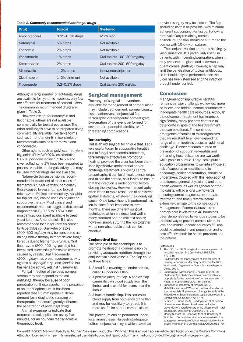
Although a large number of antifungal drugs are available for systemic mycoses, only few are effective for treatment of corneal ulcers. The commonly recommended drugs are given in Table 2.
However, except for natamycin and fluconazole, others are not available commercially for topical ocular use. The other antifungals have to be prepared using commercially available injectable forms such as amphotericin-B, miconazole, or raw materials such as clotrimazole and voriconazole.
Other agents such as polyhexamethylene biguanide (PHMB) 0.02%, chlorhexidine 0.02%, povidone iodine 1.5 to 5% and silver sulfadiazine 1% have been reported to possess variable antifungal activity and may be used if other drugs are not available.
Natamycin 5% suspension is recom-mended for treatment of most cases of filamentous fungal keratitis, particularly those caused by Fusarium sp. Topical miconazole 1% (not commercially available for topical use) can be used as adjunct or supportive therapy. Most clinical and experimental evidence suggests that topical amphotericin-B (0.15 to 0.5%) is the most efficacious agent available to treat yeast keratitis. Amphotericin-B is also recommended for fungal keratitis caused by Aspergillus sp. Oral ketaconazole(200–600 mg/day) may be considered as an adjunctive therapy in more severe fungal keratitis due to filamentous fungus. Oral fluconazole (200–400 mg per day) has been used successfully for severe keratitis caused by yeasts. Oral itraconazole (200 mg/day) has broad-spectrum activity against all Aspergillus sp. and Candida but has variable activity against Fusarium sp.
Fungal infection of the deep corneal stroma may not respond to topical antifungal therapy because of poor penetration of these agents in the presence of an intact epithelium. It has been reported that a 5 mm epithelial debri-dement (as a diagnostic scraping or therapeutic procedure) greatly enhances the penetration of antifungal drugs.
Animal experiments indicate that frequent topical application (every five minutes) for an hour can readily achieve therapeutic level.
Surgical management The range of surgical interventions available for management of corneal ulcer may include debridement, corneal biopsy, tissue adhesives, conjunctival flap, tarsorraphy, or therapeutic corneal graft. Evisceration of the eye is performed for severe pain, panophthalmitis, or life- threatening complications.
TarsorrhaphyThis is an old surgical technique that is still very useful today. In suppurative keratitis due to fungal and bacterial infections, tarsorrhapy is effective in promoting healing, provided the ulcer has been steri-lised by effective antibacterial and/or antifungal treatment. Following central tarsorrhaphy, it can be difficult to instil drops and to see the cornea, so it is vital to ensure that the infection is under control before closing the eyelids. However, tarsorrhaphy often leads to rapid resolution of persistent epithelial defects, whatever the underlying cause. Once tarsorrhaphy is performed it is left in place for at least one to three months. There are different surgical techniques which are described well in many standard ophthalmic text books; however, simply suturing the lids together with a non-absorbable stitch can be effective.
Conjunctival flapThe principle of this technique is to promote healing of a corneal lesion by providing adequate nutrition through the conjunctival blood vessels. The flap could be three types:
1 A total flap covering the entire cornea,
called Gunderson’s flap. 2 A pedicle (racquet) flap. A pedicle flap
carries its own blood supply from the limbus and is useful for ulcers near the limbus.
3 A bucket handle flap. This carries its blood supply from both ends of the flap and may be less likely to retract. It is more useful for central corneal ulcers.
This procedure can be performed under local anaesthesia. Harvesting adequate bulbar conjunctiva in eyes which have had
previous surgery may be difficult. The flap should be as thin as possible, with minimal adherent subconjunctival tissue. Following removal of any remaining corneal epithelium, the flap should be sutured to the cornea with 10-0 nylon sutures.
The conjunctival flap promotes healing by vascularisation. It is particularly useful in patients with impending perforation, when it may preserve the globe and allow subse-quent corneal grafting. However, a flap may limit the penetration of topical antibiotics, so it should only be performed once the ulcer has been sterilised and the infection brought under control.
ConclusionManagement of suppurative keratitis remains a major challenge worldwide, more so in low- and middle-income countries with inadequate health care resources. Although the outcome of treatment has improved significantly, many patients continue to deteriorate in spite of the best treatment that can be offered. The continued emergence of strains of microorganisms that are resistant to an ever-expanding range of antimicrobials poses an additional challenge. Further research related to prevention of suppurative keratitis and enhancing host resistance are two worth-while goals to pursue. Large-scale public education programmes to sensitise those at risk of suppurative keratitis, and to encourage earlier presentation, should be undertaken. Coupled with this, education of practioners, general physicians, and other health workers, as well as general ophthal-mologists, will go a long way towards ensuring correct diagnosis, appropriate treatment, and timely referral before extensive damage to the cornea occurs. Management of corneal abrasions at primary care levels within 48 hours has been demonstrated by various studies to be the best way to prevent corneal ulcers in low- and middle-income countries.3-6 This could be adopted in any population and is cost effective both for health providers and the patient.
References1 Allan BD, Dart JK. Strategies for the management of
microbial keratitis. Br J Ophthalmol 1995;79: 777–786.
2 Guidelines for the management of corneal ulcer at primary, secondary and tertiary health care facilities. World Health Organization, South East Asia Regional Office, 2004.
3 Upadhyay M, Karmacharya S, Koirala S, et al. The Bhaktapur Eye Study: Ocular trauma and antibiotic prophylaxis for the prevention of corneal ulceration in Nepal. Br J Ophthalmol 2001;85: 388–92.
4 Srinivasan S, Upadhyay MP, Priyadarsini B, Mahalakshmi, John P Whitcher. Corneal ulceration in south-east Asia III: prevention of fungal keratitis at the village level in South India using topical antibiotics. Br J Ophthalmol 2006;90: 1472–1475.
5 Getshen K, Srinivasan M, Upadhyay MP, et al.Corneal ulceration in south-east Asia I: a model for the prevention of bacterial ulcers at the village level in rural Bhutan. Br J Ophthalmol 2006;90: 276–78.
6 Maung N,Thant CC,Srinivasan M et al, Upadhyay M, Whitcher J. Corneal ulceration in south-east Asia II: a strategy for prevention of fungal keratitis at the village level in Myanmar. Br J Ophthalmol 2006;90: 968–70.
Drug topical Systemic
Amphotericin-B 0.15–0.5% drops IV infusion
Natamycin 5% drops Not available
Econazole 2% drops Not available
Voricanozole 1% drops Oral tablets 100–200 mg/day
Ketoconazole 2% drops Oral tablets 200–600 mg/day
Miconazole 1–2% drops Intravenous injection
Clotrimazole 1–2% ointment Not available
Fluconazole 0.2–0.3% drops Oral tablets 200 mg/day
Table 2. Commonly recommended antifungal drugs
Copyright © 2009 Madan P Upadhyay, Muthiah Srinivasan, and John P Whitcher. This is an open access article distributed under the Creative Commons Attribution License, which permits unrestricted use, distribution, and reproduction in any medium, provided the original work is properly cited.
CEHJ71_OA.indd 41 13/01/2010 12:14
42 COMMUnIty EyE HEaLtH JOURnaL | VOL 22 ISSUE 71 | DECEMBER 2009
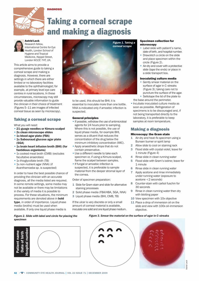
This article aims to provide a comprehensive guide to taking a corneal scrape and making a diagnosis. However, there are settings in which there are either limited or no laboratory facilities available to the ophthalmologist; for example, at primary level eye care centres in rural locations. In these circumstances, microscopy may still provide valuable information to guide the clinician in their choice of treatment (Figures 5–11 are images of infected corneal tissue as seen by microscopy).
Taking a corneal scrapeWhat you will need:• 21-guage needles or Kimura scalpel• 2x clean microscope slides • 1x blood agar plate (FBa)• 1x Sabouraud glucose agar plate
(SGa)• 1x brain heart infusion broth (BHI) (for
fastidious organisms)• 1x cooked meat broth (CMB) (excludes
facultative anaerobes)• 1x thioglycollate broth (TB)• 1x non-nutrient agar (NNA) (if
Acanthamoeba sp. is suspected)
In order to have the best possible chance of providing the clinician with an accurate diagnosis, all the media listed are required. In some remote settings, some media may not be available or there may be limitations in the variety of media it is possible to process. For these situations, the minimum requirements are denoted above in bold type, in order of importance. Liquid phase media (broths) must be used when available. If only one liquid phase media is
to be used, this should be BHI; it is essential to inoculate more than one bottle. NNA is indicated only if amoebic infection is suspected.
General principles• If possible, withdraw the use of antimicrobial
agents for 24 hours prior to sampling. Where this is not possible, the use of liquid phase media, for example BHI, serves as a diluent that reduces the concentration of the drug below the minimum inhibitory concentration (MIC).
• Apply anaesthetic drops that do not contain preservative.
• Use a different needle to take each specimen or, if using a Kimura scalpel, flame the scalpel between samples.
• If fungal or amoebic infection is suspected, it is preferable to sample material from the deeper stromal layer of the cornea.
Order of specimen preparation:
1 Slide for Gram stain and slide for alternative staining processes
2 Solid phase media (FBA/HBA, SGA, NNA) 3 Liquid phase media (BHI, CMB, TB)
If the ulcer is very discrete or only a small amount of corneal material is available, inoculate one solid and one liquid phase medium.
Specimen collection for microscopy• Label slide with patient’s name, date of birth, and hospital number. • Draw/etch a circle on the slide and place specimen within the circle (Figure 2).• Air-dry and cover with a protective slide (tape the ends) or place in a slide transport box.
Inoculating culture media• Gently smear material on the surface of agar in C-streaks (Figure 3); taking care not to puncture the surface of the agar.• Sellotape the lid of the plate to
the base around the perimeter.• Incubate inoculated culture media as
soon as possible. Refrigeration of specimens is to be discouraged and, if not being transported directly to the laboratory, it is preferable to keep samples at room temperature.
Making a diagnosis Microscopy: the Gram stain1 Air-dry and heat-fix specimen using a
Bunsen burner or spirit lamp2 Allow slide to cool on staining rack3 Flood slide with crystal violet; leave for
1 minute (Figure 4)4 Rinse slide in clean running water5 Flood slide with Gram’s iodine; leave for
1 minute6 Rinse slide in clean running water7 Apply acetone and rinse immediately
under running water (exposure to acetone <2 seconds)
8 Counter-stain with carbol fuschin for 30 seconds
9 Rinse in clean running water then dry with blotting paper
10 View specimen with 10x objective11 Place a drop of immersion oil on the
slide and view with 100x oil-immersion objective.
DIAGNOSIS
Taking a corneal scrape and making a diagnosis
Astrid LeckResearch fellow, International Centre for Eye Health, London School of Hygiene and Tropical Medicine, Keppel Street, London W1CE 7HT, UK.
Figure 1. Taking a corneal scrape
J D
art
Figure 2. Slide with label and circle for placing the specimen
Pati
ent
nam
eD
ate
of b
irth
Hos
pita
l num
ber
Astr
id L
eck
Figure 3. Smear the material on the surface of agar in C-streaks
CEHJ71_OA.indd 42 13/01/2010 12:14

• Gram positive (+ve) cocci most commonly associated with suppurative keratitis are the Staphylococci (Figure 5) and Streptococci (Figure 6, Streptococcus pneumoniae).
• Gram negative (–ve) bacilli, such as Pseudomonas sp. (Figure 7), may be associated with corneal infection.
• A definitive diagnosis of Nocardia sp (Gram variable) may be possible
• Yeast cells will stain positively.
Although not the first choice of stains for fungi, yeast cells, pseudohyphae, and fungal hyphae may be visualised in Gram-stained corneal material, typically staining negatively or Gram variable. For microscopy to provide a more definitive diagnostic tool for fungal infection, Gram stain can be destained and restained using a more appropriate stain (Figures 8 and 9).
Microscopy: additional methods Lactophenol cotton blue (LPCB) or potassium hydroxide (KOH) wet mount preparations are used to visualise fungi (Figure 10).
1 Add a drop of lactophenol cotton blue mountant to the slide.
2 Holding the coverslip between your forefinger and thumb, touch one edge of the drop of mountant with the coverslip edge, the lower it gently, avoiding air bubbles. The preparation is now ready.
3 Initial observation should be made using the low power objective (10x), switching to the higher power (40x) objective for a more detailed examination.
4 Calcofluor white and Periodic Acid Schiff reaction (PAS) staining may also be used.
Diagnostic criteriaDiagnostic criteria applied to bacterial culture• the same organism growing at the site of
inoculation on two or more solid phase cultures, or
• growth at site of inoculation on one solid phase media of an organism consistent with microscopy, or
• confluent growth on one media.
Astr
id L
eck
Figure 4. Flood the slide with crystal violet
MM
Mat
heso
nM
M M
athe
son
J D
art
MM
Mat
heso
nPA
Tho
mas
Astr
id L
eck
Figure 5. Staphylococci sp.
Figure 7.Pseudomonas sp.
Astr
id L
eck
Figure 8. Gram appearance of yeast cells (left) and pseudohyphae (right)
Figure 11.Calcofluor white preparation
Figure 6. Streptococcus pneumoniae
Astr
id L
eck
Figure 9.Fungal hyphae visible after Gram stain
Figure 10. Fungal hyphae stained with lactophenol cotton blue
Figure 12.The trophozoite form of Acanthamoeba
Diagnostic criteria applied to fungal specimens• fungal hyphae observed in corneal specimen
stained on microscopic examination, or • growth at site of inoculation on solid
culture media
Amoebic infectionsThe cyst form of Acanthamoeba sp. canbe visualised in corneal material using a direct fluorescent technique such as calcofluor white (Figure 11), haemotoxylin
and eosin, LPCB, or PAS. If corneal infection with Acanthamoeba sp. is suspected, inoculate corneal material onto non-nutrient agar in a demarcated area of the plate. In the laboratory, the square of agar where the specimen was inoculated will be excised and inverted onto an NNA plate seeded with a lawn of E.coli. Growth of the trophozoite form is imperative to confirm viability of the organism and thus prove it to be the organism responsible for infection (Figure 12).
Copyright © 2009 Astrid Leck. This is an open access article distributed under the Creative Commons Attribution License, which permits unrestricted use, distribution, and reproduction in any medium, provided the original work is properly cited.
CEHJ71_OA.indd 43 13/01/2010 12:14

44 COMMUnIty EyE HEaLtH JOURnaL | VOL 22 ISSUE 71 | DECEMBER 2009
IntroductionWe know that many of the most common causes of corneal blindness: vitamin A deficiency, trachoma, and ophthalmia neonatorum, can be prevented by simple primary health care measures. Some condi-tions are more difficult to prevent, but can be effectively treated, such as suppurative keratitis (page 39) or herpes simplex keratitis. However, some patients will still develop blinding corneal disease. What can be done for them?
At present, the best hope for curing corneal blindness is a corneal transplant, also known as a corneal graft.
In contrast with most other transplants, corneal grafting is relatively straightforward. Although additional surgical training is needed, the operation itself requires little equipment beyond a standard cataract set and a good operating microscope. Because there are no blood vessels in the cornea, the likelihood of rejection is less than for other transplants.
In this article, we want to provide guidance to eye care workers who want to know who should be referred for a graft and what complications they may need to manage after patients have had their operation.
What is corneal grafting?In this operation, the central 7–8 mm of the patient’s own diseased cornea is removed. A similar-sized disc of donor cornea is then inserted and sutured into position. In most cases, the full thickness of the cornea, including epithelium, stroma, and endothelium, is transplanted; this is known as a penetrating graft. However, it is also possible to trans-
plant just the outer, anterior layers (stroma and epithelium) or the inner, posterior layers (endothelium and Descemet’s membrane).
Indications and prognosesAny corneal disorder that causes visual impairment may be an indication for a corneal graft. However, the prognosis for corneal grafting varies greatly.
The risk of rejection is higher if the cornea is vascularised (contains blood vessels), or is inflamed or perforated (as is liable to occur in severe suppurative keratitis, for example). The same holds true for any eye that has generalised disease of the ocular surface.
Table 1 lists some of the different diagnoses that may be treated by a corneal graft, along with their prognoses. Conditions with a “very poor” prognosis should not be treated by corneal grafting as this is a waste of valuable transplant tissue.
The visual prognosis for a corneal graft is also affected by the state of the rest of the eye, not just the cornea and ocular surface. Uncontrolled intraocular pressure (IOP) is a contraindication to grafting, as IOP control is likely to be worsened by a corneal graft. Patients with known posterior segment disease affecting the retina or optic nerve are also unlikely to benefit.
A corneal graft requires much more postoperative care than a cataract extraction. This entails repeated clinic visits and using expensive eye drops frequently; therefore, the patient must value their new cornea and be motivated to take care of it.
Patients are most likely to value their new cornea if the grafted eye is their better eye. In general, this means that patients with unilateral disease and perfect vision in the other eye are poor candidates. There are exceptions: for example, if the eye is painful
as well as visually impaired. Corneal grafting in children has a very
poor prognosis and requires more intense postoperative care; it should therefore be considered carefully.
Outcomes Studies from South India1 showed that69 per cent of grafted corneas were clear at two years after surgery. In East Africa,2
87 per cent of grafts for keratoconus survived for at least two years, compared to 65 per cent performed for other diagnoses.
Unfortunately, a clear graft does not guarantee good vision. Patients may have coexisting problems, such as glaucoma, cataract, or amblyopia. Because corneal grafting alters the shape of the cornea, it often causes significant astigmatism, which can be difficult to correct with spectacles.
Visual outcomes in East Africa2 were much better for patients with keratoconus than for those with other diagnoses. In patients grafted for keratoconus, 33 per cent were <6/60 in both eyes preoperatively, compared to 5 per cent post-operatively; 78 per cent could see 6/18 or better following surgery. This data showed that corneal grafting is an effective cure for blindness caused by keratoconus.
This also demonstrates the importance of monitoring and reporting the outcomes of corneal graft operations.
ComplicationsThe main causes of graft failure in both South India1 and East Africa2 were graft rejection and infectious keratitis. Both of these complications are treatable and often preventable. In many cases, graft failure could have been prevented by early and effective management at a local eye clinic.
Corneal grafting: what eye care workers need to know
David YorstonConsultant Ophthalmologist, Gartnavel Hospital, 1053 Great Western Road, Glasgow G12 0YN, Scotland.
Prashant GargAssociate Director, Cornea and anterior segment services, LV Prasad Eye Institute, Hyderabad, India.
Diagnosis Prognosis
Keratoconus Excellent
Corneal dystrophies, such as lattice, granular, Fuch’s Excellent
Corneal scar – healed ulcer Moderate
Bullous keratopathy – aphakic or pseudophakic Moderate
Herpes simplex keratitis Moderate
Corneal scar: active ulcer/keratitis, threatened perforation(grafting may be done to salvage the eye, rather than vision)
Poor
Corneal scar: trachoma Very poor
Ocular surface disorder: chemical burn, Stevens-Johnson syndrome Very poor
Mooren’s ulcer Very poor
Re-graft Very poor
Table 1. Indicaions for corneal graft and their prognoses
CORNEAL GRAFTINGD
avid
Yor
ston
Figure 1. Keratoconus. Note the central scarring, with thinning and ectasia
CEHJ71_OA.indd 44 13/01/2010 12:14

RejectionGraft rejection is caused by an immune reaction directed against the foreign endothelial cells of the transplanted cornea. Approximately 20–30 per cent of penetrating grafts will have a rejection episode at some time. The most common symptoms of graft rejection are blurred vision, light sensitivity, redness, and pain; patients should be advised to attend an eye clinic immediately if they develop any of these symptoms. Rejection is recognised by the appearance of corneal oedema in a previously clear graft. The oedema usually spreads upwards across the graft from the inferior edge of the transplanted cornea. The eye is often inflamed and a line of keratic precipitates may be seen at the edge of the oedematous cornea.
Preventing rejection begins with selection for surgery. Some diagnoses, such as keratoconus and other corneal dystro-phies, have a very low risk of rejection. Following surgery, rejection is prevented by topical steroid drops. These are used at different frequencies for varying lengths of time, depending on the underlying diagnosis and the perceived risk of rejection. Whatever steroid regime is used, the drops should never be suddenly stopped, but should always be tailed off gradually. The usual duration of steroid therapy following a full thickness graft is six months in phakic patients and twelve months in pseudo-phakic or aphakic patients.
With prompt diagnosis and immediate treatment, graft rejection can often be reversed. The recommended management of graft rejection is intensive steroid treatment, initially in the form of hourly topical steroid drops. The use of systemic steroids, such as 500 mg methylpred-nisolone, has been shown to make little difference to the outcome and the authors therefore do not recommend it.
InfectionThe second common cause of graft failure is suppurative keratitis. This presents in the same way as any other microbial keratitis
(page 39). Patients with corneal grafts are at increased risk of corneal infection because they have reduced corneal sensation and are often on long-term topical steroids. The management of infec-tious keratitis is the same in these patients as it is for anyone else. A scraping should be taken for gram stain and culture if available (see page 42). Intensive antibiotic treatment, either monotherapy with a topical fluoroquinolone (such as ofloxacin) or combination treatment with a cepha-losporin (such as cefuroxime) and an aminoglycoside (such as gentamicin) is started immediately after the scraping has been taken, and is continued for at least 48 hours. In some settings, anti-fungal treatment may be required.
Loose sutureThe most common predisposing factor for infectious keratitis following a corneal graft is a loose suture. With time, as the graft wound heals, the very fine sutures either break or become loose. In both cases, they will erode through the corneal epithelium. This destroys the barrier effect of the epithelium and allows microorganisms to get into the cornea where they cause infection. A loose suture also promotes the growth of blood vessels into the cornea, which can lead to rejection.
All loose or broken sutures which protrude through the epithelium should be removed immediately. They can be detected by staining the cornea with fluorescein. Eye workers are sometimes reluctant to remove a corneal suture for fear that it may be the only thing holding the cornea together! This is an understandable reaction; however, if the stitch is loose or broken, then it cannot be providing any support to the wound. It is not serving any useful purpose and greatly increases the risk of complications, and it should be removed urgently.
To remove the suture, give local anaes-thetic drops three times and wait three to five minutes. Cut the suture with the sharp edge of a 26G needle. Using a pair of very fine forceps, grasp the end of the stitch and pull it gently out of the eye. Always give antibiotic drops for five days afterwards to prevent infection.
Summary• Most corneal blindness can be prevented,
but for those patients who have bilateral visual impairment caused by corneal disease, a corneal graft is the only hope of restoring sight.
• The best candidates for a corneal graft are patients with keratoconus or other corneal dystrophy, in whom about 90 per cent of grafts will remain clear for at least two years.
• Good postoperative care is essential. Patients must remain on topical steroids for a long time, and these should never be stopped suddenly.
• Graft rejection is often reversible if it is treated immediately with intensive topical steroids.
• All loose sutures should be removed immediately to reduce the risk of microbial keratitis.
References1 Dandona L, Naduvilath TJ, Janarthanan M, Ragu K, Rao
GN. Survival analysis and visual outcome in a large series of corneal transplants in India. Br J Ophthalmol 1997; 81: 726–31.
2 Yorston D, Wood M, Foster A. Penetrating keratoplasty in Africa: graft survival and visual outcome. Br J Ophthalmol 1996;80: 890–4.
Figure 2. Graft rejection. Note oedema in the lower two-thirds of the graft (Figure 2a) along with multiple keratic precipitates (Figure 2b)
Figure 3. A broken stitch has caused blood vessels to grow into the corneal graft, increasing the risk of rejection. It is surrounded by infiltrate, which may indicate infection
Figure 2a Figure 2b
Dav
id Y
orst
on
Dav
id Y
orst
on
Dav
id Y
orst
on
Copyright © 2009 David Yorston and Prashant Garg. This is an open access article distributed under the Creative Commons Attribution License, which permits unrestricted use, distribution, and reproduction in any medium, provided the original work is properly cited.
CEHJ71_OA.indd 45 13/01/2010 12:14

46 COMMUnIty EyE HEaLtH JOURnaL | VOL 22 ISSUE 71 | DECEMBER 2009
EYE BANKING
Eye banking: an introduction
While prevention is the most desirable way to control corneal blindness, once a cornea has lost its transparency, a corneal trans-plant, or graft, is a patient’s best chance to regain vision in the affected eye(s). However, the biggest limiting factor is the worldwide shortage of donated corneas.
In low- and middle-income countries, where the magnitude of corneal blindness is greatest, the availability of donated corneas is very low. This is due in large part to the lack of local eye banks. Efforts are under way to develop eye banks of optimal standards in many low- and middle-income countries, with countries like India and Philippines making notable progress. Myanmar, Ethiopia (see box right), and Kenya are examples where high quality eye banks have been established. However, this is still not enough to meet the need for corneas.
What is an eye bank?Eye banks are the institutions responsible for collecting (harvesting) and processing donor corneas, and for distributing them to trained corneal graft surgeons.1 Eye banks are regulated and part of the local health system; they may be attached to a hospital or housed in a separate building.
Cornea harvesting is the surgical removal from a deceased person of either the whole eye (enucleation) or the cornea (in situ corneal excision). This can be done by appropriately trained eye care personnel (eye bank technicians, ophthalmology residents, ophthalmologists, or general practitioners) in a variety of settings, including hospitals, homes, and funeral grounds.
Before harvestingCorneas can be harvested up to twelve hours after death, but ideally within six hours. The person who will harvest the cornea must first do the following:• Obtain written consent from the senior
next of kin of the deceased.• Verify the death certificate and ensure
there is a stated cause of death.• Review the donor’s medical and social
history to ensure they have no contra-indications to donation. (This is done by studying medical records, interviewing the physician under whose care the donor
was, and interviewing close family members. Each eye bank must have a list of such contraindications, which are available from other well-established eye banks.)
• Obtain information about any blood loss occurred prior to and at time of death, and whether the donor received infusion/transfusion of crystalloids, colloids, and blood; these are used to calculate plasma dilution.
During harvestingAseptic methods must be adhered to, including maintaining a sterile field while performing enucleation or in-situ corneal excision.2 Standard protocols include:
• pen torch examination of the eyes for foreign objects and other defects
• preparing the face and eyes of the donor using povidone iodine
• employing aseptic techniques for in situ corneal excision or enucleation
• immediate preservation of the excised eye
or cornea in an appropriate cornea preservation medium
• drawing blood to screen the donor for infectious diseases. Each eye bank must decide the most appropriate serological tests needed but at a minimum they must test for HIV, hepatits B, and syphilis.
Storing donated corneasWhole eyes can be stored in a moist chamber at two to eight degrees Celsius. This is the simplest and least expensive way to store whole eyes, but the eyes have to be used within 48 hours. Such a storage method may be suitable for some eye banks with limited resources.
Excised corneas can be stored in inter-mediate-term preservation media, such as McCary Kaufman medium (MK medium) or Optisol, both maintained at four degrees Celsius. Corneas can be stored for 96 hours in the MK medium and ten days in Optisol.
With the availability of MK medium and
Gullapalli N RaoChairman, LV Prasad Eye Institute (LVPEI), LV Prasad Marg, Banjara Hills, Hyderabad, 500 034 India. Email: [email protected]
Usha GopinathanAssociate Executive Director, LVPEI and Vice President, Eye Bank Association of India. Email: [email protected]
The Eye Bank of Ethiopia in Addis Ababa has been in existence since 2003. It is associated with Menelik II Referral Hospital, a tertiary referral centre, where most of the transplants are done. The eye bank also sends corneas to two university referral hospitals in northwestern and southern Ethiopia. Between 130 and 150 corneas are harvested (using in situ corneal excision) and used in 90–120 transplants every year. There are five corneal transplant surgeons in Ethiopia. Cornea donation is encouraged in a variety of ways, including media campaigns with well-known personalities such as the president of Ethiopia and athlete Haile Gabreselassi. So far, 6,000 Ethiopians, including Mr Gabreselassi, have pledged their corneas, and next-of-kin consent is being used increasingly. The eye bank is funded by ORBIS International Ethiopia and Addis Ababa City Government Health Bureau; it also raises funds locally. (Elmien Wolvaardt Ellison)
The Eye Bank of Ethiopia
a technician at work in the Eye Eye Bank of Ethiopia
a group of children on a visit to the Eye Bank of Ethiopia
Eye
Ban
k of
Eth
iopi
aEy
e B
ank
of E
thio
pia
CEHJ71_OA.indd 46 13/01/2010 12:14
OBITUARY PROF BARRIE JONES
Dear Editor,It was with great sadness that we learnt of the death of Prof Barrie Jones on 19 August 2009 in Tauranga, New Zealand, aged 88.
He was admired worldwide for his work in developing the concept of prevention of blindness and for establishing the International Centre for Eye Health (publishers of the Community Eye Health Journal) in 1981. He was in fact responsible for encouraging the launch of the first issues of this journal. However, your readers may not be aware of his major contributions, earlier in his career as first clinical professor of ophthalmology in the University of London, to the science and management of corneal and external eye disease.
Barrie first addressed the range of virus infections of the eye seen in London, writing about adenovirus infection and corneal involvement by the vaccinia virus. He conducted much laboratory research on herpes simplex infection, and randomised trials of interferon, idoxuridine, trifluorothy-midine, adenine arabinoside, and ultimately acyclovir. A rational approach was developed to the management of different stages of herpetic keratitis, including mechanical debridement, when to use corticosteroids,
and the role of corneal grafting.1
Although a much less common cause of keratitis than in tropical countries, fungi nonetheless caused serious corneal infec-tions in London. Barrie and his colleagues cultivated and tested the sensitivity of every fungus isolate to many different potential new drugs. He emphasized that the varia-tions in sensitivity within each species were so great that it was necessary to base rational therapy on the results of sensitivity testing of each patient’s own fungus.2
Barrie Jones was widely known as an authority on many aspects of trachoma. He developed methods of isolation, culture, and laboratory diagnosis. In extensive field studies in Tunisia, Iraq, and Iran he eluci-
dated the seasonal dynamics of transmission of blinding hyperendemic trachoma, recognising the importance of multicyclic re-infection and the role of flies in transmission in these countries. He carried out trials of early potential vaccines and of chemotherapy.3
He investigated and wrote about an astonishing range of other corneal condi-tions, including infections by amoebae, vernal and other allergic types of keratocon-juntivitis, Thygeson’s superficial punctate keratitis, pemphigoid, and dry eye syndromes. In the operating theatre, he pioneered new surgery of the lacrimal canaliculi and duct and improved the techniques, postoperative management, and outcomes of corneal grafting.
When, around 1980, he turned the full energy of his thinking to the questions of blindness prevention, it was informed by this rich background of laboratory and clinical experience.
Gordon Johnson
Recommended reading1 Jones BR. Symposium on Herpes Simplex eye disease.
Trans Ophthalmol Soc UK 1977: 97; 305 – 3552 Jones BR. Principles in the management of oculomy-
cosis. XXXI Edward Jackson Memorial Lecture. Am J Ophthalmol 1975: 79; 719 - 751
3 Jones BR. The prevention of blindness from trachoma. The Bowman Lecture 1975. Trans Ophthalmol Soc UK 1975: 95; 16 - 33
Optisol, eye banks should ideally switch over from enucleation to in situ corneal excision procedures. This will enable better viability of donated corneas during storage. With increased resistance to the antibiotics used in preservation media, inclusion of alter-native antibiotics must be considered.3
After corneas reach the eye bank, they are examined using a slit lamp to check for corneal and stromal pathology. The endothelial cell density is also examined by specular microscope; this is necessary as donor corneas with a low number of endothelial cells are likely to fail soon after surgery. The processing of whole eyes must be done within a laminar flow hood maintained in sterile conditions.
The suitability of a cornea for transplan-tation is assessed by the corneal surgeon, who will consider the donor screening report, slit lamp and specular microscopic results, and serology reports. Following processing and evaluation of corneas and serological testing, transplantable corneas are trans-ported to hospitals individually sealed and packaged, maintaining the cold chain at four degrees Celcius. The vial containing the cornea must be labelled properly with the eye bank name, tissue number, name of the preservative medium, medium lot number, expiry date of the medium, and date and time of the donor’s death. The surgeon must also be provided with the donor screening, tissue evaluation, and serology reports. It is important that the eye bank follows a fair and equitable system of tissue distribution.
StandardsEye banks should develop and adhere to acceptable standards. This reduces the risk that grafts will fail or that infection will be transmitted. It may help to refer to the technical guidelines and acceptable minimum medical standards of the European Eye Banking Association (see Useful Resources, page 38).
Finding donorsEven with an effective eye bank, finding enough people willing to donate their corneas can be difficult.
Public awareness programmes play an important role. They must emphasise that corneas can be donated by anyone, whatever their age, religion, or gender, and that neither enucleation nor in situ corneal excision causes disfigurement of the face or any delays in funeral arrangements. Family pledging is also becoming more important as family consent is usually needed before eyes or corneas can be removed.
Some of these problems may be circum-vented by favourable legislation for eye donation, such as a ‘required request’ law. This law requires hospital authorities to identify potential cornea donors and obtain consent from bereaved family members. Another law employed in some countries, such as the United States and Ethiopia, is a ‘presumed consent’ law. Under this law, every person who dies while in hospital is presumed to be an eye donor unless this is actively rejected by their next of kin.
Hospital cornea retrieval programmes can meet some of the immediate need. In these programmes, trained eye donation counsellors approach family members of the deceased and motivate them to consider eye donation. Training these counsellors in the art of grief counselling assists them in approaching family members at an appropriate time, sharing their grief, and preparing them to take the positive step of giving permission for eye donation on behalf of their loved one.
References1 Rao GN. What is eye banking? Indian J Ophthalmol
1996;44: 1–2.2 Mannis MJ, Reinhart WJ. Medical standards for eye
banks. In Brightbill F (ed): Corneal surgery, theory, technique and tissue, pp 531–548. St Louis: CV Mosby, 1993.
3 Gopinathan U, Agarwal V, Sharma S, Rao GN. Donor corneoscleral rim contamination by gentamicin resistant organisms. Indian J Ophthalmol 1994;42: 71–74.
Barrie Jones, after his retirement, discussing the progress of the clinical trial of ivermectin with Prof adenike abiose. nIGERIa
an eye donation counsellor speaks to family members of a deceased person. InDIa
Ram
ayam
ma
Inte
rnat
iona
l Eye
Ban
k, L
VP
Copyright © 2009 Gullapalli N Rao and Usha Gopinathan. This is an open access article distributed under the Creative Commons Attribution License, which permits unrestricted use, distribution, and reproduction in any medium, provided the original work is properly cited.
Copyright © 2009 Gordon Johnson. This is an open access article distributed under the Creative Commons Attribution License, which permits unrestricted use, distribution, and reproduction in any medium, provided the original work is properly cited.
CEHJ71_OA.indd 47 13/01/2010 12:14

48 COMMUnIty EyE HEaLtH JOURnaL | VOL 22 ISSUE 71 | DECEMBER 2009
Lanc
e B
elle
rs/IC
EH
Next issue
The next issue of the Community Eye Health Journal will be on children and eye care
Community Eye Health
JournalSupported by:Journal
The lateDr Hans Hirsch
Community Eye Health Journal newsLast issue of 2009Due to budgetary constraints, we regret that this is the fi nal issue of The Community Eye Health Journal in 2009, making a total of three rather than four issues. We hope that our funding situation will improve and that we will be able to resume our usual publi-cation schedule in 2010. We also welcome donations from readers who would like to support the journal and keep it going: please visit www.cehjournal.org/donate to fi nd out how you can help.
Digital video competitionWe are launching an exciting new reader competition: we want to know what the Community Eye Health Journal means to you and how it has changed the way you work. Readers are invited to submit a short digital video clip showing how you use the journal: how it has improved your work, how patients have benefi ted, or how you use it to teach others. First prize is Clinical
Ophthalmology by J Kanski (kindly donated by Elsevier), worth £128. The winning clips will be published on the Community Eye Health Journal website and in the next edition of the journal update CD.
In order to qualify, send your digital video on CD or DVD, or upload it onto YouTube (www.youtube.com) and send us the link by email. Please use only video fi le formats accepted by YouTube and include your name, contact details, and a short description of the video in your package or email. Deadline: 1 August 2010. Maximum length: Two minutes. Patient permission: If your video shows any patients,
you must get their written permission and include this in your entry. Send to: The Community Eye Health Journal, International Centre for Eye Health, London School of Hygiene and Tropical Medicine, Keppel Street, London WC1E 7HT, UK, or email [email protected] More information: www.cehjournal.org/competition
Did you know?The Access to Research Initiative (HINARI) provides not-for-profi t institutions in low- and middle-income countries with free or very low cost access to biomedical and social science journals. All staff members and students at the qualifying institutions are entitled to access the journals. To learn more and register your institution, visit www.who.int/hinari/en
MeetingsWorld Ophthalmology Congress, Berlin, 5–9 June 2010. Visit www.woc2010.org
XXI Congress of the International Society Geographical and Epidemiological Ophthalmology (ISGEO), Berlin, 3–4 June 2010. Attendees must be registered for the World Ophthalmology Congress. Visit https://www.iceh.org.uk/x/ngoq
World Congress on Refractive Error, Durban, 20–22 September and Sixth World Conference on Optometric Education, Durban, 22–24 September 2010. Abstract submissions for both close 1 March 2010. Visit www.icee2010.co.za
CoursesInternational Centre for Eye Health, UKApplication forms for all courses are available from The Registry, 50 Bedford Square, London WC1B 3DP, UK. Tel: +44 207 299 4646. For more information about all ICEH courses, email Elizabeth Mercer: [email protected]
MSc Community Eye Health, 27 September 2010 to 16 September 2011 (also available on a part-time basis over two years). Cost: UK £15,150 (overseas students) or UK £4,815 (home and EU students) Diploma Course in Community Eye Health, 17 February to 21 May 2010. Cost: UK £7,040 (overseas students) or UK £2,156
(home and EU students)Email: [email protected] or [email protected] visit www.lshtm.ac.uk/courses
Planning for VISIOn 2020, 12–16 July 2010. Cost: UK £735tropical ophthalmology, 29–31 March 2010. Cost: UK £490Public health planning for hearing impairment, 12–16 July 2010. Cost: UK £750.Email: [email protected] or visit www.iceh.org.uk
Community Eye Health Institute,South africaAll courses cost ZA R2,500 (approximately US $350). Contact Aayesha Patel, Community Eye Health Institute, University of Cape Town, Private Bag 3, RONDEBOSCH, 7700, South Africa, Tel +27 21 406 6215. Email: [email protected] visit www.cehi.uct.ac.za
Community eye health for ophthal-mology registrars, 13–15 Apriltraining of trainers for primary eye care, 31 May to 4 June 2010 and 25–29 October 2010 (two courses) Project management for Vision 2020, 26–30 July 2010 Monitoring and evaluation for Vision 2020, 23–27 August 2010Leadership and management for Vision 2020, 20–23 September 2010
Kilimanjaro Centre for Community Ophthalmology, tanzaniaAll courses paid for by various sponsors. Contact Genes Mng’anya, Good Samaritan Foundation, PO Box 2254, Moshi, Tanzania, Tel +255 27 275 3547. Email: [email protected] or visit www.kcco.net
Gender and blindness: review of the evidence and identifying research needs, 11–13 March 2010Working with management to get the support you need to achieve VISIOn 2020 goals: a course for ophthalmolo-gists, 19–23 April 2010Strengthening the capacity of national prevention of blindness coordinators, 10–14 May 2010addressing the challenges of childhood cataract in africa, 20–23 September 2010 (Nigeria)
NEWS AND NOTICES
CEHJ71_OA.indd 48 13/01/2010 12:14