Community-acquired pneumonia in children: A ...
10
Can J Infect Dis Vol 14 Suppl B November/December 2003 3B Community-acquired pneumonia in children: A multidisciplinary consensus review Donald E Low MD FRCPC 1 , James D Kellner MD MSc FRCPC 2 , Upton Allen MB MSc FAAP FRCPC 3 , Francois D Boucher MD FRCPC 4 , Thomas Kovesi MD FRCPC 5 , John Reisman MD FRCPC 5 , Ross Davidson PhD 6 , Joanne M Langley MD MSC FRCPC 7 1 Toronto Medical Laboratories and the University of Toronto, Toronto, Ontario; 2 University of Calgary and Alberta Children's Hospital, Calgary, Alberta; 3 University of Toronto and Hospital for Sick Children, Toronto, Ontario; 4 Université Laval and Centre Hospitalier Universitaire de Québec (CHUL), Quebec City and Sainte-Foy, Québec; 5 Department of Pediatrics, Children's Hospital of Eastern Ontario, Ottawa, Ontario; 6 Queen Elizabeth II Health Sciences Centre and Dalhousie University, Halifax, Nova Scotia; 7 Clinical Trials Research Center, IWK Health Center and Dalhousie University, Halifax, Nova Scotia Correspondence: Dr DE Low, Room 1479, Mount Sinai Hospital, 600 University Avenue, Toronto, Ontario M5G 1X5. Telephone 416-586-4435, fax 416-586-8746, e-mail [email protected] Reprints: Core Health Services Inc., 1800 Steeles Avenue West, Second Floor, Concord, Ontario L4K 2P3 DE Low, JD Kellner, U Allen, et al. Community-acquired pneumonia in children: A multidisciplinary consensus review. Can J Infect Dis 2003;14(Suppl B):3B-11B. Community-acquired pneumonia (CAP) is common among children and may have viral, bacterial or, occasionally, other causes. The etiology is complex, with age-related trends, and differs from that in adult CAP, necessitating different manage- ment guidelines. There is an absence of current guidelines for the management of pediatric CAP (PCAP) that take into account changing etiologies, antimicrobial-resistance issues and the use of newly licensed antimicrobials. The present review does not provide specific guidelines, but it reviews the literature and presents currrent approaches to the treatment of PCAP. To compile the review, an expert panel was convened to provide a consensus. The review discusses the etiology, diagnosis and antimicrobial treatment of PCAP as well as indications for referral to a hospital emergency department. The goal of the review is to provide those involved with treatment of PCAP in the community setting with information that can be used to make effective treatment choices. Key Words: Evidence-based review; Pediatric CAP; Treatment Pneumonie extra-hospitalière chez les enfants : consensus pluridisciplinaire La pneumonie extra-hospitalière (PEH) n’est pas rare chez les enfants et elle peut avoir une cause virale, bactérienne ou autre, parfois. Son étiologie est complexe, montre des tendances liées à l’âge et diffère de celle de la PEH chez l’adulte, d’où la nécessité d’avoir des lignes directrices propres à son traitement. Toutefois, il n’existe actuellement aucune ligne de conduite relative au traitement de la PEH chez les enfants, qui tienne compte des différences d’étiologie, de la résistance aux antimicrobiens et de l’utilisation des agents nouvellement approuvés. Le présent examen ne fournit pas de lignes directrices particulières; il fait plutôt état de la recherche documentaire sur le sujet et des différentes démarches thérapeutiques mises en œuvre chez les enfants. Pour ce faire, on a formé un groupe de travail qui avait pour tâche d’élaborer un consensus. Il s’est penché sur l’étiologie, le diagnostic et le traitement antimicrobien de la PEH en pédiatrie ainsi que sur les indications de renvoi au service d’urgence d’un centre hospitalier. L’examen avait pour but de fournir à ceux qui travaillent en milieu communautaire de l’information pertinente sur le traitement de la PEH chez les enfants afin de leur permettre de prendre des décisions éclairées en la matière. P ediatric community-acquired pneumonia (PCAP) is an acute infection of the pulmonary parenchyma acquired outside of a hospital setting. It is a common illness both in North America and in the developing world. Every year, an estimated four million deaths due to acute respiratory tract infections, mostly pneumonia, occur worldwide in children under five years of age (1). In the United States, childhood mortality rates from pneumonia have fallen by 97% over the past 50 years, likely due to improved nutrition, wider access to acute medical care and the use of antimicrobial agents (2). In addition, immunization against agents associated with lower respiratory tract infections such as measles, Streptococcus pneumoniae, Bordetella pertussis and Haemophilus influenzae type b (Hib) has contributed to the decreasing incidence. The frequency of pneumonia decreases with age; 80% of all pneumonia episodes occur in children under the age of seven years, with the peak attack rate in children aged two to four years (3). In 2000, a seven-valent pneumococcal polysaccharide-protein conjugate vaccine became available, which, unlike the previous 23-valent vaccine, showed pro- tection in children under the age of two years and will likely contribute to changes in the etiology of PCAP (4-6). ©2003 Pulsus Group Inc. All rights reserved REVIEW
Transcript of Community-acquired pneumonia in children: A ...

Can J Infect Dis Vol 14 Suppl B November/December 2003 3B
Community-acquired pneumonia in children:A multidisciplinary consensus review
Donald E Low MD FRCPC1, James D Kellner MD MSc FRCPC2, Upton Allen MB MSc FAAP FRCPC3,
Francois D Boucher MD FRCPC4, Thomas Kovesi MD FRCPC5, John Reisman MD FRCPC5,
Ross Davidson PhD6, Joanne M Langley MD MSC FRCPC7
1Toronto Medical Laboratories and the University of Toronto, Toronto, Ontario; 2University of Calgary and Alberta Children's Hospital, Calgary,Alberta; 3University of Toronto and Hospital for Sick Children, Toronto, Ontario; 4Université Laval and Centre Hospitalier Universitaire deQuébec (CHUL), Quebec City and Sainte-Foy, Québec; 5Department of Pediatrics, Children's Hospital of Eastern Ontario, Ottawa,Ontario; 6Queen Elizabeth II Health Sciences Centre and Dalhousie University, Halifax, Nova Scotia; 7Clinical Trials Research Center, IWKHealth Center and Dalhousie University, Halifax, Nova Scotia
Correspondence: Dr DE Low, Room 1479, Mount Sinai Hospital, 600 University Avenue, Toronto, Ontario M5G 1X5. Telephone 416-586-4435,fax 416-586-8746, e-mail [email protected]
Reprints: Core Health Services Inc., 1800 Steeles Avenue West, Second Floor, Concord, Ontario L4K 2P3
DE Low, JD Kellner, U Allen, et al. Community-acquired
pneumonia in children: A multidisciplinary consensus review.
Can J Infect Dis 2003;14(Suppl B):3B-11B.
Community-acquired pneumonia (CAP) is common amongchildren and may have viral, bacterial or, occasionally, othercauses. The etiology is complex, with age-related trends, anddiffers from that in adult CAP, necessitating different manage-ment guidelines. There is an absence of current guidelines forthe management of pediatric CAP (PCAP) that take intoaccount changing etiologies, antimicrobial-resistance issues andthe use of newly licensed antimicrobials. The present reviewdoes not provide specific guidelines, but it reviews the literatureand presents currrent approaches to the treatment of PCAP. Tocompile the review, an expert panel was convened to provide aconsensus. The review discusses the etiology, diagnosis andantimicrobial treatment of PCAP as well as indications forreferral to a hospital emergency department. The goal of thereview is to provide those involved with treatment of PCAP inthe community setting with information that can be used tomake effective treatment choices.
Key Words: Evidence-based review; Pediatric CAP; Treatment
Pneumonie extra-hospitalière chez les enfants :consensus pluridisciplinaire
La pneumonie extra-hospitalière (PEH) n’est pas rare chez lesenfants et elle peut avoir une cause virale, bactérienne ou autre,parfois. Son étiologie est complexe, montre des tendances liées àl’âge et diffère de celle de la PEH chez l’adulte, d’où la nécessitéd’avoir des lignes directrices propres à son traitement. Toutefois,il n’existe actuellement aucune ligne de conduite relative autraitement de la PEH chez les enfants, qui tienne compte desdifférences d’étiologie, de la résistance aux antimicrobiens et del’utilisation des agents nouvellement approuvés. Le présentexamen ne fournit pas de lignes directrices particulières; il faitplutôt état de la recherche documentaire sur le sujet et desdifférentes démarches thérapeutiques mises en œuvre chez lesenfants. Pour ce faire, on a formé un groupe de travail qui avaitpour tâche d’élaborer un consensus. Il s’est penché sur l’étiologie,le diagnostic et le traitement antimicrobien de la PEH enpédiatrie ainsi que sur les indications de renvoi au serviced’urgence d’un centre hospitalier. L’examen avait pour but defournir à ceux qui travaillent en milieu communautaire del’information pertinente sur le traitement de la PEH chez lesenfants afin de leur permettre de prendre des décisions éclairéesen la matière.
Pediatric community-acquired pneumonia (PCAP) is anacute infection of the pulmonary parenchyma acquired
outside of a hospital setting. It is a common illness both inNorth America and in the developing world. Every year, anestimated four million deaths due to acute respiratory tractinfections, mostly pneumonia, occur worldwide in childrenunder five years of age (1). In the United States, childhoodmortality rates from pneumonia have fallen by 97% over thepast 50 years, likely due to improved nutrition, wider accessto acute medical care and the use of antimicrobial agents (2).In addition, immunization against agents associated with
lower respiratory tract infections such as measles,Streptococcus pneumoniae, Bordetella pertussis and Haemophilusinfluenzae type b (Hib) has contributed to the decreasingincidence. The frequency of pneumonia decreases with age;80% of all pneumonia episodes occur in children under theage of seven years, with the peak attack rate in children agedtwo to four years (3). In 2000, a seven-valent pneumococcalpolysaccharide-protein conjugate vaccine became available,which, unlike the previous 23-valent vaccine, showed pro-tection in children under the age of two years and will likelycontribute to changes in the etiology of PCAP (4-6).
©2003 Pulsus Group Inc. All rights reserved
REVIEW

There are several guidelines for the management ofpneumonia in adults (7-9). However, data derived fromthe adult pneumonia setting cannot be applied directly topediatric pneumonia, partly as a consequence of the struc-tural differences between the immature and mature respi-ratory tracts and the differences among etiological agents.The Canadian guidelines for pediatric pneumonia pub-lished in 1997 (10) were the first comprehensive guide-lines to address treatment of pneumonia in children.Since then, the roles of Chlamydia pneumoniae andMycoplasma pneumoniae in the etiology of pneumoniahave been further elucidated, antimicrobial resistance pat-terns have continued to change and several new agentshave been approved for use in children. Hence, a WorkingGroup, made up of Canadian physicians from several dif-ferent fields of expertise including pediatric respirologists,infectious disease specialists and microbiologists under-took to compile a consensus review. The goal was to pro-vide up-to-date information on pediatric pneumonia inthe ambulatory setting, in immunocompetent childrenolder than one month without underlying cardiorespirato-ry disease, such that management of disease in these chil-dren could be undertaken more successfully.
The review is based on a comprehensive literaturesearch. Relevant articles cited in the previously publishedguidelines were obtained. An additional Medline searchfor relevant articles published between 1996 and March2003 was performed. The MeSH search terms included“pneumonia”, “respiratory tract infections”, “pneumonitis”,“etiology”, “diagnosis”, “therapy”, “management”, “antibi-otics”, “resistance”, “radiology”, “microbiology” and “bio-chemistry”. The etiology, physical diagnosis, laboratoryinvestigations and therapy of PCAP are reviewed. Thepresent review is not intended as a practice guideline fortreatment of PCAP, and specific recommendations basedon grades of evidence have not been provided (ie, A=goodevidence to support a recommendation for use; B=moder-ate evidence to support a recommendation; C=poor evi-dence to support a recommendation; D=moderateevidence to support a recommendation against use;E=good evidence to support a recommendation againstuse) (11). However, the quality of evidence has beengraded, where possible, using the Infectious DiseaseSociety of America grading system for assessing levels ofevidence (11,12). Level 1 represents data obtained fromone or more properly randomized controlled trials. LevelII represents evidence from the following: one or morewell-designed clinical trials without randomization;cohort or case-controlled studies; data obtained from mul-tiple time-series; or dramatic results from uncontrolledexperiments. Level III represents evidence from the opin-ions of respected authorities based on clinical experience,descriptive studies or reports of expert committees.
ETIOLOGY OF PNEUMONIAIdentifying the etiology of pneumonia is much more diffi-cult in infants and young children than in older childrenand adults due to the difficulty of obtaining a lower respi-ratory tract secretion and because invasive methods ofdiagnosis cannot be routinely used. If a respiratory specimenis obtained, the paucity of rapid diagnostic tests affectsmanagement because these tests are usually insensitive or
not easily available to the practicing physician caring forthe child with pneumonia. Blood cultures have a low sen-sitivity (below 20%) and have been shown to be depend-ent on disease severity (Level I and II evidence) (13-15).In a prospective study of 136 children with PCAP hospi-talized in the United Kingdom, the most useful laboratorydiagnostic tests were found to be serology and viralimmunofluorescence (Level II evidence) (16). Directimmunofluorescence tests and enzyme-linked immunosor-bent assays for viruses are becoming more readily availablein most Canadian pediatric centres. Serology has beenused in the diagnosis of M pneumoniae and C pneumoniae infections, and in research settings to docu-ment the epidemiology of more common respiratorypathogens such as S pneumoniae, H influenzae andMoraxella catarrhalis (13,17-20). The polymerase chainreaction (PCR) is available in some Canadian centres toidentify infection due to B pertussis, C pneumoniae, M pneumoniae and respiratory viruses. However, serology,fluorescent assays and PCR are not always easily accessibleto all primary caregivers and these tests have not alwaysproved to be successful in establishing etiology. In onerecent study (18), when extensive diagnostic testing wasperformed, the etiology was determined in 85% of cases(Level II evidence). However, in another study (13) usingsimilar diagnostic tests, a pathogen was identified in only43% of cases (Level I evidence). Such discrepancies haveled to uncertainty as to the true prevalence of specificorganisms.
Although various studies indicate that specific etiologiesare influenced by the age of the child, some of these studiesare flawed by the lack of testing for all possible agents.Nevertheless, there are consistent trends. Neonatal bacterialpathogens, often acquired from the genital tract of themother, including Group B streptococci, enteric flora suchas Escherichia coli and, less commonly, Listeria monocytogenescan sometimes cause pneumonia in the first three monthsof life (21). From three weeks to three months, Chlamydiatrachomatis, respiratory syncytial virus (RSV), parainfluenzavirus, S pneumoniae, B pertussis and Staphylococcus aureus arecommonly found (22). In a Canadian study of 71 hospital-ized infants less than six months of age, the cause of pneu-monia was found to be a respiratory virus in 37% of cases,to C trachomatis in 17% of cases, and to C trachomatisand/or Ureaplasma urealyticum in 21% of cases (Level IIevidence) (20). C trachomatis is most commonly found inafebrile pneumonia patients who do not have wheezingwhereas if wheezing is present, a viral etiology is morelikely (20). C trachomatis and U urealyticum acquired peri-natally can present as an afebrile pneumonitis up to 19weeks of age (20).
Viruses are identified most often in children under theage of five years, with RSV being the most common (peakincidence two to seven months) (Level II evidence)(18,23). Other respiratory viruses such as adenovirus, rhi-novirus, parainfluenza and influenza virus have also beenidentified in younger children (22).
Bacterial pneumonia becomes more likely after sixmonths of age, with S pneumoniae being the most com-monly identified bacterial cause of pneumonia in all agegroups (Levels I and II evidence) (13,18,19). Pneumoniacaused by Group A Streptococcus, S aureus and H influenzae
Low et al
Can J Infect Dis Vol 14 Suppl B November/December 20034B

is less frequent. Hib bacteremia in association with pneu-monia has been almost eliminated in Canada since theintroduction of universal infant immunization programs,and hospitalization for x-ray positive pneumonia hasdecreased by 30% in children under the age of two yearsfollowing Hib vaccination (24-26). However, strains ofother serotypes or nontypeable strains may cause infec-tion. Dual infections, mostly concomitant viral and bacte-rial infections, have been noted in several studies toaccount for up to 30% of cases (Level II evidence)(18,20,23,27).
Until recently, M pneumoniae and C pneumoniae werenot thought to cause pneumonia in children before the ageof five years. However, studies using molecular diagnostictechniques on respiratory tract specimens have shown thatyounger children are also at risk, although less so than chil-dren over five years of age. In a study of 70 children on theIndian subcontinent under the age of five years with acuterespiratory tract infections, M pneumoniae and C pneumoniaeinfection were seen in 30% and 3% of cases, respectively(Level III evidence) (28). In Italy, the prevalence of M pneumoniae and C pneumoniae in 196 children aged twoto five years was 15% and 3%, respectively (Level III evi-dence) (29). The frequency of pneumonia due to theseorganisms increases with age. Diagnostic serology identified42% of children between the ages of five and nine years and67% of children over the age of 10 years to have pneumo-nia due to M pneumoniae and C pneumoniae, with the lattermore commonly isolated in the older age group (Level IIevidence) (30).
Pneumonia due to less common etiological agents mayalso occur in some groups. Tuberculosis infection in chil-dren may sometimes present as pneumonia. AlthoughCanada has one of the lowest reported rates of tuberculosisin the world, cases of tuberculosis are still seen in all groupsof patients. However, a high incidence of disease is reportedin some Canadian Aboriginal communities, in inner citypopulations, and in children of immigrants or refugeesfrom high-incidence areas such as Africa, Asia, LatinAmerica, the Caribbean and the Middle East (31). Inchildren with these risk factors and signs of lower respira-tory tract disease, the diagnosis of Mycobacterium tuberculosismust be pursued. Nonspecific signs predominate in youngchildren, but in the child older than six years, a localizedpleural effusion may occur with cough, chest pain, tachyp-nea and fever.
Insidious or acute onset Pneumocystis carinii pneumoniamay be the first clinical illness in infants with unrecog-nized HIV infection acquired during the perinatal period.P carinii pneumonia most commonly presents betweenages three to six months but can occur as early as fourweeks of age and has a high mortality. Pneumonia mayalso occur in children with unsuspected congenitalimmune deficiency. The management of children withpneumonia and known immunosuppression is beyond thescope of this review.
CLINICAL ASSESSMENTPneumonia is an infection of the pulmonary parenchyma.Consolidation of airspaces results in inadequate airexchange, which presents clinically as respiratory distressand increased work of breathing. Signs of respiratory
distress including tachypnea, subcostal retractions, cough,crackles and decreased breath sounds should be consideredas possible predictors of pneumonia. The predictive value ofthese signs is greater if more than one is present and if thechild is febrile or cyanotic (32). The best predictor of pneu-monia in children is tachypnea (Level II evidence) (33). Infebrile children up to two years of age, the presence oftachypnea has a 70% sensitivity and a 40% to 70% speci-ficity that pneumonia is present (Level II evidence)(34,35). However, in children with a disease duration of lessthan three days, tachypnea has only a 55% sensitivity (35).Age-specific criteria for tachypnea are shown in Table 1(36,37). The clinician should document the respiratory rateby observing the child in a calm or sleeping state for 60 s,because shorter observation periods will yield falsely elevat-ed readings (38). When neither tachypnea, respiratory dis-tress, crackles nor reduced breath sounds are present, chestradiographs are unlikely to be positive (Level II evidence)(33,38).
Presence of fever (temperature greater than two standarddeviations above age-related norms) is also a good screenfor pneumonia, with a reported sensitivity of 94% and anegative predictive value of 97% (Level II evidence) (39).Assessment of oxygenation by pulse oximetry in the childwith distress is valuable because oxygenation gives a goodindication of the severity of disease (40), and the respiratoryrate does not correlate well with oxygenation (37). In pneu-monia, cyanosis is a late and uncommon sign indicatingsevere hypoxia (37,40).
In assessing the severity of pneumonia, overall clinicalappearance and behaviour including alertness and willing-ness to accept food should be taken into consideration.Subcostal retractions and other evidence of labouredbreathing increase the likelihood of a more severe form ofpneumonia (Level III evidence) (36).
In a small proportion of children under five years of age,pneumonia cannot always be accurately diagnosed based onthe presence or absence of clinical findings specific to therespiratory tract (38). Some signs like crackles and wheez-ing can be present in other lower respiratory tract diseases,such as bronchiolitis and asthma, or may be due to trans-mitted sounds from the upper airway. Conversely, no aus-cultatory signs may be present in the infant with a bacteriallobar pneumonia. In a study of 146 febrile children underthe age of five years with leukocytosis and no clinical evi-dence of pneumonia or other major infectious source, achest radiograph revealed pneumonia in 26% of the children(41). Also, in acutely ill and febrile children, pneumoniamay present as pain referred to the abdomen (42,43).
Pediatric CAP
Can J Infect Dis Vol 14 Suppl B November/December 2003 5B
TABLE 1Age-specific criteria for tachypnea
Approximate normalrespiratory rates Tachypnea threshold
Age (breaths/min) (breaths/min)
<2 months 34–50 60
2–12 months 25–40 50
1–5 years 20–30 40
>5 years 15–25 20
Data from references 36 and 37

RADIOLOGICAL ASSESSMENTSThere is no clear consensus as to when a chest radiographis necessary. Although chest radiographs are useful insome isolated cases, they have not consistently beenshown to influence management decisions or to signifi-cantly affect time to recovery (Level I evidence) (44,45).Chest radiographs are useful only when clinical findingsare ambiguous, when a complication such as a pleural effu-sion is suspected, or when pneumonia is prolonged andunresponsive to antimicrobials (Level II evidence)(46,47). However, because no auscultatory signs may bepresent in a proportion of febrile children with leukocytosis(41), chest radiography should be considered in childrenunder the age of five years without an alternative majorsource of infection but with a temperature of 39°C orhigher, and a white blood cell (WBC) count of 20×109/Lor higher. In general, the perceived need for a chest radi-ograph is likely to be determined by the clinician’s experi-ence in auscultating a child’s chest and the level ofcomfort that the clinician has in arriving at a diagnosis(48,49). Further, there appears to be considerable interob-server variability in the diagnosis of pneumonia from achest radiograph (50).
Chest radiographs have not been reliably shown to dis-tinguish viral from bacterial pneumonia (Level II evidence)(23,46,51,52). However, there is evidence of their useful-ness in distinguishing between pneumonia and bronchioli-tis. In a study of 522 children aged two to 59 monthsrandomly allocated to have a chest radiograph, pneumoniaand upper respiratory tract infections were diagnosed moreoften and bronchiolitis less often in the chest radiographgroup (Level I evidence) (44). In the wheezing child, achest radiograph is more likely to yield a potentially clini-cally significant finding such as an infiltrate or air leak,when fever or localized findings are also present, or whenthere is no family history of asthma (53). In the absence ofpersisting symptoms or signs, a follow-up chest radiographperformed one month after the initial diagnosis is unlikelyto demonstrate clinically significant findings (54) and neednot be obtained.
LABORATORY ASSESSMENTSThe use of laboratory data to confirm the diagnosis of pneu-monia is hampered by the difficulty of obtaining a lowerrespiratory tract specimen in children. Children under sevenyears of age usually cannot produce sputum, and if they do,it is likely to be a specimen contaminated with normal oraland respiratory flora. Cultures of nasopharyngeal swabs forbacterial identification are not of value, and most laborato-ries will only process such specimens for the presence orabsence of Group A streptococcus causing pharyngitis.Although culture of lung aspirates in children who havenot received antibiotics can determine bacterial etiology(55,56), this procedure is considered too invasive and israrely used today. Blood cultures, although accurate indetermining bacterial etiology, have a low sensitivity (lessthan 20%) (Levels I and II evidence) (13-15,18) and maynot be useful in the outpatient setting. However, theyshould be performed on any child with more severe pneu-monia requiring hospital care who is febrile, and in younginfants (eg, less than three months of age) (57). If sputum isavailable, Gram stain of high quality samples (fewer than
10 squamous epithelial cells and greater than 25 WBCs/lowpower field) can assist in diagnosis. In children with pleuraleffusions, cultures before antibiotic therapy have beenfound to be of benefit in determining bacterial etiology(58). A WBC count of greater than 15×109/L to 20×109/Land a temperature of higher than 39°C is suggestive of bacterial infection in a child under the age of five years(10,57,59).
Many laboratories offer viral culture and immunofluo-rescence assays for viruses such as influenza and RSV.Some laboratories also offer rapid PCR tests for respirato-ry viruses (eg, RSV), which have a specificity and sensi-tivity of greater than 95% and can be performed onnasopharyngeal aspirates (60). However, neither culturesnor serological testing for specific pathogens such as M pneumoniae or C pneumoniae are recommended routine-ly for the diagnosis of outpatient pneumonia in children.These and other tests such as viral cultures, detection ofviral antigens and cold agglutinins are useful only whenthe results might alter management (61). Depending onthe age of the patient, the severity of disease and epidemi-ological factors (eg, exposure of young infants to pertus-sis), such testing may be helpful if available. For example,a positive rapid immunofluorescence test for RSV in aninfant with characteristics of bronchiolitis would confirmthe diagnosis of viral bronchiolitis and rule out, in the vastmajority of cases, bacterial pneumonia.
TREATMENT CONSIDERATIONSClinical management of the child with suspected lower respiratory tract infection is directed toward identificationof those who would benefit from antimicrobial therapy andthose who might require admission to hospital. Hospitaladmission is necessary for children who need respiratorytherapy or support, intravenous antimicrobials or in whomthere is an inability to maintain oral hydration.
Antimicrobial therapyAn infectious etiology is not found in a significant propor-tion of PCAP cases, and even when the etiology is deter-mined, a bacterial cause may only be associated with 50%or less of cases (Level II evidence) (62). Thus, the clinicianwho decides that antimicrobial treatment is warrantedmust choose an empirical treatment approach (63). Thepresence of symptoms and signs of sepsis, even in theabsence of severe respiratory symptoms, suggests bacterialinfection (22). Localized chest pain signifies pleural irrita-tion that, in an otherwise healthy child, is indicative ofbacterial pneumonia (22). Epidemiological factors are alsouseful considerations in predicting etiology, because RSVand influenza virus are mostly found in the winter andspring seasons.
In children who appear likely to have uncomplicatedviral pneumonia and relatively mild disease, careful obser-vation rather than antimicrobial therapy may be appropri-ate (64). Treatment should take into account the age of thechild as well as clinical and epidemiological factors and,finally, the results of the chest radiograph (22).
Randomized controlled trials of antibiotic treatmentfor pneumonia in children do not use placebo, making itdifficult to determine the relative benefits of treatmentcompared with no treatment. In general it is recommended
Low et al
Can J Infect Dis Vol 14 Suppl B November/December 20036B
that antibiotics be considered for children with clinicalfeatures of pneumonia when a bacterial cause is consid-ered likely and if a chest radiograph is positive showinginfiltrates or consolidation. However, the child who hasRSV bronchiolitis may not need an antimicrobial even ifthe chest radiogram shows an infiltrate (65). Antibioticsare not recommended for the treatment of asthma excer-bations in children, because the vast majority of exacerba-tions are triggered by viral infections. Severe asthmaexacerbations have sometimes been associated withMycoplasma and Chlamydia infections, and in such casestreatment with appropriate antibiotics should be considered (66).
Antibiotic resistanceBecause antimicrobial therapy is empirical for the mostpart, one of the issues to be considered is the growing prob-lem of antimicrobial resistance and, thus, knowledge oflocal, prevailing resistance patterns may be helpful in guid-ing empirical therapy.
The prevalence of penicillin-nonsusceptible S pneumoniaestrains can vary dramatically from country to countryand even from region to region. In Canada, the overallprevalence of resistant strains according to region rangesfrom approximately 7% to 20%, with a national mean of13% (67). In a study of Canadian children attendingdaycare centres in the Toronto region in 1999, 44% werecolonized with S pneumoniae strains of which 17% werenonsusceptible to penicillin and 3% were highly resist-ant (68). In another earlier study involving 11 pediatrictertiary care centres across Canada, 6.8% of isolates werenonsusceptible to penicillin with 1.7% showing high-level resistance (69). The most dramatic changeobserved in Canada over the past five years has been theincrease in the ratio of intermediately resistant S pneu-moniae strains (penicillin minimum inhibitory concen-tration [MIC] 0.12-1 mg/L) to high-level resistant strains(penicillin MIC 2 mg/L or greater) (67,70). A US studyof pediatric pneumonia isolates demonstrated a signifi-cant increase in penicillin-intermediate and highlyresistant strains and ceftriaxone intermediately resistantstrains between 1993 and 1999 (71). Of concern is thedisproportionate loss of activity among the oralcephalosporins observed in penicillin-nonsusceptiblestrains (67). Also associated with penicillin resistance isresistance against other unrelated antimicrobial classes(67,68,72). The prevalence of these multidrug-resistantstrains has increased steadily in Canada. In 1999, 6.7%of S pneumoniae strains were resistant to the macrolidesin vitro, and rates of resistance to tetracycline andtrimethoprim/sulfamethoxazole were 6.9% and 11.6%,respectively (72).
In H influenzae, beta-lactam resistance is mediatedthrough the production of a beta-lactamase. Although fiveyears ago, more than 40% of H influenzae strains produced abeta-lactamase, rendering them resistant to the aminopeni-cillins, the current prevalence of beta-lactamase-producingstrains has decreased to 20% (73). M catarrhalis, on the other hand, is considered to be uniformly resistant to ampi-cillin, because more than 95% of all strains are beta-lacta-mase producers, but is generally susceptible to all otheragents (74).
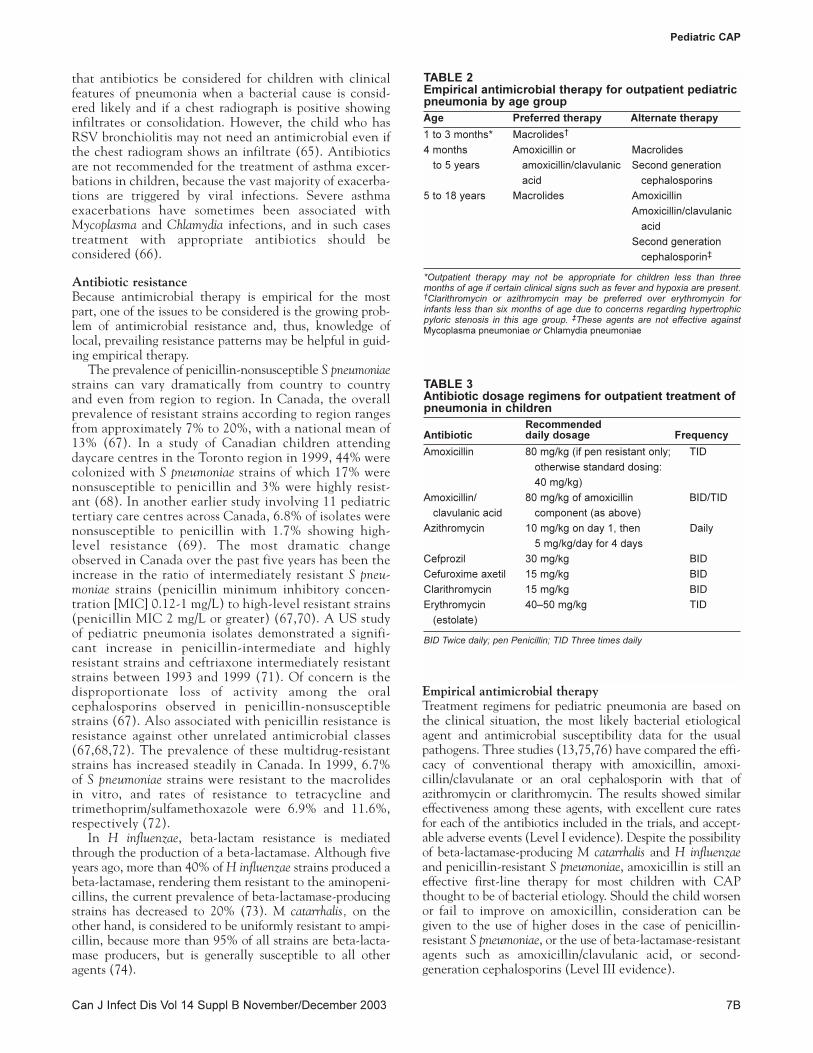
Empirical antimicrobial therapy Treatment regimens for pediatric pneumonia are based onthe clinical situation, the most likely bacterial etiologicalagent and antimicrobial susceptibility data for the usualpathogens. Three studies (13,75,76) have compared the effi-cacy of conventional therapy with amoxicillin, amoxi-cillin/clavulanate or an oral cephalosporin with that ofazithromycin or clarithromycin. The results showed similareffectiveness among these agents, with excellent cure ratesfor each of the antibiotics included in the trials, and accept-able adverse events (Level I evidence). Despite the possibilityof beta-lactamase-producing M catarrhalis and H influenzaeand penicillin-resistant S pneumoniae, amoxicillin is still aneffective first-line therapy for most children with CAPthought to be of bacterial etiology. Should the child worsenor fail to improve on amoxicillin, consideration can begiven to the use of higher doses in the case of penicillin-resistant S pneumoniae, or the use of beta-lactamase-resistantagents such as amoxicillin/clavulanic acid, or second-generation cephalosporins (Level III evidence).
Pediatric CAP
Can J Infect Dis Vol 14 Suppl B November/December 2003 7B
TABLE 2Empirical antimicrobial therapy for outpatient pediatricpneumonia by age group
Age Preferred therapy Alternate therapy
1 to 3 months* Macrolides†
4 months Amoxicillin or Macrolides
to 5 years amoxicillin/clavulanic Second generation
acid cephalosporins
5 to 18 years Macrolides Amoxicillin
Amoxicillin/clavulanic
acid
Second generation
cephalosporin‡
*Outpatient therapy may not be appropriate for children less than threemonths of age if certain clinical signs such as fever and hypoxia are present.†Clarithromycin or azithromycin may be preferred over erythromycin forinfants less than six months of age due to concerns regarding hypertrophicpyloric stenosis in this age group. ‡These agents are not effective againstMycoplasma pneumoniae or Chlamydia pneumoniae
TABLE 3Antibiotic dosage regimens for outpatient treatment ofpneumonia in children
Recommended Antibiotic daily dosage Frequency
Amoxicillin 80 mg/kg (if pen resistant only; TID
otherwise standard dosing:
40 mg/kg)
Amoxicillin/ 80 mg/kg of amoxicillin BID/TID
clavulanic acid component (as above)
Azithromycin 10 mg/kg on day 1, then Daily
5 mg/kg/day for 4 days
Cefprozil 30 mg/kg BID
Cefuroxime axetil 15 mg/kg BID
Clarithromycin 15 mg/kg BID
Erythromycin 40–50 mg/kg TID
(estolate)
BID Twice daily; pen Penicillin; TID Three times daily

Recommendations and dosage regimens for empiricalantimicrobial therapy are listed in Tables 2 and 3, respec-tively. Because S pneumoniae is the most common bacterialcause of pneumonia in children under five years of age,beta-lactam antibiotics are the first-line agents. High doseamoxicillin (80 mg/kg/day) or amoxicillin/clavulanic acidmay be appropriate if penicillin-resistant S pneumoniae issuspected based on local prevalence rates; otherwise, aregular dose (40 mg) will suffice (Level III evidence). Ifthe patient is allergic to beta-lactam agents, or if therapywith these agents is not successful, consultation with apediatric specialist is appropriate. The macrolides, eryth-romycin, azithromycin and clarithromycin should be con-sidered as first-line agents in patients five years of age andolder because of the increasing frequency of pneumoniacaused by M pneumoniae and C pneumoniae (Level I evidence) (77,78). Alternative choices in this age groupinclude amoxicillin, amoxicillin/clavulanic acid and avariety of second-generation cephalosporins. However,none of these agents are effective against M pneumoniae orC pneumoniae.
Patients on empiric therapy as well as those not onantibiotics because a viral cause was considered likelyshould be followed up within 24 h to 72 h, either by phoneor by return visit, to ensure that there is no deteriorationand to confirm efficacy of treatment (Level III evidence).
Pathogen-directed antimicrobial therapyIf a specific bacterial etiological agent is isolated from achild with pneumonia, antimicrobial therapy may bedirected specifically toward that agent. Table 4 describesthe most appropriate antimicrobial therapy for the treat-ment of specific bacterial agents. Isolates from upper air-ways specimens should not be used to direct therapybecause these will also contain normal respiratory tract flo-ra. Antimicrobial agents are not recommended for treat-ment of viral pneumonia.
Referral to a hospital emergency departmentAlthough various criteria have been used to determine thelikelihood of a severe outcome in the case of adult patientswith CAP (77), similar prediction rules do not exist forchildren. Tachypnea is used as a criterion by the WorldHealth Organization to identify children who should bereferred for medical assessment, and in addition, ‘danger signs’
such as inability to feed, seizures, lethargy or cyanosis pre-dict which children need hospital care (36,78). The physi-cian’s clinical judgement will play a major role indetermining the need for referral to the emergency depart-ment for possible admission, diagnostic testing or specialistassessment. Some considerations for referral are outlinedbelow (Level III evidence).High-risk of a complicated outcome: During the first yearof life the signs of respiratory disease may be subtle. In addi-tion, the risk of a complicated outcome is highest amongyoung infants as well as children with existing comorbidi-ties. Extrapulmonary spread of infection rarely occurs,except in young infants, in whom meningitis or metatasticseptic foci may occur with certain etiological types of pneu-monia, notably S aureus. Clinical judgement should be usedin deciding whether or not infants aged less than threemonths with pneumonia need to be hospitalized, but thosewith fever or hypoxia indicate the need for a careful work-up and are more likely to need hospitalization. Childrenrequiring oxygen or intravenous therapy may be consideredcandidates for hospital admission.Presence of comorbid conditions: The management ofchildren with certain comorbid conditions (eg, immunode-ficiency, cardiac or pulmonary disease) is beyond the scopeof this review. These children may present with manifesta-tions of more severe disease and deteriorate quickly. In thisregard, supportive treatment may be needed in the form ofoxygen therapy and respiratory support.Presence of complications: Initial evaluation by the pri-mary caregiver may reveal the presence of hypoxia, dehydra-tion, circulatory collapse or respiratory failure, all of whichindicate the need for resuscitation and emergent care.Presence of a complicated pneumonia such as pleural effu-sion, lung abscess and/or pneumatocele are also indicationsfor hospitalization, as is evidence of extrapulmonary spread ofinfection, such as septic arthritis or meningitis.Inability to administer therapy effectively: It may benecessary for some children to be referred for possibleadmission to hospital if the clinician has concerns abouthis or her ability to effectively treat the pneumonia on anoutpatient basis. This may be due to several factors,including poor compliance, as well as the inability of theinfant to tolerate oral therapy. The latter may be due todecreased oral intake and vomiting accompanying thepneumonia.
Low et al
Can J Infect Dis Vol 14 Suppl B November/December 20038B
TABLE 4Antimicrobial outpatient therapy for pediatric pneumonia by pathogen
Pathogen Preferred therapy
Streptococcus pneumoniae sensitive to penicillin (MIC < 0.1 µg/mL) Amoxicillin
S pneumoniae resistant to penicillin (MIC ≥ 0.1 µg/mL) Amoxicillin (high dose)
Haemophilus influenzae type b sensitive to ampicillin Amoxicillin
H influenzae type b resistant to ampicillin Amoxicillin/clavulanic acid or second-generation cephalosporin
or azithromycin or clarithromycin
Staphylococcus aureus Cloxacillin
Streptococcus pyogenes Penicillin
Mycoplasma pneumoniae Macrolide
Chlamydia pneumoniae Macrolide
MIC Minimum inhibitory concentration

Failed therapy: Children who have failed initial therapyshould be referred for possible admission. Although not allof these children will require admission to hospital, somemay require an initial period of parenteral therapy followedby a step-down to oral therapy.Recurrent pneumonia: Children may have recurrentpneumonia for several reasons. These include the possibil-ity of immunodeficiency states, mucociliary transportdefects such as cystic fibrosis or immotile cilia syndrome, orrecurrent aspiration. Some children with recurrent or per-sistent radiographic changes in a fixed location may haveunderlying mass lesions, congenital abnormalities or a foreign body. Bronchoscopy or other investigations may berequired in these situations. Studies have shown that themost common cause of ‘recurrent pneumonia’ or ‘recurrentbronchitis’ in children is undiagnosed asthma (79).Children with recurrent pneumonia should be evaluated todetermine whether they have asthma.
CONCLUSIONSThe etiology of CAP in children is complex and likely tochange with the advent of improved vaccines against majorpathogens. Breathing rate and other respiratory signs are usu-ally good indicators of pneumonia, but in cases of uncer-tainty or if complications are present, a chest radiographmay help to confirm the diagnosis. Laboratory tests may dis-tinguish between viral and bacterial pneumonia but are notalways readily available, and some of the more commontests are neither timely nor accurate. However, where thereis doubt, a combination of clinical signs together withresults of the chest radiograph and laboratory tests canestablish a diagnosis. These considerations can be used todetermine the necessity for empiric antimicrobial treatmentand prevailing antimicrobial susceptibility patterns shouldguide treatment choices.
ACKNOWLEDGEMENTS: The authors gratefully acknowl-edge the contribution of Dr Barbara Law and Dr Marc Lebel to thecontent and editing of the manuscript. Sponsored by an unre-stricted educational grant from Pfizer Canada Inc. Preparation ofthis manuscript was coordinated by Core Health Services Inc.
Pediatric CAP
Can J Infect Dis Vol 14 Suppl B November/December 2003 9B
REFERENCES1. Mulholland K. Magnitude of the problem of childhood
pneumonia. Lancet 1999;354:590-2.2. Dowell SF, Kupronis BA, Zell ER, Shay DK. Mortality from
pneumonia in children in the United States, 1939 through1996. N Engl J Med 2000;342:1399-407.
3. Murphy TF, Henderson FW, Clyde WA Jr, Collier AM, DennyFW. Pneumonia: An eleven-year study in a pediatric practice.Am J Epidemiol 1981;113:12-21.
4. Shinefield HR, Black S. Efficacy of pneumococcal conjugatevaccines in large scale field trials. Pediatr Infect Dis J2000;19:394-7.
5. Black SB, Shinefield HR, Ling S, et al. Effectiveness ofheptavalent pneumococcal conjugate vaccine in childrenyounger than five years of age for prevention of pneumonia.Pediatr Infect Dis J 2002;21:810-5.
6. Pelton SI, Dagan R, Gaines BM, et al. Pneumococcalconjugate vaccines: Proceedings from an interactive symposiumat the 41st Interscience Conference on Antimicrobial Agentsand Chemotherapy. Vaccine 2003;21:1562-71.
7. Mandell LA, Marrie TJ, Grossman RF, Chow AW, Hyland RH.Summary of Canadian guidelines for the initial management ofcommunity-acquired pneumonia: An evidence-based update by
the Canadian Infectious Disease Society and the CanadianThoracic Society. Can Respir J 2000;7:371-82.
8. Bartlett JG, Dowell SF, Mandell LA, File TM Jr, Musher DM, Fine MJ. Practice guidelines for the management ofcommunity-acquired pneumonia in adults. Infectious DiseasesSociety of America. Clin Infect Dis 2000;31:347-82.
9. Niederman MS, Mandell LA, Anzueto A, et al. Guidelines forthe management of adults with community-acquiredpneumonia. Diagnosis, assessment of severity, antimicrobialtherapy, and prevention. Am J Respir Crit Care Med2001;163:1730-54.
10. Jadavji T, Law B, Lebel MH, Kennedy WA, Gold R, Wang EE. A practical guide for the diagnosis and treatment of pediatricpneumonia. CMAJ 1997;156:S703-11.
11. Kish MA. Guide to development of practice guidelines. ClinInfect Dis 2001;32:851-4.
12. Woolf SH, Battista RN, Anderson GM, Logan AG, Wang E.Assessing the clinical effectiveness of preventive maneuvers:Analytic principles and systematic methods in reviewingevidence and developing clinical practice recommendations. A report by the Canadian Task Force on the Periodic HealthExamination. J Clin Epidemiol 1990;43:891-905.
13. Wubbel L, Muniz L, Ahmed A, et al. Etiology and treatment ofcommunity-acquired pneumonia in ambulatory children.Pediatr Infect Dis J 1999;18:98-104.
14. Waterer GW, Wunderink RG. The influence of the severity ofcommunity-acquired pneumonia on the usefulness of bloodcultures. Respir Med 2001;95:78-82.
15. Delport SD, Brisley T. Aetiology and outcome of severecommunity-acquired pneumonia in children admitted to apaediatric intensive care unit. S Afr Med J 2002;92:907-11.
16. Drummond P, Clark J, Wheeler J, Galloway A, Freeman R,Cant A. Community acquired pneumonia – A prospective UKstudy. Arch Dis Child 2000;83:408-12.
17. Principi N, Esposito S, Blasi F, Allegra L. Role of Mycoplasmapneumoniae and Chlamydia pneumoniae in children withcommunity-acquired lower respiratory tract infections. ClinInfect Dis 2001;32:1281-9.
18. Juven T, Mertsola J, Waris M, et al. Etiology of community-acquired pneumonia in 254 hospitalized children. PediatrInfect Dis J 2000;19:293-8.
19. Vuori E, Peltola H, Kallio MJ, Leinonen M, Hedman K.Etiology of pneumonia and other common childhood infectionsrequiring hospitalization and parenteral antimicrobial therapy.SE-TU Study Group. Clin Infect Dis 1998;27:566-72.
20. Davies HD, Matlow A, Petric M, Glazier R, Wang EE.Prospective comparative study of viral, bacterial and atypicalorganisms identified in pneumonia and bronchiolitis inhospitalized Canadian infants. Pediatr Infect Dis J 1996;15:371-5.
21. Correa AG, Starke JR. Infections of the lower respiratory tractin children. In: Niederman MS, Sarosi GA, Glassroth J, eds.Respiratory Infections. Philadelphia: Lippincott Williams andWilkins, 2001:155-80.
22. McIntosh K. Community-acquired pneumonia in children. N Engl J Med 2002;346:429-37.
23. Korppi M, Heiskanen-Kosma T, Jalonen E, et al. Aetiology ofcommunity-acquired pneumonia in children treated inhospital. Eur J Pediatr 1993;152:24-30.
24. Scheifele D, Gold R, Law B, et al. Decline in Haemophilusinfluenzae type B invasive infections at five Canadian pediatriccentres. Can Commun Dis Rep 1993;19:88-91.
25. Scheifele D, Halperin S. Haemophilus influenzae type B diseasecontrol using PENTACEL, Canada, 1998-1999. Can CommunDis Rep 2000;26:93-6.
26. Scheifele D, Halperin S, Vaudry W, et al. Historic lowHaemophilus influenzae type B case tally – Canada 2000. CanCommun Dis Rep 2001;27:149-50.
27. Paisley JW, Lauer BA, McIntosh K, Glode MP, Schachter J, Rumack C. Pathogens associated with acute lower respiratorytract infection in young children. Pediatr Infect Dis 1984;3:14-9.

Low et al
Can J Infect Dis Vol 14 Suppl B November/December 200310B
28. Pandey A, Chaudhry R, Nisar N, Kabra SK. Acute respiratorytract infections in Indian children with special reference toMycoplasma pneumoniae. J Trop Pediatr 2000;46:371-4.
29. Esposito S, Bosis S, Cavagna R, et al. Characteristics ofStreptococcus pneumoniae and atypical bacterial infections inchildren 2-5 years of age with community-acquired pneumonia.Clin Infect Dis 2002;35:1345-52.
30. Heiskanen-Kosma T, Korppi M, Laurila A, Jokinen C, KleemolaM, Saikku P. Chlamydia pneumoniae is an important cause ofcommunity-acquired pneumonia in school-aged children:Serological results of a prospective, population-based study.Scand J Infect Dis 1999;31:255-9.
31. Nobert E, Chernick V. Tuberculosis: 5. Pediatric disease. CMAJ1999;160:1479-82.
32. Margolis PA, Ferkol TW, Marsocci S, et al. Accuracy of theclinical examination in detecting hypoxemia in infants withrespiratory illness. J Pediatr 1994;124:552-60.
33. Leventhal JM. Clinical predictors of pneumonia as a guide toordering chest roentgenograms. Clin Pediatr (Phila)1982;21:730-4.
34. Taylor JA, Del Beccaro M, Done S, Winters W. Establishingclinically relevant standards for tachypnea in febrile childrenyounger than 2 years. Arch Pediatr Adolesc Med 1995;149:283-7.
35. Palafox M, Guiscafre H, Reyes H, Munoz O, Martinez H.Diagnostic value of tachypnoea in pneumonia definedradiologically. Arch Dis Child 2000;82:41-5.
36. World Health Organization. Acute respiratory infections inchildren: Case management in small hospitals in developingcountries. In: WHO, ed. WHO/ARI/90.5. Geneva: 1990.
37. Berman S, Simoes EA, Lanata C. Respiratory rate andpneumonia in infancy. Arch Dis Child 1991;66:81-4.
38. Margolis P, Gadomski A. Does this infant have pneumonia?JAMA 1998;279:308-13.
39. Zukin DD, Hoffman JR, Cleveland RH, Kushner DC, HermanTE. Correlation of pulmonary signs and symptoms with chestradiographs in the pediatric age group. Ann Emerg Med1986;15:792-6.
40. Wang EE, Milner RA, Navas L, Maj H. Observer agreement forrespiratory signs and oximetry in infants hospitalized with lowerrespiratory infections. Am Rev Respir Dis 1992;145:106-9.
41. Bachur R, Perry H, Harper MB. Occult pneumonias: Empiricchest radiographs in febrile children with leukocytosis. AnnEmerg Med 1999;33:166-73.
42. Ravichandran D, Burge DM. Pneumonia presenting with acuteabdominal pain in children. Br J Surg 1996;83:1707-8.
43. Jona JZ, Belin RP. Basilar pneumonia simulating acuteappendicitis in children. Arch Surg 1976;111:552-3.
44. Swingler GH, Hussey GD, Zwarenstein M. Randomisedcontrolled trial of clinical outcome after chest radiograph inambulatory acute lower-respiratory infection in children. Lancet1998;351:404-8.
45. Bushyhead JB, Wood RW, Tompkins RK, Wolcott BW, Diehr P.The effect of chest radiographs on the management and clinicalcourse of patients with acute cough. Med Care 1983;21:661-73.
46. Alario AJ, McCarthy PL, Markowitz R, Kornguth P, RosenfieldN, Leventhal JM. Usefulness of chest radiographs in childrenwith acute lower respiratory tract disease. J Pediatr1987;111:187-93.
47. Bachur R, Perry H, Harper M. Empiric chest radiographs infebrile children with leukocytosis. Ann Emerg Med 1999;33:480.
48. Kramer MS, Roberts-Brauer R, Williams RL. Bias and ‘overcall’in interpreting chest radiographs in young febrile children.Pediatrics 1992;90:11-3.
49. Green SM, Rothrock SG. Evaluation styles for well-appearingfebrile children: are you a “risk-minimizer” or a “test-minimizer”?Ann Emerg Med 1999;33:211-4.
50. Young M, Marrie TJ. Interobserver variability in theinterpretation of chest roentgenograms of patients with possiblepneumonia. Arch Intern Med 1994;154:2729-32.
51. Tew J, Calenoff L, Berlin BS. Bacterial or nonbacterial
pneumonia: Accuracy of radiographic diagnosis. Radiology1977;124:607-12.
52. Swingler GH. Radiologic differentiation between bacterial andviral lower respiratory infection in children: A systematicliterature review. Clin Pediatr (Phila) 2000;39:627-33.
53. Roback MG, Dreitlein DA. Chest radiograph in the evaluationof first time wheezing episodes: Review of current clinicalpractice and efficacy. Pediatr Emerg Care 1998;14:181-4.
54. Heaton P, Arthur K. The utility of chest radiography in thefollow-up of pneumonia. N Z Med J 1998;111:315-7.
55. Silverman M, Stratton D, Diallo A, Egler LJ. Diagnosis of acutebacterial pneumonia in Nigerian children. Value of needleaspiration of lung of countercurrent immunoelectrophoresis.Arch Dis Child 1977;52:925-31.
56. Vuori-Holopainen E, Peltola H. Reappraisal of lung tap: Reviewof an old method for better etiologic diagnosis of childhoodpneumonia. Clin Infect Dis 2001;32:715-26.
57. Hickey RW, Bowman MJ, Smith GA. Utility of blood cultures inpediatric patients found to have pneumonia in the emergencydepartment. Ann Emerg Med 1996;27:721-5.
58. Hardie W, Bokulic R, Garcia VF, Reising SF, Christie CD.Pneumococcal pleural empyemas in children. Clin Infect Dis1996;22:1057-63.
59. Shuttleworth DB, Charney E. Leukocyte count in childhoodpneumonia. Am J Dis Child 1971;122:393-6.
60. Ghildyal R, Hogg G, Meanger J. Detection and subgrouping ofrespiratory syncytial virus directly from nasopharyngeal aspirates.Clin Microbiol Infect 1997;3:120-3.
61. Skerrett SJ. Diagnostic testing for community-acquiredpneumonia. Clin Chest Med 1999;20:531-48.
62. Heiskanen-Kosma T, Korppi M, Jokinen C, et al. Etiology ofchildhood pneumonia: Serologic results of a prospective,population-based study. Pediatr Infect Dis J 1998;17:986-91.
63. McCracken GH Jr. Diagnosis and management of pneumonia inchildren. Pediatr Infect Dis J 2000;19:924-8.
64. Bradley JS. Management of community-acquired pediatricpneumonia in an era of increasing antibiotic resistance andconjugate vaccines. Pediatr Infect Dis J 2002;21:592-8.
65. Hall CB, Powell KR, Schnabel KC, Gala CL, Pincus PH. Risk ofsecondary bacterial infection in infants hospitalized withrespiratory syncytial viral infection. J Pediatr 1988;113:266-71.
66. Esposito S, Blasi F, Arosio C, et al. Importance of acuteMycoplasma pneumoniae and Chlamydia pneumoniae infections inchildren with wheezing. Eur Respir J 2000;16:1142-6.
67. Zhanel GG, Karlowsky JA, Palatnick L, Vercaigne L, Low DE,Hoban DJ. Prevalence of antimicrobial resistance in respiratorytract isolates of Streptococcus pneumoniae: Results of a Canadiannational surveillance study. The Canadian Respiratory InfectionStudy Group. Antimicrob Agents Chemother 1999;43:2504-9.
68. Kellner JD, Ford-Jones EL. Streptococcus pneumoniae carriage inchildren attending 59 Canadian child care centers. TorontoChild Care Centre Study Group. Arch Pediatr Adolesc Med 1999;153:495-502.
69. Scheifele D, Halperin S, Pelletier L, Talbot J. Invasivepneumococcal infections in Canadian children, 1991-1998:Implications for new vaccination strategies. Canadian PaediatricSociety/Laboratory Centre for Disease Control ImmunizationMonitoring Program, Active (IMPACT). Clin Infect Dis2000;31:58-64.
70. Davidson RJ, Canadian Bacterial Surveillance Network, Low DE. A cross Canada survillance of antimicrobial resistancein respiratory tract pathogens. Can J Infect Dis 1999;10:128-33.
71. Kaplan SL, Mason EO Jr, Wald E, et al. Six year multicentersurveillance of invasive pneumococcal infections in children.Pediatr Infect Dis J 2002;21:141-7.
72. Chen D, McGeer A, de Azavedo JC, Low DE, The CanadianBacterial Surveillance Network. Decreased susceptibility ofStreptococcus pneumoniae to fluoroquinolones in Canada. N EnglJ Med 1999;341:233-9.

Pediatric CAP
Can J Infect Dis Vol 14 Suppl B November/December 2003 11B
73. Low DE. Antimicrobial drug use and resistance amongrespiratory pathogens in the community. Clin Infect Dis 2001;33(Suppl 3):S206-13.
74. Zhanel GG, Karlowsky JA, Low DE, Hoban DJ. Antibioticresistance in respiratory tract isolates of Haemophilus influenzaeand Moraxella catarrhalis collected from across Canada in 1997-1998 [In Process Citation]. J Antimicrob Chemother2000;45:655-62.
75. Block S, Hedrick J, Hammerschlag MR, Cassell GH, Craft JC.Mycoplasma pneumoniae and Chlamydia pneumoniae in pediatriccommunity-acquired pneumonia: Comparative efficacy andsafety of clarithromycin vs. erythromycin ethylsuccinate.Pediatr Infect Dis J 1995;14:471-7.
76. Harris JA, Kolokathis A, Campbell M, Cassell GH, Hammerschlag MR. Safety and efficacy of azithromycin in thetreatment of community-acquired pneumonia in children.Pediatr Infect Dis J 1998;17:865-71.
77. Fine MJ, Auble TE, Yealy DM, et al. A prediction rule toidentify low-risk patients with community-acquired pneumonia[see comments]. N Engl J Med 1997;336:243-50.
78. Klein JO. Bacterial pneumonias. In: Feigin RD, Cherry JD, eds.Textbook of Pediatric Infectious Diseases. Philadelphia: WBSaunders Company, 1998:274-84.
79. Eigen H, Laughlin JJ, Homrighausen J. Recurrent pneumonia inchildren and its relationship to bronchial hyperreactivity.Pediatrics 1982;70:698-704.

Submit your manuscripts athttp://www.hindawi.com
Stem CellsInternational
Hindawi Publishing Corporationhttp://www.hindawi.com Volume 2014
Hindawi Publishing Corporationhttp://www.hindawi.com Volume 2014
MEDIATORSINFLAMMATION
of
Hindawi Publishing Corporationhttp://www.hindawi.com Volume 2014
Behavioural Neurology
EndocrinologyInternational Journal of
Hindawi Publishing Corporationhttp://www.hindawi.com Volume 2014
Hindawi Publishing Corporationhttp://www.hindawi.com Volume 2014
Disease Markers
Hindawi Publishing Corporationhttp://www.hindawi.com Volume 2014
BioMed Research International
OncologyJournal of
Hindawi Publishing Corporationhttp://www.hindawi.com Volume 2014
Hindawi Publishing Corporationhttp://www.hindawi.com Volume 2014
Oxidative Medicine and Cellular Longevity
Hindawi Publishing Corporationhttp://www.hindawi.com Volume 2014
PPAR Research
The Scientific World JournalHindawi Publishing Corporation http://www.hindawi.com Volume 2014
Immunology ResearchHindawi Publishing Corporationhttp://www.hindawi.com Volume 2014
Journal of
ObesityJournal of
Hindawi Publishing Corporationhttp://www.hindawi.com Volume 2014
Hindawi Publishing Corporationhttp://www.hindawi.com Volume 2014
Computational and Mathematical Methods in Medicine
OphthalmologyJournal of
Hindawi Publishing Corporationhttp://www.hindawi.com Volume 2014
Diabetes ResearchJournal of
Hindawi Publishing Corporationhttp://www.hindawi.com Volume 2014
Hindawi Publishing Corporationhttp://www.hindawi.com Volume 2014
Research and TreatmentAIDS
Hindawi Publishing Corporationhttp://www.hindawi.com Volume 2014
Gastroenterology Research and Practice
Hindawi Publishing Corporationhttp://www.hindawi.com Volume 2014
Parkinson’s Disease
Evidence-Based Complementary and Alternative Medicine
Volume 2014Hindawi Publishing Corporationhttp://www.hindawi.com


![Community acquired pneumonia [cap] in children](https://static.fdocuments.us/doc/165x107/5454e4c4af795946778b8712/community-acquired-pneumonia-cap-in-children.jpg)