Community acquired pneumonia
48
Community- Acquired Pneumonia SAMIR EL ANSARY
-
Upload
dr-mohamed-maged-kharabish -
Category
Health & Medicine
-
view
82 -
download
4
Transcript of Community acquired pneumonia
Community-Acquired Pneumonia
SAMIR EL ANSARY
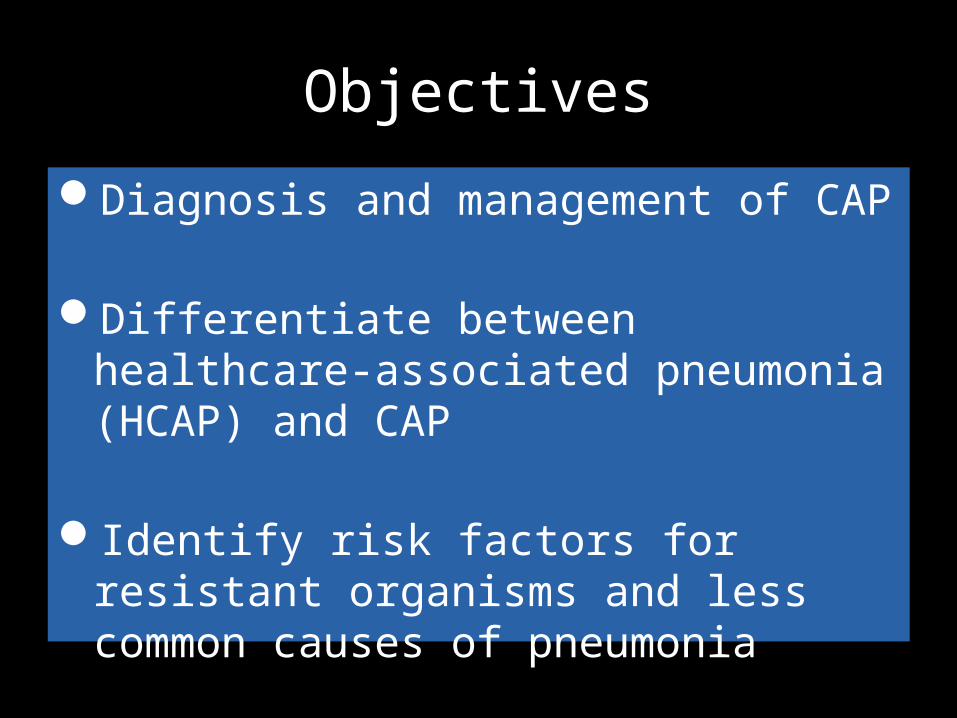
Objectives
Diagnosis and management of CAP
Differentiate between healthcare-associated pneumonia (HCAP) and CAP
Identify risk factors for resistant organisms and less common causes of pneumonia

CAP - Epidemiology
Very common 5 million cases/year in North AmericaAt least 1 million hospitalizations/year
9th leading cause of infectious death in US30 day morality for hospitalized patients is up
to 23%$17 billion/year in healthcare costs in US

Which of these patients have community-acquired pneumonia (CAP)?
34 yo hospital employee, previously healthy, admitted for acute pneumonia.
56 yo man admitted with CHF, noted to have pneumonia the day after admission.
76 yo bedridden man transferred from a nursing home for acute confusion, noted to have a new infiltrate on CXR.
✔
✔

Alphabet Soup of Terms
• CAP: Community-acquired pneumonia– Outside of hospital or extended-care facility
• HCAP: Healthcare-associated pneumonia– Long-term or extended care facility, hemodialysis, outpatient
chemo, wound care, etc.• HAP: Hospital-acquired pneumonia
– ≥ 48 h from admission
• VAP: Ventilator-associated pneumonia– ≥ 48 h from endotracheal intubation

What differentiates acute bronchitis (or other causes of fever/cough) from pneumonia : – Clinical definition requires chest imaging – air space disease.
HCAP includes the following patients with pneumonia: hospitalized in an acute care hospital for more than 2 days within 90 days of the pneumonia; resided in a long-term care facility (e.g., nursing home); received recent parenteral antimicrobial therapy, chemotherapy, or wound care within 30 days of pneumonia; or received treatment in a hospital or hemodialysis clinic.
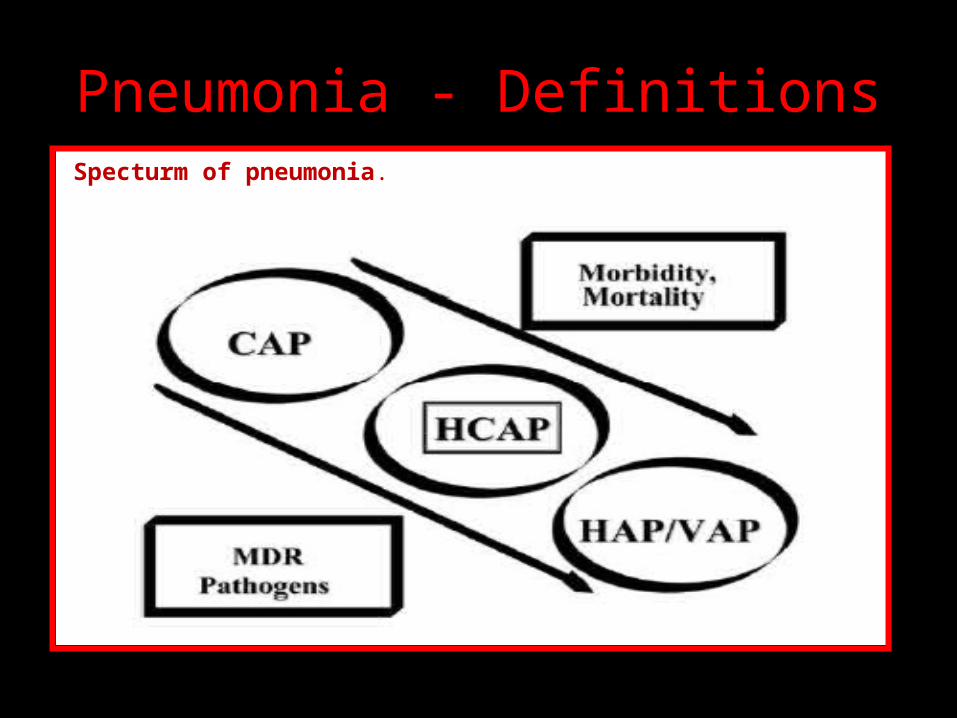
Pneumonia - DefinitionsSpecturm of pneumonia.
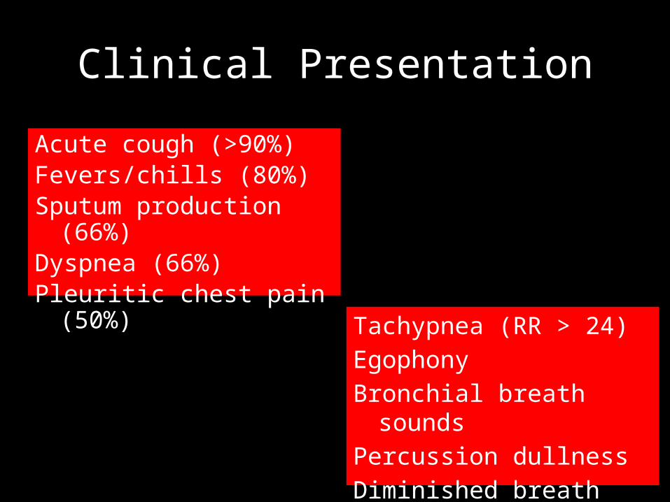
Clinical Presentation
Acute cough (>90%)Fevers/chills (80%)Sputum production (66%)Dyspnea (66%)Pleuritic chest pain (50%)
Tachypnea (RR > 24)
Egophony
Bronchial breath sounds
Percussion dullness
Diminished breath sounds

Clinical Presentation
Acute cough (>90%)Fevers/chills (80%)Sputum production (66%)Dyspnea (66%)Pleuritic chest pain (50%)
Tachypnea (RR > 24)EgophonyBronchial breath soundsPercussion dullnessDiminished breath sounds
Lung physical examSensitivity 47-69% ; Specificity 58-75%

CXR
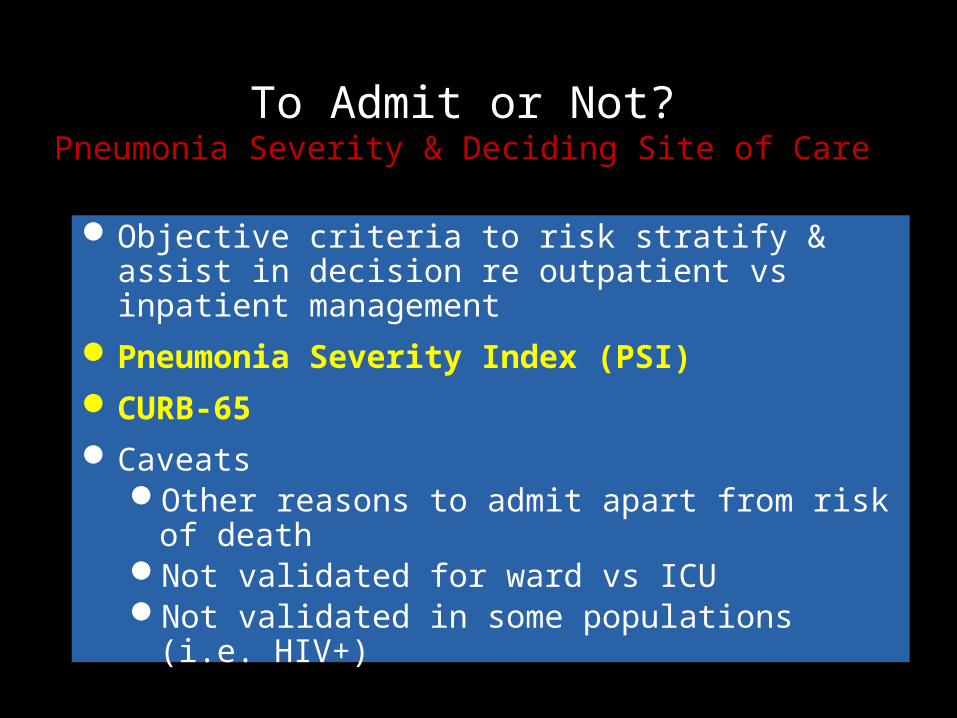
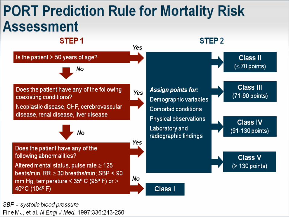
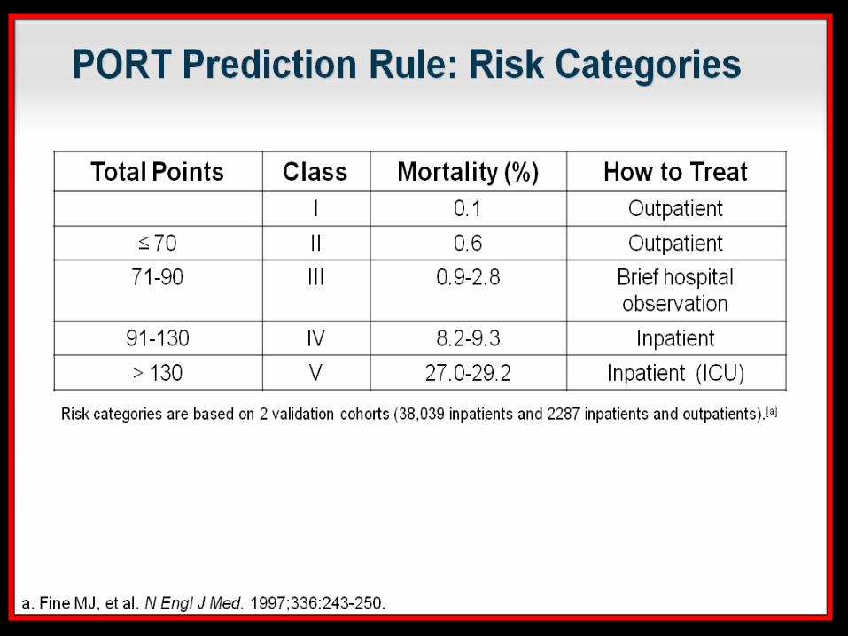
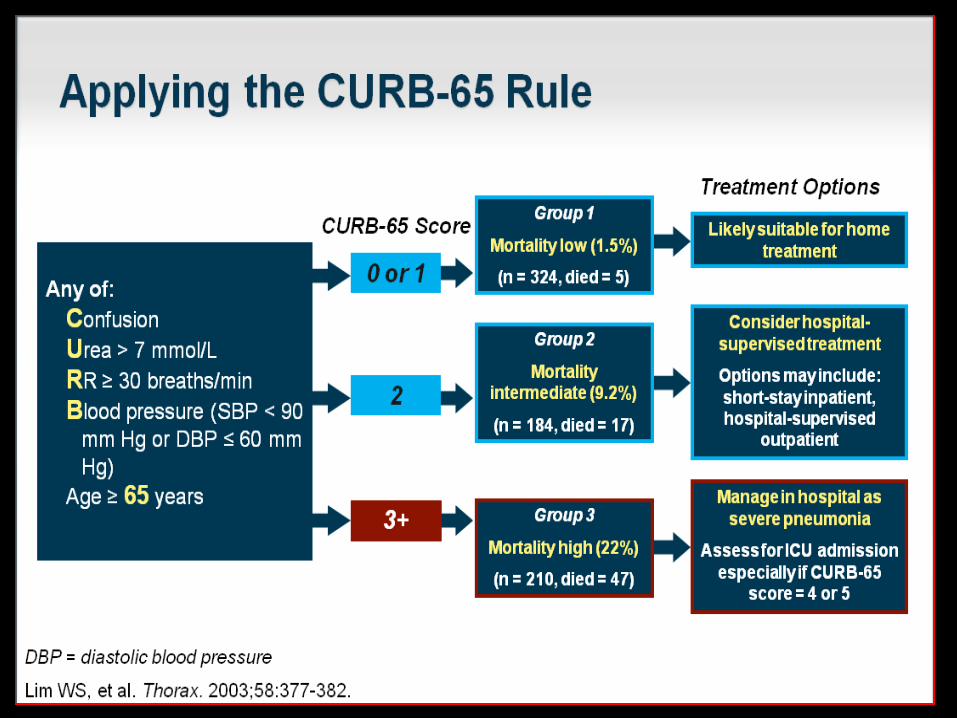
To Admit or Not?Pneumonia Severity & Deciding Site of Care
Objective criteria to risk stratify & assist in decision re outpatient vs inpatient management
Pneumonia Severity Index (PSI)CURB-65Caveats
Other reasons to admit apart from risk of deathNot validated for ward vs ICUNot validated in some populations (i.e. HIV+)


Criteria for Severe CAP(Admit to ICU)
Minor criteriaRespiratory rate ≥30 breaths/minPaO2/FiO2 ratio ≥ 250Multilobar infiltratesConfusion/disorientationUremia (BUN ≥20 mg/dL)Leukopenia (WBC <4000 cells/mm3)Thrombocytopenia (platelets <100,000 cells/mm3)Hypothermia (core T <36C)Hypotension requiring aggressive fluid resuscitation
Major criteriaInvasive mechanical ventilationSeptic shock with the need for vasopressors

Microbiology
TYPICAL– Streptococcus pneumoniae– Haemophilus influenzae – Moraxella catarrhalis– Klebsiella pneumoniae
ATYPICAL– Mycoplasma pneumoniae– Chlamydophila pneumoniae– Legionella pneumophila
2/3 are typical; 1/3 are atypical

Microbiology of CAP among hospitalized patients
Outpatient Streptococcus pneumoniaeMycoplasma pneumoniaeHaemophilus influenzaeChlamydophila pneumoniaeRespiratory viruses
Inpatient (Ward) S. pneumoniaeM. pneumoniaeH. influenzaeC. PneumoniaeLegionella speciesRespiratory virusesAspiration
Inpatient (ICU) S. pneumoniaeLegionella spp.Staphylococcus aureusGram-negative bacilli

Age-specific Rates of Hospital
Admission by Pathogen
Marsten. Community-based pneumonia incidence study group.Arch Intern Med 1997;157:1709-18
Two-thirds of deaths due to a known CAP pathogen is due to Strep pneumoniae.

Comorbidities & Associated Pathogens
Alcoholism
COPD and/or Tobacco
Strep pneumoniae Oral anaerobes Klebsiella pneumoniae Acinetobacter spp M. tuberculosis
Haemophilus influenzae Pseudomonas aeruginosa Legionella spp S. pneumoniae Moraxella catarrhalis Chlamydophila pneumoniae

Aspiration
Lung Abscess
Structural lung disease (e.g. bronchiectasis)
Advanced HIV
Gram-negative enteric pathogens Oral anaerobes
CA-MRSA Oral anaerobes, microaerophilic streptococci,
Actinomyces, Nocardia spp Endemic fungi M. tuberculosis, atypical mycobacteria
P. aeruginosa Burkholderia cepacia S. aureus
Pneumocystis jirovecii Cryptococcus Histoplasma Tuberculosis Aspergillus P. aeruginosa

MRSAModern-day CAP pathogen
Must consider MRSA, MSSA coverage in severe CAP, esp during flu season!

MRSA CAPClinical Features
Cavitary infiltrate or necrosisRapidly increasing pleural effusionGross hemoptysis (not just blood-streaked)Concurrent influenzaNeutropeniaErythematous rashSkin pustulesYoung, previously healthy patientSevere pneumonia during summer months

Is sputum culture helpful?
Sputum Gram stain and cultureLow sensitivity (25-40%)Considered optional for
outpatientsBlood culture
Positive < 10%May help guide antibiotic therapy
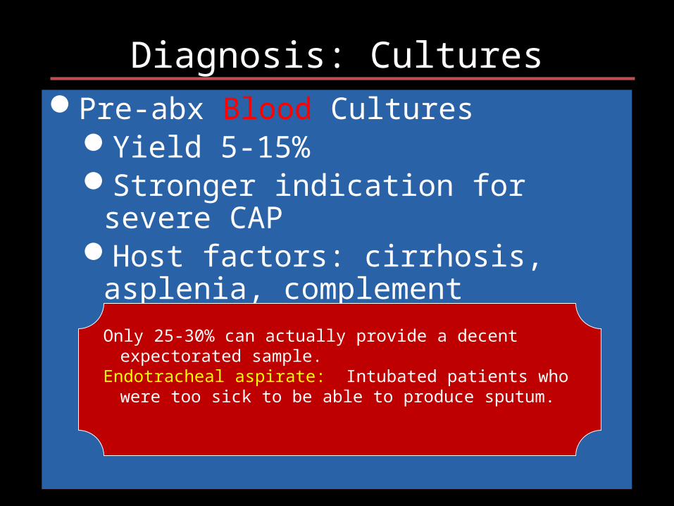
Diagnosis: CulturesPre-abx Blood Cultures
Yield 5-15%Stronger indication for severe CAPHost factors: cirrhosis, asplenia,
complement deficiencies, leukopenia
Only 25-30% can actually provide a decent expectorated sample.Endotracheal aspirate: Intubated patients who were too sick to be
able to produce sputum.

Diagnosis: Cultures
Pre-abx expectorated sputum Gs & CxYield can be variableDepends on multiple factors: specimen
collection, transport, speed of processing, use of cytologic criteria
Adequate samplePre-abx endotracheal aspirate Gs & CxPleural effusions > 5 cm on lateral upright CXR

Diagnosis: Other testing
Urinary antigen testsS. pneumoniae
L. pneumophila serogroup 1
60-80% sensitive, >90% specific in adults
Pros: rapid (15 min), simple, more sensitive than Cx, can detect Pneumococcus after abx started
Cons: no susceptibility data, not helpful in patients with recent CAP (prior 3 months)
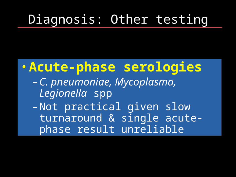
Diagnosis: Other testing
• Acute-phase serologies– C. pneumoniae, Mycoplasma, Legionella
spp– Not practical given slow turnaround &
single acute-phase result unreliable

Diagnosis: Other testing
• Influenza testing– Hospitalized patients: Severe respiratory illness (T> 37.8°C with
SOB, hypoxia, or radiographic evidence of pneumonia) without other explanation and suggestive of infectious etiology should get screened during season
– NP swab or nasal wash/aspirate– Rapid flu test (15 min) - Distinguishes A vs B
• Sensitivity 50-70%; specificity >90%– Respiratory virus DFA & culture - reflex subtyping for A– Respiratory viral PCR panel - reflex subtyping for A– Epidemic Influenza PCR panel – screens for A & B with reflex
subtyping for A

Outpatient Empiric CAP Abx
• Healthy; no abx x past 3 months– Macrolide: azithromycin– 2nd choice: doxycycline
• Comorbidities; abx x past 3 mon– Respiratory fluoroquinolone: Moxifloxacin, levofloxacin 750
mg, gemifloxacin– Beta-lactam (preferred: amoxicillin 1 g3 or amox/clav 2 g2;
alternative: ceftriaxone, cefuroxime 500 mg2), + macrolide
• Regions with >25% high-level macrolide-resistant S. pneumo (MIC ≥16), consider alternative agents
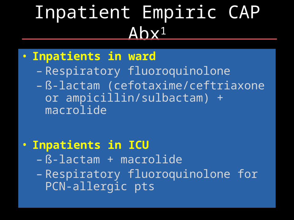
Inpatient Empiric CAP Abx1
• Inpatients in ward– Respiratory fluoroquinolone– ß-lactam (cefotaxime/ceftriaxone or
ampicillin/sulbactam) + macrolide
• Inpatients in ICU– ß-lactam + macrolide– Respiratory fluoroquinolone for PCN-allergic pts
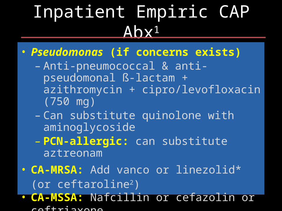
Inpatient Empiric CAP Abx1
• Pseudomonas (if concerns exists)– Anti-pneumococcal & anti-pseudomonal ß-
lactam + azithromycin + cipro/levofloxacin (750 mg)
– Can substitute quinolone with aminoglycoside– PCN-allergic: can substitute aztreonam
• CA-MRSA: Add vanco or linezolid* (or ceftaroline2)• CA-MSSA: Nafcillin or cefazolin or ceftriaxone
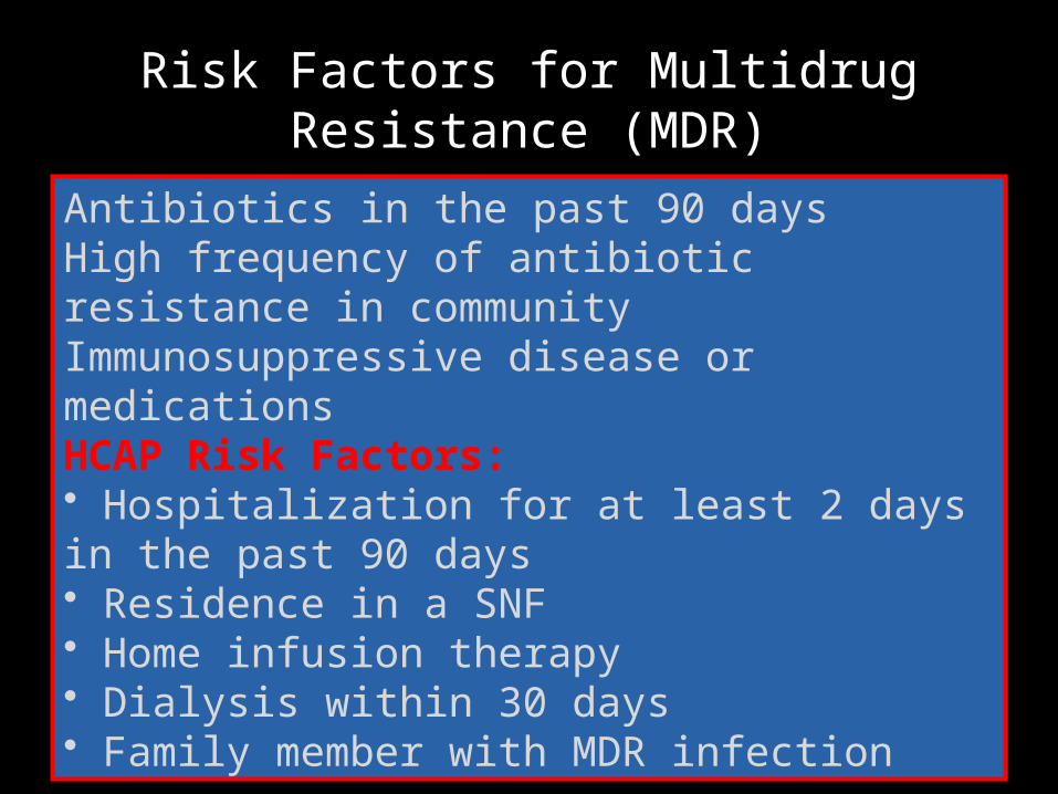
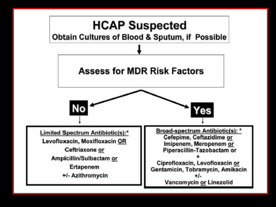
Risk Factors for Multidrug Resistance (MDR)
Antibiotics in the past 90 daysHigh frequency of antibiotic resistance in communityImmunosuppressive disease or medicationsHCAP Risk Factors:• Hospitalization for at least 2 days in the past 90 days• Residence in a SNF• Home infusion therapy• Dialysis within 30 days• Family member with MDR infection
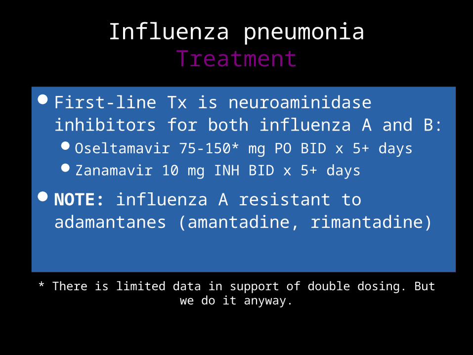
Influenza pneumoniaTreatment
First-line Tx is neuroaminidase inhibitors for both influenza A and B:Oseltamavir 75-150* mg PO BID x 5+ daysZanamavir 10 mg INH BID x 5+ days
NOTE: influenza A resistant to adamantanes (amantadine, rimantadine)
* There is limited data in support of double dosing. But we do it anyway.

Antiviral Therapy for Influenza
CDC Guidelines for Influenza 2012-2013
Should be started ASAP in:Anyone hospitalized with suspected or confirmed
influenzaAnyone with severe, complicated or progressive
respiratory illnessAnyone at higher risk of complications from influenza

Individuals at Higher Risk for Influenza Complications
CDC Guidelines for Influenza 2012-2013
• Extremes of age: children <2, adults ≥65 years• Comorbid conditions:
– Chronic pulmonary– Cardiovascular (except HTN alone)– Renal, hepatic, hematologic, metabolic (DM)– Neurologic, neuromuscular (cerebral palsy, epilepsy, CVA, SCI)
• Immunosuppression (caused by meds, HIV infection)• Pregnant or post-partum (<2 wks) women• Persons <19 years on long-term aspirin• American Indians & Alaskan Natives• Morbidly obese (BMI ≥40)• Residents in NH or chronic-care facilities
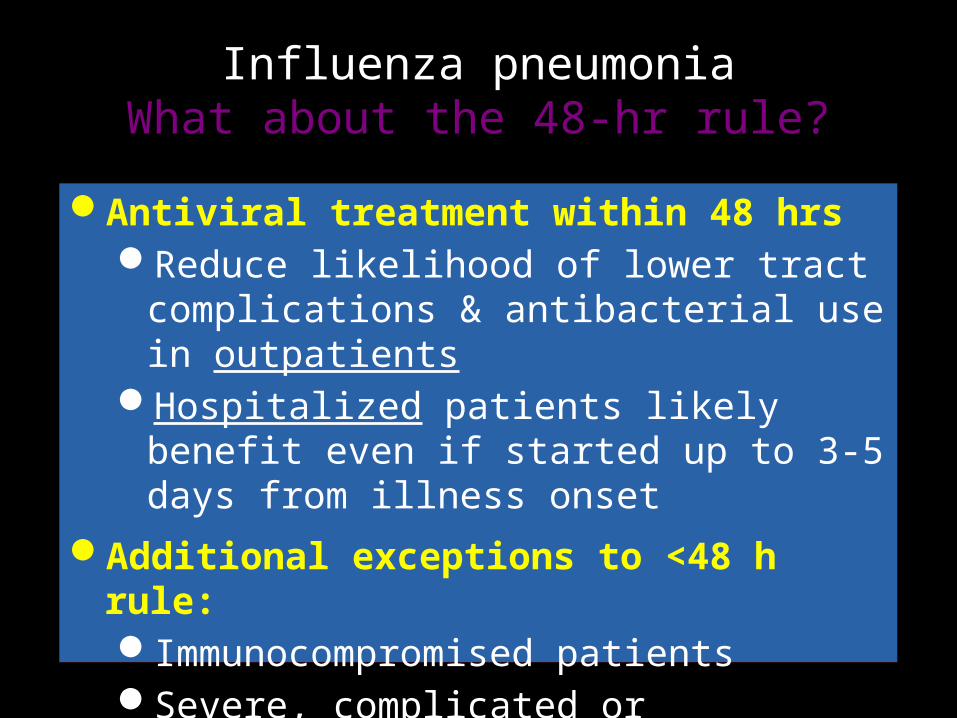
Influenza pneumoniaWhat about the 48-hr rule?
Antiviral treatment within 48 hrsReduce likelihood of lower tract complications &
antibacterial use in outpatientsHospitalized patients likely benefit even if
started up to 3-5 days from illness onset Additional exceptions to <48 h rule:
Immunocompromised patientsSevere, complicated or progressive illness

Follow-up ResponseExpected improvement?
Clinical improvement w/ effective abx: 48-72 hrsFever can last 2-5 days with Pneumococcus, longer with
other etiologies, esp Staph aureusCXR clearing
If healthy & <50 yo, 60% have clear CXR x 4 wksIf older, COPD, bacteremic, alcoholic, etc. only 25% with clear
CXR x 4 wksSwitch from IV to PO
Hemodynamically stable, improving clinicallyAble to ingest meds with working GI tract
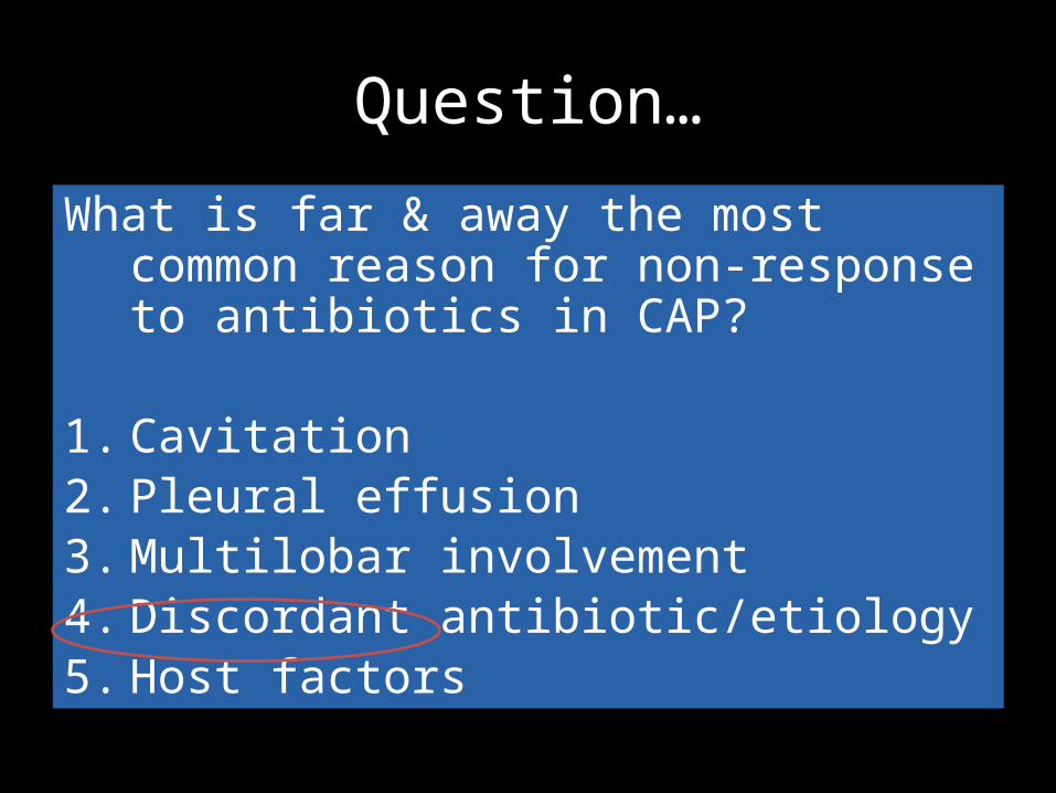
Question…
What is far & away the most common reason for non-response to antibiotics in CAP?
1. Cavitation2. Pleural effusion3. Multilobar involvement4. Discordant antibiotic/etiology5. Host factors

• • A 58 y/o man with advanced liver disease, construction worker in outdoor excavation• C/O acute fever, cough, pleuritic chest pain, WBC 23,000.• CXR and chest CT show RML nodule. No response to Unasyn + Levo.• Concern for pneumococcal pneumonia.


Coccidioides immitis
- Endemic to the desert southwest
- Dissemination more common in non-Caucasians, pregnant, immunocompromised
- Acute & chronic pulmonary syndromes (“valley fever”—fever, cough, arthralgias, Erythema nodosum)
- Diagnosis based on serology, culture, or histopathology
NW Infections: Coccidioides
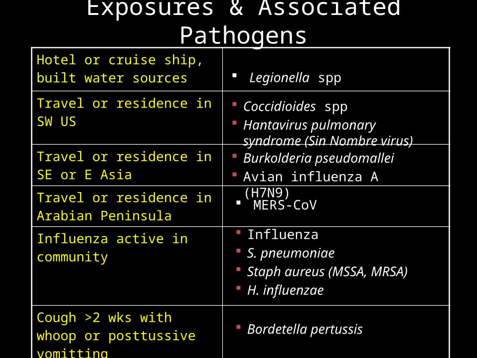
Exposures & Associated PathogensHotel or cruise ship, built water sources
Travel or residence in SW US
Travel or residence in SE or E Asia
Travel or residence in Arabian Peninsula
Influenza active in community
Cough >2 wks with whoop or posttussive vomitting
Legionella spp
Coccidioides spp Hantavirus pulmonary syndrome
(Sin Nombre virus) Burkolderia pseudomallei Avian influenza A (H7N9)
MERS-CoV
Bordetella pertussis
Influenza S. pneumoniae Staph aureus (MSSA, MRSA) H. influenzae
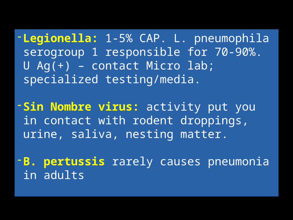
- Legionella: 1-5% CAP. L. pneumophila serogroup 1 responsible for 70-90%. U Ag(+) – contact Micro lab; specialized testing/media.
- Sin Nombre virus: activity put you in contact with rodent droppings, urine, saliva, nesting matter.
- B. pertussis rarely causes pneumonia in adults

Zoonotic Exposures & Associated Pathogens
Bat or bird droppings
Histoplasma capsulatum
Birds Chlamydophila psittaci Avian influenza (H7N9)
Rabbits Francisella tularensis
Farm animals or parturient cats
Coxiella burnetti (Q fever)

Take Home Points
Ask patients about co-morbidities and travel/other potential exposures when they present with a respiratory illness
Evaluate patients for MDR risk factors when managing patients in the community with respiratory illness