Communication - University of Waterloocamj/pdf/2011/Effect of Chemistry_MMTA_2011.pdfCommunication...
7
Communication Effect of Chemistry on Nonisothermal Tempering and Softening of Dual-Phase Steels S.S. NAYAK, V.H. BALTAZAR HERNANDEZ, and Y. ZHOU In sequel to our recent report, [1] we demonstrate in this article the effect of chemistry on nonisothermal tem- pering and softening behavior of dual-phase steels. The martensite morphology and tempered structure were analyzed using electron microscopy. Electron energy loss spectrometry and energy-dispersive X-ray spec- troscopy were used for a compositional analysis. It was observed that characteristic of the tempered structure in dual-phase (DP) steels was a function of prior mar- tensite structure and chemistry, which in turn influence the extent of softening. DOI: 10.1007/s11661-011-0868-8 ȑ The Minerals, Metals & Materials Society and ASM International 2011 An important issue with DP steel, which hampers its use in practical applications, is heat-affected zone (HAZ) softening that occurs in welding because of the tempering of the martensite phase in the base metal (BM). [2–8] Tempering of martensite is well documented in the literature, but it has been addressed mostly to fully martensitic steels subjected to isothermal tempering treatment. [9–11] In the context of explaining the temper- ing of martensite that causes softening in DP steel, we recently compared the characteristics of martensite tempering in DP steels subjected to isothermal and nonisothermal tempering, and the consequent effect on softening behavior. [1] Nonisothermal tempering cycle was achieved in resistance spot welding (RSW). We observed fine cementite in nonisothermally tempered DP steel unlike the coarser and spheroidized cementite in isothermal tempering. In addition, the extent of soften- ing was observed to vary with steel chemistry and martensite structure, which likely indicates a dependence of tempering behavior on chemistry. Thus, the objective of the current article was to support our recent study by investigating the effect of chemistry on the nonisother- mal tempering of martensite in DP steels and its effect on softening. The starting materials were three zinc-coated 1.2-mm thick DP steel sheets. DP steels used in the study were designated, according to their alloying level and, in turn, carbon equivalent (CE) calculated using Yurioka for- mula, [12] as follows: lean (DP L ), moderate (DP M ), and rich (DP R ), which are listed in Table I. All chemistries listed are in wt pct. The mechanical properties and volume fraction of martensite of the DP steels are also included in the table. A standard metallographic analysis was used for calcu- lating the volume fraction ( f m ). Figure 1 illustrates the nonisothermal tempering thermal cycle (RSW) used in the study; the details of this are reported elsewhere. [1,5] Figure 2 illustrates a schematic cross section of resis- tance spot-welded DP steel showing different zones of a weldment. It is to be noted that HAZ is subdivided at the location of Ac 1 as upper-critical and subcritical regions [5] ; however, the study was focused on subcritical HAZ (location ‘‘a’’ in Figure 2) where maximum softening occurs in the DP steel welds [5] and BM (location ‘‘b’’ in Figure 2), where martensite does not get affected in nonisothermal tempering. The microstructure of the subcritical HAZ and BM (location ‘‘a’’ and ‘‘b’’ in Figure 2, respectively) of the DP steels were analyzed using a JEOL7000F (Japan Electron Optics Ltd., Tokyo, Japan) scanning electron microscope (SEM) and a Philips CM12 (Philips, Irvine, CA) transmission electron microscope (TEM) operated at 120 kV. TEM samples of the BM were prepared by standard twin-jet electropolishing, whereas carbon extraction replicas were made for structural analysis of the precipitated carbides (at subcritical HAZ) in non- isothermally tempered specimens. Sample preparation details were reported in our recent study. [1] The com- positions of carbides were examined using electron energy loss spectrometry (EELS) and TEM energy- dispersive X-ray spectroscopy (EDX). EELS analysis was conducted in a TITAN II 800-300 (FEI, Hillsboro, OR) cryo in situ TEM operated at 300 kV. A Shimadzu HMV-2000 (Shimadzu Corporation, Kyoto, Japan) hardness tester was used to measure Vickers microh- ardness using a load of 200 g with a 15-second dwell time keeping 200 lm spacing between subsequent indentations. The carbide particle size was measured using high-magnification SEM images and TEM images of extracted replicas. Representative SEM micrographs (Figure 3(a) through (c)) illustrating the ferrite matrix (a) and martensite (a¢) in the BM microstructure delineated the distinct morphology of a¢-phase in all the three DP steels (Table I). It may be noted that a and a¢ has body- centered cubic and body-centered tetragonal crystal structure, respectively. For example, the martensite blocks were defined finely in DP L steel (Figure 3(a)), whereas a solid featureless morphology of the a¢ phase was observed in DP M and DP R steels (Figure 3(b) and (c)). A comparison of all three BM microstructures indicated a larger grain size of a¢ in DP L , whereas a smaller grain size was observed in DP M followed by DP R steel. Bright-field images of a¢ and corresponding S.S. NAYAK, Postdoctoral Fellow, and Y. ZHOU, Director and Professor, are with the Center for Advanced Materials Joining, Department of Mechanical and Mechatronics Engineering, University of Waterloo, Waterloo, Ontario N2L 3G1, Canada. Contact e-mails: [email protected] and [email protected] V.H. BALTAZAR HERNANDEZ, formerly Graduate Student, with the Department of Mechanical Engineering, University of Waterloo, is now Professor, with the MPyM-EPMM Academic Unit of Engineering, Autonomous University of Zacatecas, C.P. 98000 Zacatecas, Mexico. Manuscript submitted April 13, 2011. Article published online August 31, 2011 3242—VOLUME 42A, NOVEMBER 2011 METALLURGICAL AND MATERIALS TRANSACTIONS A
Transcript of Communication - University of Waterloocamj/pdf/2011/Effect of Chemistry_MMTA_2011.pdfCommunication...

CommunicationEffect of Chemistry on NonisothermalTempering and Softeningof Dual-Phase Steels
S.S. NAYAK, V.H. BALTAZAR HERNANDEZ,and Y. ZHOU
In sequel to our recent report,[1] we demonstrate in thisarticle the effect of chemistry on nonisothermal tem-pering and softening behavior of dual-phase steels. Themartensite morphology and tempered structure wereanalyzed using electron microscopy. Electron energyloss spectrometry and energy-dispersive X-ray spec-troscopy were used for a compositional analysis. It wasobserved that characteristic of the tempered structure indual-phase (DP) steels was a function of prior mar-tensite structure and chemistry, which in turn influencethe extent of softening.
DOI: 10.1007/s11661-011-0868-8� The Minerals, Metals & Materials Society and ASMInternational 2011
An important issue with DP steel, which hampers itsuse in practical applications, is heat-affected zone(HAZ) softening that occurs in welding because of thetempering of the martensite phase in the base metal(BM).[2–8] Tempering of martensite is well documentedin the literature, but it has been addressed mostly tofully martensitic steels subjected to isothermal temperingtreatment.[9–11] In the context of explaining the temper-ing of martensite that causes softening in DP steel, werecently compared the characteristics of martensitetempering in DP steels subjected to isothermal andnonisothermal tempering, and the consequent effect onsoftening behavior.[1] Nonisothermal tempering cyclewas achieved in resistance spot welding (RSW). Weobserved fine cementite in nonisothermally tempered DPsteel unlike the coarser and spheroidized cementite inisothermal tempering. In addition, the extent of soften-ing was observed to vary with steel chemistry andmartensite structure, which likely indicates a dependenceof tempering behavior on chemistry. Thus, the objectiveof the current article was to support our recent study byinvestigating the effect of chemistry on the nonisother-
mal tempering of martensite in DP steels and its effecton softening.The starting materials were three zinc-coated 1.2-mm
thick DP steel sheets. DP steels used in the study weredesignated, according to their alloying level and, in turn,carbon equivalent (CE) calculated using Yurioka for-mula,[12] as follows: lean (DPL), moderate (DPM), andrich (DPR), which are listed in Table I. All chemistrieslisted are in wt pct.The mechanical properties and volume fraction of
martensite of the DP steels are also included in the table.A standard metallographic analysis was used for calcu-lating the volume fraction ( fm). Figure 1 illustrates thenonisothermal tempering thermal cycle (RSW) used inthe study; the details of this are reported elsewhere.[1,5]
Figure 2 illustrates a schematic cross section of resis-tance spot-welded DP steel showing different zones of aweldment. It is to be noted that HAZ is subdivided atthe location of Ac1 as upper-critical and subcriticalregions[5]; however, the study was focused on subcriticalHAZ (location ‘‘a’’ in Figure 2) where maximumsoftening occurs in the DP steel welds[5] and BM(location ‘‘b’’ in Figure 2), where martensite does notget affected in nonisothermal tempering.The microstructure of the subcritical HAZ and BM
(location ‘‘a’’ and ‘‘b’’ in Figure 2, respectively) of theDP steels were analyzed using a JEOL7000F (JapanElectron Optics Ltd., Tokyo, Japan) scanning electronmicroscope (SEM) and a Philips CM12 (Philips, Irvine,CA) transmission electron microscope (TEM) operatedat 120 kV. TEM samples of the BM were prepared bystandard twin-jet electropolishing, whereas carbonextraction replicas were made for structural analysis ofthe precipitated carbides (at subcritical HAZ) in non-isothermally tempered specimens. Sample preparationdetails were reported in our recent study.[1] The com-positions of carbides were examined using electronenergy loss spectrometry (EELS) and TEM energy-dispersive X-ray spectroscopy (EDX). EELS analysiswas conducted in a TITAN II 800-300 (FEI, Hillsboro,OR) cryo in situ TEM operated at 300 kV. A ShimadzuHMV-2000 (Shimadzu Corporation, Kyoto, Japan)hardness tester was used to measure Vickers microh-ardness using a load of 200 g with a 15-second dwelltime keeping 200 lm spacing between subsequentindentations. The carbide particle size was measuredusing high-magnification SEM images and TEM imagesof extracted replicas.Representative SEM micrographs (Figure 3(a)
through (c)) illustrating the ferrite matrix (a) andmartensite (a¢) in the BM microstructure delineated thedistinct morphology of a¢-phase in all the three DP steels(Table I). It may be noted that a and a¢ has body-centered cubic and body-centered tetragonal crystalstructure, respectively. For example, the martensiteblocks were defined finely in DPL steel (Figure 3(a)),whereas a solid featureless morphology of the a¢ phasewas observed in DPM and DPR steels (Figure 3(b) and(c)). A comparison of all three BM microstructuresindicated a larger grain size of a¢ in DPL, whereas asmaller grain size was observed in DPM followed byDPR steel. Bright-field images of a¢ and corresponding
S.S. NAYAK, Postdoctoral Fellow, and Y. ZHOU, Director andProfessor, are with the Center for Advanced Materials Joining,Department of Mechanical and Mechatronics Engineering, Universityof Waterloo, Waterloo, Ontario N2L 3G1, Canada. Contacte-mails: [email protected] and [email protected]. BALTAZAR HERNANDEZ, formerly Graduate Student, withthe Department of Mechanical Engineering, University of Waterloo, isnow Professor, with the MPyM-EPMMAcademic Unit of Engineering,Autonomous University of Zacatecas, C.P. 98000 Zacatecas, Mexico.
Manuscript submitted April 13, 2011.Article published online August 31, 2011
3242—VOLUME 42A, NOVEMBER 2011 METALLURGICAL AND MATERIALS TRANSACTIONS A

selected area diffraction (SAD) patterns (insets) in allDP steels are illustrated in Figures 3(d) and (f). Inaccordance with SEM observation, coarser lath mor-phology was observed in DPL steel (Figure 3(d)) withrelatively larger blocks compared with that in the DPM
steel (Figure 3(e)) with SAD patterns confirming 011½ �a0and �111
� �a0 zone axes in DPL and DPM steels, respec-
tively. Interestingly, DPR steel was observed to containtwinned plates of a¢ (Figure 3(f)) as confirmed by the
indexed SAD pattern showing streaks of 23
�5353
� �twin
reflections along with the 011½ �a0 zone axis. This obser-
vation was in accordance with a¢ carbon content: lowcarbon a¢ (DPL= 0.273 wt pct and DPM = 0.269 wtpct) consisted laths whereas twinned morphology wasobserved in high-carbon a¢ (DPR = 0.360 wt pct).[9,11]
The high density of dislocations observed in the BMof DPM steel (Figure 4(a)) was a typical characteristicfeature of low-carbon martensite laths.[13] The SADpatterns shown in Figures 4(b) and (c), taken from thelocations indicated by circles ‘‘b’’ and ‘‘c’’ in Fig-ure 4(a), were indexed to contain spots from 001½ �a0 and�113� �
a zone axes, respectively, confirming the presenceof a¢-phase and a matrix in DPM steel. It is to be notedthat TEM bright-field images of the DPM steel BMdelineated the microstructural features distinctly, i.e.,random arrays of dislocations in the a-matrix, a¢-phase,prior-c grain boundaries (a/a¢ boundaries), and blockboundaries (marked by a white dotted line), whichseparate a¢-blocks containing parallel laths.[1] It was alsoconcluded that a¢-phase in the DPM steel has micro-structural features similar to low-carbon martensiticsteel.Figure 5 illustrates representative SEM images of
tempered structure and bright-field images of carbidesin replicas of all nonisothermally tempered DP steels. Aclose observation of the tempered structures suggested aseverely decomposed a¢ in DPL steel (Figure 5(a)) and abroken lathy appearance in DPM (Figure 5(b)) and DPR
steels (Figure 5(c)). Even though carbide precipitationwas observed to be distributed thoroughly in all tem-pered martensite, coarser carbides were observed in DPL
steel, whereas finer carbides precipitated in DPR steel.The finer carbides were associated with the higher carboncontent, leading to the twinned substructure of a¢-phasein DPR steel (Figure 3(f)), which further confirms thatthe tempering characteristic of a¢ in DP steel is similar tothat of martensitic steel with similar carbon content.[14]
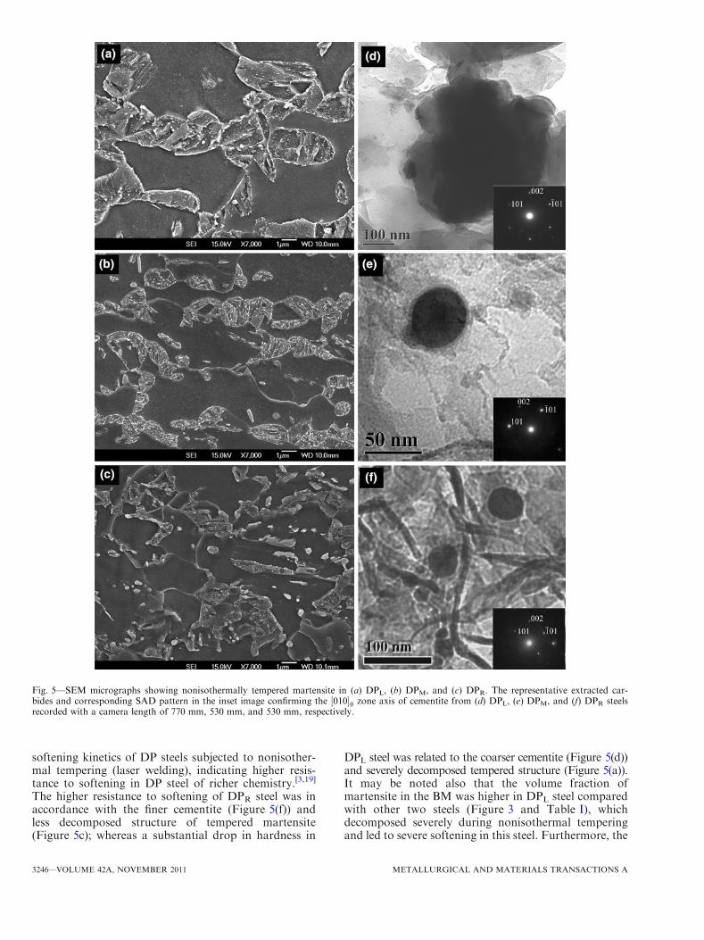
The inset SAD patterns showed spots from the 010½ � zoneaxis of orthorhombic structure corroborating precipita-tion of cementite (h), irrespective of DP steel chemistry(Figure 5(d) and (f)); however, the average carbide sizeof cementite in nonisothermally tempered DP steelssuggested a trend: DPL (300± 23 nm) fi DPM (45±14 nm) fi DPR (30± 12 nm).Figure 6 shows a dark-field image of a cementite
particle obtained, using (1) (010) diffraction spot, fromnonisothermally tempered DPM steel (Figure 6(a)); (2)EELS spectrum of the cementite indicating peaks of
Table I. Details of the DP Steels Studied: CE was used to categorize the DP steels
Steel DPL DPM DPR
C 0.150 0.132 0.147Mn 1.500 1.907 1.719Cr 0.021 0.161 0.612CE 0.390 0.475 0.525fm (pct) 54 48 40a¢-carbon content (calculated wt pct) 0.273 0.269 0.360Hardness (VHN) 330 301 2810.2 pct yield strength (MPa) 674 534 524Ultimate tensile strength (MPa) 1061 979 820Total elongation (pct) 12 15 18
Fig. 1—Thermal cycle of nonisothermal tempering implemented onDP steels[1,5].
Fig. 2—Schematic cross-section of resistance spot weld showing thedifferent zones: the fusion zone (FZ), HAZ, and BM. The HAZ isdivided into upper- and subcritical HAZ[5].
METALLURGICAL AND MATERIALS TRANSACTIONS A VOLUME 42A, NOVEMBER 2011—3243

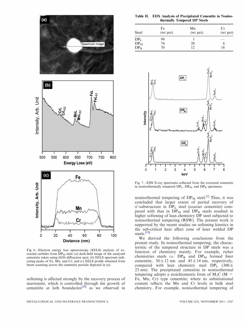
substitutional elements viz. Fe, Mn, and Cr (Figure 6(b));and (3) intensity profile made across the cementite(Figure 6(c)) following the path of the electron beam(marked as spectrum image in Figure 6(a)). Quantifica-tion of the spectrum indicated a relatively largerconcentration of Fe (80 wt pct) in cementite comparedwith Mn (16 wt pct) and Cr (4 wt pct). The distribution
of alloying elements was consistent within the cementitewith Fe, having a high-intensity profile confirming theEELS spectrum analysis. An EDX analysis of thecementite in all three DP steels (Figure 7) indicatedcharacteristic peaks of only Fe in the DPL specimen;however, additional Mn and Cr peaks were observed inthe spectrums of DPM and DPR steels. Supporting the
Fig. 3—SEM images showing the BM microstructure of (a) DPL, (b) DPM, and (c) DPR steels, and the BF images of the a¢ phase containing theinset SAD patterns showing spots from 011½ �a0 , �111
� �a0 , and 011½ �a0 zone axes in (d) DPL, (e) DPM, and (f) DPR steels taken with a camera length
of 770 mm, 530 mm, and 1100 mm, respectively.
3244—VOLUME 42A, NOVEMBER 2011 METALLURGICAL AND MATERIALS TRANSACTIONS A

EELS result, the peaks of Cr in cementite were lessintense than that of Mn in DPM steel, whereas strongerpeaks of Cr were observed in the cementite of the DPR
steel. A summary of the relative percentage of substit-utionals within cementite in DPL, DPM, and DPR steelsis listed in Table II.
An EELS and EDX analysis suggested that thesubstitutional composition of precipitated cementite innonisothermal tempering reflects the substitutional levelin the bulk DP steel chemistry. For example, cementiteof DPR steel comprised a high Cr and Mn content,suggesting the precipitation of substitutional richcementite. This reflects the Cr and Mn levels availablefor partitioning from bulk composition (Table I). Thepresence of Mn and Cr substitutionals in cementiteprecipitated in nonisothermally tempered DP steelssuggested that thermodynamically stable cementiteadopts the stoichiometric form of M3C (M = Fe, Mn,Cr), which is consistent with the previous reports.[10,14–16] In this work, nonisothermal tempering of DPR steelresulted cementite with a stoichiometric form of(Fe0.7Mn0.12Cr0.18)3C.
The stoichiometric form of cementite is derived fromthe concentration of alloying elements within cementite(Cr, Mn) or at the h/a interface (Si). In this case, thealloying elements controlled the growth rate of cement-ite by restricting the diffusion of carbon, thus reducingthe cementite coarsening.[17] Studies on composition ofcementite with substitutional elements (Fe, Mn, and Cr)in isothermally tempered martensitic steels indicatedthat the partition of alloying elements between cementiteand matrix is negligible in the temperature range of623 K to 823 K (350 �C to 550 �C) i.e., paraequilibriumstate of cementite in which carbon diffusion domi-
nates[10,11,14,16,18]; however, increasing the temperingtemperature to 723 K (450 �C) resulted in enrichmentof cementite with Cr and Mn.[14] A subsequent increasein tempering temperature to 923 K (650 �C) resulted ina gradual increase in Mn concentration with theenrichment strongly retarding the growth of cement-ite.[10] A similar effect was observed with Cr addition.[11]
In the current study, the substitutional content in theprecipitated cementite was related to the richness of steelchemistry, which suggested a restriction in diffusion ofcarbon even at high temperature, i.e., 923 K (650 �C),which was further supported by insufficient time avail-able for diffusion of carbon into cementite in noniso-thermal tempering of DP steels because of rapid heatingin nonisothermal tempering.[1] Thus, considering thecementite size and compositional analysis, it was con-cluded that cementite precipitation in the DPL steel wascontrolled by diffusion of carbon because the cementitecomposition (Table II) is near to the bulk steel chem-istry (Table I). In addition, it is coarser in size, whereasprecipitate coarsening in the DPM and DPR alloysis significantly reduced because of the diffusion ofsubstitutional elements that controls the coarseningmechanism.The effect of DP steel chemistry on softening behavior
is illustrated in Figure 8. The plot of the hardnessdifference in BM and nonisothermally tempered speci-mens (Figure 8(a)) suggested a trend in the extentof softening similar to that of the cementite size(Figure 5(d) through (f)) i.e., DPL (high) fi DPM fiDPR (low). The normalized softening (Figure 8(b))suggested that DP steel of rich chemistry has a higherresistance to softening in nonisothermal tempering,[1]
which was supported well by previous studies on the
Fig. 4—(a) Bright-field image delineating prior c grain boundary in BM of DPM steel, and the corresponding SAD patterns for (b) a¢-phase and(c) a-matrix confirming [011]a¢ and �113
� �a zone axes, respectively. The camera length used for the diffraction pattern was 530 mm.
METALLURGICAL AND MATERIALS TRANSACTIONS A VOLUME 42A, NOVEMBER 2011—3245
softening kinetics of DP steels subjected to nonisother-mal tempering (laser welding), indicating higher resis-tance to softening in DP steel of richer chemistry.[3,19]
The higher resistance to softening of DPR steel was inaccordance with the finer cementite (Figure 5(f)) andless decomposed structure of tempered martensite(Figure 5c); whereas a substantial drop in hardness in
DPL steel was related to the coarser cementite (Figure 5(d))and severely decomposed tempered structure (Figure 5(a)).It may be noted also that the volume fraction ofmartensite in the BM was higher in DPL steel comparedwith other two steels (Figure 3 and Table I), whichdecomposed severely during nonisothermal temperingand led to severe softening in this steel. Furthermore, the
Fig. 5—SEM micrographs showing nonisothermally tempered martensite in (a) DPL, (b) DPM, and (c) DPR. The representative extracted car-bides and corresponding SAD pattern in the inset image confirming the 010½ �h zone axis of cementite from (d) DPL, (e) DPM, and (f) DPR steelsrecorded with a camera length of 770 mm, 530 mm, and 530 mm, respectively.
3246—VOLUME 42A, NOVEMBER 2011 METALLURGICAL AND MATERIALS TRANSACTIONS A
softening is affected strongly by the recovery process ofmartensite, which is controlled through the growth ofcementite at lath boundaries[20] as we observed in
nonisothermal tempering of DPM steel.[1] Thus, it wasconcluded that larger extent of partial recovery ofa¢-substructure in DPL steel (coarser cementite) com-pared with that in DPM and DPR steels resulted inhigher softening of lean chemistry DP steel subjected tononisothermal tempering (RSW). The present work issupported by the recent studies on softening kinetics inthe sub-critical heat affect zone of laser welded DPsteels.[19]
We derived the following conclusions from thepresent study. In nonisothermal tempering, the charac-teristic of the tempered structure in DP steels was afunction of chemistry mainly. For example, richerchemistries steels i.e. DPR and DPM formed finercementite, 30± 12 nm and 45± 14 nm, respectively,compared with lean chemistry steel DPL (300±23 nm). The precipitated cementite in nonisothermaltempering adopts a stoichiometric form of M3C (M =Fe, Mn, Cr) type cementite; where its substitutionalcontent reflects the Mn and Cr levels in bulk steelchemistry. For example, nonisothermal tempering of
Fig. 6—Electron energy loss spectroscopy (EELS) analysis of ex-tracted carbides from DPM steel: (a) dark-field image of the analyzedcementite taken using (010) diffraction spot; (b) EELS spectrum indi-cating peaks of Fe, Mn, and Cr; and (c) EELS profile obtained frombeam scanning across the cementite particle depicted in (a).
Fig. 7—EDS X-ray spectrums collected from the extracted cementitein nonisothermally tempered DPL, DPM, and DPR specimens.
Table II. EDX Analysis of Precipitated Cementite in Noniso-
thermally Tempered DP Steels
SteelFe(wt pct)
Mn(wt pct)
Cr(wt pct)
DPL 99 1 —DPM 74 20 6DPR 70 12 18
METALLURGICAL AND MATERIALS TRANSACTIONS A VOLUME 42A, NOVEMBER 2011—3247

DPR steel resulted cementite with a stoichiometric formof (Fe0.7Mn0.12Cr0.18)3C. The trend in extent of soften-ing follows the trend of the cementite size: DPL (high) fiDPM fi DPR (low).
The authors acknowledge support from Auto21(Network of Centres for Excellence, Canada), The Ini-tiative for Automotive Manufacturing Innovation(IAMI), ArcelorMittal Dofasco, International ZincAssociation (IZA), and Huys Industries in Canada.V.H. Baltazar Hernandez acknowledges the supportfrom CONACYT Mexico and the Autonomous Uni-versity of Zacatecas Mexico.
REFERENCES1. V.H. Baltazar Hernandez, S.S. Nayak, and Y. Zhou: Metall.
Mater. Trans. A, 2011, vol. 42, pp. 3115–29.2. S.K. Panda, N. Sreenivasan, M.L. Kuntz, and Y. Zhou: J. Eng.
Mater. Tech., 2008, vol. 130, pp. 041003-1–041003-9.3. M. Xia, E. Biro, Z. Tian, and Y. Zhou: ISIJ Int., 2008, vol. 48, pp.
809–14.4. V.H. Baltazar Hernandez, M.L. Kuntz, M.I. Khan, and Y. Zhou:
Sci. Tech. Weld. Joining, 2008, vol. 13, pp. 769–76.5. V.H. Baltazar Hernandez, S.K. Panda, Y. Okita, and Y. Zhou:
J. Mater. Sci., 2010, vol. 45, pp. 1638–47.6. V.H. Baltazar Hernandez, S.K. Panda, M.L. Kuntz, and Y. Zhou:
Mater. Lett., 2010, vol. 64, pp. 769–76.7. M.I. Khan, M.L. Kuntz, and Y. Zhou: Sci. Tech. Weld. Joining,
2008, vol. 13, pp. 49–59.8. E. Biro and A. Lee: Sheet Metal Welding Conference XII, AWS,
Livonia, MI, 2006.9. G.B. Olson and W.S. Owen: Martensitic Nucleation, ASM Inter-
national, Materials Park, OH, 1992, p. 261.10. G. Miyamoto, J.C. Oh, K. Hono, T. Furuhara, and T. Maki: Acta
Mater., 2007, vol. 55, pp. 5027–38.11. J. Chance and N. Ridley: Metall. Trans. A, 1981, vol. 12A, pp.
1205–13.12. N. Yurioka, H. Suzuki, S. Ohshita, and S. Saito: Weld. J., 1983,
vol. 62, pp. 147s–153s.13. G. Tomas: Metall. Trans., 1971, vol. 2, pp. 2373–85.14. R.C. Thomson and M.K. Miller: Acta Mater., 1998, vol. 46,
pp. 2203–13.15. P. SCAF, S. Wiesen, and U. Gonser: Acta Mater., 1992, vol. 40,
pp. 373–79.16. G. Ghosh and G.B. Olson: Acta Mater., 2002, vol. 50, pp. 2009–
2119.17. A. Nagao, K. Hayashi, K. Oi, S. Mitao, and N. Shikanai: Mater.
Sci. Forum, 2007, vols. 539–543, pp. 4720–25.18. R.C. Thomson and H.K.D.H. Bhadeshia: Mater. Sci. Tech., 1994,
vol. 10, pp. 205–08.19. E. Biro, J.R. McDermid, J.D. Embury, and Y. Zhou: Metall.
Mater. Trans. A, 2010, vol. 41A, pp. 2348–56.20. S. Takaki, S. Iizuka, K. Tomimura, and Y. Tokunaga: Mater.
Trans. JIM, 1991, vol. 32, pp. 207–13.
Fig. 8—Effects of chemistry on softening: (a) Vickers microhardness(HV) of the BM and nonisothermally tempered DP steels and (b)normalized softening of the DPL, DPM, and DPR steels taken fromRef. 1.
3248—VOLUME 42A, NOVEMBER 2011 METALLURGICAL AND MATERIALS TRANSACTIONS A