Common Inflammatory Rashes Seen by - dermpathmd.com Dermatology/Red Rashes.pdf · Common...
46
Transcript of Common Inflammatory Rashes Seen by - dermpathmd.com Dermatology/Red Rashes.pdf · Common...

Common Inflammatory Rashes Seen by Dermatologists Atopic dermatitis Seborrheic dermatitis Psoriasis Tinea
Atopic dermatitis
Pathogenesis: immune mediated Epidemiology:
10% of children Most present before age 7 Atopic diathesis: 75% have a personal or family history
of allergic disease
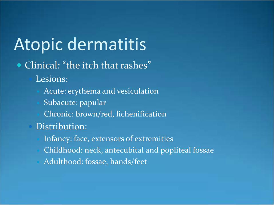
Atopic dermatitis Clinical: “the itch that rashes”
Lesions: Acute: erythema and vesiculation Subacute: papular Chronic: brown/red, lichenification
Distribution: Infancy: face, extensors of extremities Childhood: neck, antecubital and popliteal fossae Adulthood: fossae, hands/feet
Atopic dermatitis
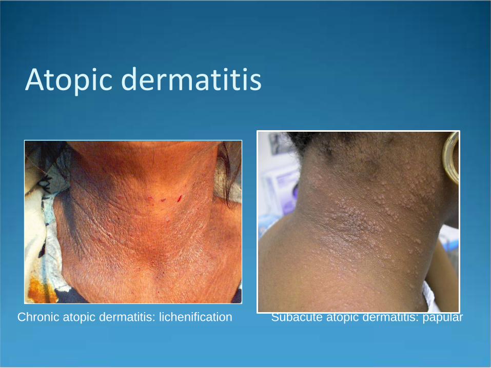
Chronic atopic dermatitis: lichenification Subacute atopic dermatitis: papular
Atopic dermatitis
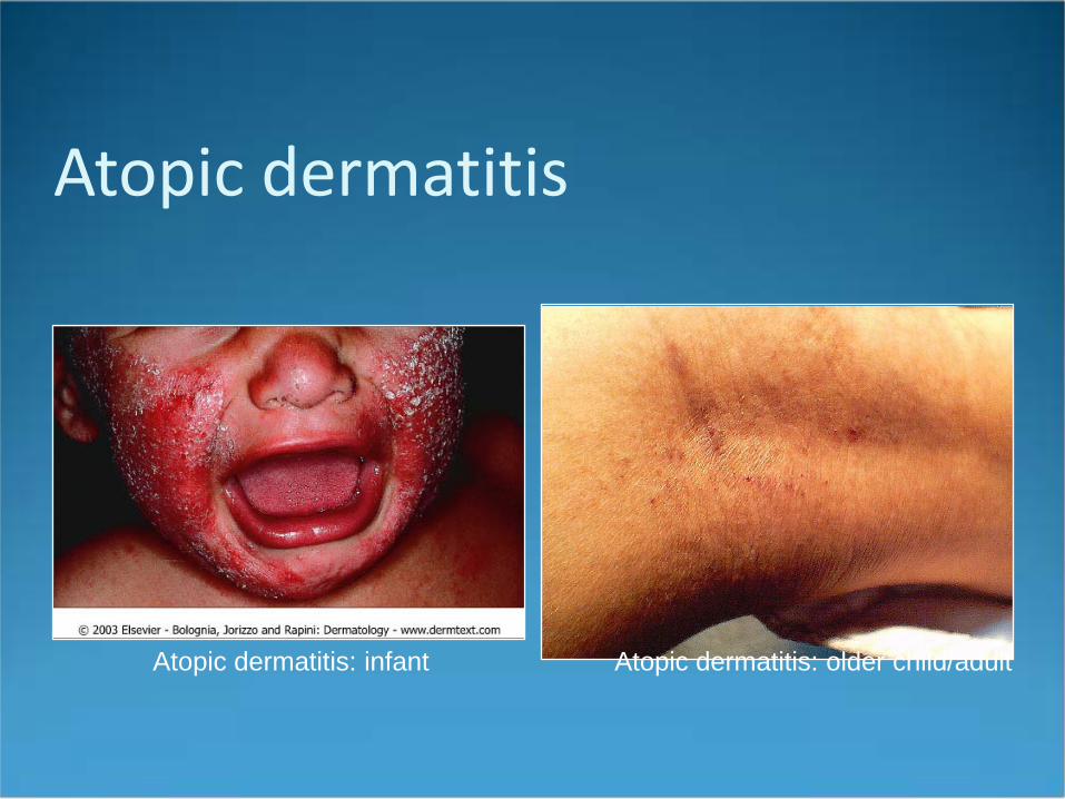
Atopic dermatitis: infant Atopic dermatitis: older child/adult
Atopic dermatitis Clinical:
Other findings: Pityriasis alba Dennie-Morgan lines, allergic shiners Keratosis Pilaris Icthyosis Vulgaris Hyperlinear palms
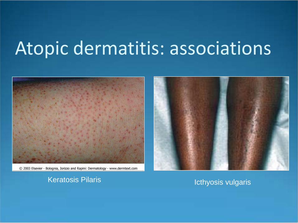
Atopic dermatitis: associations
Keratosis Pilaris Icthyosis vulgaris
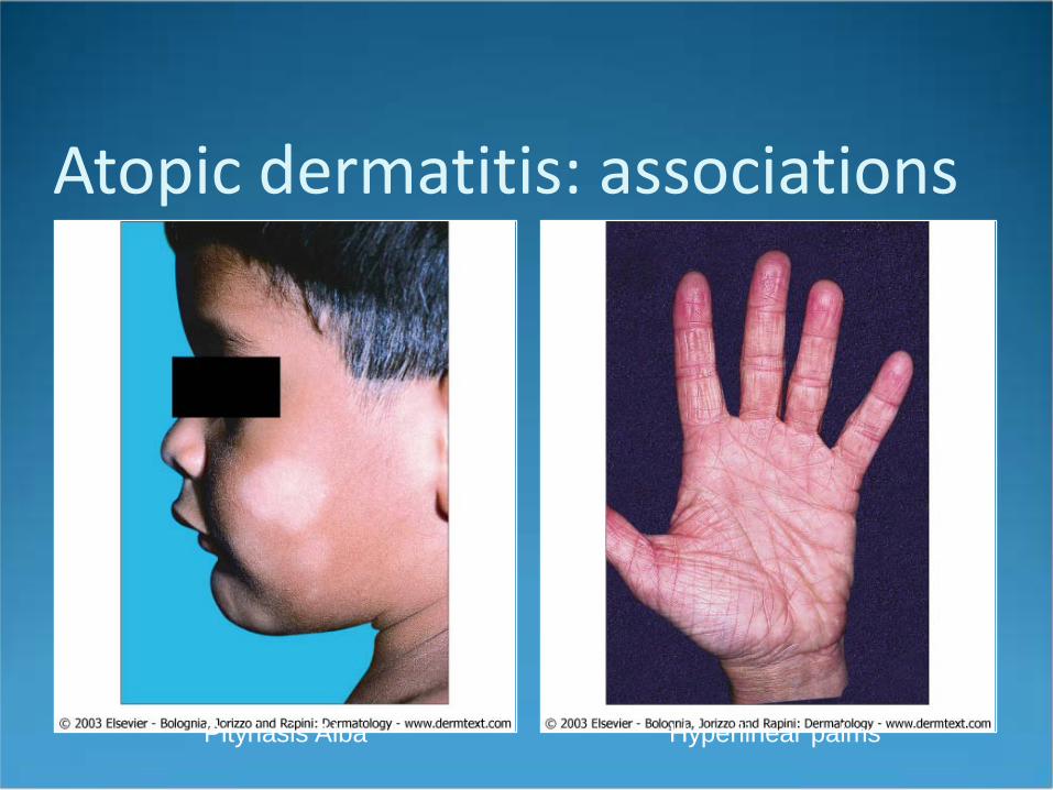
Atopic dermatitis: associations
Pityriasis Alba Hyperlinear palms
Atopic dermatitis Diagnosis
Clinical: pruritis, rash, chronicity, atopy Supportive: elevated IgE, eosinophilia, RAST tests
Differential Diagnosis Allergic/contact dermatitis Seborrheic dermatitis Infections (fungal) Congenital disorders (e.g. Netherton’s, SCID, Wiskott-
Aldrich, Chediak-Higashi)
Atopic dermatitis Treatments
“Soak and Grease”: hydration, steroid, +/-occlusion Lowest potency possible
Role of immunomodulators unclear Vaseline or other emollients additionally
Wet pajamas Sauna suit
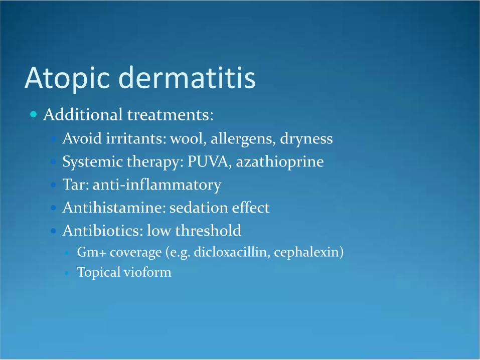
Atopic dermatitis Additional treatments:
Avoid irritants: wool, allergens, dryness Systemic therapy: PUVA, azathioprine Tar: anti-inflammatory Antihistamine: sedation effect Antibiotics: low threshold
Gm+ coverage (e.g. dicloxacillin, cephalexin) Topical vioform
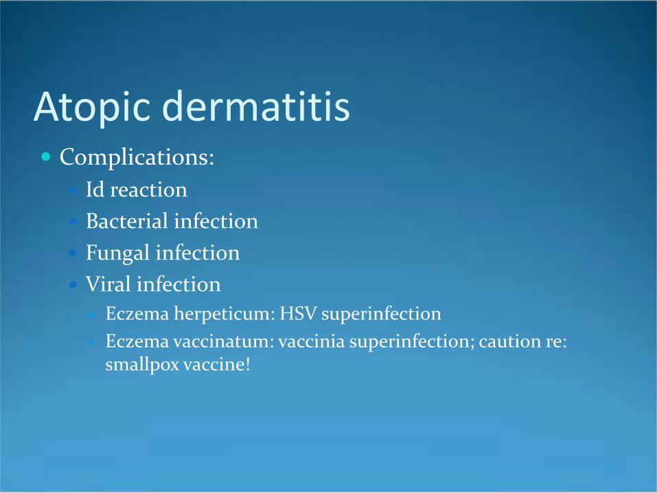
Atopic dermatitis Complications:
Id reaction Bacterial infection Fungal infection Viral infection
Eczema herpeticum: HSV superinfection Eczema vaccinatum: vaccinia superinfection; caution re:
smallpox vaccine!
Atopic dermatitis
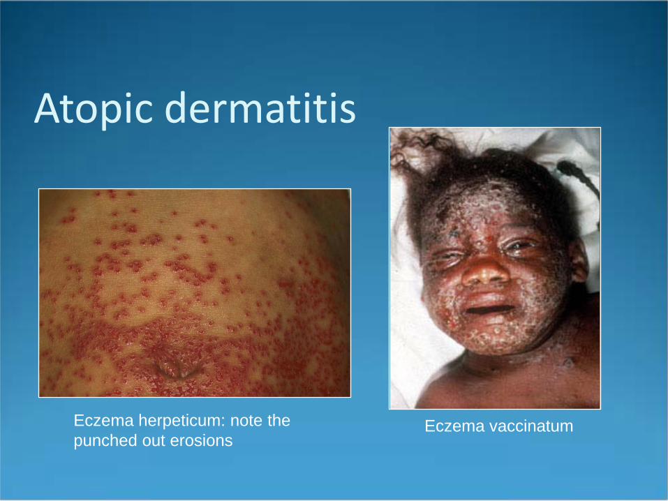
Eczema herpeticum: note the punched out erosions
Eczema vaccinatum
Atopic dermatitis What three components make-up the atopic diathesis? Where would you expect to see eczema in a 4-month
old? Bonus: at what age to children have a coordinated scratch?
Should I use steroids on infected appearing lesions? What is the concern about the smallpox vaccine and
eczema?
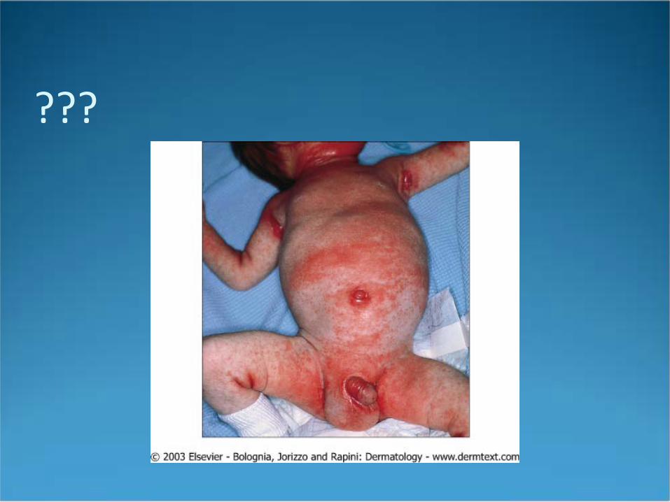
???

Seborrheic Dermatitis Pathogenesis: reaction to ubiquitous yeast,
pityrosporum ovale Epidemiology:
Affects 3-5% of healthy population Bimodal peaks: infancy (2-10 weeks, post-puberty) Severe in HIV and Parkinson’s disease
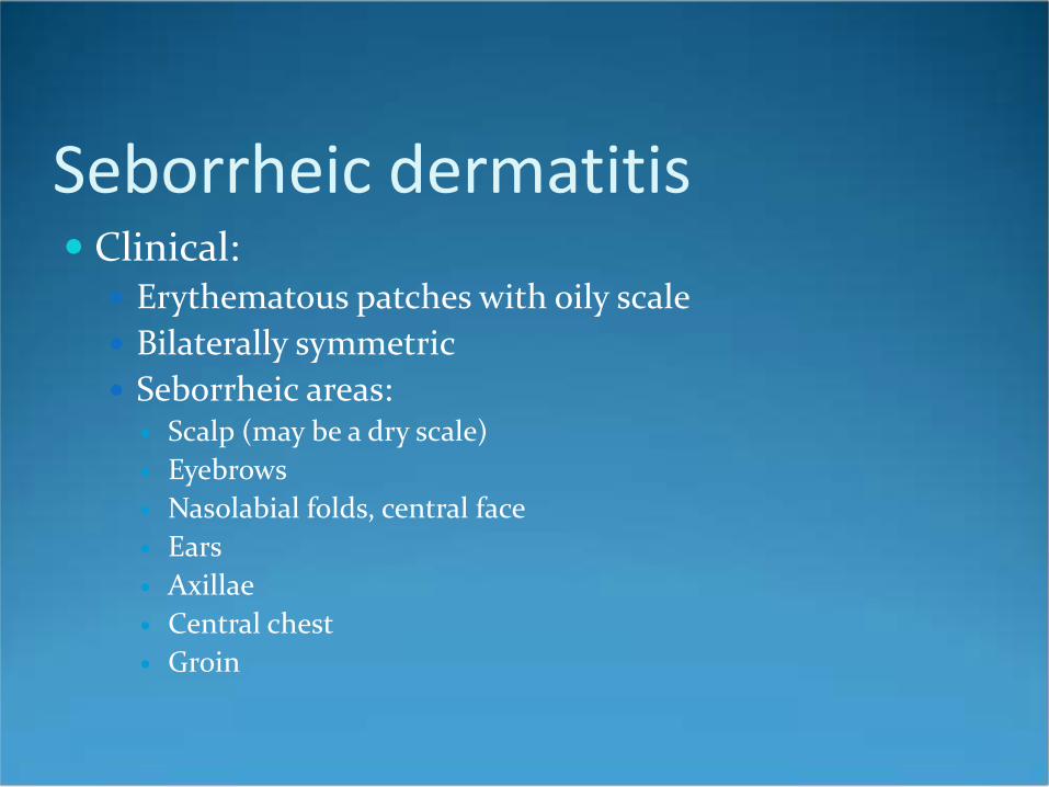
Seborrheic dermatitis Clinical:
Erythematous patches with oily scale Bilaterally symmetric Seborrheic areas:
Scalp (may be a dry scale) Eyebrows Nasolabial folds, central face Ears Axillae Central chest Groin
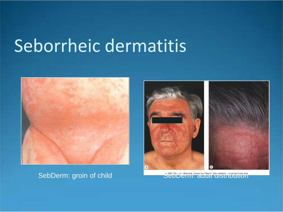
Seborrheic dermatitis
SebDerm: groin of child SebDerm: adult distribution
Seborrheic dermatitis Diagnosis:
Clinical Distribution very helpful
Differential diagnosis: Atopic dermatitis Psoriasis (overlap?) Lupus Rosacea Congenital (e.g. Histiocytosis, Acrodermatitis
enteropathica)
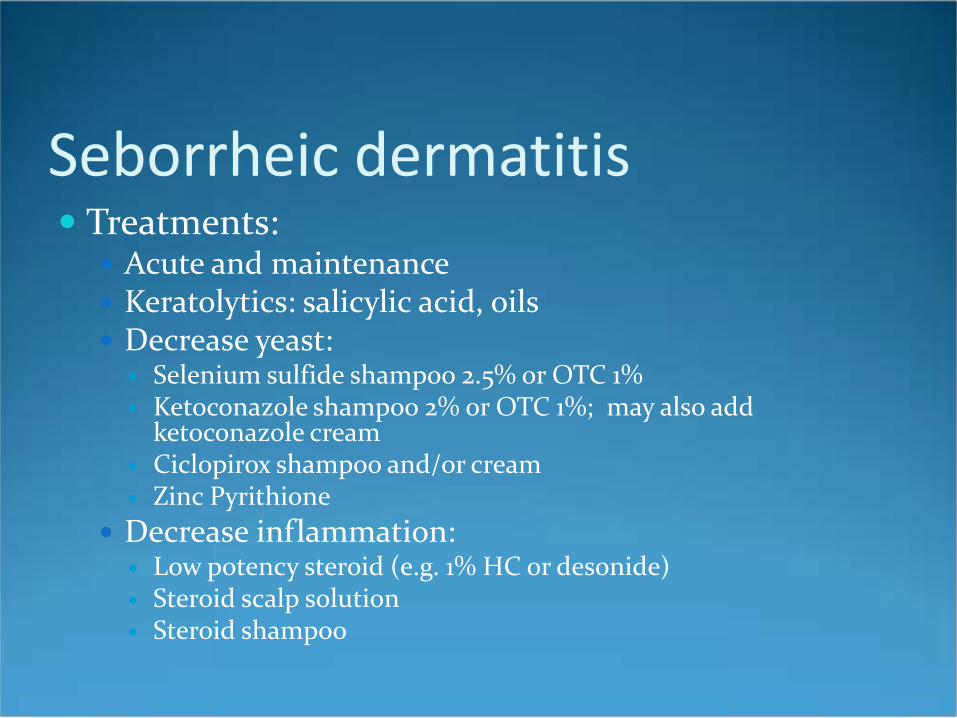
Seborrheic dermatitis Treatments:
Acute and maintenance Keratolytics: salicylic acid, oils Decrease yeast:
Selenium sulfide shampoo 2.5% or OTC 1% Ketoconazole shampoo 2% or OTC 1%; may also add
ketoconazole cream Ciclopirox shampoo and/or cream Zinc Pyrithione
Decrease inflammation: Low potency steroid (e.g. 1% HC or desonide) Steroid scalp solution Steroid shampoo
Seborrheic dermatitis Complications:
Post inflammatory hypopigmentation Cradle cap: thick, adherent scale of seb derm on the
scalp in infancy; resolves by age 1 Treatment: frequent washings with baby shampoo or anti-
fungals
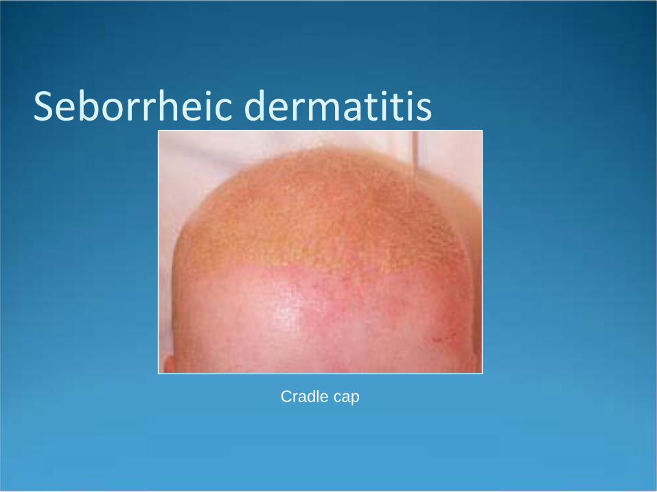
Seborrheic dermatitis
Cradle cap
Seborrheic dermatitis True or false: Seborrheic dermatitis may affect the eye
lid margins? At what age does seborrheic dermatitis of infancy
usually resolve? What is the presumed causative organism of
seborrheic dermatitis?
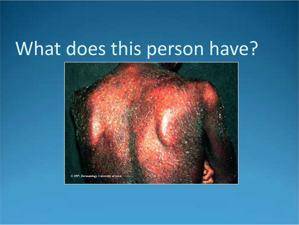
What does this person have?
Psoriasis Pathogenesis
T-cell mediated Increased epidermal turnover Infectious?
Epidemiology Affects 1% of the population Familial history, twin studies Age of onset usually in 20s
Younger age, more severe presentation
Psoriasis Clinical: “classic”
Sharply demarcated erythematous plaques with thick, silvery scale
Locations: scalp, elbows, knees; ears, intergluteal cleft Nails involved in close to 50%
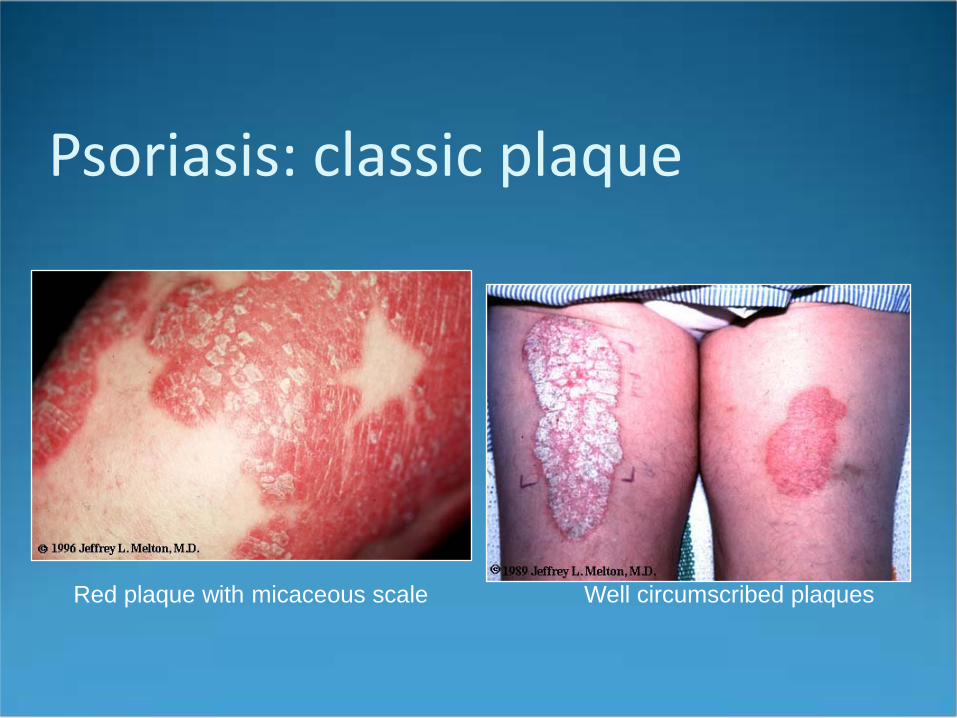
Psoriasis: classic plaque
Red plaque with micaceous scale Well circumscribed plaques
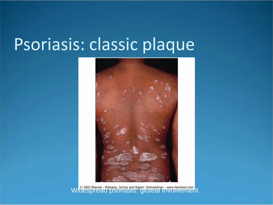
Psoriasis: classic plaque
Widespread psoriasis: gluteal involvement
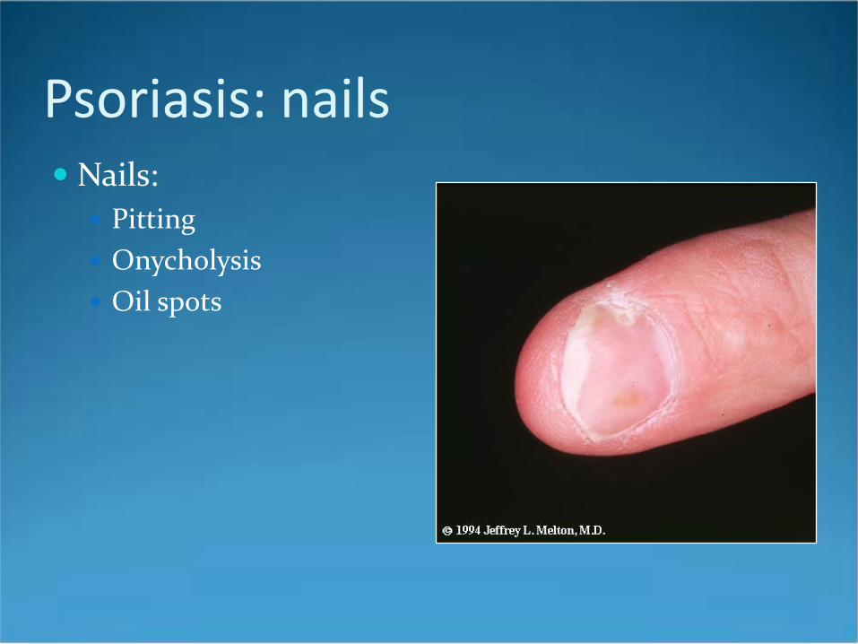
Psoriasis: nails Nails:
Pitting Onycholysis Oil spots
Psoriasis Clinical:
Variants Guttate Erythrodermic Pustular:
Palmoplantar Localized Generalized (Von Zombusch)
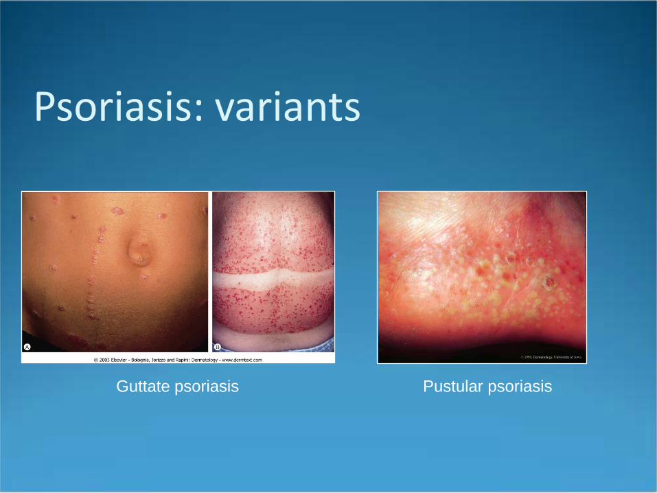
Psoriasis: variants
Guttate psoriasis Pustular psoriasis
Psoriasis Treatments: cell turnover, inflammation
Topical: Steroids Tar Vitamin D Retinoids UV light: UVB, excimer laser
Psoriasis Treatments:
Systemic: Methotrexate Acitretin Cyclosporine PUVA Biologics
Psoriasis Complications
Acute exacerbations: DRUGS, infection Staph aureus colonization Arthritis: 5-10%
1) asymmetric monarthritis 2) DIP joint disease 3) RA-like: DIPs and MCPs 4) ankylosing spondylitis: sacroilitis, HLA-B27 5) arthritis mutilans: osteolysis of the digits
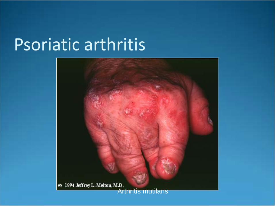
Psoriatic arthritis
Arthritis mutilans
Psoriasis What is Koebner’s phenomenon? What is Auspitz sign? What is the mechanism of methotrexate? Name two drugs that may exacerbate a psoriasis flare?
Tinea Pathogenesis: superficial skin infection with
dermatophyte organism Microsporum Epidermophyton Trichophyton
Epidemiology: very common! Sources include other humans, animals, and plant matter
Tinea Diagnosis: clinical and KOH Differential diagnosis: depends on location
Tinea capitis: SebDerm, alopecia areata, discoid lupus, folliculitis
Tinea cruris: SebDerm, candida Tinea corporis: nummular eczema, psoriasis, lupus Tinea pedis: ezcema, psoriasis, bacterial infn ETC….
Tinea: corporis Pathogenesis:
T. rubrum T. mentagrophytes M. canis
Clinical: Annular, scaly patch Single or multiple or polycyclic Leading scale
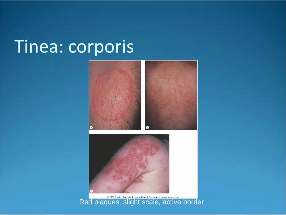
Tinea: corporis
Red plaques, slight scale, active border
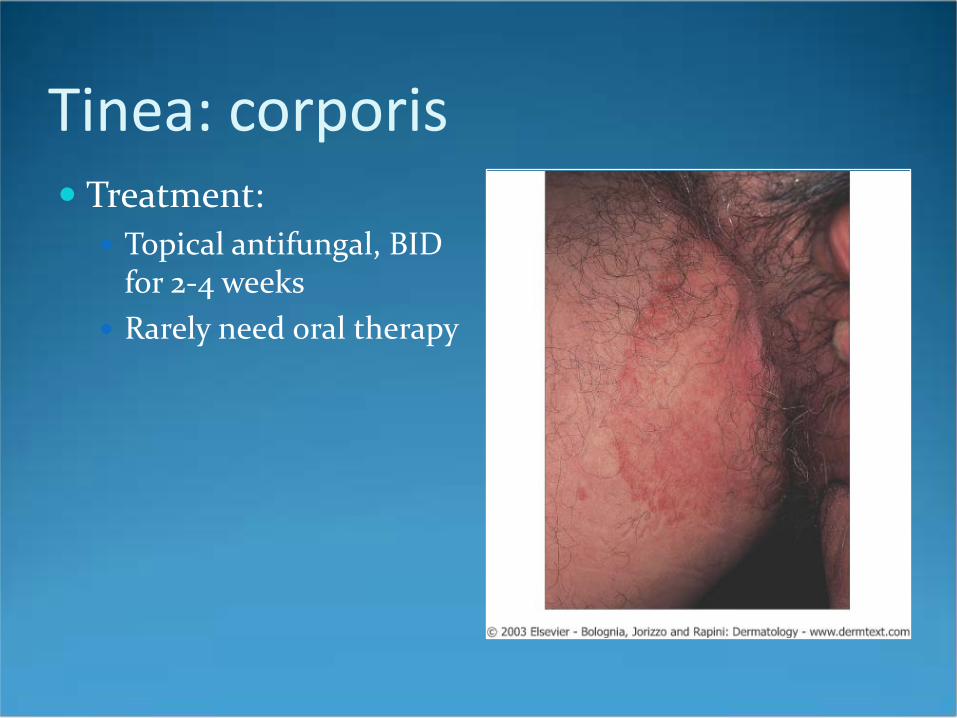
Tinea: corporis Treatment:
Topical antifungal, BID for 2-4 weeks
Rarely need oral therapy
Tinea: capitis Pathogenesis:
T. tonsurans M. canis M. andouinni
Clinical: Seborrheic Black dot Kerion
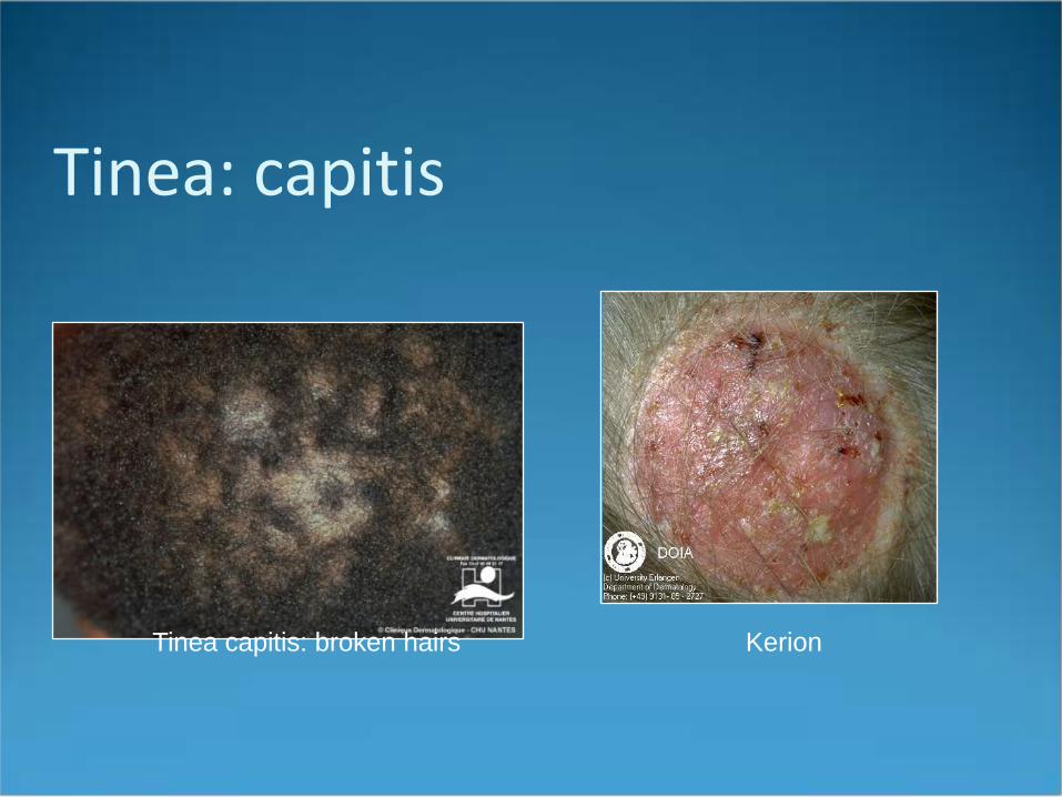
Tinea: capitis
Tinea capitis: broken hairs Kerion
Tinea: capitis Treatment:
Oral therapy indicated. Usually require 6-12 weeks. Choice depends on organism
KOH: endothrix, ectothrix, favus Wood’s lamp: green flourescence Culture
Tinea What is the most common cause of tinea capitis in the
United States? What dermatophyte causes “tinea” versicolor? Your wood’s lamp exam of the scalp flouresces green.
What is the most likely organism? What would you call a dermatophyte infection of the
hands?