
COMMISSIONING THROUGH EVALUATION Standards for … · COMMISSIONING THROUGH EVALUATION Standards...
42
1 COMMISSIONING THROUGH EVALUATION Standards for the Provision of Stereotactic Ablative Radiotherapy To be read in conjunction with the NHS England Radiotherapy Service Specification Service Specification No. Enter CRG document code Service Radiotherapy Commissioner Lead Kim Fell Provider Lead Period 12 months Date of Review 1. Population Needs 1.1 National/local context and evidence base Stereotactic Ablative Radiotherapy (SABR), also known as Stereotactic Body Radiotherapy (SBRT), is an emerging novel radiation technology. The American College of Radiology (ACR) and the American Society for Radiation Oncology (ASTRO) define SABR as “an external beam radiation therapy method used to very precisely deliver a high dose of radiation to an extra-cranial target within the body, using either a single dose or a small number of fractions.” 1 Specialised treatment planning techniques result in a high dose to the target with steep dose gradients resulting in rapid dose fall off beyond the target. This results in high biologically effective dose (BED) while providing normal tissue sparing, and is the basis for development of SABR. The technique requires specialist positioning equipment and/or imaging (stereotaxis) to confirm correct targeting (accuracy). Using a small number of fractions provides the opportunity for savings compared with conventional fractionation or surgical alternatives, and may free up capacity within NHS radiotherapy departments. The treatment can be delivered using either standard linear accelerators or specially designed devices which are dedicated to delivering stereotactic treatments. This document specifically refers to SABR for patients with: • oligometastatic cancer (3 or fewer sites of metastatic disease) • cancer that has recurred in a site treated previously treated with radiotherapy • patients with hepatocellular carcinoma
Transcript of COMMISSIONING THROUGH EVALUATION Standards for … · COMMISSIONING THROUGH EVALUATION Standards...

1
COMMISSIONING THROUGH EVALUATION
Standards for the Provision of Stereotactic Ablative Radiotherapy
To be read in conjunction with the NHS England Radiotherapy Service Specification
Service Specification No.
Enter CRG document code
Service Radiotherapy
Commissioner Lead Kim Fell
Provider Lead
Period 12 months
Date of Review
1. Population Needs
1.1 National/local context and evidence base
Stereotactic Ablative Radiotherapy (SABR), also known as Stereotactic Body Radiotherapy (SBRT), is an emerging novel radiation technology. The American College of Radiology (ACR) and the American Society for Radiation Oncology (ASTRO) define SABR as “an external beam radiation therapy method used to very precisely deliver a high dose of radiation to an extra-cranial target within the body, using either a single dose or a small number of fractions.” 1 Specialised treatment planning techniques result in a high dose to the target with steep dose gradients resulting in rapid dose fall off beyond the target. This results in high biologically effective dose (BED) while providing normal tissue sparing, and is the basis for development of SABR. The technique requires specialist positioning equipment and/or imaging (stereotaxis) to confirm correct targeting (accuracy). Using a small number of fractions provides the opportunity for savings compared with conventional fractionation or surgical alternatives, and may free up capacity within NHS radiotherapy departments. The treatment can be delivered using either standard linear accelerators or specially designed devices which are dedicated to delivering stereotactic treatments. This document specifically refers to SABR for patients with: • oligometastatic cancer (3 or fewer sites of metastatic disease) • cancer that has recurred in a site treated previously treated with radiotherapy • patients with hepatocellular carcinoma
2
There is now evidence from multiple non-randomised retrospective studies demonstrating that SABR is associated with local control rates of ~90% and can be given with minimal toxicity.2 Some early findings suggest it may be possible to delay the need for systemic therapy and improve progression-free survival using SABR.3 Further data is required to determine whether these benefits translate into an overall survival benefit. Nationally; the context supporting delivery is set out in the guidance from the National Radiotherapy Implementation Group; (Stereotactic Body Radiotherapy: Guidelines for Commissioners, Providers and clinicians in England 2011). This guidance sets out that Stereotactic Ablative radiotherapy (SABR) should be provided in the context of clinical trials and clinical studies to ensure that the evidence base for this treatment continues to accrue.
2. Outcomes
2.1 NHS Outcomes Framework Domains & Indicators
Domain
1
Preventing people from dying prematurely X
Domain
2
Enhancing quality of life for people with long-
term conditions
X
Domain
3
Helping people to recover from episodes of ill-
health or following injury
Domain
4
Ensuring people have a positive experience of
care
X
Domain
5
Treating and caring for people in safe
environment and protecting them from
avoidable harm
X
The CtE Programme will evaluate a number of key questions including:
1. Efficacy:
Local control
Progression free survival
Overall survival
Freedom from widespread metastatic disease not amenable to further SABR
3
(Oligomets CtE only)
2. Toxicity:
CTCAE v4 (site-specific measure)
EQ5D
Visual analogue pain score (if painful bony metastases present)
3. Radiotherapy planning/dosimetry:
Immediate investigation of any radiotherapy treatment plans leading to severe toxicity
Assessment of ongoing treatment plan quality (i) at different centres over time, (ii) using different plan/treat methods
4. Costs:
What is the actual cost, and relative cost effectiveness, of treatment with SABR for the clinical indications covered within the CtE programme?
Are there any factors from the experience of provision within centres participating in the scheme that should be taken into account in terms of future service provision.
Should the service become routinely commissioned by the NHS?
3. Scope
3.1 Aims and objectives of service
The indications for extra-cranial SABR can be divided into those for which there is a continuously evolving evidence base demonstrating the safety and clinical efficacy of SABR, and indications that are potentially interesting but should only be conducted in a clinical trial. The current SABR indications for which evidence is rapidly accumulating are listed in Table 1. It is the intention that this group of patients should receive SABR within this Commissioning through Evaluation (CtE) proposal, which will serve to improve access to SABR within the UK and enable data collection to further expand the current evidence base. The design and set-up of appropriate clinical trials to investigate the safety and efficacy of SABR for other indications is strongly encouraged and should be promoted. Clinical trials take precedence over any CtE programme and, as such, should a clinical trial be available to the same patient cohort as this programme, that cohort of patients will no longer be eligible for treatment as part of this scheme. It is possible that additional indications will be added to the programme in due course. The conditions under consideration include benign spinal conditions and renal cancer.
4
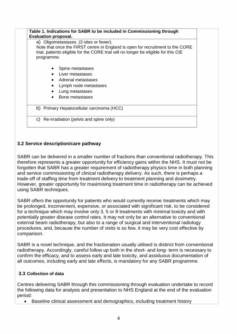
Table 1. Indications for SABR to be included in Commissioning through Evaluation proposal.
a) Oligometastases. (3 sites or fewer). Note that once the FIRST centre in England is open for recruitment to the CORE trial, patients eligible for the CORE trial will no longer be eligible for this CtE programme.
Spine metastases
Liver metastases
Adrenal metastases
Lymph node metastases
Lung metastases
Bone metastases
b) Primary Hepatocellular carcinoma (HCC)
c) Re-irradiation (pelvis and spine only)
3.2 Service description/care pathway
SABR can be delivered in a smaller number of fractions than conventional radiotherapy. This therefore represents a greater opportunity for efficiency gains within the NHS. It must not be forgotten that SABR has a greater requirement of radiotherapy physics time in both planning and service commissioning of clinical radiotherapy delivery. As such, there is perhaps a trade-off of staffing time from treatment delivery to treatment planning and dosimetry. However, greater opportunity for maximising treatment time in radiotherapy can be achieved using SABR techniques. SABR offers the opportunity for patients who would currently receive treatments which may be prolonged, inconvenient, expensive, or associated with significant risk, to be considered for a technique which may involve only 3, 5 or 8 treatments with minimal toxicity and with potentially greater disease control rates. It may not only be an alternative to conventional external beam radiotherapy, but also to a range of surgical and interventional radiology procedures, and, because the number of visits is so few, it may be very cost effective by comparison. SABR is a novel technique, and the fractionation usually utilised is distinct from conventional radiotherapy. Accordingly, careful follow up both in the short- and long- term is necessary to confirm the efficacy, and to assess early and late toxicity, and assiduous documentation of all outcomes, including early and late effects, is mandatory for any SABR programme.
3.3 Collection of data
Centres delivering SABR through this commissioning through evaluation undertake to record the following data for analysis and presentation to NHS England at the end of the evaluation period:
Baseline clinical assessment and demographics, including treatment history
5
Dosimetric data for each patient including D95% PTV, max and min PTV dose, max and volume dose delivered to all clinically relevant OAR, margins used, IGRT method used.
Acute toxicity (CTCAE v 4.0)
Late toxicity (CTCAE v 4.0)
Quality of life (EQ-5D)
Local control, PFS and OS at 1 and 2 years post treatment
Visual analogue pain score (if relevant; particularly for spinal or bony treatments)
The consent process for SABR should include permission for data collection and analysis by NHS England or institutions acting on their behalf.
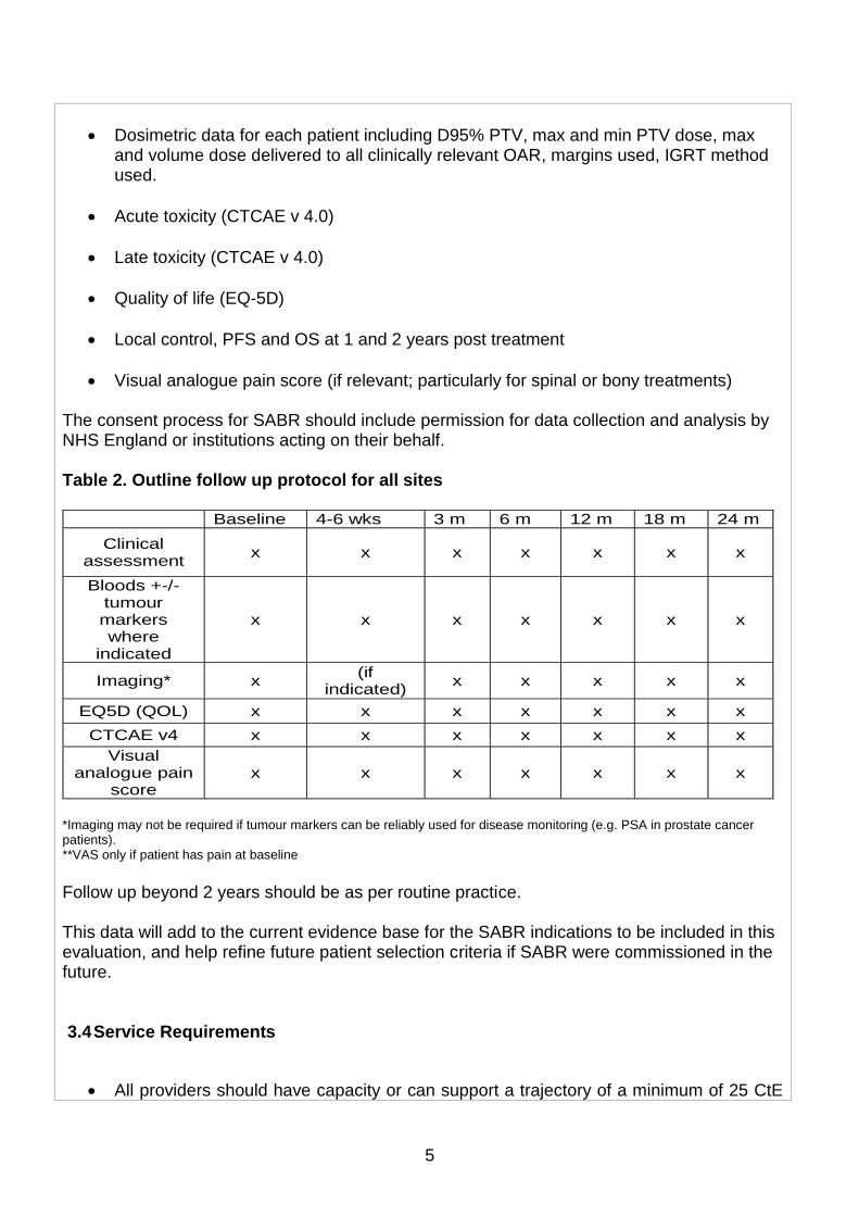
Table 2. Outline follow up protocol for all sites Baseline 4-6 wks 3 m 6 m 12 m 18 m 24 m
Clinical assessment
x x x x x x x
Bloods +-/- tumour markers where
indicated
x x x x x x x
Imaging* x (if
indicated) x x x x x
EQ5D (QOL) x x x x x x x
CTCAE v4 x x x x x x x
Visual analogue pain
score x x x x x x x
*Imaging may not be required if tumour markers can be reliably used for disease monitoring (e.g. PSA in prostate cancer patients). **VAS only if patient has pain at baseline
Follow up beyond 2 years should be as per routine practice. This data will add to the current evidence base for the SABR indications to be included in this evaluation, and help refine future patient selection criteria if SABR were commissioned in the future. 3.4 Service Requirements
All providers should have capacity or can support a trajectory of a minimum of 25 CtE

6
oligometastases cases per year in the first year of the CtE programme.
All providers must demonstrate that a minimum 24% inverse planned IMRT has been achieved consistently over the last year.
All providers must ensure all patients treated within the CtE are subject to a multidisciplinary team (MDT) approach to patient selection and treatment – all cases must be discussed at an appropriate MDT. (Site specific MDT and SABR Planning group).
All providers must comply with the full radiotherapy service specification.
Networked arrangements between RT providers that enable the RT service within this network to achieve around 2m population (geographical coverage) must be in place.
The referral pathways to your treatment centre should be defined to illustrate the networked referral arrangements which will lead to this required geographical coverage.
All providers must be IOG compliant for the range of primary tumours within the CtE, have a specialised MDT and offer the full range of treatments options for the indications agreed, offering genuine choice between clinically suitable options
Providers must have an adequate Technical Multi-professional Radiotherapy SABR Team present and able to deliver SABR radiotherapy to the standards set out in the RT QA CtE SABR pre-assessment document.
Providers will collect the audit clinical outcome data through their own collection process for all SABR CtE patients treated and these data must be available for evaluation.
Providers should have an externally accredited quality management system (e.g BSI) in place.
The age profile of the equipment to be used in the treatment of SABR patients must comply with the requirements within the service specification. Contingency arrangements to manage times of equipment breakdown and staff leave must be in place.
Providers should have a minimum of two subspecialist Clinical Oncologists with
7
experience in treating SABR patients. Providers should demonstrate a commitment to ongoing development of the SABR service and should describe the workforce / equipment available to take this initiative forward in their organisation and evidence of attendance at SABR meetings / conferences and training events by team members.
Providers should identify and provide additional support for patients and their carers who are required to travel distances for their treatment.
Providers should have an identified healthcare professional lead in place able to provide individual expert advice and support for the whole SABR patient pathway.
Patients eligible for the randomised CORE (Conventional care Or Radio ablation in the treatment of Extracranial metastases) trial in oligometastatic disease will be excluded from the CtE programme once the first centre recruiting to the CORE trial is open.
Providers should ensure continued clinical competency and continued commitment to developing the service.
In addition participating centres in the Re-irradiation CtE must:
• Maintain the surgical links / MDT arrangements to complex surgery
• Build on existing expertise in the treatment of Re-irradiation Pelvis and Spine using SABR
• Build on the RT QA process in this area
• Maintain the networked referral arrangements to include geographical coverage
In addition participating centres in the HCC CtE must:
• Build on the existing expertise in the treatment of HCC using SABR
• Maintain the surgical links / MDT arrangements to complex surgery
• Maintain the networked referral arrangements to include geographical coverage
3.5 Any acceptance and exclusion criteria and thresholds
The SABR work force needs to be truly multidisciplinary in its approach. Careful consideration should be given from the outset to the provision of adequate staffing and the education and training of staff in this new technology and associated techniques. Doses of up to 60Gy in up to 8 fractions (maximum) should be used as per UK SABR guidelines. Appropriate education and continuing education of professionals directly involved in SABR
8
procedures should be given a high priority. Training should include QA, planning, treatment delivery and verification technologies and techniques. Safety considerations should also be included in the training for these new techniques. The training of professionals should involve the ‘normal’ and ‘unusual’ circumstances likely to occur in the radiotherapy process.
4. Applicable Service Standards
4.1 Applicable national standards e.g. NICE, Royal College
Key guidance is provided in the National Radiotherapy Implementation Group Report: “Stereotactic Body Radiotherapy: Guidelines for Commissioners, Providers and clinicians in England 2011” 4.2 Interdependencies with other services/providers
5. Applicable Service Standards
5.1 Applicable national standards e.g. NICE
5.2 Applicable standards set out in Guidance and/or issued by a competent body (e.g. Royal Colleges)
6. Applicable quality requirements and CQUIN goals
6.1 Applicable quality requirements (See Schedule 4 Parts A-D)
6.2 Applicable CQUIN goals (See Schedule 4 Part E)
7. Location of Provider Premises
The Provider’s Premises are located at:
7. Individual Service User Placement
9
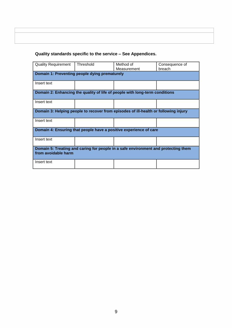
Quality standards specific to the service – See Appendices.
Quality Requirement
Threshold Method of Measurement
Consequence of breach
Domain 1: Preventing people dying prematurely
Insert text
Domain 2: Enhancing the quality of life of people with long-term conditions
Insert text
Domain 3: Helping people to recover from episodes of ill-health or following injury
Insert text
Domain 4: Ensuring that people have a positive experience of care
Insert text
Domain 5: Treating and caring for people in a safe environment and protecting them from avoidable harm
Insert text

10
Detailed Programme Requirements
SABR for Oligometastases (General) The term oligometastases refers to patients with a limited number of metastatic sites (usually considered to be patients with ≤ 5 metastatic sites). Historically, once a cancer has acquired the ability to metastasise it has generally been considered incurable, other than in a few rare chemo-sensitive exceptions. Subsequent treatment has therefore focused on systemic therapy (e.g. chemotherapy or endocrine therapy) given with palliative intent. However, it is now established that in many cancers, long term survival is possible after radical treatment of limited sites of metastatic disease. For example, in a series of mixed primary sites, 26% of patients remained alive 10 years after pulmonary metastectomy 4. Similarly 5 year survival rates following chemotherapy and surgery for liver metastases in colorectal cancer have now improved to 40-50% 5. This supports the concept that an oligometastatic disease state exists, whereby radical treatment of all active disease sites may result in improved disease free survival, and potentially, improved overall survival. The oligometastatic phenotype is being increasingly identified with the more widespread use of sensitive imaging procedures, such as PET-CT and MRI. An indication of incidence is 15% of all NSCLC will develop single organ oligometastatic disease. SABR may be used to treat a variety of oligometastatic sites and is particularly indicated in situations where other modalities of local therapy, such as surgery, are unsuitable. A number of retrospective and prospective cohort studies have reported outcomes of using SABR for the treatment of oligometastatic disease. This data has recently been reviewed 6,7. In general these studies demonstrate high rates of local control, together with acceptable rates of toxicity (≤ 10% G3 or higher). Common oligometastatic disease sites suitable for SABR include the treatment of lung, adrenal, liver, bone, vertebral and lymph node metastases. The aim of SABR in patients with oligometastases is to:
achieve local control at the metastatic site and prevent the clinical sequelae of disease progression at that site
improve disease free survival with the aim of enabling the patient to defer systemic therapy, or at least delay time until the next systemic therapy is required, thus maximising quality of life
improve overall survival. It is however acknowledged that at present the overall survival benefit of SABR in this context is not proven and requires further evaluation in clinical trials. Eligibility criteria for SABR for oligometastatic disease The following patient eligibility criteria for the oligometastatic disease to be treated within this commissioning through evaluation proposal:
Metastatic carcinoma with either a histologically or cytologically proven primary site or a male patient with a PSA>50 and clinical evidence of prostate cancer
11
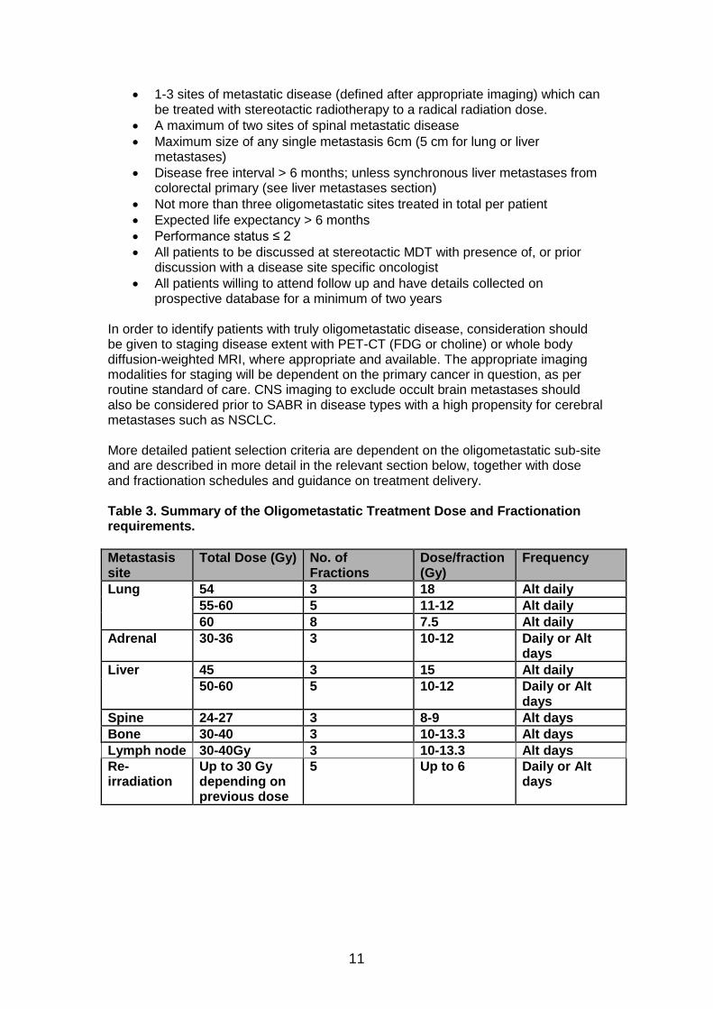
1-3 sites of metastatic disease (defined after appropriate imaging) which can be treated with stereotactic radiotherapy to a radical radiation dose.
A maximum of two sites of spinal metastatic disease
Maximum size of any single metastasis 6cm (5 cm for lung or liver metastases)
Disease free interval > 6 months; unless synchronous liver metastases from colorectal primary (see liver metastases section)
Not more than three oligometastatic sites treated in total per patient
Expected life expectancy > 6 months
Performance status ≤ 2
All patients to be discussed at stereotactic MDT with presence of, or prior discussion with a disease site specific oncologist
All patients willing to attend follow up and have details collected on prospective database for a minimum of two years
In order to identify patients with truly oligometastatic disease, consideration should be given to staging disease extent with PET-CT (FDG or choline) or whole body diffusion-weighted MRI, where appropriate and available. The appropriate imaging modalities for staging will be dependent on the primary cancer in question, as per routine standard of care. CNS imaging to exclude occult brain metastases should also be considered prior to SABR in disease types with a high propensity for cerebral metastases such as NSCLC. More detailed patient selection criteria are dependent on the oligometastatic sub-site and are described in more detail in the relevant section below, together with dose and fractionation schedules and guidance on treatment delivery. Table 3. Summary of the Oligometastatic Treatment Dose and Fractionation requirements.
Metastasis site
Total Dose (Gy) No. of Fractions
Dose/fraction (Gy)
Frequency
Lung 54 3 18 Alt daily 55-60 5 11-12 Alt daily 60 8 7.5 Alt daily
Adrenal 30-36 3 10-12 Daily or Alt days
Liver 45 3 15 Alt daily 50-60 5 10-12 Daily or Alt
days Spine 24-27 3 8-9 Alt days Bone 30-40 3 10-13.3 Alt days Lymph node 30-40Gy 3 10-13.3 Alt days Re-irradiation
Up to 30 Gy depending on previous dose
5 Up to 6 Daily or Alt days
12
SABR for spine metastases Spinal metastases are a common development in patients with advanced cancer, some of whom may survive for several years. Bone metastases commonly cause severe pain (requiring strong opiate analgesia), pathological fractures and, in spinal metastases there is a risk of paraplegia due to spinal cord compression. Complex spinal surgery may be necessary to treat tumours, fractures of the spinal column or spinal cord compression. Radiotherapy is established as an effective way of giving pain relief in patients with spine metastases and can prevent the neurological sequelae of cord compression. SABR has been established as an effective and well tolerated way of delivering radiotherapy to spinal metastases and can deliver a much higher, and hence more effective, dose to the tumour. A recent systematic review including over 1400 patients, showed analgesic benefit in 79%, local control of the tumour in 90% and a risk of myelopathy of <0.5% 8. Indications for spine SABR
Oligometastatic disease from previous malignancy (see specific eligibility below)
Re-irradiation where the position of the spinal metastasis would benefit from the increased precision and image-guidance of SABR (See re-irradiation section)
Patient eligibility Inclusion criteria
Patients with metastatic carcinoma, melanoma or sarcoma (lymphoma and myeloma excluded). Histology from initial primary or metastases is mandatory.
WHO performance status ≤2
Ambulatory without severe co morbidity
Life expectancy of more than six months
A maximum of two sites of spinal metastatic disease requiring treatment for pain relief or tumour control
Assessment by spinal SABR multi-disciplinary team (MDT) and discussion regarding the need for prophylactic vertebroplasty if concerns over vertebral stability (this is most relevant for re-irradiation or if pre-existent evidence of vertebral collapse)
No spinal instability
No cord compression
No chemotherapy within 28 days. Targeted therapies should be stopped a minimum of 14 days prior to SABR. Concurrent hormonal therapy is allowed.
No previous radiotherapy to the site of metastases. If prior radiotherapy to site, please see re-irradiation section
Patient availability for follow up to assess radiotherapy related morbidity, pain and functional ability
Disease free interval between primary treatment and development of metastases of six months or more
Pre-treatment patient assessment and consent Where the indication is oligometastatic disease, patients should be fully staged with CT chest/abdo/pelvis and PET scan (where indicated) to rule out more widespread disease. Other imaging (bone scan, whole body DW-MRI) may be useful in certain cases.
13
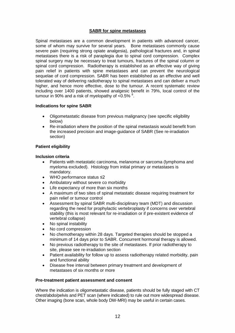
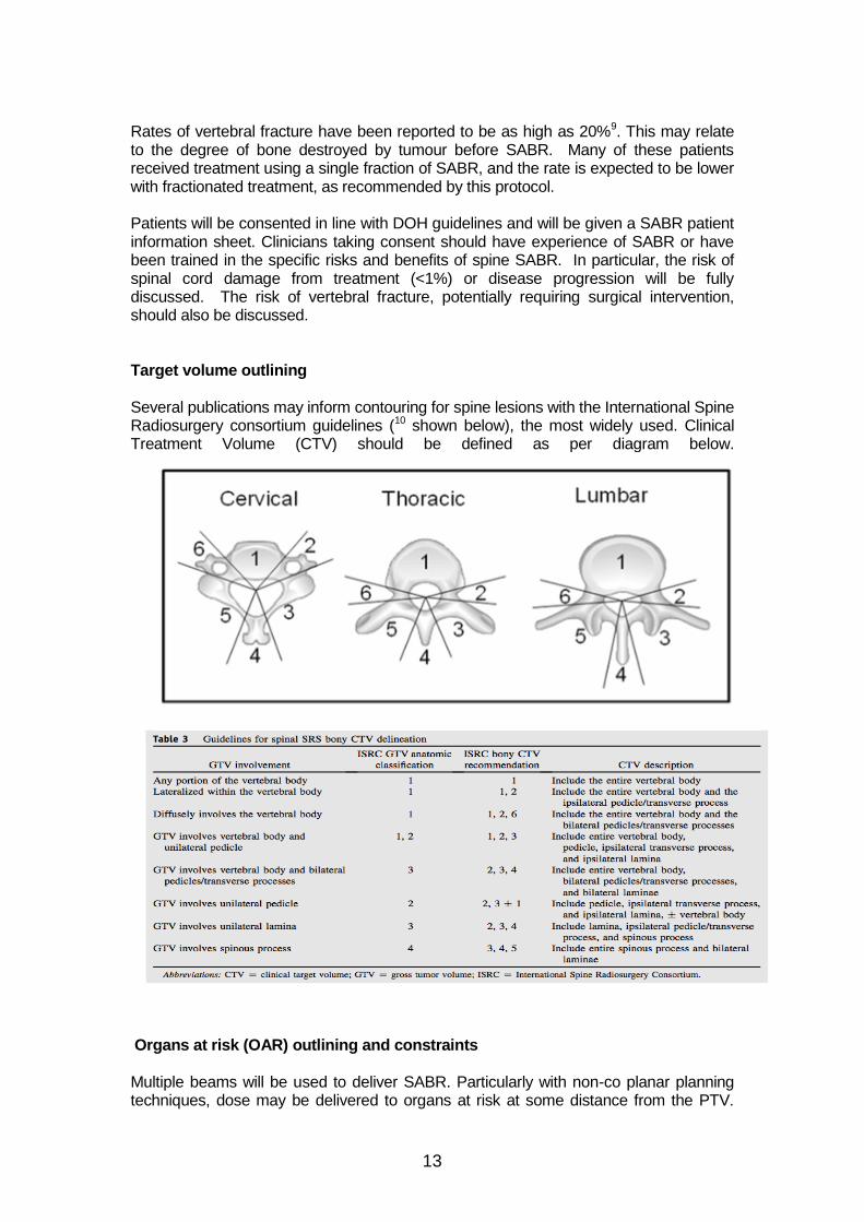
Rates of vertebral fracture have been reported to be as high as 20%9. This may relate to the degree of bone destroyed by tumour before SABR. Many of these patients received treatment using a single fraction of SABR, and the rate is expected to be lower with fractionated treatment, as recommended by this protocol. Patients will be consented in line with DOH guidelines and will be given a SABR patient information sheet. Clinicians taking consent should have experience of SABR or have been trained in the specific risks and benefits of spine SABR. In particular, the risk of spinal cord damage from treatment (<1%) or disease progression will be fully discussed. The risk of vertebral fracture, potentially requiring surgical intervention, should also be discussed. Target volume outlining Several publications may inform contouring for spine lesions with the International Spine Radiosurgery consortium guidelines (10 shown below), the most widely used. Clinical Treatment Volume (CTV) should be defined as per diagram below.
Organs at risk (OAR) outlining and constraints Multiple beams will be used to deliver SABR. Particularly with non-co planar planning techniques, dose may be delivered to organs at risk at some distance from the PTV.
14
Therefore every potential organ through which beams can traverse should be contoured. This may include lungs, heart, kidneys, liver, bowel, thyroid, larynx, chest wall and oesophagus (see Appendix 2). Dose constraints given in the Appendix 1 must be respected. If these constraints cannot be met, please contact the SABR CtE QA team. Radiotherapy Doses and Dose Prescription The doses advised for spinal SABR are based on the experience reported from multiple centres over a decade. The recommended doses show good levels of efficacy with a very low risk of spinal cord damage. Recommended dose prescription should be to ensure that 95% of the PTV receives 24-27 Gy in 3 fractions delivered alternate days, but coverage/dose must be reviewed to keep within the spinal cord tolerance detailed above and the prescribed dose or coverage should be compromised to achieve this. This prescribed dose could be viewed as being at least 3 times the conventional dose (8 Gy single fraction). Follow-up after treatment Follow up should be as per CtE requirements (Table 2 above and CtE Monitoring Form).

15
SABR for liver metastases Background The liver is a common site of metastases from many solid organ malignancies including colorectal, breast and lung cancer. In patients with oligometastatic liver disease, systemic therapy, generally in combination with surgical resection where appropriate, is standard of care. In disease types such as colorectal cancer, this combined approach had improved 5 year survival rates to ~ 50% 5. However, less than 20% of patients will be suitable for surgery. This may be as a result of co-morbidities or due to an unfavourable distribution of disease within the liver. For this patient group, there are a number of alternative ablative techniques available, delivered with the goal of improving time to disease progression and overall survival. The evidence base for the treatment of liver metastases with ablative therapy, as described below is strongest in colorectal cancer, where the role for local therapy is well established. Radiofrequency ablation (RFA) is the most established ablative technique and is routinely used in clinical practice. However, it is not recommended for the treatment of metastases situated close to large vessels or in the dome of the liver, and is less effective when used for treating lesions > 3cm in size 13. For this group, SABR is an alternative technique with a rapidly expanding body of evidence supporting its use 14. High rates of local control (60-90% at 2 years), together with preservation of liver function and an acceptable toxicity profile (<10% G3 toxicity) have led to it being increasingly integrated into the therapeutic pathway. At present its use is generally reserved for patients unsuitable for both surgery and RFA. For patients with small (<4cm), favourably located lesions, which would be suitable for treatment with either RFA or SABR, the optimal technique is yet to be established. This is the basis for a currently recruiting international randomised trial (RAS trial). A variety of SABR fractionation regimes have been used, ranging from 1-10 fractions. Dose escalation studies have shown that higher doses (up to 75 Gy in 3 fractions) can be efficacious and well tolerated. A dose response relationship for colorectal liver metastases has been demonstrated, with increasing rates of local control dependent on BED. However the dose that can be safely delivered without exceeding normal tissue tolerances is often limited by tumour volume and/or proximity to adjacent organs at risk. SABR is most suitable for lesions <6 cm in size, ideally situated at least 5mm away from the closest visceral organ at risk to reduce the risk of toxicity. Patients with liver metastases which fall outwith the therapeutic options of resective surgery or RFA, should be considered for SABR if they meet the eligibility criteria listed below. The most suitable modality of local therapy should be discussed in a specialist Hepatobiliary MDT on an individual patient basis. Indications
The treatment of liver metastases from colorectal cancer where the colorectal MDT and hepatobiliary MDT have agreed that SABR is indicated to achieve local control (i.e. synchronous or metachronous).
The treatment of liver metastases from non-colorectal cancer that meet the oligometastatic eligibility criteria listed above (i.e. metachronous only).
16
Eligibility criteria Inclusion criteria
1-3 liver metastases unequivocally seen on contrast enhanced CT and/or MRI in patients with previously histologically confirmed malignancy
Metastases unresectable, patient unfit for or declines surgery or presence of extra-hepatic disease making hepatic surgery an inappropriate treatment option
Maximum size of any single metastasis 5 cm
Childs-Pugh Class A
Adequate hepatic function defined as: >700 cc normal liver (liver volume less gross tumour volume, (GTV), bilirubin <3 x upper limit of normal, INR<1.3, ALT < 5x upper limit of normal, platelet count >80)
Performance status ≤ 2
Life expectancy > 6 months
Patients must have recovered from the effects of previous surgery, radiotherapy or chemotherapy with a minimum of 4 weeks break prior to SABR
Suitability for treatment established in Hepatobiliary MDT and Stereotactic MDT
All patients willing to attend follow up and have details collected on prospective database
Exclusion criteria
Active hepatitis or clinically significant liver failure (encephalopathy, oesophageal varices, portal hypertension)
Prior abdominal radiotherapy precluding SABR, that is any previous radiation therapy in which a mean dose to the liver of 15Gy in conventional fractionation was delivered or previous doses to critical normal structures that would make re-irradiation unsafe. Prior pelvic radiation is permitted, as long as there is no overlap between pelvic and liver radiation fields
Clinically apparent ascites
Central nervous system metastases
Pre-treatment patient assessment This should include: baseline blood tests including FBC, Coagulation Screen, U and E's, LFTs including albumin, and calcium. Tumour markers should be performed if relevant. A diagnostic liver MRI should be performed to aid tumour definition (unless contraindications to MRI), in addition to a contrast-enhanced CT. A DMSA scan may be required if the renal dose is likely to be significant. Patients will be consented in line with DOH guidelines and will be given a SABR patient information sheet. Clinicians taking consent should have experience of SABR or have been trained in the specific risks and benefits of spine SABR.
Target volume outlining Accurate tumour delineation can be challenging and both the contrast CT and MRI (if possible and no contra-indications) should be used to aid tumour delineation.

17
Radiology input should be sought to aid tumour and/or organ at risk definition where necessary.
Gross tumour volume (GTV) will be defined by contrast CT and MRI and includes all definable disease
Clinical Target Volume (CTV) = GTV + 5mm isotropically, edited to liver contours (ie CTV does not extend beyond the liver border)
The planning target volume (PTV) margin will be dependent on delivery technique and immobilization systems and will be pre-determined by centres following internal auditing of their treatment uncertainties. Organs at risk (OAR) outlining and constraints The following OAR will be outlined in their entirety on the planning CT scan: normal liver, kidneys, stomach, duodenum and small bowel, spinal cord, oesophagus, heart, lungs, and chest wall (see Appendix 2). Dose constraints given in Appendix 1 should be respected. If these constraints cannot be met, please contact the SABR CtE QA team. The exception to this is the chest wall constraint. Exceeding chest wall tolerance predicts for a higher incidence of chest wall pain and rib fracture for which the patient should be consented. PTV dose should not be limited due to exceeding chest wall tolerance.
Dose and prescription
Two fractionation regimes are available depending on the clinical scenario and physician choice. The highest BED achievable within the planning constraints should be used as there is evidence to show that local control correlates with BED. However in all cases OAR doses must be respected and if planning constraints cannot be met, the prescription dose should be reduced accordingly.
45 Gy in 3 fractions prescribed to prescription isodose covering 95% of PTV.
50-60 Gy in 5 fractions may be used when a larger PTV volume is being treated in order to achieve OAR constraint, when the PTV is within 1 cm of small bowel/visceral OAR or adjacent to chest wall/ribs. ≥95% of the PTV will receive the prescription dose.
Care on treatment Patients who have a large volume of stomach or duodenum treated should be
prescribed anti-emetics. 5HT3 antagonists are preferred.
Patients whose PTV is close to the stomach/duodenum should be prescribed prophylactic proton pump inhibitors and continue taking them for at least 3 months following treatment.
Follow up Follow up should be as per CtE requirements (Table 2 above and CtE Monitoring Form).
18
SABR for adrenal metastases Background The adrenal gland is a common site of metastases from many solid organ malignancies. 40% of lung and breast carcinomas will give rise to adrenal metastases, with the commonest malignancy to metastasise to the adrenal gland being melanoma (50% of patients)15. The most common presentation of adrenal metastases is in the context of widespread metastatic disease but in a minority of patients the adrenal gland will be the only site of disease relapse, or part of an oligometastatic presentation, where ablative treatment of all active sites of disease may be appropriate. Historically the gold standard for radical treatment of adrenal metastases has been surgical adrenalectomy. Despite the risk of systemic relapse in malignancies such as lung cancer, a meta-analysis of the surgical data suggests that up to 25% of patients will become long term survivors post adrenalectomy suggesting local therapy may be of benefit16. There are a limited number of published series on SABR for the treatment of adrenal metastases. No prospective trials have been published but a number of retrospective series exist. Local control rates vary from 55-100% at 1-2 years. The relatively poor overall survival rates reflect the propensity of patients to relapse at distant disease sites after SABR. Reported dose fractionation regimes range from 16-48 Gy in 1-10 fractions. Given the lack of prospective trials, it is reasonable to conclude that the optimal dose fractionation regime for adrenal metastases is yet to be elucidated. Whilst a dose response relationship for SABR for primary NSCLC and liver metastases has been shown to exist, no Phase I dose escalation studies have been performed in the treatment of adrenal metastases. Decisions on dose must therefore be made using the best available evidence which is predominantly single centre series and extrapolating from evidence in the treatment of similar abdominal targets, such as liver metastases. The relatively modest dose of 48Gy/8# used by Katoh et al who reported 100% local control rates at 1 year, equates to a BED of 76 Gy (assuming an α/β ratio of 10 and with the caveat that the linear quadratic equation may not be robust at large fraction sizes). Caution should be used however in interpreting these outcomes due to the small number of patients in this series. A similar BED (79 Gy) is delivered using the fractionation 36Gy/3#, with the advantage of delivering treatment with fewer fractions over a shorter treatment period. Casamassima et al report a 2 year local control rate of 90% in a relatively large series (n=48), with this as the most commonly utilized prescription dose, prescribed to the 70% isodose 17. However they do not report on the exact proportion of patients in this series that received this dose, with 8 patients receiving radiosurgery in a single fraction. It is therefore difficult to evaluate with certainty the efficacy of this dose regime. Given the proximity of the adrenal gland to relatively radiosensitive normal tissues such as small bowel, kidney and liver, it is likely that it may be difficult to achieve higher doses than this without compromising late normal tissue toxicity. Therefore a dose of 30-36Gy/3# daily or on alternate days will be used for use in the commissioning evaluation programme. Ideally lesions should be > 5mm from the closest visceral OAR. Toxicity including gastric and duodenal perforation has been reported and all normal tissue constraints are absolute and should be respected.
19
Indications
Oligometastatic disease with radiological evidence of adrenal metastases
Patient unfit for surgical adrenalectomy, refuses surgery or surgery is deemed inappropriate in context of disease management
Eligibility criteria Inclusion criteria
Metastatic carcinoma with either a histologically or cytologically proven primary site,
Maximum size 6cm
Disease free interval > 6 months
Expected life expectancy > 6 months
Performance status ≤ 2
Discussed at stereotactic MDT with presence of, or prior discussion with, a disease site specific oncologist
All patients willing to attend follow up and have details collected on prospective database
No chemotherapy within 28 days. Targeted therapies should be stopped a minimum of 14 days prior to SABR. Concurrent hormonal therapy is allowed.
Exclusion criteria
Prior abdominal radiotherapy precluding SABR
Caution in patients with renal impairment or one functioning kidney (consider DMSA)
Pre-treatment assessment
Random cortisol level
PET-CT , or other scans as clinically indicated, to confirm oligometastatic disease should be performed in malignancies
Consider DMSA if renal dose likely to be significant
Patients should be consented in accordance with DOH guidance and given a SABR patient information sheet.
Target volume definition
GTV = gross tumour volume as defined on all available imaging.
GTV = CTV. No additional margin is added for microscopic disease.
PTV margin is dependent on local treatment uncertainties Organs at risk (OAR) outlining and constraints
This will be identical to those for liver metastasis treatment (see above).
Dose and prescription 30-36 Gy in 3 fractions prescribed so that the 36 Gy isodose covers 95% of the PTV. Dose maximum is 51.4 Gy (equivalent to prescribing to 70% isodose). D max must lie within the GTV. Follow up Follow up should be as per CtE requirements (Table 2 above and CtE Monitoring Form).
20
SABR for lung metastases Background In selected patients resection of lung metastases can lead to long term survival and even cure. The International Registry of Lung Metastases (1997) looking at the outcome of 5206 patients undergoing metastatectomy treated in 18 European and North American centres between 1955 and 1995 suggested that, in patients undergoing complete resection, the overall 5, 10, and 15 year survival was 36%, 26%, and 22% respectively. In a multivariate analysis, factors associated with improved outcome included: complete resection, solitary metastasis, histology (patients with germ cell tumours did best), and a disease free interval (DFI) of more than 3 years. In patients with resectable tumours, a DFI less than 36 months and multiple metastases were independent adverse risk factors. SABR provides an attractive non-invasive alternative to metastectomy in patients who are not suitable surgical candidates on the basis of co-morbidities, inadequate lung function or disease status. SABR for the treatment of early stage medically inoperable NSCLC has a well-established safety and efficacy record, with high rates of LC (>90% at 2 years) and minimal toxicity despite being an often elderly and co-
morbid patient group 18 .
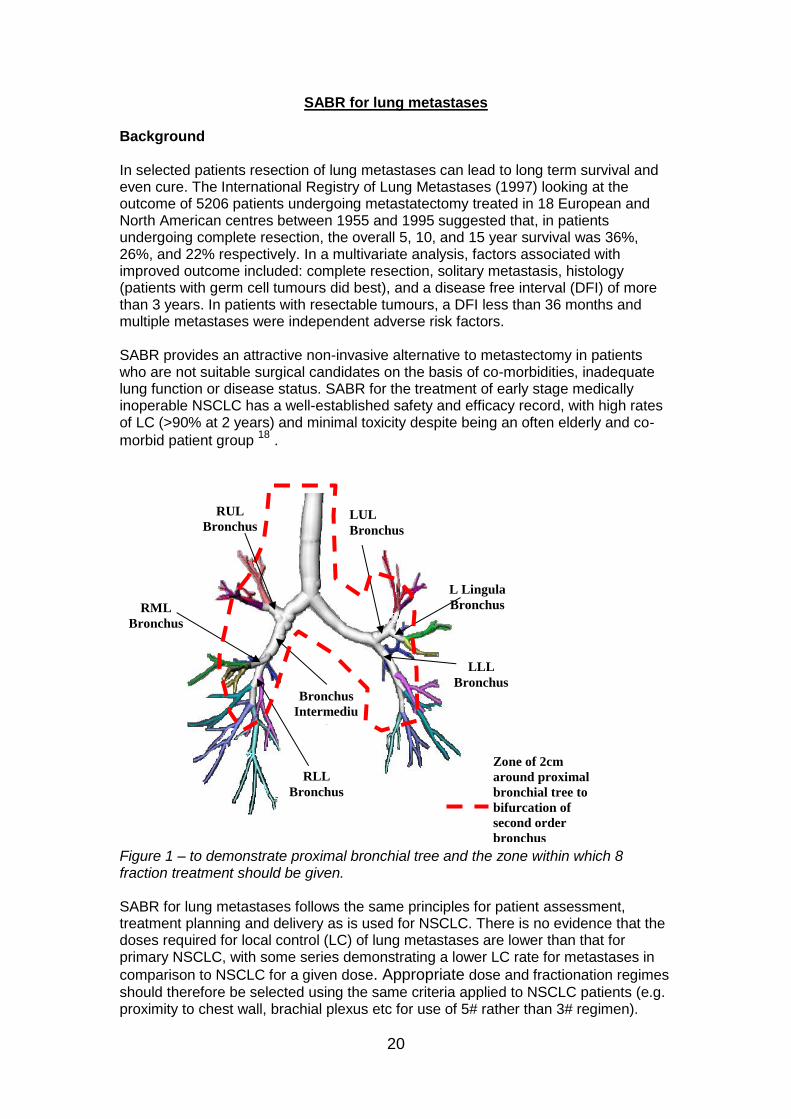
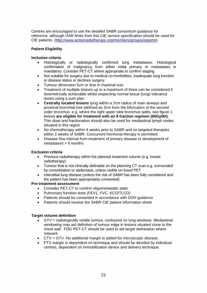
Figure 1 – to demonstrate proximal bronchial tree and the zone within which 8 fraction treatment should be given. SABR for lung metastases follows the same principles for patient assessment, treatment planning and delivery as is used for NSCLC. There is no evidence that the doses required for local control (LC) of lung metastases are lower than that for primary NSCLC, with some series demonstrating a lower LC rate for metastases in
comparison to NSCLC for a given dose. Appropriate dose and fractionation regimes
should therefore be selected using the same criteria applied to NSCLC patients (e.g. proximity to chest wall, brachial plexus etc for use of 5# rather than 3# regimen).
Bronchus
Intermediu
s
RUL
Bronchus
RLL
Bronchus
LUL
Bronchus
LLL
Bronchus
L Lingula
Bronchus RML
Bronchus
Zone of 2cm
around proximal
bronchial tree to
bifurcation of
second order
bronchus
21
Centres are encouraged to use the detailed SABR consortium guidance for reference, although OAR limits from this CtE service specification should be used for CtE patients. (http://www.actionradiotherapy.org/members/groups/experts). Patient Eligibility
Inclusion criteria
Histologically or radiologically confirmed lung metastases. Histological confirmation of malignancy from either initial primary or metastases is mandatory. Consider PET-CT where appropriate to confirm staging.
Not suitable for surgery due to medical co-morbidities, inadequate lung function or disease status or declines surgery.
Tumour dimension 5cm or less in maximal size
Treatment of multiple lesions up to a maximum of three can be considered if dosimetrically achievable whilst respecting normal tissue (lung) tolerance doses using a sum plan
Centrally located lesions lying within a 2cm radius of main airways and proximal bronchial tree (defined as 2cm from the bifurcation of the second order bronchus. e.g. where the right upper lobe bronchus splits, see figure 1 below) are eligible for treatment with an 8 fraction regimen (60Gy/8#). This dose and fractionation should also be used for mediastinal lymph nodes situated in this region
No chemotherapy within 4 weeks prior to SABR and no targeted therapies within 2 weeks of SABR. Concurrent hormonal therapy is permitted.
Disease free interval from treatment of primary disease to development of metastases > 6 months
Exclusion criteria
Previous radiotherapy within the planned treatment volume (e.g. breast radiotherapy)
Tumour that is not clinically definable on the planning CT scan e.g. surrounded by consolidation or atelectasis, unless visible on fused PET
Interstitial lung disease (unless the risk of SABR has been fully considered and the patient has been appropriately consented)
Pre-treatment assessment
Consider PET-CT to confirm oligometastatic state
Pulmonary function tests (FEV1, FVC, KCO/TLCO)
Patients should be consented in accordance with DOH guidance.
Patients should receive the SABR CtE patient information sheet Target volume definition
GTV = radiologically visible tumour, contoured on lung windows. Mediastinal windowing may aid definition of tumour edge in lesions situated close to the chest wall. FDG PET-CT should be used to aid target delineation where relevant.
CTV = GTV. No additional margin is added for microscopic disease.
PTV margin is dependent on technique and should be decided by individual centres, dependent on immobilization device and delivery technique.
22
Organs at risk (OAR) outlining and constraints The following organs should be outlined for lung treatments: normal lung (used to create Lung-GTV), proximal airways, proximal trachea, proximal bronchial tree, proximal bronchial tree + 2cm, spinal cord, oesophagus, heart, brachial plexus. Liver and chest wall should also be delineated where appropriate (see Appendix 2). Dose constraints given in the Appendix 1 must be respected. If these constraints cannot be met, please contact the SABR CtE QA team. Dose and fractionation options for lung metastases SABR
54 Gy in 3 fractions – for lesions away from chest wall and mediastinum.
55-60 Gy in 5 fractions - for lesions where any part of the PTV is in contact with chest wall or where OAR dose constraints can’t be met with the 3 fraction regimen.
60 Gy in 8 fractions for central lung/mediastinal lesions (see criteria below)
Target coverage: aim for the dose received by 95% of the PTV to receive the prescribed dose (and 99% of the PTV to receive >90% of the prescribed dose).
Maximum dose and dose conformity should be as detailed in the UK SABR Consortium guidelines.
Treatment should be delivered on an alternate day basis with a minimum of 40 hours between fractions.
Follow up
Follow up should be as per CtE requirements (Table 2 above and CtE Monitoring Form).
23
SABR for lymph node and bone oligometastases (non-spine) Background There is now a burgeoning evidence base supporting the role of SABR in oligometastases of lymph node and bone. A variety of doses and techniques have been used but similarly good outcomes observed. Patient selection for treatment of oligometastatic lymph nodes and bone (non-spine) metastases
Patients should meet all the general oligometastatic eligibility criteria stated above.
Patients in whom there is overlap with previous radiation in the pelvis or spine should be considered in the re-irradiation section below.
Abdominal lymph node metastases within 5mm of stomach/duodenum/small bowel will be challenging to treat safely to an ablative dose.
Patients who biologically require irradiation of a whole nodal field (e.g. supra-clavicular recurrence from breast cancer) are excluded.
Pre-treatment patient assessment and consent Patients should be fully staged with CT chest/abdo/pelvis and PET scan (where indicated) to rule out more widespread disease. Other imaging (bone scan, whole body DW-MRI) may be useful in certain cases. Patients will be consented in line with DOH guidelines and will be given a SABR patient information sheet. Clinicians taking consent should have experience of SABR or have been trained in the specific risks and benefits of spine SABR.
Target volume outlining
The GTV should be outlined, with a CTV margin applied as clinically required to cover microscopic spread. For example, this may be a 2-3 millimetres for bony metastases, but CTV may identical to GTV for nodal metastases. Organs at risk (OAR) outlining and constraints Multiple beams and/or beam angles will be used to deliver SABR. Therefore every potential organ which beams can traverse should be contoured. This may include lungs, heart, kidneys, liver, bowel, thyroid, larynx, chest wall, and oesophagus (see Appendix 2). Dose constraints given in the Appendix 1 must be respected. If these constraints cannot be met, please contact the SABR CtE QA team Radiotherapy Doses and Dose Prescription There is no consensus as yet for the optimal dose to lymph node and bone (non-spine) metastasis. The UK clinical experience is mostly based on 30 Gy in 3 fractions

24
alternate daily, which appears efficacious and well tolerated 19. This dose delivers a BED of 180 Gy for an alpha/beta of 2 Gy (e.g. prostate) but for an alpha/beta of 10 Gy delivers 60 Gy. Patients for whom the a/b ratio is likely to be >6 Gy may benefit from careful dose escalation to 36-40 in 3 fractions 20 but the evidence base for this is less robust. Dose: 30-40 Gy in 3 fractions alternate daily 95% of the PTV should receive the prescription dose but this should be compromised where needed to remain within OAR tolerance, i.e. PTV D95=prescription dose Follow-up after treatment Follow up should be as per CtE requirements (Table 2 above and CtE Monitoring Form).
25
SABR for Re-irradiation (spine and pelvis/para-aortic) Re-growth of tumour in a previously irradiated area is a difficult problem and the therapeutic options may be few. Uncontrolled tumour growth, however, will certainly cause morbidity, deterioration in quality of life and, ultimately, death. Re-treatment in this scenario is not without risk, but the improved accuracy and precision of SABR makes it an attractive modality which may be able to improve outcomes without a protracted daily regimen of treatment. Within the CtE process, re-irradiation is limited to the spine and pelvic regions. This also includes the para-aortic region in patients who have previously had this area irradiated as part of an extended pelvic field (e.g. certain patients previously irradiated for cervical cancer). Patient Consent It is vital that patients undergoing re-irradiation treatments discuss the potential risks and benefits of treatment with a SABR specialist clinician. In particular, the risks of permanent spinal cord damage and bowel damage should be discussed. The patient should receive the CtE information sheet. Re-irradiation of the spine Re-irradiation of the spine is not uncommon in several solid malignancies where re-growth of tumour at a previously irradiated site can cause pain and spinal cord compression. Some recovery of spinal cord tolerance is noted in animal studies and clinical experience would suggest this is so in humans too. The tolerance of the spinal cord to profoundly hypo-fractionated radiotherapy is incompletely understood but Saghal has suggested a method for calculating remaining cord tolerance using conversion to 2 Gy equivalent dose 21. The dose delivered varies significantly within and between series but one series has shown a significant dose-response for spinal/para-spinal re-irradiation with a local failure rate at one year of 45% for 20 Gy in 5 fractions vs. 26% for 30 Gy in 5 fractions 22. Mantel et al have recently reviewed the available case series and phase II trials of spinal reirradiation with SABR 23. Multiple different dose-fractionations were used, but the majority were treated with around 30 Gy in 3-5 fractions. The reported incidence of clinically significant myelopathy was low (two patients across six studies [containing 330 patients in total]). The risk of vertebral fracture may be higher in the re-irradiation setting and hence spinal stability and the need for prophylactic vertebroplasty should be discussed with a neurosurgeon/orthopaedic surgeon in most cases prior to treatment. It is noted that approximately half of post-SABR vertebral fractures are progression of existing fracture9. Patients need to be consented for the risk of vertebral fracture and incident pain. Patient eligibility for re-irradiation of the spine with SABR
26
Metastatic carcinoma with either a histologically or cytologically proven primary site, Carcinoma of unknown primary (CUP) with histology or cytology proven metastasis or a male patient with a PSA>50 and clinical evidence of prostate cancer.
WHO performance status ≤2.
Ambulatory without severe comorbidity
Life expectancy of more than six months.
A maximum of two sites of spinal metastatic disease requiring treatment for pain relief or tumour control
Assessment by spinal SABR multi-disciplinary team (MDT) that SABR is the most appropriate modality of treatment
No current spinal instability
No cord compression
No chemotherapy within 28 days. Targeted therapies should be stopped a minimum of 14 days prior to SABR.
At least 6 months from initial radiotherapy course
All patients willing to attend follow up and have details collected on prospective database
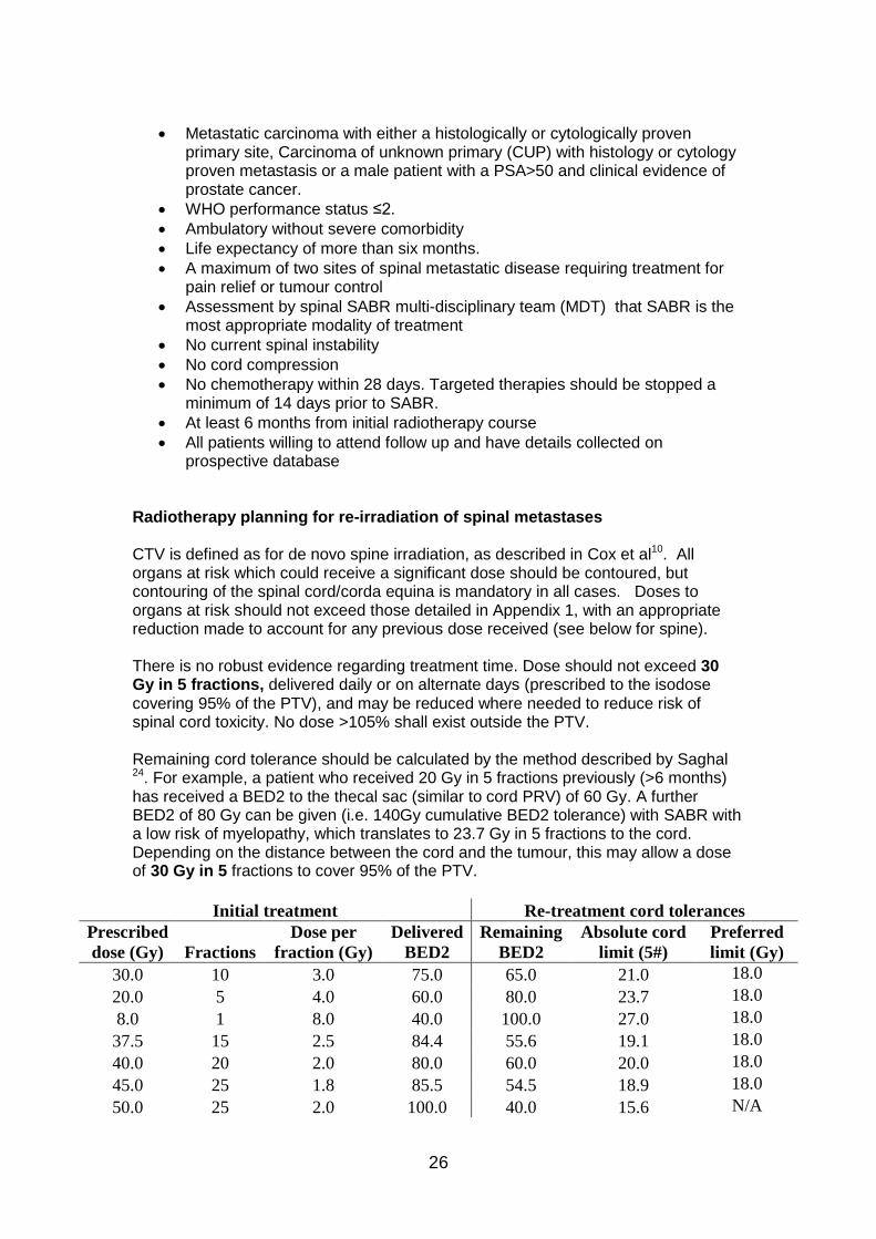
Radiotherapy planning for re-irradiation of spinal metastases CTV is defined as for de novo spine irradiation, as described in Cox et al10. All organs at risk which could receive a significant dose should be contoured, but contouring of the spinal cord/corda equina is mandatory in all cases. Doses to organs at risk should not exceed those detailed in Appendix 1, with an appropriate reduction made to account for any previous dose received (see below for spine). There is no robust evidence regarding treatment time. Dose should not exceed 30 Gy in 5 fractions, delivered daily or on alternate days (prescribed to the isodose covering 95% of the PTV), and may be reduced where needed to reduce risk of spinal cord toxicity. No dose >105% shall exist outside the PTV. Remaining cord tolerance should be calculated by the method described by Saghal 24. For example, a patient who received 20 Gy in 5 fractions previously (>6 months) has received a BED2 to the thecal sac (similar to cord PRV) of 60 Gy. A further BED2 of 80 Gy can be given (i.e. 140Gy cumulative BED2 tolerance) with SABR with a low risk of myelopathy, which translates to 23.7 Gy in 5 fractions to the cord. Depending on the distance between the cord and the tumour, this may allow a dose of 30 Gy in 5 fractions to cover 95% of the PTV.
Initial treatment Re-treatment cord tolerances
Prescribed
dose (Gy) Fractions
Dose per
fraction (Gy)
Delivered
BED2
Remaining
BED2
Absolute cord
limit (5#)
Preferred
limit (Gy)
30.0 10 3.0 75.0 65.0 21.0 18.0
20.0 5 4.0 60.0 80.0 23.7 18.0
8.0 1 8.0 40.0 100.0 27.0 18.0
37.5 15 2.5 84.4 55.6 19.1 18.0
40.0 20 2.0 80.0 60.0 20.0 18.0
45.0 25 1.8 85.5 54.5 18.9 18.0
50.0 25 2.0 100.0 40.0 15.6 N/A

27
Follow up Follow up should be as per CtE requirements (Table 2 above and CtE Monitoring Form). Re-Irradiation of the Pelvis/para-aortic region Background Abusaris et al have published a cohort of 27 patients who underwent re-irradiation to the pelvis to a median dose of 34 Gy in 1-10 fractions. There was no toxicity (acute or late) higher than grade 2 and 96% recorded symptom improvement (mostly pain or bleeding). Several other studies 25-28 have used doses of 36 Gy in 6 fractions, 30 in 5 or 36 Gy in 3 fractions for re-irradiation of pelvic recurrence, largely in gynaecological, rectal or prostate cancer. Toxicity is low in these series and treatment efficacious with high rates of local control and 40-50% of patients progression-free at 2-3 years. Patient selection
Patients with pelvic or para-aortic nodal, bony, soft tissue recurrence or positive margin after maximal surgery in the pelvis
Life expectancy >6 months
No significant toxicity from previous radiation
>6 months since initial radiation treatment
Histologically confirmed malignancy
WHO performance status ≤2
Ambulatory without severe comorbidity, particularly no significant bowel disease.
No chemotherapy within 28 days. Targeted therapies should be stopped a minimum of 14 days prior to SABR (concurrent hormone therapy is permitted.
Patient availability for follow up to assess radiotherapy related morbidity, pain and functional ability for two years.
Assessment in a specialist SABR and site-specific MDTs Radiotherapy planning for pelvic/para-aortic re-irradiation CTV will be defined on an individual basis, taking into account clinical knowledge of disease spread. OAR definition – bowel should be delineated in all cases, either as ‘bowel bag’ (peritoneal cavity) or individual loops. Other organs such as bladder, urethra, sacral nerve roots, should be considered. Most series in the literature do not report either the optimization constraints used or the DVH parameters achieved. The dose constraints given in Appendix 1 should be considered absolute maximums. Where an organ or tissue received a significant dose from the initial radiation course, a BED tolerance remaining calculation should be made and equivalent tolerance in 5 fractions determined to aid planning.

28
Dose 30 Gy in 5 fractions should be prescribed to 95% of the PTV volume, delivered on alternate days. Follow up Follow up should be as per CtE requirements (Table 2 above and CtE Monitoring Form).

29
SABR for primary liver tumours Hepatocellular carcinoma is the commonest primary liver tumour, accounting for ~ 75% of cases worldwide. For primary liver tumours, transplant or surgery are the primary curative modalities of treatment. However for the majority, surgery is not feasible due to disease burden or location, co-morbidities or inadequate functional liver reserve. For patients with early stage but inoperable disease, SABR has been used for both indications with promising long term control rates and a favourable toxicity profile. In 2011, there were 4,348 new cases of liver cancer in the UK and in 2012 there were 4,514 deaths from liver cancer in the UK. The liver cancer incidence and mortality rates have increased overall (tripled) since the early 1970s. 43% of the cases are diagnosed in subjects aged>75years old. Hepatocellular carcinoma HCC usually arises within a cirrhotic liver. Underlying liver impairment is common and an important factor in determining management. SABR may be used as potentially curative therapy in patients with early stage disease who are unsuitable for surgical resection, liver transplant or radiofrequency ablation (RFA). Higher levels of toxicity have been reported in patients with more severe degrees of functional liver impairment. There are several staging classifications for HCC, in addition to TNM, which aim to
enable the better prediction of outcome by incorporating functional parameters. The
Barcelona Clinic Liver Cancer system is the most widely used, incorporating the
Childs-Pugh score for the assessment of liver dysfunction. For the purposes of this
commissioning through evaluation process, only patients with Childs Pugh Class A
should be treated. SABR in this group has demonstrated excellent local control rates
with minimal toxicity.
Summary of described outcomes from recent published data from Aitken KL, Hawkins MA. The role of radiotherapy and chemoradiation in the management of primary liver tumours Clin Oncol (R Coll Radiol). 2014 Sep;26(9):569-80. doi: 10.1016/j.clon.2014.05.016. Epub 2014 Jun 18.
30
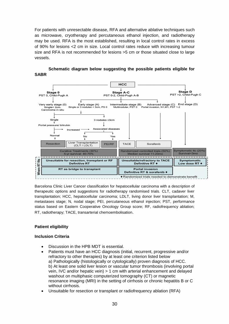
For patients with unresectable disease, RFA and alternative ablative techniques such
as microwave, cryotherapy and percutaneous ethanol injection, and radiotherapy
may be used. RFA is the most established, resulting in local control rates in excess
of 90% for lesions <2 cm in size. Local control rates reduce with increasing tumour
size and RFA is not recommended for lesions >5 cm or those situated close to large
vessels.
Schematic diagram below suggesting the possible patients eligible for
SABR
Barcelona Clinic Liver Cancer classification for hepatocellular carcinoma with a description of
therapeutic options and suggestions for radiotherapy randomised trials. CLT, cadaver liver
transplantation; HCC, hepatocellular carcinoma; LDLT, living donor liver transplantation; M,
metastases stage; N, nodal stage; PEI, percutaneous ethanol injection; PST, performance
status based on Eastern Cooperative Oncology Group score; RF, radiofrequency ablation;
RT, radiotherapy; TACE, transarterial chemoembolisation.
Patient eligibility Inclusion Criteria
Discussion in the HPB MDT is essential.
Patients must have an HCC diagnosis (initial, recurrent, progressive and/or refractory to other therapies) by at least one criterion listed below a) Pathologically (histologically or cytologically) proven diagnosis of HCC. b) At least one solid liver lesion or vascular tumor thrombosis (involving portal vein, IVC and/or hepatic vein) > 1 cm with arterial enhancement and delayed washout on multiphasic computerized tomography (CT) or magnetic resonance imaging (MRI) in the setting of cirrhosis or chronic hepatitis B or C without cirrhosis.
Unsuitable for resection or transplant or radiofrequency ablation (RFA)

31
Unsuitable for or refractory to transarterial hepatic chemo-embolization (TACE) or drug eluting beads (DEB); no response to TACE or DEB
History/physical examination including examination for encephalopathy, ascites, and ECOG Performance Status 0-1
Adequate haematological and organ function as defined below: Absolute neutrophil count (ANC) ≥ 1,500 cells/mm3, Platelets ≥ 70,000 cells/mm3 Hemoglobin ≥ 8.0 g/dl (Note: The use of transfusion or other intervention to achieve Hgb ≥ 8.0 g/dl is acceptable.)Total bilirubin < 2 mg/dL Internationalized Normal Ratio (INR) < 1.7; Albumin ≥ 28 g/L AST and ALT < 6 times ULN
BCLC stage: Intermediate (B) or advanced (C)
Liver volume minus intrahepatic GTV > 700 cc. and Intrahepatic tumor GTV/liver volume ratio <80%.
Maximum dimension 5 cm
Childs-Pugh Class A only (Childs Pugh scoring system classification)
Life expectancy > 6 months
Patients must have recovered from the effects of previous surgery, radiotherapy or chemotherapy with a minimum of 4 weeks break prior to SABR
Suitability for treatment established in Hepatobiliary MDT and Stereotactic MDT
All patients willing to attend follow up and have details collected on prospective database for a minimum of two years.
Exclusion criteria
Active hepatitis or clinically significant liver failure (encephalopathy, oesophageal varices, portal hypertension)
Prior abdominal radiotherapy precluding SABR, that is any previous radiation therapy in which a mean dose to the liver of 15Gy in conventional fractionation was delivered or previous doses to critical normal structures that would make re-irradiation unsafe. Prior pelvic radiation is permitted, as long as there is no overlap between pelvic and liver radiation fields
Clinically apparent ascites
Any one hepatocellular carcinoma > 6 cm
More than 5 discrete intrahepatic parenchymal foci of HCC
Direct tumour extension into the stomach, duodenum, small bowel or large bowel
Extrahepatic metastases or malignant nodes (that enhance with typical features of HCC) > 3.0 cm in sum of maximal diameters (e.g. 2 lung lesions>2 cm )
Active hepatitis, Prior liver transplant.
Pre-treatment patient assessment This should include: baseline blood tests including FBC, Coagulation Screen, U and E's, LFTs including albumin, and calcium. Tumour markers should be performed if relevant. A diagnostic liver MRI should be performed to aid tumour definition (unless contraindications to MRI), in addition to a contrast-enhanced CT. A DMSA scan may be required if the renal dose is likely to be significant.

32
Patients will be consented in line with DOH guidelines and will be given a SABR patient information sheet. Clinicians taking consent should have experience of SABR or have been trained in the specific risks and benefits of spine SABR.
Target volume outlining
Tumour volume and normal tissue definition should be discussed with radiology and joint contouring is recommended. The GTV is defined as all parenchymal enhancing disease. Clinical target volume (CTV) = GTV The appropriate PTV margin will be dependent on local treatment uncertainties. Organs at risk (OAR) outlining and constraints The following OAR will be outlined in their entirety on the planning CT scan: normal liver, kidneys, stomach, duodenum and small bowel, spinal cord, oesophagus, heart, lungs, and chest wall (see Appendix 2). Dose constraints given in Appendix 1 should be respected. If these constraints cannot be met, please contact the SABR CtE QA team. The exception to this is the chest wall constraint. Exceeding chest wall tolerance predicts for a higher incidence of chest wall pain and rib fracture for which the patient should be consented. PTV dose should not be limited due to exceeding chest wall tolerance.
Radiotherapy Doses and Dose Prescription
Treatment planning and tolerances as per liver metastases section. Prescription
doses of 40-50 Gy in 5 fractions should be delivered. Aim for 95% of the PTV to
receive 95% of the prescription dose (D95>95). Allow down to 90% if necessary to
spare OARs (D95>90). Maximum dose should lie within the PTV, and must be within
130% of the prescription dose (D0.1cc <130%).
The final PTV dose is based on the volume of normal liver irradiated, as well as
proximity to duodenum, stomach and small bowel. If normal tissue constrains cannot
be met, lowering the PTV dose in the overlap area with GI tract should be considered
initially, followed by dose reduction in the total prescribed dose (see flowchart).
If tumour is located away from luminal GI structures, the PTV dose prescription
should be as high as possible based on mean liver dose.
33
Care on treatment
Patients who have a large volume of stomach or duodenum treated should be
prescribed anti-emetics. 5HT3 antagonists are preferred.
Patients whose PTV is close to the stomach/duodenum should be prescribed
prophylactic proton pump inhibitors.and continue taking them for at least 3 months
following treatment.
Follow up Follow up should be as per CtE requirements (Table 2 above and CtE Monitoring
Form). LFTs, coagulation screen and tumour markers (if appropriate) should be
measured at each visit.
34
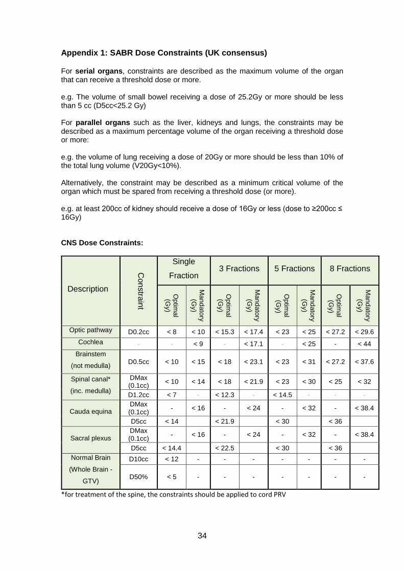
Appendix 1: SABR Dose Constraints (UK consensus) For serial organs, constraints are described as the maximum volume of the organ that can receive a threshold dose or more. e.g. The volume of small bowel receiving a dose of 25.2Gy or more should be less than 5 cc (D5cc<25.2 Gy) For parallel organs such as the liver, kidneys and lungs, the constraints may be described as a maximum percentage volume of the organ receiving a threshold dose or more: e.g. the volume of lung receiving a dose of 20Gy or more should be less than 10% of the total lung volume (V20Gy<10%). Alternatively, the constraint may be described as a minimum critical volume of the organ which must be spared from receiving a threshold dose (or more). e.g. at least 200cc of kidney should receive a dose of 16Gy or less (dose to ≥200cc ≤ 16Gy) CNS Dose Constraints:
Description
Con
stra
int
Single
Fraction 3 Fractions 5 Fractions 8 Fractions
Optim
al
(Gy)
Mand
ato
ry
(Gy)
Optim
al
(Gy)
Mand
ato
ry
(Gy)
Optim
al
(Gy)
Mand
ato
ry
(Gy)
Optim
al
(Gy)
Mand
ato
ry
(Gy)
Optic pathway D0.2cc < 8 < 10 < 15.3 < 17.4 < 23 < 25 < 27.2 < 29.6
Cochlea - - < 9 - < 17.1 - < 25 - < 44
Brainstem
(not medulla) D0.5cc < 10 < 15 < 18 < 23.1 < 23 < 31 < 27.2 < 37.6
Spinal canal*
(inc. medulla)
DMax (0.1cc)
< 10 < 14 < 18 < 21.9 < 23 < 30 < 25 < 32
D1.2cc < 7 - < 12.3 - < 14.5 - - -
Cauda equina DMax (0.1cc)
- < 16 - < 24 - < 32 - < 38.4
D5cc < 14 < 21.9 < 30 < 36
Sacral plexus DMax (0.1cc)
- < 16 - < 24 - < 32 - < 38.4
D5cc < 14.4 < 22.5 < 30 < 36
Normal Brain
(Whole Brain -
GTV)
D10cc < 12 - - - - - - -
D50% < 5 - - - - - - -
*for treatment of the spine, the constraints should be applied to cord PRV
35
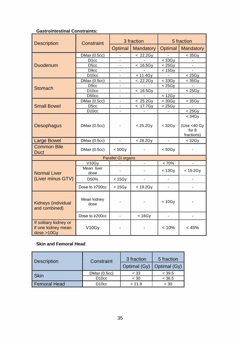
Gastrointestinal Constraints:
Description Constraint 3 fraction 5 fraction
Optimal Mandatory Optimal Mandatory
Duodenum
DMax (0.5cc) - < 22.2Gy - < 35Gy
D1cc - - < 33Gy -
D5cc - < 16.5Gy < 25Gy -
D9cc - - < 15Gy -
D10cc - < 11.4Gy - < 25Gy
Stomach
DMax (0.5cc) - < 22.2Gy < 33Gy < 35Gy
D5cc - - < 25Gy -
D10cc - < 16.5Gy - < 25Gy
D50cc - - < 12Gy -
Small Bowel DMax (0.5cc) - < 25.2Gy < 30Gy < 35Gy
D5cc - < 17.7Gy < 25Gy -
D10cc - - - < 25Gy
Oesophagus DMax (0.5cc) - < 25.2Gy < 32Gy
< 34Gy
(Use <40 Gy for 8
fractions)
Large Bowel DMax (0.5cc) - < 28.2Gy - < 32Gy
Common Bile Duct
DMax (0.5cc) < 50Gy - < 50Gy -
Parallel GI organs
Normal Liver (Liver minus GTV)
V10Gy - - < 70% -
Mean liver dose
- - < 13Gy < 15.2Gy
D50% < 15Gy - - -
Dose to ≥700cc < 15Gy < 19.2Gy - -
Kidneys (individual and combined)
Mean kidney dose
- - < 10Gy -
Dose to ≥200cc - < 16Gy - -
If solitary kidney or if one kidney mean dose >10Gy
V10Gy - - < 10% < 45%
Skin and Femoral Head
Description Constraint 3 fraction 5 fraction
Optimal (Gy) Optimal (Gy)
Skin DMax (0.5cc) < 33 < 39.5
D10cc < 30 < 36.5
Femoral Head D10cc < 21.9 < 30
36
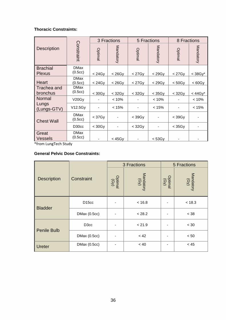
Thoracic Constraints:
Description
Con
stra
int
3 Fractions 5 Fractions 8 Fractions
Optim
al
Mand
ato
ry
Optim
al
Mand
ato
ry
Optim
al
Mand
ato
ry
Brachial Plexus
DMax (0.5cc) < 24Gy < 26Gy < 27Gy < 29Gy < 27Gy < 38Gy*
Heart DMax (0.5cc) < 24Gy < 26Gy < 27Gy < 29Gy < 50Gy < 60Gy
Trachea and bronchus
DMax (0.5cc) < 30Gy < 32Gy < 32Gy < 35Gy < 32Gy < 44Gy*
Normal Lungs (Lungs-GTV)
V20Gy - < 10% - < 10% - < 10%
V12.5Gy - < 15% - < 15% - < 15%
Chest Wall
DMax (0.5cc)
< 37Gy - < 39Gy - < 39Gy -
D30cc < 30Gy - < 32Gy - < 35Gy -
Great Vessels
DMax (0.5cc) - < 45Gy - < 53Gy - -
*from LungTech Study General Pelvic Dose Constraints:
Description Constraint
3 Fractions 5 Fractions
Optim
al
(Gy)
Mand
ato
ry
(Gy)
Optim
al
(Gy)
Mand
ato
ry
(Gy)
Bladder
D15cc - < 16.8 - < 18.3
DMax (0.5cc) - < 28.2 - < 38
Penile Bulb
D3cc - < 21.9 - < 30
DMax (0.5cc) - < 42 - < 50
Ureter DMax (0.5cc) - < 40 - < 45
37
Appendix 2: Organ at risk delineation
Further guidance on delineation, including organs not listed below, can be found in the online Radiation Therapy Oncology Group (RTOG) normal tissue atlases, available at: https://www.rtog.org/CoreLab/ContouringAtlases.aspx
Liver metastasis and adrenal treatments:
Normal liver
The normal liver is defined as the portion of liver not radiographically involved by gross tumour. (Liver volume minus GTV). It is required that there is at least 700cc of normal liver.
Kidneys
Both kidneys will be contoured separately. A combined renal volume structure should also be formed.
Stomach
The stomach should be contoured from gastro-oesophageal junction to duodenum using mediastinal windowing.
Duodenum and small bowel
Duodenum and small bowel (jejunum/ileum) will be contoured as separate structures to include the mucosal bowel wall and all luminal contents. Individual loops of small bowel should be outlined on all slices from 2 cm above to 2 cm below the PTV for co-planar beam arrangements, not including colon and duodenum. If non-coplanar beams are used, bowel should be contoured 10-15cm cranially and caudally from the PTV.
Spinal canal
The spinal canal will be contoured at least 10 cm superiorly and inferiorly to the extent of the PTV and taken to represent the cord. A centre dependent margin will be added to create the PRV dependent on immobilization technique. Note – this is different to cord contouring for spine SABR – see above and technical specification for details.
Oesophagus
The oesophagus should be contoured from the cricoid cartilage to the gastro-oesophageal junction using mediastinal windowing.
Heart
The heart will be contoured along with the pericardial sac using mediastinal windowing. The superior aspect for the purpose of contouring is defined as the superior aspect of the pulmonary artery (as seen on coronal reconstruction of the CT) and the caudal border should be defined by the lowest part of the left ventricle's inferior wall that is distinguishable from the liver.
Lungs
Normal lung will consist of both lungs contoured as one structure Chest wall
The chest wall will be defined as the 3 cm rind of the ipsilateral hemi-thorax outside the lungs and contoured at least 5 cm above and below the PTV.
38
Hepatocellular carcinoma treatment:
Spinal Canal
Outline the spinal canal from 2cm above to 2cm below the PTV. If non-coplanar beams are used, a greater length should be outlined.
Liver
The whole liver should be outlined.
Normal liver
Liver contour minus the GTV contour (use treatment planning system (TPS) operators to perform the function).
Kidneys
Both kidneys should be outlined separately.
Stomach
The whole stomach should be outlined.
Duodenum
The whole of the duodenum from below the pylorus to the fourth part of duodenum (up to the ligament of Treitz) should be outlined.
Small Bowel
Individual loops of small bowel should be outlined on all slices from 2 cm above to 2 cm below the PTV not including colon and duodenum. If non-coplanar beams are used, consider outlining further loops.
Colon
Large Bowel- The transverse colon should be outlined.
Common bile duct (CBD) and bifurcations
These ducts should be identified as tubular structures (lumen density equivalent to water). The expected location of the bile ducts is, the CHD (common hepatic duct) is anterior to the portal vein, lateral to the hepatic artery, and surrounded by fat in the porta hepatis, and the CBD was within or adjacent to the parenchyma of the pancreatic head. If there is uncertainty on the location, the portal vein can be contoured from the splenic confluence to the first bifurcation of the left and right portal veins, then expanded by 15 mm to define the central bile ducts structure.
Skin
defined as 0.5 cm inner rind of body contour contoured if adjacent to PTV and in regions receiving more than 10Gy.
Chest wall
defined as 3 cm inner rind of the body contoured if adjacent to PTV and in regions receiving more than 10Gy.

39
Oesophagus
contour the normal circumference of oesophagus from the gastro-oesophageal junction to carina.
Lung treatments: Lungs
Normal lung will consist of both lungs contoured as one structure (including all inflated and collapsed regions of lung), contoured on lung windows, considered together as one organ, minus the GTV or ITV.
Proximal Trachea
Contouring of the proximal trachea should begin at least 10 cm superior to the extent of the PTV or 5 cm superior to the carina (whichever is more superior) and continue inferiorly to the superior aspect of the proximal bronchial tree.
Proximal Bronchial Tree
The proximal bronchial tree will include the most inferior 2 cm of distal trachea and the proximal airways on both sides. The following airways will be included according to standard anatomical relationships: the distal 2 cm of trachea, the carina, the right and left main stem bronchi, the right and left upper lobe bronchi, the intermedius bronchus, the right middle lobe bronchus, the lingular bronchus, and the right and left lower lobe bronchi. Contouring of the lobar bronchi will end immediately at the site of a segmental bifurcation
Proximal bronchial tree plus 2 cm
An artificial structure 2 cm larger in all directions from the proximal bronchial tree should be created. If the GTV falls within this artificial structure it is not considered to be peripheral and the patient should NOT be treated with lung SABR using the 3 to 5 fraction schedules. These patients need to be treated with 8 fractions.
Great vessels - see RTOG atlas. Spinal canal
The spinal canal will be contoured at least 10 cm superiorly and inferiorly to the extent of the PTV and taken to represent the cord.
Oesophagus
The oesophagus should be contoured from the cricoid cartilage to the gastro-oesophageal junction using mediastinal windowing.
Heart
The heart will be contoured along with the pericardial sac using mediastinal windowing. The superior aspect for the purpose of contouring is defined as the superior aspect of the pulmonary artery (as seen on coronal reconstruction of the CT) and the caudal border should be defined by the lowest part of the left ventricle's inferior wall that is distinguishable from the liver.
Brachial plexus

40
For treatment of lesions other than those in the lower lobes, the major trunks of the ispsilateral brachial plexus will be contoured by using the subclavian and axillary vessels as a surrogate for identifying the location of the plexus on head and neck windowing. These vessels will be contoured from the bifurcation of the brachiocephalic trunk into the jugular/subclavian veins (or carotid/subclavian arteries), following along the route of the subclavian vein to the axillary vein and ending after the neurovascular structures cross the 2nd rib.
Liver
For lower lobe tumours, at least 1000cc of liver must be outlined. Chest Wall
The chest wall will be defined as the 2 cm rind of the ipsilateral hemi-thorax outside the lungs and contoured at least 5 cm above and below the PTV.

41
References:
1. Potters L, Kavanagh B, Galvin JM, et al. American Society for
Therapeutic Radiology and Oncology (ASTRO) and American College of
Radiology (ACR) practice guideline for the performance of stereotactic
body radiation therapy. Int J Radiat Oncol Biol Phys. Feb 1
2010;76(2):326-332.
2. Tree AC, Khoo VS, Eeles RA, et al. Stereotactic body radiotherapy for
oligometastases. Lancet Oncol. Jan 2013;14(1):e28-37.
3. Decaestecker K, De Meerleer G, Lambert B, et al. Repeated stereotactic
body radiotherapy for oligometastatic prostate cancer recurrence. Radiat
Oncol. 2014;9:135.
4. Long-term results of lung metastasectomy: prognostic analyses based on
5206 cases. The International Registry of Lung Metastases. J Thorac
Cardiovasc Surg. Jan 1997;113(1):37-49.
5. Fernandez FG, Drebin JA, Linehan DC, Dehdashti F, Siegel BA,
Strasberg SM. Five-year survival after resection of hepatic metastases
from colorectal cancer in patients screened by positron emission
tomography with F-18 fluorodeoxyglucose (FDG-PET). Ann Surg. Sep
2004;240(3):438-447; discussion 447-450.
6. Corbin KS, Hellman S, Weichselbaum RR. Extracranial oligometastases:
a subset of metastases curable with stereotactic radiotherapy. J Clin
Oncol. Apr 10 2013;31(11):1384-1390.
7. Tree AC, Khoo VS, Eeles RA, et al. Stereotactic body radiotherapy for
oligometastases. Lancet Oncol. Jan 2013;14(1):e28-37.
8. Hall WA, Stapleford LJ, Hadjipanayis CG, Curran WJ, Crocker I, Shu
HK. Stereotactic body radiosurgery for spinal metastatic disease: an
evidence-based review. Int J Surg Oncol. 2011;2011:979214.
9. Sahgal A, Atenafu EG, Chao S, et al. Vertebral compression fracture
after spine stereotactic body radiotherapy: a multi-institutional analysis
with a focus on radiation dose and the spinal instability neoplastic score. J
Clin Oncol. Sep 20 2013;31(27):3426-3431.
10. Cox BW, Spratt DE, Lovelock M, et al. International Spine Radiosurgery
Consortium consensus guidelines for target volume definition in spinal
stereotactic radiosurgery. Int J Radiat Oncol Biol Phys. Aug 1
2012;83(5):e597-605.
11. Timmerman RD. An overview of hypofractionation and introduction to
this issue of seminars in radiation oncology. Semin Radiat Oncol. Oct
2008;18(4):215-222.
12. Benedict SH, Yenice KM, Followill D, et al. Stereotactic body radiation
therapy: the report of AAPM Task Group 101. Med Phys. Aug
2010;37(8):4078-4101.
13. Weng M, Zhang Y, Zhou D, et al. Radiofrequency ablation versus
resection for colorectal cancer liver metastases: a meta-analysis. PLoS
One. 2012;7(9):e45493.
14. Hoyer M, Swaminath A, Bydder S, et al. Radiotherapy for liver
metastases: a review of evidence. Int J Radiat Oncol Biol Phys. Mar 1
2012;82(3):1047-1057.
42
15. Chawla S, Chen Y, Katz AW, et al. Stereotactic body radiotherapy for
treatment of adrenal metastases. Int J Radiat Oncol Biol Phys. Sep 1
2009;75(1):71-75.
16. Tanvetyanon T, Robinson LA, Schell MJ, et al. Outcomes of
adrenalectomy for isolated synchronous versus metachronous adrenal
metastases in non-small-cell lung cancer: a systematic review and pooled
analysis. J Clin Oncol. Mar 1 2008;26(7):1142-1147.
17. Casamassima F, Livi L, Masciullo S, et al. Stereotactic radiotherapy for
adrenal gland metastases: university of Florence experience. Int J Radiat
Oncol Biol Phys. Feb 1 2012;82(2):919-923.
18. Solda F, Lodge M, Ashley S, Whitington A, Goldstraw P, Brada M.
Stereotactic radiotherapy (SABR) for the treatment of primary non-small
cell lung cancer; systematic review and comparison with a surgical
cohort. Radiother Oncol. Oct 2013;109(1):1-7.
19. Aitken K TA, Taylor H, Hawkins M, van As N. Stereotactic body
radiation therapy for extra-cranial metastases – can we change the
therapeutic paradigm? Paper presented at: NCRI2013; Liverpool.
20. Jereczek-Fossa BA, Beltramo G, Fariselli L, et al. Robotic image-guided
stereotactic radiotherapy, for isolated recurrent primary, lymph node or
metastatic prostate cancer. Int J Radiat Oncol Biol Phys. Feb 1
2012;82(2):889-897.
21. Sahgal A, Ma L, Weinberg V, et al. Reirradiation human spinal cord
tolerance for stereotactic body radiotherapy. Int J Radiat Oncol Biol Phys.
Jan 1 2012;82(1):107-116.
22. Damast S, Wright J, Bilsky M, et al. Impact of dose on local failure rates
after image-guided reirradiation of recurrent paraspinal metastases. Int J
Radiat Oncol Biol Phys. Nov 1 2011;81(3):819-826.
23. Mantel F, Flentje M, Guckenberger M. Stereotactic body radiation
therapy in the re-irradiation situation--a review. Radiat Oncol. 2013;8:7.
24. Sahgal A, Ma L, Weinberg V, et al. reirradiation HUMAN Spinal Cord
Tolerance for Stereotactic Body Radiotherapy. Int J Radiat Oncol Biol
Phys. Oct 15 2010.
25. Jereczek-Fossa BA, Beltramo G, Fariselli L, et al. Robotic Image-Guided Stereotactic Radiotherapy, for Isolated Recurrent Primary, Lymph Node or Metastatic Prostate Cancer. Int J Radiat Oncol Biol Phys. Jan 27 2011.
26. Dewas S, Bibault JE, Mirabel X, et al. Robotic image-guided reirradiation
of lateral pelvic recurrences: preliminary results. Radiat Oncol. 2011;6:77.
27. Deodato F, Macchia G, Grimaldi L, et al. Stereotactic radiotherapy in
recurrent gynecological cancer: a case series. Oncol Rep. Aug
2009;22(2):415-419.
28. Defoe SG, Bernard ME, Rwigema JC, Heron DE, Ozhasoglu C, Burton S.
Stereotactic body radiotherapy for the treatment of presacral recurrences
from rectal cancers. J Cancer Res Ther. Oct-Dec 2011;7(4):408-411.


