Commentary How the python heart separates pulmonary and...
7
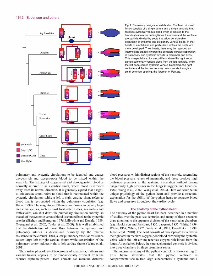
1611 Introduction The vertebrate cardiovascular system has undergone a striking evolutionary change that attended the evolution of air breathing and the subsequent rise in metabolism which occurred during the evolution of endothermy (Fig. 1). The ancestral heart of the early chordates, currently represented by cephalochordates, is merely a contractile dorsal aorta that propagates blood (Rähr, 1979), while proper cardiac function evolved among hagfish and lampreys (Burggren et al., 1997). Therefore, within vertebrates, the evolution of the cardiovascular system may be viewed as a progression from a serial arrangement of the cardiac chambers in cartilaginous and bony fishes to a complete division of the systemic and pulmonary circuits by means of the four-chambered heart of mammals and birds (e.g. Holmes, 1975). As the high metabolism of endothermic mammals and birds requires high blood flows, the hearts of these animals can achieve high heart rates and blood pressures, which exert large demands on the left cardiac ventricle to deliver forceful and fast contractions (Lillywhite et al., 1999; Seymour and Blaylock, 2000). Conversely, the requirement for effective gas exchange across the lung necessitates a thin epithelium, which weakens the blood–air barrier. To avoid oedema or structural damage to the lung, the thin pulmonary epithelium could only evolve in concert with a reduction in pulmonary blood pressure (e.g. Burggren, 1982). In terms of the overall cardiac anatomy, most ectothermic tetrapods, lungfishes and many air-breathing fish, which all have considerably lower aerobic demands than birds and mammals, represent an intermediate evolutionary stage. In most of these vertebrates, the heart contains two separate atria, one that receives oxygen-rich blood from the lungs and a second that receives oxygen-poor blood from the systemic circulation. The two atria empty their blood into a single ventricle (Fig. 1). Extant reptiles are particularly interesting in this context because they derive all of their gas exchange over the lungs and because their ancestors form evolutionary stem groups for both mammals and birds. This implies that the four-chambered cardiac design and the ability to separate systemic and pulmonary blood pressures evolved independently in mammals and birds (Seymour et al., 2004). Extant reptiles are a paraphyletic group that, apart from the crocodiles, is represented by turtles, the tuatara as well as lizards and snakes (squamates). While the anatomy of the cardiac ventricle differs substantially amongst these groups, they all have cardiac ventricles that are incompletely divided. In most cases, this cardiovascular design causes systolic blood pressures in the The Journal of Experimental Biology 213, 1611-1617 © 2010. Published by The Company of Biologists Ltd doi:10.1242/jeb.030999 Commentary How the python heart separates pulmonary and systemic blood pressures and blood flows Bjarke Jensen 1 , Jan M. Nielsen 2 , Michael Axelsson 3 , Michael Pedersen 4 , Carl Löfman 5 and Tobias Wang 1, * 1 Zoophysiology, Department of Biological Sciences, Aarhus University, Building 1131, Universitetsparken 8000, Aarhus, Denmark, 2 Department of Cardiology B, Aarhus University Hospital, Skejby Sygehus, Denmark, 3 Department of Zoology, University of Gothenburg, Gothenburg, Sweden, 4 MR Research Center, Aarhus University Hospital, Denmark and 5 Department of Obstetrics and Gynaecology, Sahlgrenska Academy, Sahlgrenska University Hospital, Sweden *Author for correspondence ([email protected]) Accepted 2 February 2010 Summary The multiple convergent evolution of high systemic blood pressure among terrestrial vertebrates has always been accompanied by lowered pulmonary pressure. In mammals, birds and crocodilians, this cardiac separation of pressures relies on the complete division of the right and left ventricles by a complete ventricular septum. However, the anatomy of the ventricle of most reptiles does not allow for complete anatomical division, but the hearts of pythons and varanid lizards can produce high systemic blood pressure while keeping the pulmonary blood pressure low. It is also known that these two groups of reptiles are characterised by low magnitudes of cardiac shunts. Little, however, is known about the mechanisms that allow for this pressure separation. Here we provide a description of cardiac structures and intracardiac events that have been revealed by ultrasonic measurements and angioscopy. Echocardiography revealed that the atrioventricular valves descend deep into the ventricle during ventricular filling and thereby greatly reduce the communication between the systemic (cavum arteriosum) and pulmonary (cavum pulmonale) ventricular chambers during diastole. Angioscopy and echocardiography showed how the two incomplete septa, the muscular ridge and the bulbuslamelle – ventricular structures common to all squamates – contract against each other in systole and provide functional division of the anatomically subdivided ventricle. Washout shunts are inevitable in the subdivided snake ventricle, but we show that the site of shunting, the cavum venosum, is very small throughout the cardiac cycle. It is concluded that the python ventricle is incapable of the pronounced and variable shunts of other snakes, because of its architecture and valvular mechanics. Supplementary material available online at http://jeb.biologists.org/cgi/content/full/213/10/1611/DC1 Key words: cardiovascular, heart, reptile. THE JOURNAL OF EXPERIMENTAL BIOLOGY
-
Upload
nguyendien -
Category
Documents
-
view
215 -
download
1
Transcript of Commentary How the python heart separates pulmonary and...
1611
IntroductionThe vertebrate cardiovascular system has undergone a strikingevolutionary change that attended the evolution of air breathing andthe subsequent rise in metabolism which occurred during theevolution of endothermy (Fig.1). The ancestral heart of the earlychordates, currently represented by cephalochordates, is merely acontractile dorsal aorta that propagates blood (Rähr, 1979), whileproper cardiac function evolved among hagfish and lampreys(Burggren et al., 1997). Therefore, within vertebrates, the evolutionof the cardiovascular system may be viewed as a progression froma serial arrangement of the cardiac chambers in cartilaginous andbony fishes to a complete division of the systemic and pulmonarycircuits by means of the four-chambered heart of mammals andbirds (e.g. Holmes, 1975). As the high metabolism of endothermicmammals and birds requires high blood flows, the hearts of theseanimals can achieve high heart rates and blood pressures, whichexert large demands on the left cardiac ventricle to deliver forcefuland fast contractions (Lillywhite et al., 1999; Seymour andBlaylock, 2000). Conversely, the requirement for effective gasexchange across the lung necessitates a thin epithelium, whichweakens the blood–air barrier. To avoid oedema or structuraldamage to the lung, the thin pulmonary epithelium could only
evolve in concert with a reduction in pulmonary blood pressure(e.g. Burggren, 1982).
In terms of the overall cardiac anatomy, most ectothermictetrapods, lungfishes and many air-breathing fish, which all haveconsiderably lower aerobic demands than birds and mammals,represent an intermediate evolutionary stage. In most of thesevertebrates, the heart contains two separate atria, one that receivesoxygen-rich blood from the lungs and a second that receivesoxygen-poor blood from the systemic circulation. The two atriaempty their blood into a single ventricle (Fig.1). Extant reptiles areparticularly interesting in this context because they derive all oftheir gas exchange over the lungs and because their ancestors formevolutionary stem groups for both mammals and birds. This impliesthat the four-chambered cardiac design and the ability to separatesystemic and pulmonary blood pressures evolved independently inmammals and birds (Seymour et al., 2004).
Extant reptiles are a paraphyletic group that, apart from thecrocodiles, is represented by turtles, the tuatara as well as lizardsand snakes (squamates). While the anatomy of the cardiac ventriclediffers substantially amongst these groups, they all have cardiacventricles that are incompletely divided. In most cases, thiscardiovascular design causes systolic blood pressures in the
The Journal of Experimental Biology 213, 1611-1617© 2010. Published by The Company of Biologists Ltddoi:10.1242/jeb.030999
Commentary
How the python heart separates pulmonary and systemic blood pressures and bloodflows
Bjarke Jensen1, Jan M. Nielsen2, Michael Axelsson3, Michael Pedersen4, Carl Löfman5 and Tobias Wang1,*1Zoophysiology, Department of Biological Sciences, Aarhus University, Building 1131, Universitetsparken 8000, Aarhus, Denmark,
2Department of Cardiology B, Aarhus University Hospital, Skejby Sygehus, Denmark, 3Department of Zoology, University ofGothenburg, Gothenburg, Sweden, 4MR Research Center, Aarhus University Hospital, Denmark and 5Department of Obstetrics and
Gynaecology, Sahlgrenska Academy, Sahlgrenska University Hospital, Sweden*Author for correspondence ([email protected])
Accepted 2 February 2010
SummaryThe multiple convergent evolution of high systemic blood pressure among terrestrial vertebrates has always been accompaniedby lowered pulmonary pressure. In mammals, birds and crocodilians, this cardiac separation of pressures relies on the completedivision of the right and left ventricles by a complete ventricular septum. However, the anatomy of the ventricle of most reptilesdoes not allow for complete anatomical division, but the hearts of pythons and varanid lizards can produce high systemic bloodpressure while keeping the pulmonary blood pressure low. It is also known that these two groups of reptiles are characterised bylow magnitudes of cardiac shunts. Little, however, is known about the mechanisms that allow for this pressure separation. Herewe provide a description of cardiac structures and intracardiac events that have been revealed by ultrasonic measurements andangioscopy. Echocardiography revealed that the atrioventricular valves descend deep into the ventricle during ventricular fillingand thereby greatly reduce the communication between the systemic (cavum arteriosum) and pulmonary (cavum pulmonale)ventricular chambers during diastole. Angioscopy and echocardiography showed how the two incomplete septa, the muscularridge and the bulbuslamelle – ventricular structures common to all squamates – contract against each other in systole andprovide functional division of the anatomically subdivided ventricle. Washout shunts are inevitable in the subdivided snakeventricle, but we show that the site of shunting, the cavum venosum, is very small throughout the cardiac cycle. It is concludedthat the python ventricle is incapable of the pronounced and variable shunts of other snakes, because of its architecture andvalvular mechanics.
Supplementary material available online at http://jeb.biologists.org/cgi/content/full/213/10/1611/DC1
Key words: cardiovascular, heart, reptile.
THE JOURNAL OF EXPERIMENTAL BIOLOGY
1612
pulmonary and systemic circulations to be identical and causesoxygen-rich and oxygen-poor blood to be mixed within theventricle. The mixing of oxygenated and deoxygenated blood isnormally referred to as a cardiac shunt, where blood is directedaway from its normal direction. It is generally agreed that a right-to-left cardiac shunt refers to blood that is recirculated within thesystemic circulation, while a left-to-right cardiac shunt refers toblood that is recirculated within the pulmonary circulation (e.g.Hicks, 1998). The magnitude of these shunt flows can be very largeand some species, such as most freshwater turtles, sea snakes andrattlesnakes, can shut down the pulmonary circulation entirely, sothat all of the systemic venous blood is shunted back to the systemicarteries (Shelton and Burggren, 1976; Lillywhite and Donald, 1989;Overgaard et al., 2002; Taylor et al., 2009). It is well establishedthat the distribution of blood flow between the systemic andpulmonary arteries is determined primarily by the relativeresistance in the circuits. Thus, a low pulmonary vascular resistancecauses large left-to-right cardiac shunts while constriction of thepulmonary artery induces right-to-left cardiac shunts (Wang et al.,2001).
The cardiac physiology of two groups of squamates, pythons andvaranid lizards, appears to be fundamentally different from the‘normal reptilian pattern’. Both animals can maintain different
blood pressures within distinct regions of the ventricle, resemblingthe blood pressure values of mammals, and these produce highperfusion pressures in the systemic circulation without havingdangerously high pressures in the lungs (Burggren and Johansen,1982; Wang et al., 2002; Wang et al., 2003). Here we describe theunique physiology of the python heart and provide a structuralexplanation for the ability of the python heart to separate bloodflows and pressures throughout the cardiac cycle.
The anatomy of the python heartThe anatomy of the python heart has been described in a numberof studies over the past two centuries and many of these accountsdraw attention to the apparent division of the ventricular chambers(e.g. Hopkinson and Pancoast, 1837; Jacquart, 1855; White, 1959;White, 1968; White, 1976; Webb et al., 1971; Farrell et al., 1998;Jensen et al., 2010). The heart consists of two separate atria, wherethe right atrium receives oxygen-poor blood carried by the systemicveins, while the left atrium receives oxygen-rich blood from thelungs. As explained below, the single, elongated ventricle is dividedinto three chambers by three prominent septa.
The internal anatomy of the python ventricle is shown in Fig.2.This figure illustrates that the python ventricle iscompartmentalised in two large subchambers; a systemic and a
B. Jensen and others
Fig.1. Circulatory designs in vertebrates. The heart of mostfishes consists of a single atrium and a single ventricle thatreceives systemic venous blood which is ejected to thebranchial circulation. In lungfishes the atrium and the ventricleare partially divided by septa that allow considerableseparation of systemic and pulmonary venous blood. In thehearts of amphibians and particularly reptiles the septa aremore developed. Their hearts, then, may be regarded asintermediate stages towards the complete cardiac separationof pulmonary and systemic circuits in mammals and birds.This is especially so for crocodilians where the right aortacarries pulmonary venous blood from the left ventricle, whilethe left aorta carries systemic venous blood from the rightventricle and the two aortae may communicate through asmall common opening, the foramen of Panizza.
THE JOURNAL OF EXPERIMENTAL BIOLOGY

1613Function of the python heart
pulmonary chamber (cavum arteriosum and cavum pulmonale,respectively), with a third shallow chamber, the cavum venosum,situated between the other two. Three different septa areresponsible for this division and while all three are found in theventricle of all squamates and testudines, they are particularlyconspicuous and well developed in the heart of pythons and varanidlizards (Greil, 1903; Acolat, 1943; Webb et al., 1971; Webb, 1979;Van Mierop and Kutsche, 1985; Farrell et al., 1998; Jensen et al.,2010). The vertical septum runs from the apex approximately three-quarters of the length towards the base of the ventricle, and therebycompletely partitions the caudal part of the ventricle into a rightand left side. The other two septa, the muscular ridge and thebulbuslamelle, divide the base of the ventricle during ventricularcontraction (Fig.2).
The cavum venosum is conceptually important because bloodentering the cavum pulmonale during diastole passes through thecavum venosum, while the blood that is ejected from the cavumarteriosum during cardiac contraction must traverse the cavumvenosum to reach the aortic arches (Fig.2). All squamate ventriclescontain the cavum venosum, but it is uniquely reduced in pythons(Acolat, 1943; Webb et al., 1971; Jensen et al., 2010) and while
blood streams necessarily cross there, the volumes that areexchanged between the two blood streams are at all times smallcompared with the volume of the python ventricle.
Flow and pressure separation in the python heartThe remarkable ability of the python heart to separate blood flowsand pressures was discovered recently in two studies (Wang etal., 2002; Wang et al., 2003). Using an in situ heart preparationwhere inflow of physiological saline into the right and left atriacould be manipulated independently at different outflowpressures, it was shown that perfusate entering the systemicchamber of the ventricle, the cavum arteriosum, was almostexclusively ejected into the systemic arches, while perfusateentering the ventricle from the right atrium was directed towardsthe pulmonary circulation (Fig.3). These findings implied afunctional division of systemic and pulmonary inflowsthroughout the cardiac cycle, which means that the heart isfunctionally divided during both filling and ejection. It alsobecame evident that the left systemic side of the perfused pythonventricle could generate much higher pressures than the rightpulmonary side. When catheters were inserted into both sides of
Fig.2. Anatomy of the python heart (Python regius).(A)Internal view of the dorsal half of the heart. Arrowsindicate the position of the vertical septum (VS). Notethat the ventricle to the left of the VS has a densermyocardium. (B)Caudal view of the base. (C)Cranialview of the caudal three-quarters of the ventricleshowing that the cavum venosum (CV) is a shallowchamber. avv, atrioventricular valves; BL, bulbuslamelle;C, cranial; CA, cavum arteriosum; cds, cul-de-sac of thecavum pulmonale; CP, cavum pulmonale; D, dorsal;IAS, interatrial septum; L, left; LAo, left aorta; LAt, leftatrium; MR, muscular ridge; PA, pulmonary artery; R,right; RAo, right aorta; RAt, right atrium; VP, pulmonaryvein.
Pressure in left atrium (kPa)
Flo
w (
ml m
in–1
kg–1
)
0 0.2 0.4 0.6 0.80
20
40
60
80
Systemic flow
Pulmonary flow
0
20
40
60
80
100
Blo
od p
ress
ure
(cm
H2O
)
120
RAo
CA
CP
LPA
1 sFig.3. Arterial flows of an in situ perfused Python molurus heart, where theentire heart was perfused with and immersed in Ringer solution at 30°C(for details, see Wang et al., 2002). Systemic flow is very responsive toincreased return to the left atrium, indicated by atrial pressure, whilepulmonary flow is essentially unaffected, which indicates an effectivediastolic separation of venous returns. 1kPa10.2cmH2O.
Fig.4. Simultaneous measurements of intraventricular pressures andarterial pressure in the systemic and pulmonary circulation in ananaesthetised P. molorus. CA, cavum arteriosum; CP, cavum pulmonale;LPA, left pulmonary artery; RAo, right aortic arch. 1cmH2O0.098kPa.Taken from Wang et al. (Wang et al., 2003).
THE JOURNAL OF EXPERIMENTAL BIOLOGY

1614
the ventricle as well as into one of the pulmonary arteries and oneof the systemic arches, it was unequivocally demonstrated thatsystemic blood pressures are much higher than pulmonarypressures (Fig.4). These measurements were performed in bothanaesthetised and fully recovered Burmese python (Pythonmolurus) and were subsequently repeated on other species ofpythons (e.g. Zaar et al., 2007).
While the recordings of flows and pressure providedunequivocal evidence for pressure and flow separation in the
pythons, our understanding of the anatomy underlying thephenomenon, based primarily on inspection of preserved hearts,has remained somewhat speculative. To understand the dynamicsof the cardiac structures in vivo, we have used echocardiographyand angioscopy of in situ perfused hearts of pythons to visualisethe ventricular septa and the atrioventricular valves during thecardiac cycle. Together with previous anatomical studies onpreserved hearts, these images reveal how the muscular ridgeseparates the cavum arteriosum from the cavum pulmonale
B. Jensen and others
Fig.5. Images from supplementary material Movie1(angioscopy) showing the cavum pulmonale of a 12kgP. molorus looking cranially from a caudal position. Theentire heart was perfused with and immersed in Ringersolution at 30°C (for details, see Wang et al., 2002).Down is left and right is ventral. The angioscope wasinserted through the apical and right ventricular wall.(A)Diastole. A small opening (arrow) is formed betweenthe bulbuslamelle (BL) and the muscular ridge (MR) inthe cranial part of the cavum pulmonale and the valvesof the pulmonary artery (vap) are closed. (B)Earlysystole. The opening is closed and the lumen of thepulmonary artery (PA) is seen through the open valves.
A B
C D
Fig.6. Images from supplementary material Movie2 (echocardiography, not synchronised with electrocardiogram, ECG) showing transverse scans of theventricle of a 4kg P. molorus. The snake was anaesthetised with 1.5% isoflurane and images were obtained with a vivid 7 echocardiographic system (GEHealthcare) using an 11MHz phased array paediatric transducer operating at a frame rate of approximately 150Hz. (A)Late diastole. The vertical septum(VS) divides the cavum arteriosum (CA) from the cavum pulmonale (CP). The CP has a ventral part, which leads directly to the pulmonary artery, withouttrabeculae and a dorso-lateral part, the cul-de-sac (cds), with a low density of trabeculae. The CA consists of a luminal core enclosed by a high density oftrabeculae. (B)Mid-systole. The bulbuslamelle (BL) presses against the prominent muscular ridge (MR) and the two septa separate the CP from the CV andCA. (C)End-systole. All arterial valves are closed. (D)Schematic drawing of the ventral surface of the python heart showing the planes of scanning in A–C.D, dorsal; L, left; LAo, left aorta; LAt, left atrium; PA, pulmonary artery; RAo, right aorta; RAt, right atrium; V, ventricle; vap, valves of PA; vcp, posterior venacava; vla, valves of LAo; vra, valves of RAo; vp, pulmonary vein.
THE JOURNAL OF EXPERIMENTAL BIOLOGY

1615Function of the python heart
during cardiac contraction and clarify the significance of theatrioventricular valves for flow separation during cardiac filling.
How the python heart separates blood flows duringventricular contraction
Inspection of preserved hearts has revealed that the well-developedvertical septum divides the posterior end of the ventricle and theprominent muscular ridge fits perfectly onto the bulbuslamelle(e.g. Hopkinson and Pancoast, 1837; Brücke, 1852; Greil, 1903;Acolat, 1943; Webb et al., 1971; Webb, 1979; Van Mierop andKutsche, 1985; Farrell et al., 1998; Jensen et al., 2010). Althoughthese anatomical descriptions were performed before theventricular pressure separation had been measured in vivo, it wasspeculated that the muscular ridge and the vertical septumprovided the basis for functional separation of intraventricularblood flows and pressures during cardiac contraction. It wasenvisaged that the muscular ridge abuts the bulbuslamelle duringventricular contraction to partition the ventricle (Brücke, 1852;Greil, 1903; White, 1959; White, 1976). We were able to verifythis mechanism by inserting a high-resolution angioscope into theperfused in situ heart (see supplementary material Movie1; Fig.5).In these experiments, the spontaneously beating heart remained insitu and was perfused with saline at physiologically relevantpressures (for details, see Wang et al., 2002). The angioscopecould be inserted through the ventricular wall causing littledamage and allowing an intraventricular visualisation of thebeating heart.
A view from the apical end of the cavum pulmonale takentowards the pulmonary outflow tract for different parts of thecardiac cycle is presented in Fig.5 (see also supplementarymaterial Movie1). During diastole, when blood enters the cavumpulmonale via the cavum venosum, blood flows through theopening between the crest of the muscular ridge and thebulbuslamelle on the ventricular wall (Fig.5A). As ventricularsystole starts, the bulbuslamelle immediately abuts the muscularridge, separating the cavum pulmonale from the rest of theventricle during the isometric contraction and allowing pressurein the cavum pulmonale to increase (Fig.5B). This is rapidlyfollowed by the opening of the pulmonary arterial valves,allowing oxygen-poor blood to be ejected into the pulmonaryartery from the cavum pulmonale (Fig.5B; supplementarymaterial Movie1).
We have also visualised the functional separation of the cavumpulmonale from the rest of the ventricle, by the muscular ridgejoining to the bulbuslamelle during cardiac contraction, withechocardiography on pythons lightly anaesthetised by inhalation ofisoflurane. Transverse sections of the ventricle taken byechocardiography are presented in Fig.6 and supplementaryMovie2. This figure shows that the prominent muscular ridge isstatic, as would be expected from the longitudinal orientation of themyocardium of the muscular ridge (Kashyap, 1975), and closelyopposed by the bulbuslamelle throughout the cardiac cycle. Thecavum venosum, therefore, remains as a narrow passage at alltimes.
Thus, our echocardiography and angioscope studies demonstratethat the ventricular pressure separation during ventricularcontraction can be attributed to the muscular ridge abutting thebulbuslamelle, as had been envisaged in early anatomicaldescriptions (Brücke, 1852; Greil, 1903; Acolat, 1943). Bycontacting each other, the muscular ridge and bulbuslamelleseparate the cavum pulmonale from the rest of the ventricle,allowing the development of low pressures in the pulmonary
circulation, while the more muscular cavum arteriosum cangenerate a much higher pressure in the systemic circulation (Zaaret al., 2007; Jensen et al., 2010). Consistent with the importance ofthe MR, we recently showed that pythons with cardiacmalformations and exceptionally poorly developed MR did notexhibit ventricular pressure separation (Jensen and Wang, 2009).The images also show that the closure provided by thebulbuslamelle and the muscular ridge occurs prior to ventricularshortening (see supplementary material Movie1). It is possible thatthe bulbuslamelle depolarises earlier than most of the ventriclebecause it is located immediately caudal to the atrioventricularfunnel where the depolarising currents are propagated and wherethe atrioventricular musculature merges (Laurens, 1915; Buchanan,1956; Farrell et al., 1998).
How the python heart separates blood flows duringventricular filling
The mechanisms responsible for the separation of blood flowsduring systole cannot explain how flows are separated duringventricular filling when oxygen-poor blood enters the cavumpulmonale from the right atrium, whilst oxygen-rich blood entersthe cavum arteriosum through the left atrium. Studies on the insitu perfused hearts, however, showed that the inflow from theright atrium is selectively directed towards the pulmonarycirculation, whereas inflow through the left atrium is directedtowards the systemic arteries (Fig.3) (Wang et al., 2002). Earlyanatomical studies of hearts from pythons and other snakes hadalready shown that the atrioventricular valves are very large andthat they appeared to fit the underlying septa. On this basis, it wassuggested that the atrioventricular valves are forced caudallytowards the muscular ridge and vertical septum during ventricularfilling, which would effectively guide the oxygen-poor bloodfrom the right atrium into the cavum pulmonale, while oxygenrich-blood is directed from the left atrium into the cavumarteriosum (Hopkinson and Pancoast, 1837; White, 1968; Webb,1979). Our in vivo visualisation by echocardiography confirmsthis action of the atrioventricular valves. Mid-horizontal andtransverse echocardiographical scans of the ventricle at variousstages of diastole and systole are shown in Figs6 and 7 (see alsosupplementary material Movies2 and 3). As shown in Fig.7, theatrioventricular valves fill the atrioventricular opening duringsystole, i.e. the valves are closed. However, when the ventriclerelaxes the atrioventricular valves rapidly descend onto thevertical septum, stretching the interatrial septum while bothvalves wrap around the vertical septum (Fig.7B,C; supplementarymaterial Movie3). At the start of ventricular contraction, theatrioventricular valves ascend back into the atrioventricularorifice (Fig.7D). The size of the lumen between theatrioventricular valves and the vertical septum, through which thecavum arteriosum and the cavum venosum communicate insystole and often referred to as the interventricular canal (White,1959; Webb et al., 1971; Farrell et al., 1998), is therefore reducedsignificantly by the deep descent of the atrioventricular valves.
The deflection of the atrioventricular valves occurs prior to atrialcontraction as verified by the P-wave of the electrocardiogram(ECG), which implies that ventricular relaxation rather than atrialcontraction causes both atrioventricular valves to invert rapidlyonto the vertical septum (Fig.7). This observation is consistent withthe substantial diastolic suction of the excised turtle ventricle(Katz, 1930; Kraner, 1959), although atrial contractions appear tocontribute substantially to ventricular filling in squamates(Johansen, 1959; Johansen and Burggren, 1984).
THE JOURNAL OF EXPERIMENTAL BIOLOGY

1616
Cardiac shunts in the functionally divided heartIn pythons, the functional size of the cavum venosum is reducedand the interventricular canal is diminished in diastole because ofthe large movement of the atrioventricular valves. This wouldcontribute to a substantial reduction in the wash-out shunt whenblood from the cavum arteriosum is ejected into the aortic archesthrough the cavum venosum (e.g. Heisler et al., 1983). Thus, whenthe bulbuslamelle and the muscular ridge separate the cavumvenosum from the cavum pulmonale early in systole, residualvenous blood in the cavum venosum will be ‘washed out’, alongwith the arterial blood of the cavum arteriosum, into the aortae(Heisler and Glass, 1985; Hicks, 1998). Since the cavum venosumis a minor chamber even in diastole, the residual volume is alwayssmall and the washout shunt, therefore, will only constitute a smallproportion of cardiac output (Figs2, 6 and 7) [see Retzius cited byBrücke (Brücke, 1852)] (Webb et al., 1971; van Mierop andKutsche, 1985; Wang et al., 2003).
The functional morphology of the python, as described here,explains how blood flows can remain separate throughout thecardiac cycle and indicates that the degree of mixing betweenarterial and venous blood (i.e. the cardiac shunts) is rather small.Consistent with this proposal, arterial oxygen levels remain higheven when systemic venous oxygen concentrations are low duringdigestion (e.g. Overgaard and Wang, 2002). Consequently, the
architecture of the python ventricle seems to prevent the largevariations in cardiac shunting that are associated with theventilatory and metabolic states of other snakes (e.g. Lillywhite andDonald, 1989; Wang et al., 1998; Wang et al., 2003). It appears,however, that the vertical septum is raised and close to theatrioventricular orifice in most squamates and since theatrioventricular valves of pythons are similar to those of othersnakes, it is likely that the dynamic action of the atrioventricularvalves in most snakes and lizards may be similar to those of pythons(Acolat, 1943; Webb et al., 1971; Holmes, 1975; Webb, 1979; VanMierop and Kutsche, 1984; Van Mierop and Kutsche, 1985; Farrellet al., 1998). This would help to explain the low degrees of shuntingin other snakes during times of high metabolic demand (Wang etal., 1998).
GlossaryAtrioventricular valves
The two valves that guard the opening between the atria and the ventricle.
BulbuslamelleOpposed to the muscular ridge, this ventricular septum separates thecavum pulmonale and cavum venosum during ventricular systole.
Cavum arteriosumThe left-sided ventricular chamber that receives oxygenated blood fromthe left atrium; i.e. the ‘systemic side’ of the ventricle.
B. Jensen and others
A B
C D
Fig.7. Images from supplementary material Movie3 (echocardiography) showing horizontal scans of the ventricle and atria of a 4kg P. molurus. The snakewas anaesthetised with 1.5% isoflurane and images were obtained with a vivid 7 echocardiographic system using an 11MHz phased array paediatrictransducer operating at a frame rate of approximately 150Hz. The white bar shows the synchronised ECG recordings (grey trace in the bottom left corner).Recordings were triggered at the QRS complex. (A)Late systole. The left and right atrioventricular valves completely separate the ventricle from the atria.The bulbuslamelle (BL) presses against the prominent muscular ridge (MR) and the two septa separate the cavum pulmonale (CP) from the cavumvenosum (CV) and cavum arteriosum (CA). (B)Ventricular relaxation. The atrioventricular valves descend rapidly onto the vertical septum (VS) and therebyseparate the CA from the CV. (C)Mid-diastole. As ventricular filling progresses, the VS becomes more slender and both the MR and the BL becomediscernible. (D)Diastole–systole transition. Ventricular contraction forces the atrioventricular valves off the vertical septum and towards the atrioventricularorifice. Ventricular filling has reached the extent where sinusoids within the MR and BL become discernible. C, cranial; IAS, interatrial septum; LAt, leftatrium; lav, left atrioventricular valve; P, P-wave of ECG; QRS, QRS complex of ECG; R, right; RAt, right atrium; rav, right atrioventricular valve; s, sinusoid;T, T-wave of ECG.
THE JOURNAL OF EXPERIMENTAL BIOLOGY

1617Function of the python heart
Cavum pulmonaleThe right-sided chamber of the ventricle that receives deoxygenated bloodfrom the right atrium.
Cavum venosumThe small central ventricular chamber that fills with deoxygenated bloodin diastole and ejects oxygenated blood in systole; i.e. the ‘pulmonaryside’ of the ventricle.
ECGElectrocardiogram.
Interatrial septumMyocardial septum that completely divides the left and right atria and towhich the atrioventricular valves attach.
Interventricular canalVentricular space between the vertical septum and the atrioventricularvalves.
Left-to-right cardiac shuntThe recirculation of oxygenated blood returning from the lungs to thepulmonary circulation, or systemic bypass.
Muscular ridgeOpposed to the bulbuslamelle, this ventricular septum separates the cavumpulmonale and cavum venosum during ventricular systole.
Right-to-left cardiac shuntThe recirculation of oxygen-poor venous blood returning from the lungs tothe pulmonary circulation.
Vertical septumA ventricular septum of compact myocardium that divides the caudal two-thirds of the ventricle into the cavum arteriosum and cavum pulmonale.
AcknowledgementsWe are grateful to Per Henriksen for help with the figures. This study wassupported by the Danish Research Council.
ReferencesAcolat, L. (1943). Contribution à l’anatomie comparée du cæur, et en particulier du
ventricule, chez les batraciens et chez les reptiles. Thèses a la Faculté des Sciencesde Nancy, Besançon. 127 pp.
Brücke, E. (1852). Beiträge zur vergleichenden Anatomie und Physiologie des Gefäss-Systemes. Denkschrift der Kaiserliche Akademie der Wissenschaften in Wien,Mathematisch-Naturwissenschaftliche Klasse 3, 335-367 (with Taf. XVIII-XXIII).
Buchanan, J. G. (1956). The gross and minute anatomy of the heart of the lizard,Leiolopisma grande (Gray). Trans. R. Soc. NZ 84, 103-120.
Burggren, W. W. (1982). Pulmonary blood plasma filtration in reptiles: A ‘wet’vertebrate lung? Science 215, 77-78.
Burggren, W. W. and Johansen, K. (1982). Ventricular hemodynamics in the monitorlizard Varanus exanthematicus: pulmonary and systemic pressure separation. J.Exp. Biol. 96, 343-354.
Burggren, W., Farrell, A. P. and Lillywhite, H. B. (1997). Vertebrate cardiovascularsystems. In Handbook of Comparative Physiology (ed. W. Dantzler), pp. 215-308.New York: Oxford University Press.
Farrell, A. P., Gamperl, A. K. and Francis, E. T. B. (1998). Comparative aspects ofheart morphology. In Biology of the Reptilia, vol. 19 (Morphology G) (ed. C. Gansand A. S. Gaunt), pp. 375-424. Ithaca, New York: Society for the Study ofAmphibians and Reptiles.
Greil, A. (1903). Beiträge zur vergleichenden Anatomie und Entwicklungsgeschichtedes Herzens und des Truncus Arteriosus der Wirbelthiere. Morph. Jahrbuch 31, 123-210.
Heisler, N. and Glass, M. L. (1985). Mechanisms and regulation of central vascularshunts in reptiles. In Cardiovascular Shunts: Phylogenetic, Ontogenetic and ClinicalAspects (ed. K. Johansen and W. W. Burggren), pp. 334-354. Copenhagen:Munksgaard.
Heisler, N., Neumann, P. and Maloiy, G. M. O. (1983). The mechanism ofintracardiac shunting in the lizard, Varanus exanthematicus. J. Exp. Biol. 105, 15-31.
Hicks, J. W. (1998). Cardiac shunting in reptiles: mechanisms, regulation andphysiological functions. In Biology of the Reptilia, vol. 19 (Morphology G) (ed. C.Gans and A. S. Gaunt), pp. 425-483. Ithaca, New York: Society for the Study ofAmphibians and Reptiles.
Holmes, B. E. (1975). A reconsideration of the phylogeny of the tetrapod heart. J.Morph. 147, 209-228.
Hopkinson, J. P. and Pancoast, J. (1837). On the visceral anatomy of the Python(Cuvier), described by Daudin as the Boa Reticulata. Trans. Am. Phil. Soc. 5, 121-136.
Jacquart, H. (1855). Mémoire sur les organes de la circulation chez le serpent Python.Ann. Sci. Nat. 4, 321-364.
Jensen, B. and Wang, T. (2009). Hemodynamic consequences of cardiacmalformations in two juvenile ball pythons (Python regius). J. Zoo. Wildlife Med. 40,752-756.
Jensen, B., Nyengaard, J. R., Pedersen, M. and Wang, T. (2010). Anatomy of thepython heart. Anat. Sci. Int. doi: 10.1007/s12565-010-0079-1.
Johansen, K. (1959). Circulation in the three-chambered snake heart. Circ. Res. 7,828-832.
Johansen, K. and Burggren, W. W. (1984). Venous return and cardiac filling invaranid lizards. J. Exp. Biol. 113, 389-399.
Kashyap, H. V. (1975). Muscular architecture of the ophidian ventricle. J. Herp. 9, 93-102.
Katz, L. N. (1930). The role played by the ventricular relaxation process in filling theventricle. Am. J. Physiol. 95, 542-553.
Kraner, J. C. (1959). Effects of increased residual volume, increased output resistanceand autonomic drugs on ventricular suction in the turtle. Circ. Res. 7, 101-106.
Laurens, H. (1915). The connecting systems of the reptile heart. Anat. Rec. 9, 427-445.Lillywhite, H. B. and Donald, J. A. (1989). Pulmonary blood flow regulation in an
aquatic snake. Science 245, 293-295.Lillywhite, H. B., Zippel, K. C. and Farrell, A. P. (1999). Resting and maximal heart
rates in ectothermic vertebrates. Comp. Biochem. Physiol. 124A, 369-382.Overgaard, J. and Wang, T. (2002). Increased blood oxygen affinity during digestion
in the snake Python molurus. J. Exp. Biol. 205, 3327-3334.Overgaard, J., Stecyk, J. A. W., Farrell, A. P. and Wang, T. (2002). Adrenergic
control of the cardiovascular system in the turtle Trachemys scripta. J. Exp. Biol.205, 3335-3345.
Rähr, H. (1979). The circulatory system of amphioxus (Branchiostoma lanceolatum(Pallas)). Acta. Zool. 60, 1-18.
Seymour, R. S. and Blaylock, A. J. (2000). The principle of Laplace and scaling ofventricular wall stress and blood pressure in mammals and birds. Physiol. Biochem.Zool. 73, 389-405.
Seymour, R. S., Bennett-Stamper, C. L., Johnston, S. D., Carrier, D. R. and Grigg,G. C. (2004). Evidence for endothermic ancestors of crocodiles at the stem ofarchosaur evolution. Physiol. Biochem. Zool. 77, 1051-1067.
Shelton, G. and Burggren, W. (1976). Cardiovascular dynamics of the cheloniaduring apnoea and lung ventilation. J. Exp. Biol. 64, 323-343.
Taylor, E. W., Andrade, D. V., Abe, A. S., Leite, C. A. C. and Wang, T. (2009). Theunequal influence of the left and the right vagi on the control of the heart andpulmonary artery in the rattlesnake, Crotalus durissus. J. Exp. Biol. 212, 145-151.
Van Mierop, L. H. S. and Kutsche, L. M. (1984). Comparative anatomy andembryology of the ventricles and arterial pole of the vertebrate heart. In CongenitalHeart Disease: Causes and Processes (ed. J. J. Nora and A. Takao), pp. 459-479.Mount Kisco, NY: Futura Publishing.
Van Mierop, L. H. S. and Kutsche, L. M. (1985). Some aspects of comparativeanatomy of the heart. In Cardiovascular Shunts: Phylogenetic, Ontogenetic andClinical Aspects (ed. K. Johansen and W. W. Burggren), pp. 38-56. Copenhagen:Munksgaard.
Wang, T., Abe, A. S. and Glass, M. L. (1998). Effects of temperature on lung andblood gases in the south american rattlesnake Crotalus durissus terrificus. Comp.Biochem. Physiol. 121A, 7-11.
Wang, T., Warburton, S., Abe, A. and Taylor, T. (2001). Vagal control of heart rateand cardiac shunts in reptiles: relation to metabolic state. Exp. Physiol. 86, 777-784.
Wang, T., Altimiras, J. and Axelsson, M. (2002). Intracardiac flow separation in an insitu perfused heart from Burmese python Python molurus. J. Exp. Biol. 205, 2715-2723.
Wang, T., Altimiras, J., Klein, W. and Axelsson, M. (2003). Ventricularhaemodynamics in Python molurus: separation of pulmonary and systemicpressures. J. Exp. Biol. 206, 4241-4245.
Webb, G. J. W. (1979). Comparative cardiac anatomy of the Reptilia. III. The heart ofcrocodilians and an hypothesis on the completion of the interventricular septum ofcrocodilians and birds. J. Morph. 161, 221-240.
Webb, G. J. W., Heatwole, H. and de Bavay, J. (1971). Comparative cardiacanatomy of the Reptilia. I. The chambers and septa of the varanid ventricle. J.Morph. 134, 335-350.
White, F. N. (1959). Circulation in the reptilian heart (squamata). Anat. Rec. 135, 129-134.
White, F. N. (1968). Functional anatomy of the heart of reptiles. Am. Zool. 8, 211-219.White, F. N. (1976). Circulation. In Biology of the Reptilia, vol. 5 (Physiology A) (ed. C.
Gans), pp. 275-334. New York: Academic Press.Zaar, M., Overgaard, J., Gesser, H. and Wang, T. (2007). Contractile properties of
the functionally divided python heart: two sides of the same matter. Comp. Biochem.Physiol. 146, 163-173.
THE JOURNAL OF EXPERIMENTAL BIOLOGY