Color-to-Grayscale Conversion Using a Smart Phone Camera ...
16
Marquee University e-Publications@Marquee School of Dentistry Faculty Research and Publications Dentistry, School of 9-1-2015 Color-to-Grayscale Conversion Using a Smart Phone Camera for Value Comparison Seok-Hwan Cho Marquee University, [email protected] Accepted version. e Journal of Prosthetic Dentistry, Vol. 114, No. 3 (September 2015): 462-463. DOI. © 2015 Editorial Council for the Journal of Prosthetic Dentistry. Published by Mosby, Inc. Used with permission.
Transcript of Color-to-Grayscale Conversion Using a Smart Phone Camera ...
Marquette Universitye-Publications@MarquetteSchool of Dentistry Faculty Research andPublications Dentistry, School of
9-1-2015
Color-to-Grayscale Conversion Using a SmartPhone Camera for Value ComparisonSeok-Hwan ChoMarquette University, [email protected]
Accepted version. The Journal of Prosthetic Dentistry, Vol. 114, No. 3 (September 2015): 462-463.DOI. © 2015 Editorial Council for the Journal of Prosthetic Dentistry. Published by Mosby, Inc. Usedwith permission.

NOT THE PUBLISHED VERSION; this is the author’s final, peer-reviewed manuscript. The published version may be accessed by following the link in the citation at the bottom of the page.
The Journal of Prosthetic Dentistry, Vol. 115, No. 4 (April 2016): pg. 489-494. DOI. This article is © Elsevier and permission has been granted for this version to appear in e-Publications@Marquette. Elsevier does not grant permission for this article to be further copied/distributed or hosted elsewhere without the express permission from Elsevier.
1
Effect of Toothbrushing On Shade
and Surface Roughness of
Extrinsically Stained Pressable
Ceramics
Lessly A. Garza Resident, Graduate Prosthodontics, Marquette University School
of Dentistry
Milwaukee, WI
Geoffrey Thompson Assistant Professor and Program Director, Postgraduate Program
in Prosthodontics, Marquette University School of Dentistry
Milwaukee, WI
Seok-Hwan Cho Assistant Professor and Director, Predoctoral Prosthodontics and
Biomaterials, Department of General Dental Sciences, Marquette
University School of Dentistry
Milwaukee, WI
David W. Berzins Associate Professor and Director, Department of General Dental
Sciences, Graduate Dental Biomaterials, Marquette University
School of Dentistry
Milwaukee, WI

NOT THE PUBLISHED VERSION; this is the author’s final, peer-reviewed manuscript. The published version may be accessed by following the link in the citation at the bottom of the page.
The Journal of Prosthetic Dentistry, Vol. 115, No. 4 (April 2016): pg. 489-494. DOI. This article is © Elsevier and permission has been granted for this version to appear in e-Publications@Marquette. Elsevier does not grant permission for this article to be further copied/distributed or hosted elsewhere without the express permission from Elsevier.
2
Abstract
Statement of problem
The effect of toothbrushing on extrinsically stained pressable ceramic
materials is unknown.
Purpose
The purpose of this in vitro study was to investigate the effects of
toothbrushing on the shade and surface roughness of extrinsically stained,
pressable ceramics.
Material and methods
Two materials, leucite-based (IPS Empress Esthetic [EE]; Ivoclar
Vivadent AG) and lithium disilicate-based ceramic (IPS e.max Press [EP];
Ivoclar Vivadent AG), were studied. For each material, 24 disk-shaped
specimens, 10 mm (diameter)×3 mm (height) were fabricated. Three
different methods (n=8) of applying extrinsic stains were performed on each
material: glazed only (G, control group); stained then glazed (SG); and
stained and glazed together (T). The specimens were brushed with a
multistation brushing machine under a load of 1.96 N at a rate of 90 strokes
per minute with a soft and straight toothbrush (Oral-B #35) and a 1:1
toothpaste and distilled water slurry. Shade and roughness were measured at
baseline and at 72, 144, 216, and 288 hours, which is equivalent to 3, 6, 9,
and 12 years of simulated toothbrushing for 2 minutes twice a day. A
repeated measures ANOVA with staining technique as a fixed factor was used
to evaluate shade and roughness (α=.05).
Results
For EE groups, no significant change was found after 12 years of
simulated toothbrushing regarding shade and surface roughness, irrespective
of staining techniques (P>.05). However, EP groups demonstrated a
significant shade change and an increase in surface roughness after 12 years
of simulated toothbrushing. Shade change was found to depend on the
method of applying stain. For the EP-SG technique, a significant shade
change was observed only at the 9- to 12-year interval (P=.047). However,
the EP-T technique demonstrated a significant difference in shade between
baseline and 3 years (P=.005) and in the 6- to 9-year interval (P=.005).
Surface roughness was only significantly affected at baseline and 3 years for
the EP-T group (P=.005).
Conclusions
For the shade and surface roughness of the EE groups, no statistically
significant difference was found after 12 years of toothbrushing, irrespective
of the staining technique. The shade and surface roughness of the EP groups
were significantly statistically affected by toothbrushing time; only shade
changes were found to depend on technique.
Clinical Implications
Even though a statistically significant change was found, 12 years of
toothbrushing did not produce a clinically relevant shade change or increase
in the surface roughness of extrinsically stained and/or glazed IPS Empress
Esthetic and IPS e.max Press pressable ceramics.

NOT THE PUBLISHED VERSION; this is the author’s final, peer-reviewed manuscript. The published version may be accessed by following the link in the citation at the bottom of the page.
The Journal of Prosthetic Dentistry, Vol. 115, No. 4 (April 2016): pg. 489-494. DOI. This article is © Elsevier and permission has been granted for this version to appear in e-Publications@Marquette. Elsevier does not grant permission for this article to be further copied/distributed or hosted elsewhere without the express permission from Elsevier.
3
The ability to recreate a natural appearance in ceramic
restorations is essential for clinical success;1 and 2 fortunately, this is
now easier because of the improvements in restorative materials.3
Improved shade selection and translucency in ceramic materials have
led to an increased use of monolithic ceramic restorations.4 Because
esthetic veneering is not done for monolithic materials, custom shade
matching with surface color correction pigments may be required to
adjust the shades of ceramic restorations. This process is known as
extrinsic staining,5 and 6 which can be described as the superficial
application of stains to the outermost layers of ceramic restorations.
Stains are conventionally applied with a fine brush to recreate the
special characteristics required to mimic natural teeth.5 and 7 In contrast
with the intrinsic staining technique, extrinsic staining can be worn
away by toothbrushing over time because the stains are directly
exposed to the oral environment.7 Anil and Bolay8 showed that
extrinsic stain should be placed as deeply as possible in the restoration
to ensure its durability.
Toothbrushing is well known as a preventive strategy for
common dental diseases9 and is the most effective way to remove
plaque and consequently prevent caries.9, 10, 11 and 12 Toothpastes
contain abrasive components; therefore, dentists should prescribe the
dentifrice that is the least harmful to the natural dentition.12
Toothpaste abrasiveness is measured with relative dentin abrasivity
(RDA). The American Dental Association allows for a maximum RDA of
250.13 Several studies have shown that toothbrushing can affect
extrinsically stained feldspathic porcelain restorations.5, 6, 7, 8, 12 and 14
Anil and Bolay8 reported a significant change in shade and the
decreased surface roughness of extrinsically stained feldspathic
porcelain restorations after an equivalent of 8.5 years of
toothbrushing. Aker et al7 demonstrated that the use of a normal
toothbrush with a common dentifrice could wear away color corrective
porcelain stains applied to the surface of feldspathic porcelain
restorations in a period of 10 to 12 years unless a protective layer of
glaze was applied over the stain. Conversely, Bativala et al5 found that
the extrinsic stain layer on feldspathic porcelain restorations was
resistant to significant loss from the use of a fluoride dentifrice applied
with a soft multitufted toothbrush for at least 8.5 years of simulated
brushing. They also found that some of the stain layer on the

NOT THE PUBLISHED VERSION; this is the author’s final, peer-reviewed manuscript. The published version may be accessed by following the link in the citation at the bottom of the page.
The Journal of Prosthetic Dentistry, Vol. 115, No. 4 (April 2016): pg. 489-494. DOI. This article is © Elsevier and permission has been granted for this version to appear in e-Publications@Marquette. Elsevier does not grant permission for this article to be further copied/distributed or hosted elsewhere without the express permission from Elsevier.
4
feldspathic porcelain restorations remained for periods of up to 11.4
years, although the surface was significantly roughened.
Pressable ceramics are one of the most popular restorative
systems because they are easy to fabricate, translucent, and exhibit
marginal fit stability during firings, low shrinkage, less brittleness, and
lower porosity compared with conventional feldspathic porcelain.15 and 16
The pressable ceramic restorations are fabricated by a combination of
the lost-wax and heat-pressing methods, providing excellent marginal
fit and esthetic results. However, no studies have reported the effect
of toothbrushing on extrinsically stained pressable ceramic materials.
The purpose of this in vitro study was to investigate the effect of
toothbrushing on the shade and surface roughness of 2 types of
extrinsically stained pressable ceramics. The null hypothesis of this
study was that no change would be observed in the shade or surface
roughness of 2 stained and/or glazed pressable ceramic materials after
3, 6, 9, and 12 years of simulated toothbrushing when compared with
baseline measurements.
Material and Methods
Two materials, leucite-based (IPS Empress Esthetic [EE]; Ivoclar
Vivadent AG) and lithium disilicate-based ceramic (IPS e.max Press
[EP]; Ivoclar Vivadent AG), were studied. Specimens were produced in
wax with a metal mold in preparation for investing and pressing.
For each material, 24 disk-shaped specimens, 10 mm (diameter)×3
mm (height), were fabricated according to manufacturer specifications
and instructions (IPS Empress Esthetic [ETC1] and IPS e.max Press
[LT, shade A1]; Ivoclar Vivadent AG) (Fig. 1). Each specimen
contained a fiducial mark on the nontested side that was used for
orientation. Specimens were ground from 3.00 mm to 2.90 mm with
320-grit through 420-grit silicon carbide paper (MetLab SiC; MetLab
Corp) on a metallograph (Buehler Ltd) with water coolant to create
space for a 100-μm addition of extrinsic staining material.
Final thicknesses were measured with a digital caliper (Westward;
Grainger Inc).

NOT THE PUBLISHED VERSION; this is the author’s final, peer-reviewed manuscript. The published version may be accessed by following the link in the citation at the bottom of the page.
The Journal of Prosthetic Dentistry, Vol. 115, No. 4 (April 2016): pg. 489-494. DOI. This article is © Elsevier and permission has been granted for this version to appear in e-Publications@Marquette. Elsevier does not grant permission for this article to be further copied/distributed or hosted elsewhere without the express permission from Elsevier.
5
Figure 1. Mold for fabrication of disk-shaped specimens, 10 mm (diameter)×3 mm
(height).
Three different methods (n=8) of applying extrinsic stain were
used on each material: glazed only (G, control group); stained then
glazed (SG); and stained and glazed together (T) (Fig. 2). Based on
power analysis, a sample size of 48 achieved 80% power to detect a
large effect (f=0.45) with a significance level of .05. The glazing
material (IPS Empress universal glaze paste and IPS e.max Ceram
glaze; Ivoclar Vivadent AG) and staining material (IPS Empress
universal shade A4 and IPS e.max Ceram shade A4; Ivoclar Vivadent
AG) were applied and fired for each group according to the
manufacturer’s instructions. After adding the stain and/or glaze
materials, the specimens were measured again and ground with 320-
grit through 420-grit silicon carbide paper on a metallograph until a
final thickness of 3 mm (±30 μm) was achieved. This process
produced specimens with a glaze and/or stain layer of 100 μm (±30
μm) thickness.
Figure 2. Stain and/or glaze application techniques.

NOT THE PUBLISHED VERSION; this is the author’s final, peer-reviewed manuscript. The published version may be accessed by following the link in the citation at the bottom of the page.
The Journal of Prosthetic Dentistry, Vol. 115, No. 4 (April 2016): pg. 489-494. DOI. This article is © Elsevier and permission has been granted for this version to appear in e-Publications@Marquette. Elsevier does not grant permission for this article to be further copied/distributed or hosted elsewhere without the express permission from Elsevier.
6
Shade and surface roughness measurements were made at
baseline and subsequently after brushing the specimens on a
multistation brushing machine (Sabri Dental Enterprises Inc) (Fig. 3).
The multistation brushing machine featured 4 arms and 4 reservoirs
and allowed the simultaneous brushing of 8 specimens. Soft, straight
toothbrushes (Oral-B #35; Procter & Gamble) were used in the brush
heads. The reservoirs were filled with a solution made from 150 g of
medium abrasive 70 RDA toothpaste (Colgate Total; Colgate-Palmolive
Co) suspended in 150 mL of distilled water (1:1 ratio). The specimens
were fixed in custom polymer holders so that the fiducial mark and the
brush strokes were parallel to each other. Each specimen was brushed
a total of 288 hours with a load of 1.96 N10, 11, 14, 17 and 18 at a brushing
rate of 90 strokes per minute with interruptions at 72, 144, and 216
hours. Because 48 000 strokes was equivalent to 3 years of twice-daily
toothbrushing for 2 minutes,7 the 72, 144, 216, and 288 hours
correspond to 3, 6, 9, and 12 years of toothbrushing. Brushes and
toothpaste were replaced after every 72 hours (3 years) of simulated
brushing. The specimens were rinsed with water and dried after
brushing and before all measurements.
Figure 3. Multistation brushing machine.
A total of 48 specimens (2 materials×8 specimens×3
techniques) were evaluated for shade changes with a
spectrophotometer (CM-700D; Konica Minolta) at 5 different intervals:
baseline and after 72, 144, 216, and 288 hours of brushing. A
spectrophotometer measures the reflected or transmitted light from a
NOT THE PUBLISHED VERSION; this is the author’s final, peer-reviewed manuscript. The published version may be accessed by following the link in the citation at the bottom of the page.
The Journal of Prosthetic Dentistry, Vol. 115, No. 4 (April 2016): pg. 489-494. DOI. This article is © Elsevier and permission has been granted for this version to appear in e-Publications@Marquette. Elsevier does not grant permission for this article to be further copied/distributed or hosted elsewhere without the express permission from Elsevier.
7
specific object and provides measurements corresponding to visible
light wavelengths.19 Before spectrophotometric analysis, the
specimens were mounted in a custom mounting device to ensure
repeatability of the measurement area. At each interval,
measurements were made 3 times and the average used for data
analysis.
Surface analysis with a contact profilometer is one method of
measuring and describing the shape of a surface. The most common
terminology used to describe surface contours or roughness are Ra, Ry,
and Rz. Ra is the arithmetical mean deviation of the profile average of
the absolute values of the profile deviations from the mean line; Ry is
the sum of the highest peak and the deepest valley from the mean
line; and Rz is the average of the 5 highest peaks and the average of
the 5 deepest valleys.20 and 21 Surface roughness was evaluated with a
profilometer (Mitutoyo Surftest SV-400; Mitutoyo America Corp) at
baseline and after 72, 144, 216, and 288 hours of brushing. The
instrument was calibrated with a standard reference specimen and
then set to travel at a speed of 0.10 mm/s with a range of 600 μm
during testing. A Gaussian filter and an amplitude transmittance of
50% were selected. A diamond stylus (5 μm tip radius) was used
under a constant measuring force of 3.9 mN. Surface roughness (Ra)
was measured 3 times by orienting the fiducial notch at the 11, 12,
and 1 o’clock positions. The detector moved across the specimen,
perpendicular to the direction of the toothbrushing movement. The
surface analyzer determined a roughness profile of each specimen
based on the average of the 3 measurements.
A total of 1440 measurements (48 specimens×3 shade
measurements×3 roughness measurements×5 intervals) were
collected by 1 examiner (L.G.). A repeated measures ANOVA was used
to evaluate shade and roughness (α=.05) with time as a repeated
factor and technique as a fixed factor. Separate analyses were
performed for each material, and a Bonferroni correction was made to
control the Type I error.
NOT THE PUBLISHED VERSION; this is the author’s final, peer-reviewed manuscript. The published version may be accessed by following the link in the citation at the bottom of the page.
The Journal of Prosthetic Dentistry, Vol. 115, No. 4 (April 2016): pg. 489-494. DOI. This article is © Elsevier and permission has been granted for this version to appear in e-Publications@Marquette. Elsevier does not grant permission for this article to be further copied/distributed or hosted elsewhere without the express permission from Elsevier.
8
Results
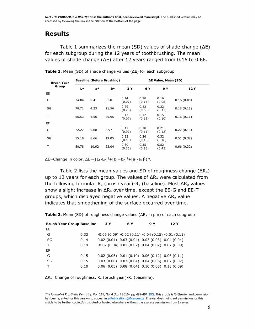
Table 1 summarizes the mean (SD) values of shade change (ΔE)
for each subgroup during the 12 years of toothbrushing. The mean
values of shade change (ΔE) after 12 years ranged from 0.16 to 0.66.
Table 1. Mean (SD) of shade change values (ΔE) for each subgroup
Brush Year
Group
Baseline (Before Brushing)
ΔE Value, Mean (SD)
L* a* b* 3 Y 6 Y 9 Y 12 Y
EE
G 74.84 0.41 6.50 0.14 (0.07)
0.20 (0.14)
0.16 (0.08)
0.16 (0.09)
SG 70.71 4.23 11.56 0.29
(0.28)
0.52
(0.65)
0.22
(0.17) 0.18 (0.11)
T 66.53 6.56 20.95 0.17
(0.07)
0.12
(0.12)
0.15
(0.10) 0.16 (0.11)
EP
G 72.27 0.68 8.97 0.12
(0.07)
0.18
(0.11)
0.21
(0.12) 0.22 (0.13)
SG 55.10 8.66 19.05 0.23
(0.13)
0.26
(0.15)
0.32
(0.16) 0.51 (0.32)
T 50.78 10.92 23.04 0.30
(0.15)
0.35
(0.13)
0.82
(0.45) 0.66 (0.32)
ΔE=Change in color, ΔE=([L1-L2]2+[b1=b2]2+[a1-a2]2)½.
Table 2 lists the mean values and SD of roughness change (ΔRa)
up to 12 years for each group. The values of ΔRa were calculated from
the following formula: Ra (brush year)-Ra (baseline). Most ΔRa values
show a slight increase in ΔRa over time, except the EE-G and EE-T
groups, which displayed negative values. A negative ΔRa value
indicates that smoothening of the surface occurred over time.
Table 2. Mean (SD) of roughness change values (ΔRa in μm) of each subgroup
Brush Year Group Baseline 3 Y 6 Y 9 Y 12 Y
EE
G 0.33 -0.06 (0.09) -0.02 (0.11) -0.04 (0.15) -0.01 (0.11)
SG 0.14 0.02 (0.04) 0.03 (0.04) 0.03 (0.03) 0.04 (0.04)
T 0.19 -0.02 (0.04) 0.01 (0.07) 0.04 (0.07) 0.07 (0.09)
EP
G 0.15 0.02 (0.05) 0.01 (0.10) 0.06 (0.12) 0.06 (0.11)
SG 0.15 0.03 (0.06) 0.03 (0.04) 0.04 (0.06) 0.07 (0.07)
T 0.10 0.06 (0.05) 0.08 (0.04) 0.10 (0.05) 0.13 (0.09)
ΔRa=Change of roughness, Ra (brush year)-Ra (baseline).

NOT THE PUBLISHED VERSION; this is the author’s final, peer-reviewed manuscript. The published version may be accessed by following the link in the citation at the bottom of the page.
The Journal of Prosthetic Dentistry, Vol. 115, No. 4 (April 2016): pg. 489-494. DOI. This article is © Elsevier and permission has been granted for this version to appear in e-Publications@Marquette. Elsevier does not grant permission for this article to be further copied/distributed or hosted elsewhere without the express permission from Elsevier.
9
Table 3 indicates the results of repeated measures ANOVA.
Analyses were performed separately for the EE and EP groups. All
assumptions, including a sphericity assumption, of the repeated
measures ANOVA were satisfied. Overall for the EE group, no
significant shade change or surface roughness change was noted after
12 years of simulated brushing, irrespective of time and staining
technique (P>.05). Conversely, the EP group revealed a significant
change in shade (P=.001) and roughness (P=.001) as a function of
brush year. The roughness change of the EP subgroups depended only
on brush year. However, shade change was found to depend on
brushing time and staining technique (P=.005). For the EP subgroup,
the 2-step technique (EP-SG) had less shade change over time
compared with the EP-T subgroups (P=.039).
Table 3. Repeated measures ANOVA (α=.05)
Group/Source of Variance Shade (ΔE) Roughness (ΔRa)
EE
Brush year .269 .141
Brush year×technique .268 .482
Technique
G versus SG .078 .085
G versus T .965 .319
SG versus T .047∗ .724
EP
Brush year .001∗ .001∗
Brush year×technique .005∗ .709
Technique
G versus SG .166 .989
G versus T .001∗ .994
SG versus T .039∗ .989
∗ Statistically significant (P<.05).
A post hoc test for comparing ΔE and ΔRa at 3-year
toothbrushing intervals in the groups EP-SG and EP-T is shown in
Table 4. In terms of shade change for EP-SG, a statistically significant
difference was noted between the 9-year and 12-year intervals
(P=.047). In the EP-T group, statistically significant shade changes
were noted at baseline and 3 years (P=.005) and between the 6-year
and 9-year intervals (P=.005). Furthermore, a statistically significant
change in surface roughness (ΔRa) was noted between base line and
the 3-year interval for the EP-T subgroup (P=.005).

NOT THE PUBLISHED VERSION; this is the author’s final, peer-reviewed manuscript. The published version may be accessed by following the link in the citation at the bottom of the page.
The Journal of Prosthetic Dentistry, Vol. 115, No. 4 (April 2016): pg. 489-494. DOI. This article is © Elsevier and permission has been granted for this version to appear in e-Publications@Marquette. Elsevier does not grant permission for this article to be further copied/distributed or hosted elsewhere without the express permission from Elsevier.
10
Table 4. Post hoc comparison (α=.05)
Group EP Shade (ΔE) Roughness (ΔRa)
EP-SG
Baseline and 3 y .253 .253
3 y and 6 y .164 .933
6 y and 9 y .058 .458
9 y and 12 y .047∗ .231
EP-T
Baseline and 3 y .005∗ .005∗
3 y and 6 y .422 .270
6 y and 9 y .005∗ .098
9 y and 12 y .426 .527
∗ Statistically significant (P<.05).
Discussion
The effect of toothbrushing on the shade and surface roughness
of extrinsically stained pressable ceramics was investigated. The null
hypothesis of the study was rejected for the EP group for both shade
and roughness; the null hypothesis was not rejected for the EE group.
Several studies of extrinsically stained feldspathic porcelain
materials have reported similar results as in the present study. Anil
and Bolay8 found a significant color change in extrinsically stained
feldspathic dental porcelain after an equivalent of 8.5 years of
toothbrushing. Aker et al7 demonstrated that toothbrushing could wear
the surface stain of feldspathic ceramic restorations over a period of
10 to 12 years unless a protective layer of glaze was applied. The
present study demonstrated that a significant shade change occurred
after an equivalent of 12 years of toothbrushing for the EP group
(P=.001). A significant difference was found between the EP-SG
and EP-T groups (P=.039). After 12 years of simulated toothbrushing,
the 2-step technique (SG) was found to be more resistant to shade
change than the T staining technique (T). This implies that the glaze
layer applied over the stain layer may play a protective role. In
contrast with EP, no significant difference in shade change was found
for EE group. This could be explained by the different chemical
composition and mechanical property of the 2 materials used: IPS
Empress universal shade/glaze and IPS e.max Ceram shade/glaze. In
addition, different firing temperatures could affect the stain stability

NOT THE PUBLISHED VERSION; this is the author’s final, peer-reviewed manuscript. The published version may be accessed by following the link in the citation at the bottom of the page.
The Journal of Prosthetic Dentistry, Vol. 115, No. 4 (April 2016): pg. 489-494. DOI. This article is © Elsevier and permission has been granted for this version to appear in e-Publications@Marquette. Elsevier does not grant permission for this article to be further copied/distributed or hosted elsewhere without the express permission from Elsevier.
11
found in the present study. However, Mulla and Weiner 22
demonstrated no difference between 2 different firing temperatures.
Previous studies5, 7 and 8 used the stain application technique
described by Lund et al,6 which relied on visual assessment for stain
application. Assessing color with the human eye is considered
inconsistent because of external variables such as light and internal
variables such as age, fatigue, sex, color blindness, personal bias, and
experience.2 Conversely, the present study used a controlled stain and
glaze application procedure that facilitated the reproducibility of
manufactured specimens. Moreover, color change (ΔE) was measured
with a spectrophotometer, which can provide a more consistent and
objective evaluation than the human eye.23
Although shade was found to be significantly affected in the EP
group, it was not clinically significant. In order to understand the
clinical significance of any shade changes, color tolerances, such as
perceptible tolerances and acceptability tolerances, must be
understood. Perceptible tolerances have been defined as the amount of
color difference that might be detected visually. Acceptability
tolerances have been defined as the alteration of color that is
considered esthetically unacceptable.1 Douglas et al1 summarized
different studies that evaluated color-matching tolerances. Most of
those studies were performed in nonclinical conditions. A study that
was performed in a clinical scenario reported a perfect color match to
have a ΔE of 3.7; barely acceptable matches were found to have ΔE of
6.8.24 A more recent clinical study by Douglas et al1 reported
perceptibility tolerances to be at 2.6 ΔE, while acceptability was 5.5
ΔE. The present study found mean ΔE values for both materials to be
as low as 0.16 and as high as 0.66 after 12 years of toothbrushing.
Although a statistically significant shade change (ΔE)was noted after
12 years of toothbrushing, the values of shade change (ΔE) were small
compared with the clinical threshold for perceptibility and acceptability.
This means that the shade change values found in this study would
not be considered clinically significant.
In terms of surface roughness, Anil and Bolay8 found a
significant decrease in the roughness of extrinsically stained
feldspathic dental porcelain after an equivalent of 8.5 years of
toothbrushing. In comparison, the present study found that mean Ra

NOT THE PUBLISHED VERSION; this is the author’s final, peer-reviewed manuscript. The published version may be accessed by following the link in the citation at the bottom of the page.
The Journal of Prosthetic Dentistry, Vol. 115, No. 4 (April 2016): pg. 489-494. DOI. This article is © Elsevier and permission has been granted for this version to appear in e-Publications@Marquette. Elsevier does not grant permission for this article to be further copied/distributed or hosted elsewhere without the express permission from Elsevier.
12
values increased slightly over time. This inconsistency may be the
result of differences in testing methods. Anil and Bolay8 used a
different brushing machine, greater brushing load (5.88 N), and harder
nylon toothbrushes, while the present study used a lighter brushing
load (1.96 N, an average obtained from the literature10, 11, 14, 17 and 18)
and a soft, straight Oral-B #35 toothbrush.14 and 18 All of the groups in
the present study became rougher, with 1 exception: the EE-G
subgroup, which possessed a higher baseline roughness than the other
samples, became smoother (-0.06 at 3 years to -0.01 at 12 years).
Patients can perceive a roughened surface (Ra) of 0.50 μm
clinically.25 The present study determined a mean Ra value of 0.32 (EE-
G), 0.18 (EE-SG), 0.26 (EE-T), 0.21 (EP-G), 0.22 (EP-SG), and 0.23
(EP-T) after 12 years toothbrushing. These numbers are below the
clinical thresholds noted above. Even though the EP group
demonstrated a statistically significant change in roughness as a
function of brushing time and techniques (P<.05), it is not considered
to be clinically significant.
This study has several limitations. First, there was a variance of
±30 μm among the groups in regard to the thickness of the stain
and/or glaze layers. However, it should be noted that ΔE and ΔRa were
outcome measures used for statistical comparisons and not absolute
values. Second, the current American Dental Association
recommendation for toothbrush replacement is every 3 to 4 months.26
This frequency could change the results of the present study.
However, in the present study, the slurry and toothbrushes were
replaced after every 3 years of simulated toothbrushing because of
experimental design and time management. If the test toothbrush
bristles lost their stiffness, it could manifest as a minimal increase in
surface roughness. Many types of toothpastes are commercially
available for toothbrushing. Some believe that toothbrush abrasion
and recession are the results of toothbrushing. However, one study
proved that abrasion was due to the effect of the dentifrice only and
had no relationship to the toothbrush.12 The composition of the slurry
used did not contain saliva or synthetic saliva and did not replicate the
oral environment. Last, no real comparisons could be made with
previous studies because of the difference in protocols and the use of
dissimilar materials. Further studies will be needed to investigate the

NOT THE PUBLISHED VERSION; this is the author’s final, peer-reviewed manuscript. The published version may be accessed by following the link in the citation at the bottom of the page.
The Journal of Prosthetic Dentistry, Vol. 115, No. 4 (April 2016): pg. 489-494. DOI. This article is © Elsevier and permission has been granted for this version to appear in e-Publications@Marquette. Elsevier does not grant permission for this article to be further copied/distributed or hosted elsewhere without the express permission from Elsevier.
13
effect of different brushing systems, such as with electronic
toothbrushes and fluoride toothpastes.
Conclusions
Within the limitations of this study, the following conclusions
may be drawn:
1. No statistically significant difference was found in the shade change
and surface roughness of the extrinsically stained EE groups after 12
years of simulated toothbrushing.
2. Statistically significant differences were found in the shade change
and surface roughness of the extrinsically stained EP groups after 12
years of simulated toothbrushing.
Acknowledgments
The authors thank Ivoclar Vivadent for providing the materials and allowing
the use of their equipment, facilities; Drs Shashikant Singhal and Thomas Hill
for help with specimen fabrication; and SABRI Inc for providing the
toothbrushing machine apparatus, without which, this project would not have
been possible.
References
1 R.D. Douglas, T.J. Steinhauer, A.G. Wee. Intraoral determination of the
tolerance of dentists for perceptibility and acceptability of shade
mismatch. J Prosthet Dent, 97 (2007), pp. 200–208 2 N. AlGhazali, G. Burnside, R.W. Smith, A.J. Preston, F.D. Jarad. Performance
assessment of Vita Easy Shade spectrophotometer on colour
measurement of aesthetic dental materials. Eur J Prosthodont Restor
Dent, 19 (2011), pp. 168–174 3 A. Dozić, C.J. Kleverlaan, A. El-Zohairy, A.J. Feilzer, G. Khashayar.
Performance of five commercially available tooth color-measuring
devices. J Prosthodont, 16 (2007), pp. 93–100 4 S. Schultheis, J.R. Strub, T.A. Gerds, P.C. Guess. Monolithic and bi-layer
CAD/CAM lithium-disilicate versus metal-ceramic fixed dental
prostheses: comparison of fracture loads and failure modes after
fatigue. Clin Oral Investig, 17 (2013), pp. 1407–1413 5 F. Bativala, S. Weiner, P. Berendsen, G.R. Vincent, J. Ianzano, W.T. Harris.
The microscopic appearance and effect of toothbrushing on

NOT THE PUBLISHED VERSION; this is the author’s final, peer-reviewed manuscript. The published version may be accessed by following the link in the citation at the bottom of the page.
The Journal of Prosthetic Dentistry, Vol. 115, No. 4 (April 2016): pg. 489-494. DOI. This article is © Elsevier and permission has been granted for this version to appear in e-Publications@Marquette. Elsevier does not grant permission for this article to be further copied/distributed or hosted elsewhere without the express permission from Elsevier.
14
extrinsically stained metal-ceramic restorations. J Prosthet Dent, 571
(1987), pp. 47–52 6 T.W. Lund, W.B. Schwabacher, R.J. Goodkind. Spectrophotometric study of
the relationship between body porcelain color and applied metallic
oxide pigments. J Prosthet Dent, 53 (1985), pp. 790–796 7 D.A. Aker, J.R. Aker, S.E. Sorensen. Toothbrush abrasion of color-corrective
porcelain stains applied to porcelain-fused-to-metal restorations.
J Prosthet Dent, 44 (1980), pp. 161–163 8 N. Anil, S. Bolay. Effect of toothbrushing on the material loss, roughness,
and color of intrinsically and extrinsically stained porcelain used in
metal-ceramic restorations: an in vitro study. Int J Prosthodont, 15
(2002), pp. 483–487 9 T. Arai, S. Kinoshita. A comparison of plaque removal by different
toothbrushes and toothbrushing methods. Bull Tokyo Med Dent Univ,
24 (1977), pp. 177–188 10 G.I. McCracken, J. Janssen, M. Swan, N. Steen, M. de Jager, P.A. Heasman.
Effect of brushing force and time on plaque removal using a powered
toothbrush. J Clin Periodontol, 30 (2003), pp. 409–413 11 G.A. Van der Weijden, M.F. Timmerman, M.M. Danser, U. Van der Velden.
Relationship between the plaque removal efficacy of a manual
toothbrush and brushing force. J Clin Periodontol, 25 (1998), pp. 413–
416 12 S. Kinoshita, T. Arai, R. Uraguchi. Abrasive properties of commonly used
dentifrices. Bull Tokyo Med Dent Univ, 26 (1979), pp. 225–242 13 J. da Costa, A. Adams-Belusko, K. Riley, J.L. Ferracane. The effect of
various dentifrices on surface roughness and gloss of resin composites.
J Dent, 38 (suppl 2) (2010), pp. e123–e128 14 J.C. Wataha, P.E. Lockwood, R.L. Messer, J.B. Lewis, D.J. Mettenburg.
Brushing-induced surface roughness of nickel-, palladium-, and gold-
based dental casting alloys. J Prosthet Dent, 99 (2008), pp. 455–460 15 S.H. Cho, W.W. Nagy, J.T. Goodman, E. Solomon, M. Koike. The effect of
multiple firings on the marginal integrity of pressable ceramic single
crowns. J Prosthet Dent, 107 (2012), pp. 17–23 16 H.J. Conrad, W.J. Seoung, I.J. Pesun. Current ceramic materials and
systems with clinical recommendations: a systematic review.
J Prosthet Dent, 98 (2007), pp. 389–404 17 A. Wiegand, J.P. Burkhard, F. Eggmann, T. Attin. Brushing force of manual
and sonic toothbrushes affects dental hard tissue abrasion. Clin Oral
Investig, 17 (2013), pp. 815–822 18 J.C. Wataha, P.E. Lockwood, M. Noda, S.K. Nelson, D.J. Mettenburg. Effect
of toothbrushing on the toxicity of casting alloys. J Prosthet Dent, 87
(2002), pp. 94–98

NOT THE PUBLISHED VERSION; this is the author’s final, peer-reviewed manuscript. The published version may be accessed by following the link in the citation at the bottom of the page.
The Journal of Prosthetic Dentistry, Vol. 115, No. 4 (April 2016): pg. 489-494. DOI. This article is © Elsevier and permission has been granted for this version to appear in e-Publications@Marquette. Elsevier does not grant permission for this article to be further copied/distributed or hosted elsewhere without the express permission from Elsevier.
15
19 J.D. Da Silva, S.E. Park, H.P. Weber, S. Ishikawa-Nagai. Clinical
performance of a newly developed spectrophotometric system on tooth
color reproduction. J Prosthet Dent, 99 (2008), pp. 361–368 20 S.A. Whitehead, A.C. Shearer, D.C. Watts, N.H. Wilson. Comparison of two
stylus methods for measuring surface texture. Dent Mater, 15 (1999),
pp. 79–86 21 C.Y. Poon, B. Bhushan. Comparison of surface roughness measurements by
stylus profiler, AFM and non-contact optical profiler. Wear, 190 (1995),
pp. 76–88 22 F.A. Mulla, S. Weiner. Effects of temperature on color stability of porcelain
stains. J Prosthet Dent, 65 (1991), pp. 507–512 23 S. Paul, A. Peter, N. Pietrobon, C.H. Hämmerle. Visual and
spectrophotometric shade analysis of human teeth. J Dent Res, 81
(2002), pp. 578–582 24 W.M. Johnston, E.C. Kao. Assessment of appearance match by visual
observation and clinical colorimetry. J Dent Res, 68 (1989), pp. 819–
822 25 C.S. Jones, R.W. Billington, G.J. Pearson. The in vivo perception of
roughness of restorations. Br Dent J, 196 (2004), pp. 42–45 26 P.M. Glaze, A.B. Wade. Toothbrush age and wear as it relates to plaque
control. J Clin Periodontol, 13 (1986), pp. 52–56
Supported by an American Academy of Fixed Prosthodontics Stanley D.
Tylman Research Grant and the Marquette University School of Dentistry Student Research Fund.
Corresponding author: Dr Seok-Hwan Cho, Marquette University School of Dentistry, 1801 W Wisconsin Ave, Milwaukee, WI 53233