Color Atlas of Clinical Laboratory Medicine First...
44
Page 1 of 44 Color Atlas of Clinical Laboratory Medicine First Version Addis Ababa, February 2015
Transcript of Color Atlas of Clinical Laboratory Medicine First...

Page 1 of 44
Color Atlas of Clinical Laboratory Medicine
First Version
Addis Ababa, February 2015

Page 2 of 44
Table Contents
1. Hematology ................................................................................................................................. 5
1.1. Cell Maturation .................................................................................................................... 5
1.2 Red Blood Cells Morphology ............................................................................................... 6
1.3 Abnormal Red Blood Cell Morphology ................................................................................ 6
1.4 White Blood Cell Morphology .............................................................................................. 9
1.5 Abnormal White Blood Cell Morphology .......................................................................... 11
2. Bacteriology .............................................................................................................................. 12
2.1 Gram Positive Cocci............................................................................................................ 12
2.2 Gram Positive Bacilli .......................................................................................................... 13
2.3. Gram Negative Cocci ......................................................................................................... 14
2.4 Gram Negative Bacilli ....................................................................................................... 14
2.5. Acid Fast Bacilli, AFB ....................................................................................................... 15
2.6 Spirochetes .......................................................................................................................... 16
3. Parasitology............................................................................................................................... 18
3.1. Intestinal Protozoa .............................................................................................................. 18
3.2. Intestinal Nematodes .......................................................................................................... 21
3.3. Intestinal Cestodes .............................................................................................................. 24
3.4.Trematodes .......................................................................................................................... 25
3.5. Blood Protozoa ................................................................................................................... 27
3.6. Tissue (Filarial) Nematodes ............................................................................................... 38
4. Urinalysis .................................................................................................................................. 39
4.1. Urine sediment cells ........................................................................................................... 39
4.2. Urine Parasites.................................................................................................................... 41
4.3. urine Sediment Crystals ..................................................................................................... 42
4.4. Urine sediment Casts .......................................................................................................... 43
Acknowledgments

Page 3 of 44
The EPHLA is deeply indebted to Tariku Bekele and Tadesse Hailu, both of whom are senior
members of our Association and with considerable laboratory experience, for their tireless effort
in assembling and systematically arranging the required information for this color atlas to be a
reality.
Even though almost all the information in the atlas is downloaded from their free internet access,
EPHLA still would like to express its appreciation to the World Health Organization for creating
such marvelous opportunity.
Preparation of laboratory color atlas was first pioneered by MSH (Ethiopian Network for
HIV/AIDS Treatment, Care & Support Program) whose management group unreservedly
encouraged us to proceed with compilation of the current atlas and therefore our gratitude also
goes to them.
Unreserved support and the funding of this project by the Ethiopian Public Health Association
(EPHA) and the CDC (through the grant # GH001039) are greatly acknowledged.
Preface Diagnostic laboratory service is not only an indispensable part but the backbone of the health care
delivery system which often determines the outcome of patient care for better or worse. Unfortunately,
Page 4 of 44
however, laboratory service in this country like the rest of many others in Africa has never received the
attention it deserves. We often hear that constraints of financial and human resources, among others,
being put forward as the reasons for the apparent gaps. It is true that there is limitation of resources but it
is not the whole truth. Frankly speaking most health care providers don't care while many others including
from the management side don't really much understand that test results generated by the labs constitute
evidence-based scientific medicine which pinpoint a better or even the right treatment option(s). Whether
we are health care providers or health managers, sooner or later we all are patients and thus beneficiaries
of laboratory services and therefore concerted effort and commitment by all towards promoting quality
laboratory standards is the order of the day. With this kind of mind set and with concerted effort from all
stakeholders, significant qualitative change in our laboratory systems is not only plausible but achievable
too. Gradually but surely such changes are on the way to many of our labs in health facilities across the
tier, thanks to the effort of governmental and non-governmental institutions.
The Ethiopian Public Health laboratory Association (EPHLA) is a non-for-profit and a non-governmental
organization established to contribute towards the national effort of strengthening public health
laboratories and to help them render quality standard diagnosis and services to the patient population. To
this end, the Association adopted several strategies, chief among them being human capacity building by
increasing access to information so that laboratorians could keep themselves abreast with current
scientific developments. It is in this spirit that this color laboratory atlas was developed which is mainly
intended for facilities in remote locations with limited or no access to scientific information.
This atlas by no means should be considered as a silver bullet and hence as a substitute to the ultimate
decision based primarily on the discretion of the technician and on further verification and/or validation
of the test procedures whenever this is applicable or feasible. This is particularly evident considering the
similarities in micro-morphological or phenotypic characteristics between less related or even unrelated
organisms depicted in the atlas. Caution must therefore be exercised against sweeping conclusion and
hence wrong diagnosis that could be detrimental to patient outcome. Thus, it is worth a while reiterating
that the color atlas is meant to serve not as the only but as one of the many possible tools at the disposal
of the lab professionals in the diagnosis of the major pathogenic agents and/or organisms that are
prevalent in the country.
This color atlas being the first of its kind to our Association, the EPHLA intends to update it through
follow up of new editions. Thus, the kind cooperation of our laboratory colleagues in providing feedback
on gaps and weaknesses noted in the publication or on any other relevant issues is vital in strengthening
and increasing the output of the EPHLA towards our common goal of achieving quality standards in all of
our labs.
EPHLA
February, 2015

Page 5 of 44
1. Hematology
1.1. Cell Maturation
Hematopoiesis is a process of blood cell production and maturation in the bone marrow. The process
begins with the pluripotent stem cell. The stem cell is capable to proliferate, differentiate and replicate.
The differentiation into a myeloid or lymphoid stem cell takes place in response of growth factors
(cytokines)

Page 6 of 44
1.2 Red Blood Cells Morphology
Normocytic Normochromic Microcytic Hypochromic
1.3 Abnormal Red Blood Cell Morphology
Target Cells Bite/Schistocyte

Page 7 of 44
Ovalocytes (Elliptocytes) Tear drop cells
Stomatocyte Sickle cells

Page 8 of 44
Roulex fromed RBCs Spherocyt
Acanthocytes (Spur Cells) Howell-Jolly Bodies

Page 9 of 44
Cabot ring (Ring shaped, thin, may be figure of 8 shaped)
1.4 White Blood Cell Morphology
Band and Segmented WBC Basophil

Page 10 of 44
Eosnophil Monocyte
Lymphocyte

Page 11 of 44
Platelets (Normal)
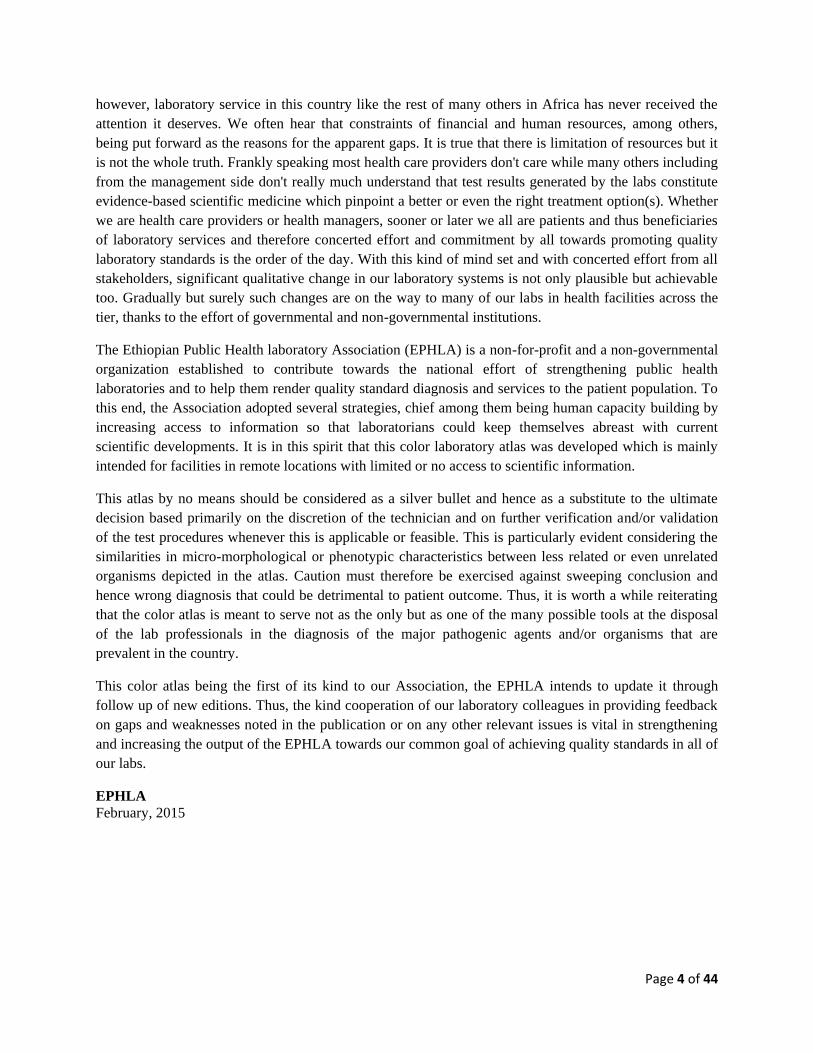
1.5 Abnormal White Blood Cell Morphology
Toxic granulation Dohle bodies Toxic granulation, vacuolization, &
Dohle bodies are seen during bacterial infections,
burns, cancer, and toxic or inflammatory states
Page 12 of 44
’
Vacuoles
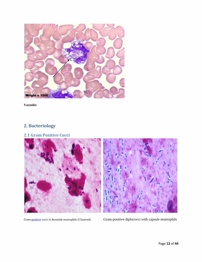
2. Bacteriology
2.1 Gram Positive Cocci
Gram-positive cocci in &outside neutrophils (Clustered) Gram-positive diplococci with capsule neutrophils

Page 13 of 44
Gram positive cocci cluster Gram positive streptococci
2.2 Gram Positive Bacilli
Gram positive Bacilli

Page 14 of 44
2.3. Gram Negative Cocci
Gram-negative diplococci phagocytized by neutrophils Gram Negative extracellular diplococcic
2.4 Gram Negative Bacilli
Gram-negative rods with capsule, Large-sized Gram-negative rods

Page 15 of 44
2.5. Acid Fast Bacilli, AFB
Fluorochrome stained smear numerous green AFB Ziehl Neelson stained AFB
Fluorochrome stained smear numerous green AFB Fuorochrome stained non AFB

Page 16 of 44
Fluorochrome stained smear showing numerous green Ziehl-Neelsen (ZN) stained Acid Fast Bacill acid-fast
acid-fast bacilli
2.6 Spirochetes
Borellia spps are long and spiral-shaped. The circular objects are red blood cells. The irregular purple
object in the top right corner is a white blood cell

Page 17 of 44
Borrelia spps stained in a blood sample with a Wright stain.

Page 18 of 44
3. Parasitology
3.1. Intestinal Protozoa
Trophozoite of E. histolytica/E. dispar in a direct wet mount stained with iodine
Cyst E. histolytica/E. dispar in an unstained
wet mount of stool
Cyst E. histolytica/E. dispar in an unstained wet
mount of stool with Iodine

Page 19 of 44
Fig. A: B. coli trophozoite in a wet mount, 400× magnification. Fig. B: B. coli trophozoite in a Mann's hematoxylin stained
smear, 500× magnification.
Figure A: B. hominis cyst-like forms in a wet mount ,
unstained
Figure B: Cryptosporidium spp. oocysts (pink arrows) in
Wet mount. A budding yeast (brown arrow) is in the same
field

Page 20 of 44
Figure A: Cryptosporidium sp. oocysts stained with trichrome. Oocysts may be detected, but should not be
confirmed by this method. Trichrome staining is inadequate for a definite diagnosis because oocysts will appear
unstained. Here the Cryptosporidium oocysts are represented by red arrows; the blue arrow represents yeast
Fig A: Immature oocyst of C. belli in an unstained
wet mount, containing a single sporoblast
Fig B: Immature oocyst of C. belli stained with safranin,
containing a single sporobla
Page 21 of 44
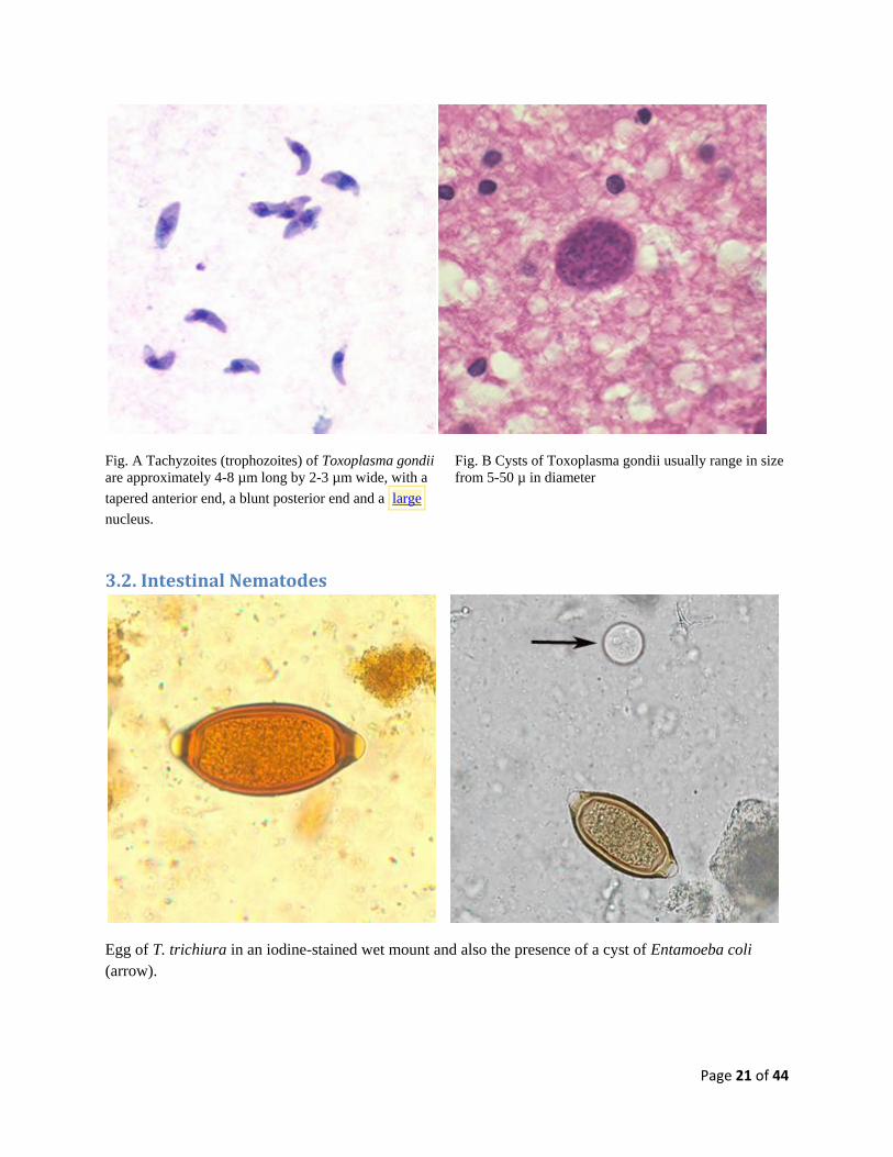
Fig. A Tachyzoites (trophozoites) of Toxoplasma gondii
are approximately 4-8 µm long by 2-3 µm wide, with a
tapered anterior end, a blunt posterior end and a large
nucleus.
Fig. B Cysts of Toxoplasma gondii usually range in size
from 5-50 µ in diameter
3.2. Intestinal Nematodes
Egg of T. trichiura in an iodine-stained wet mount and also the presence of a cyst of Entamoeba coli
(arrow).

Page 22 of 44
Fig. A: Eggs of E. vermicularis in a wet mount Fig. B: Egg of E. vermicularis in an iodine-stained wet
mount
Unfertilized and fertilized eggs of A. lumbricoides in an unstained wet mount of stool 200x

Page 23 of 44
Hookworm egg in an unstained wet mount, taken at 400x magnification.
Rhabditiform larva of S. stercoralis in unstained wet mounts of stool. Notice the short buccal canal and
the genital primordium (red arrows).
Rhabditiform larva of S. stercoralis in an unstained wet mount of stool. Notice the prominent genital
primordium (blue arrow), rhabditoid esophagus (red arrow) and short buccal canal (green arrow).

Page 24 of 44
3.3. Intestinal Cestodes
Taenia sp. eggs in unstained wet mounts. Iodine-stained wet mount of a Taenia sp. egg
Egg of H. diminuta in a wet mount stained with iodine. Four of the hooks are visible at this level of focus

Page 25 of 44
Eggs of D. latum in an iodine- stained wet mount; Eggs of D. latum in an unstained wet mount
3.4.Trematodes
Egg of S. mansoni in an unstained wet mount

Page 26 of 44
Egg of S. hematobium in an unstained wet mount
Egg of F. hepatica in an unstained wet mount, taken at 400x and 100 x magnification

Page 27 of 44
3.5. Blood Protozoa
Fig. A: Leishmania sp. amastigotes in a Giemsa-stained
tissue scraping
Fig. B : Leishmania (Viannia) panamensis amastigotes
in a Giemsa-stained tissue scraping. Identification to the
species level is not possible based on morphology and
other diagnostic techniques such isoenzyme assay or
PCR are needed.
Fig. C: Leishmania sp. amastigotes; touch-prep stained with Giemsa. Fig. D: Leishmania tropica amastigotes from an impression smear of a
biopsy specimen from a skin lesion. In this figure, amastigotes are
being freed from a rupturing macrophage. Patient had traveled to
Egypt, Africa, and the Middle East. Based on culture in NNN medium,
followed by isoenzyme analysis, the species was identified as L.
tropica.

Page 28 of 44
Fig. A : Trypansoma brucei spp. in a thick blood smear stained Fig. B : Trypansoma brucei spp in a thick
With Giemsa blood smear stained with Giemsa.
Fig. C : Trypanosoma brucei spp. in a thin blood smear
stained with Wright-Giemsa. Fig. D: Trypanosoma brucei ssp. in a thin blood smear stained
with Wright-Giemsa.
Page 29 of 44
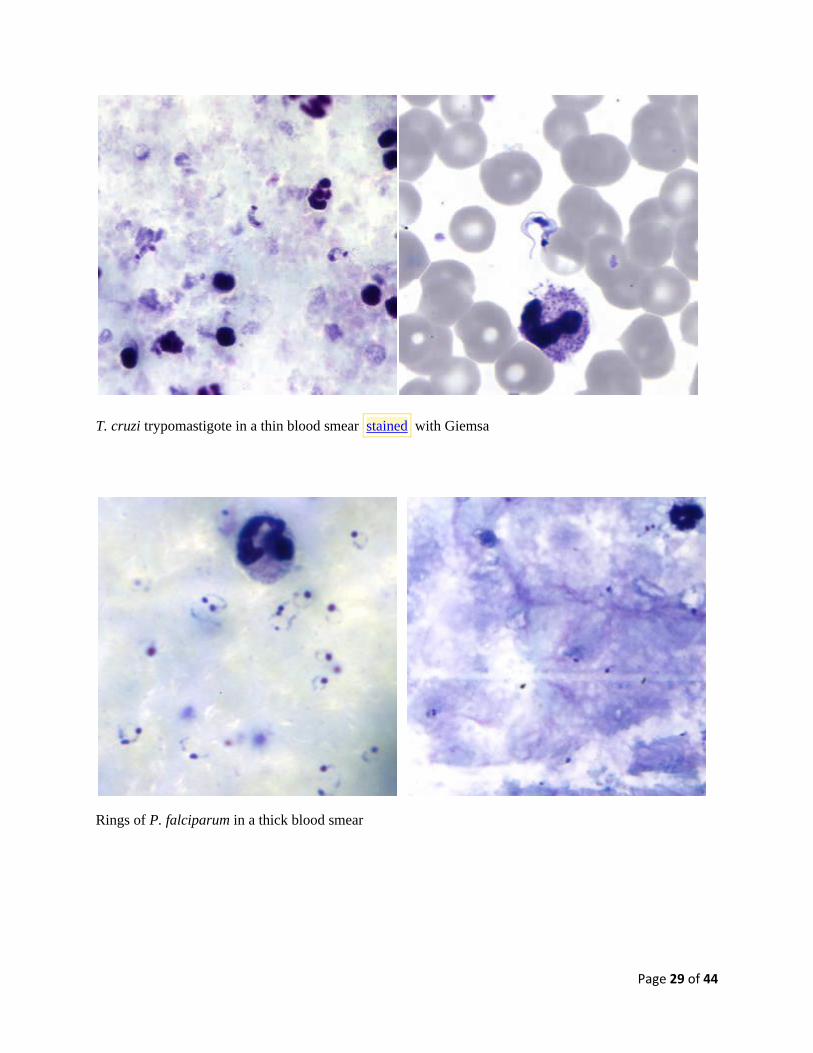
T. cruzi trypomastigote in a thin blood smear stained with Giemsa
Rings of P. falciparum in a thick blood smear
Page 30 of 44
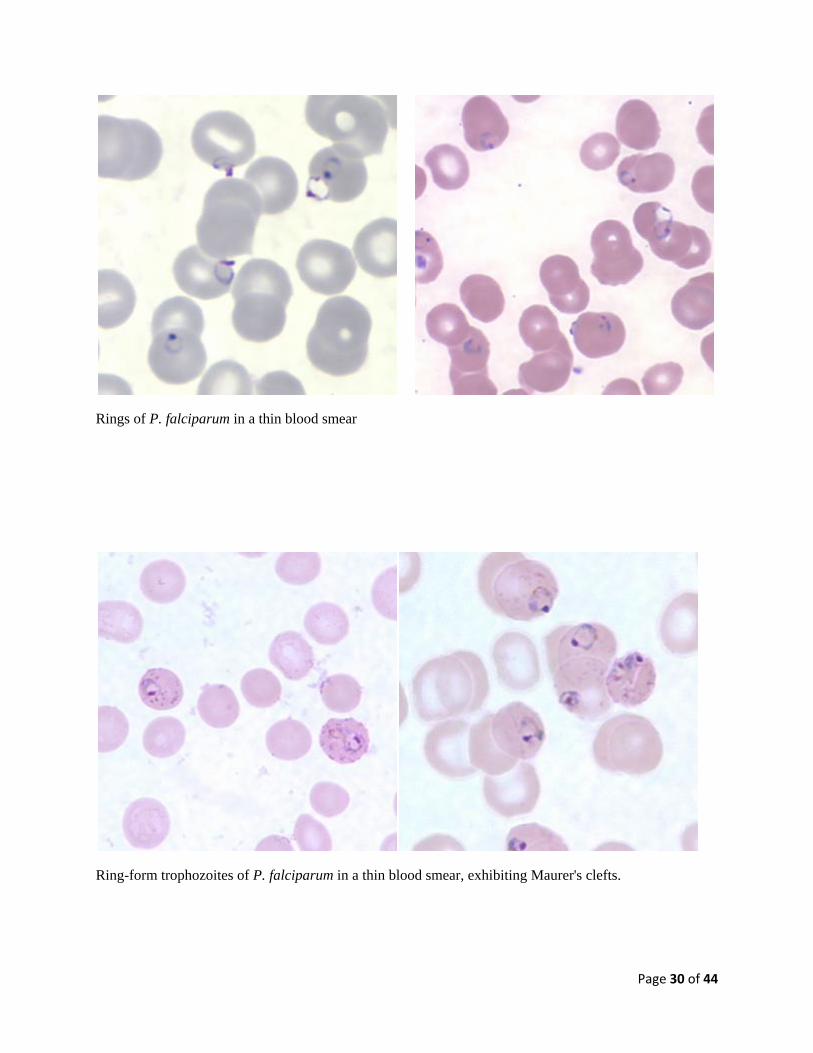
Rings of P. falciparum in a thin blood smear
Ring-form trophozoites of P. falciparum in a thin blood smear, exhibiting Maurer's clefts.

Page 31 of 44
Gametocyte of P. falciparum in a thick blood smear. Note also the presence of many ring-form
trophozoites.
Gametocyte of P. falciparum in a thin blood smear and thick Giemsa stain
Page 32 of 44
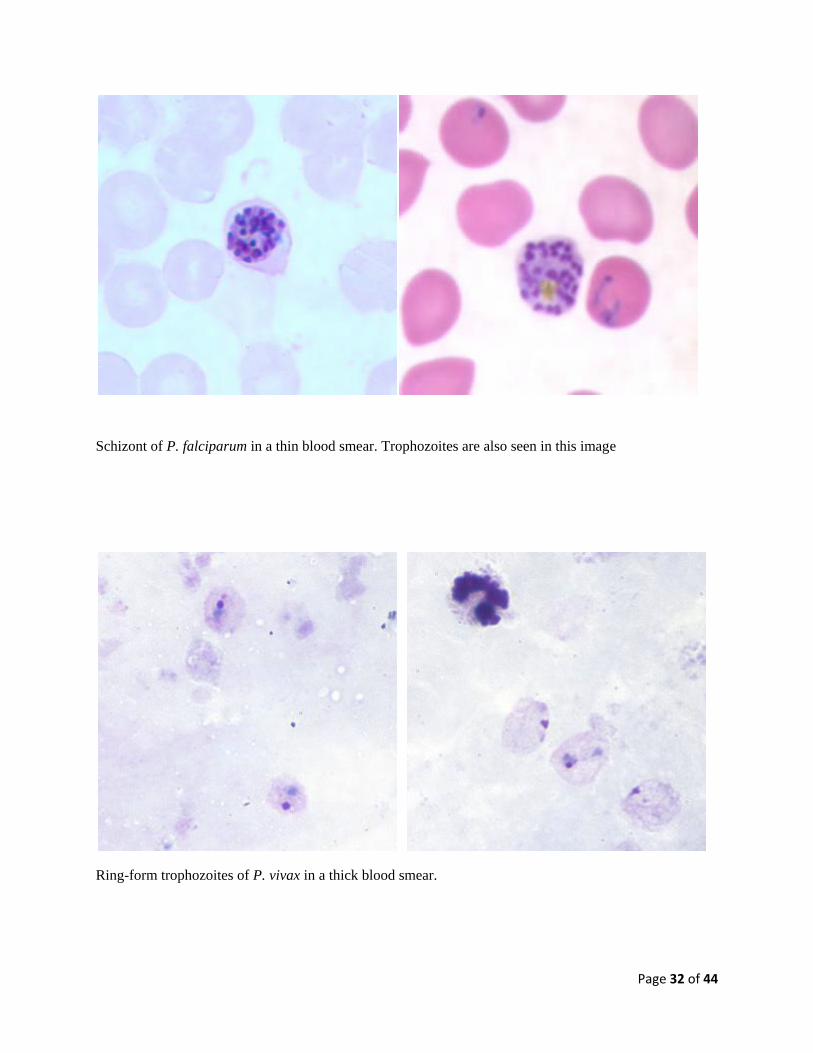
Schizont of P. falciparum in a thin blood smear. Trophozoites are also seen in this image
Ring-form trophozoites of P. vivax in a thick blood smear.

Page 33 of 44
Ring-form trophozoite of P. vivax in a thin blood smear.
Trophozoite of P. vivax in a thick blood smear. Trophozoites of P. vivax in a thin blood smear
Note the amoeboid appearance, Schüffner's dots and enlarged infected RBCs.

Page 34 of 44
Fig. 1: Gametocyte (upper) and trophozoite (lower) of P. vivax
in a thick blood smear Fig. 2: Macrogametocytes of P. vivax in a thin blood smear.
Note the enlargement of the gametocytes compared to
uninfected RBCs
Fig. 1: Schizont of P. vivax in a thick blood smears Fig. 2: Ruptured schizont of P. vivax in a thin
blood smear, showing free merozoites and pigment

Page 35 of 44
Fig. 1: Ring-form trophozoite of P. malariae in a thin blood smear. Fig. 2: Trophozoite of P. malariae in a thick blood smear.
Fig. 1: Basket-form trophozoite of P. malariae in a thin blood smear Fig. 2: Gametocyte of P. malariae in a thin blood
smear

Page 36 of 44
Fig. 1: Schizonts of P. malariae in a thick blood smear. Fig. 2: Schizont of P. malariae in a thin blood
Fig. A: Ring-form trophozoites of P. ovale in a thin blood
smear. Note the multiple-infected RBC in this image. Fig. B: Ring-form trophozoite of P. ovale in a thick
blood smear.

Page 37 of 44
Fig. 1: Infected RBCs showing developing (lower) and ring-
form (upper two) trophozoites of P. ovale in a thin blood
smear.
Fig. 2: Trophozoite of P. ovale in a thick blood smear.
Fig. 1: Gametocyte of P. ovale (red arrow) nestled between
two white blood cells in a thick blood smear. Fig. 2: Macrogametocyte of P. ovale in a thin blood smear.
Note the fimbriation.

Page 38 of 44
Fig. 1: Schizont of P. ovale in a thick blood smear. Fig. 2: Schizont (upper right) and ring-form
trophozoite (lower left) of P. ovale in a thin blood smear
3.6. Tissue (Filarial) Nematodes
Microfilaria of W. bancrofti in a thick blood smear stained with Giemsa

Page 39 of 44
Larvae of Trichinella, freed from their cysts, typically coiled. Encysted larvae of Trichinella in pressed muscle
tissue.
4. Urinalysis
4.1. Urine sediment cells
Ghost RBC RBC’s showing rouleaux

Page 40 of 44
White Blood Cells in urine with 200X and 400x magnification, respectively
Pus cell Yeasts cells (hyphae)

Page 41 of 44
4.2. Urine Parasites
Trophozoite of T.vaginalis in wet mount Trophozoites of T. vaginalis , stained with Giemsa
Egg of S. haematobium in a wet mount of urine concentrates, showing the characteristic terminal spine.

Page 42 of 44
4.3. urine Sediment Crystals
Amorphous Urate crystals Amorphous phosphate Crystals
Uric Acid Crystals Calcium Oxalate Crystals

Page 43 of 44
4.4. Urine sediment Casts
Granular Cast hyaline casts
WBC Casts Fatty Wax

Page 44 of 44
Cholesterol cast