external antennal morphometry of trichopl usia ni - Agricultural

INTRODUCTION
The ant subfamily Proceratiinae Emery, 1895 comprises three genera, Discothyrea Roger, 1863, Probolomyrmex Mayr, 1901, and Procera-tium Roger, 1863 (Bolton 2020). Brown (1957, 1958) noted that Discothyrea and Proceratium are specialized predators of arthropod eggs, es-pecially spider eggs, one of the most remarkable specializations in ant predation. Specialization for spider egg predation by Discothyrea was first documented in the field for an African species,
Colony composition, arthropod egg predation, and antennal structure of the ant Discothyrea sauteri (Hymenoptera: Formicidae)
Keiichi Masuko
Biological Laboratory, Senshu University, 2-1-1 Higashimita, Tama-ku, Kawasaki, 214-8580 Japan
Corresponding author: [email protected]
Keywords Polygyny, Japan, spider, sensilla
Copyright This article is distributed under a Creative Commons Attribution License CCBY4.0Communicating Editor
Benoit Guénard
The genera Discothyrea and Procera-tium are closely related (Ward & Fisher 2016) and are diagnosed as having “abdominal tergite IV enlarged and strongly vaulted, tergite hyper-trophied with respect to sternite IV, which is re-
ESM Electronic supplementary information for this article can be downloaded from the Asian Myrmecology website
D. oculata Emery, 1901 (Dejean & Dejean 1998; Dejean et al. 1999), whereas the Japanese spe-cies, P. itoi (Forel, 1918), preys on eggs of vari-ous arthropod taxa, including eggs of Hemiptera, Chilopoda, and Opiliones (Masuko 2019b). On the other hand, Probolomyrmex is a specialized predator of polyxenid millipedes (Ito 1998).
ASIAN MYRMECOLOGY — Volume 12, e012002, 2020ISSN 1985-1944 | eISSN: 2462-2362 DOI: 10.20362/am.012002
ABSTRACT. In an evergreen, broadleaf forest in central Japan, we collected 21 colonies of the cryp-tic ant, Discothyrea sauteri, which nests in small cavities in the soil (ca. 100 mm depth), rotting logs, and dead branches lying on the forest floor. The largest colony contained 132 workers, and one third of the collected queenright colonies were functionally polygynous, as revealed by dissection of the queens. Ant nests collected between April and November contained many globular, arthropod eggs. These eggs did not hatch in the laboratory, but we concluded that they are spider eggs, because some colonies nested in woven silk egg-sac structures and many others nested in cavities lined with silk filaments. Moreover, SEM observations showed strong similarities of these silk filaments to those of spider egg sacs. The extremely enlarged terminal segment of the antennal funiculus, the antennal club, which characterizes Discothyrea, has a group of short, hollow sensilla near its end. As these sensilla firmly contact the surface of globular prey eggs when ants hold them, the antennal shape and the dis-tribution of these sensilla may be related to lifting a prey egg using the antennae.
Citation Masuko K, 2020. Colony composition, arthropod egg predation, and antennal structure of theant Discothyrea sauteri (Hymenoptera: Formicidae). Asian Myrmecology 12: e012002
Keiichi Masuko2 of 16
duced” (Bolton 2003, p.49). This gives them the peculiar body form with an anteriorly oriented abdomen, which is related to different behaviors such as predation, nest defense, and thanatosis (see below). Worldwide, 49 and 86 extant species have been described for Discothyrea and Proc-eratium, respectively (Bolton 2020). Yet, because these ants are cryptic and collection of entire col-onies has been infrequent, detailed data on their behavior and ecology are limited to a few species (Dejean & Dejean 1998; Dejean et al. 1999; Ma-suko 2019b). In the present study, 21 colonies of the Japanese species, Discothyrea sauteri Forel, 1912, were collected from a single locality in Kanagawa Prefecture, in central Japan, permit-ting us to obtain detailed information on colony population, social structure, and colony founding, based on field data and laboratory rearing. Also, morphological traits likely related to egg preda-tion were examined using scanning electron mi-croscopy (SEM) and histological sections.
MATERIALS AND METHODS
Study area and ant sampling
Ants were collected in a small forest (ca. 35 ha) located at Cape Manazuru (35.144° N, 139.154° E), Kanagawa Prefecture, from 1978 to 1984. The natural vegetation of this area is ever-green broadleaf forest dominated by Castanop-sis sieboldii (Makino) Hatus. ex T. Yamaz. et Mashiba (Miyawaki et al. 1969). In this forest, most D. sauteri nests were excavated close to the soil surface (ca. 100 mm depth), but some were sheltered in dead branches or rotting logs lying on the forest floor (see below). As there was no effective way to locate nests from the surface, nests were encountered haphazardly by digging in soil or by dismantling dead branches and rot-ting logs. When a nest was found, as many ants as possible were collected using an aspirator, and taken to the laboratory, where they were sorted under a stereomicroscope. In addition to the ants and their brood, eggs of unidentified arthropods, likely stored as prey, were frequently noted and carefully collected.
After censusing in the laboratory, ants were then preserved in 80% ethanol or Kahle’s solution (Barbosa 1974) or reared, depending on the intended purpose. Arthropod eggs gathered in the field were also preserved in these fixatives, or used as food for ants reared in the laboratory. Some dealate gynes (queens) and workers were dissected to examine insemination, ovary devel-opment, and the number of ovarioles.
Ant rearing
For behavioral observations in the laboratory, seven colonies were reared in polystyrene con-tainers covered with a transparent glass and kept at 21–26°C. Container bottoms were covered with gypsum mixed with activated carbon pow-der. When a colony population (ants plus arthro-pod eggs) was small, a brood chamber was exca-vated in the center of the plaster floor that the ants selected for nesting. For large colonies, ants and prey eggs were not confined in such a chamber, but were placed directly in a terrarium. In addi-tion to arthropod eggs recovered from ant field nests, eggs, or hatchling spiders and centipedes were given as prey to these laboratory colonies (see below for their taxonomy).
SEM and histology
SEM (Keyence VE-8800) and histological stud-ies were conducted on 20 workers obtained from colonies collected from 1981 to 1984. These were first preserved in Kahle’s solution and then trans-ferred to 80% ethanol. SEM specimens were air-dried, mounted on stubs, and coated with gold-palladium. The same procedure was used for silk-like filaments found inside D. sauteri nests. For histological examination of worker antennae, preserved specimens were further dehydrated in an ethanol series with propylene oxide, before embedding in Agar Low Viscosity Resin (Agar Scientific). Sections (0.5–0.8 μm) were cut using a diamond knife, and then stained in a 1% methy-lene blue / 1% borax solution. Sections were ex-amined using a compound microscope (Olympus Vanox) and morphological details were imaged with a digital camera (Shimadzu Moticam-2500).
Ecology of Discothyrea sauteri 3 of 16
1-30 31–60 61–90 91–120 121–1500
3
6
9
No.
of c
olon
ies
No. of workers per colony
N = 21
Masuko, Fig. 1
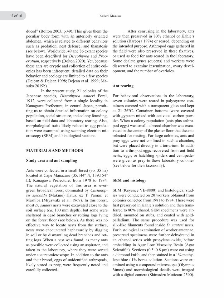
Fig. 1. Frequency distribution of the colony worker population in D. sauteri, based on 21 collections made at Manazuru between 1978 and 1984.
RESULTS
Colony composition
Among the 21 D. sauteri colonies collected, four were judged to be incomplete samples (Table 1). The number of workers per colony (including the incomplete samples) ranged from 1–132 and the median was 34 (n = 21; mean ± SD = 40.4 ± 32.1) (Fig. 1); thus, D. sauteri colonies are rather small. Only one queenless colony (coded 81-285) was noted among 17 complete or nearly complete samples. Among the other 16 colonies, 11 were monogynous and 5 were polygynous (numbers of queens ranged from 1–14). Seventeen queens from 3 monogynous and 3 polygynous colonies were dissected (Table 2). Dissections of 16 queens (one dissection failed) revealed that all were inseminated and had corpora lutea (yellow bodies) in their ovaries. Corpora lutea are remnants of follicle cells and their presence indicates that these queens had laid eggs (Torre-Bueno 1989). Moreover, the degree of ovary development, based on ovariole length, was the same in two queens of colony 83–49 and among ten queens of colony 82–60 (Fig. 2a, b). These observations confirm that polygyny in D. sauteri is functional, since multiple queens in the same nest were equally fertile. A statistical-ly significant positive correlation was obtained
between queen and worker numbers per colony (Fig. 3), which is consistent with functional po-lygyny. When two polygynous colonies, 81-77 with 14 queens and 82-245 with 2 queens, were reared in the laboratory, there was no evidence of aggressive interactions among the queens. In all dissected queens, with a single exception in colony 82–60, the number of ovarioles per ovary was three (Table 2), and the same was true for two workers that were dissected.
Colony foundation
In functionally polygynous ants, colony budding, in which one or more inseminated queens ac-companied by workers disperse from a natal nest, is common (Hölldobler & Wilson 1990). Due to small sample sizes, the nature of D. sauteri colony foundation remains unconfirmed. In the present study, no solitary foundress was collect-ed, while an incipient colony (82–290) was col-lected in early August, composed of one queen, one worker, and four eggs (Table 1). Larvae and pupae were absent. Since pupal gynes and males were observed in D. sauteri nests collected in mid or late July (Table 1), this small colony might be a fraction just split from a mother colony, contain-ing a newly inseminated queen. The short ovaries of this queen indicated that her egg-laying had

Keiichi Masuko4 of 16
just started (Fig. 2c). However, it is also possible that the queen started her colony independently during the previous year and had produced only one worker in this second year. Another small colony (82-366), collected in mid September, was also monogynous, having only seven young-er larvae and four workers, of which three were callows. One of them was totally pale, suggest-ing that it had eclosed in that nest. Here, too, the queen’s ovaries were short, but somewhat longer than those of queen 82–290 (Fig. 2c, d), suggest-ing that this colony might have started during the previous year. Because only two such inicipient
colonies were collected in the present study, it re-mains uncertain whether independent foundation, colony fission, or both occur in D. sauteri.
Nesting habits and prey eggs
Among the 21 colonies collected, 14 nested di-rectly in the soil, 6 nested in dead branches or rot-ting logs, and one in a soil-litter sample carried to the laboratory, so that its nesting site was uncer-tain (likely in the soil; colony 79–63; Table S1).
Table 1. Census of 21 colonies of D. sauteri collected in Manazuru, Kanagawa Prefecture, Japan. Colony collections (samples) were categorized as complete (C), nearly complete (N), or incomplete (I). No record exists for sex and caste of two pupae in 79-63; however, a male pupa and five worker pupae were confirmed later in laboratory rearing. Colony
codeDate of
collectionSample category Queen Worker Egg Larva Prepupa Pupa
78-45 5 Feb. 1978 C 1 84 4183-49 6 Mar. 1983 C 2 64 4979-36 25 Mar. 1979 C 1 40 1482-60 26 Mar. 1982 N 10 76 ca. 15078-112 9 Apr. 1978 C 1 46 3184-7 13 Apr. 1984 C 1 73 ca. 6081-77 6 May 1981 C 14 13281-109 27 May 1981 C 1 15 3779-63 24 June 1979 C 1 16 5 2 5 278-141 15 July 1978 I 33 ca. 25 1 gyne, 1 male81-190 21 July 1981 N 1 13 24 10 22 7 workers, 9 males82-245 23 July 1982 C 2 28 ca. 30 37 32 18 workers, 5 males81-203 29 July 1981 I 3 ca. 10 8 3 13 workers82-290 10 Aug. 1982 C 1 1 482-366 19 Sept. 1982 C 1 4 781-285 23 Sept. 1981 N 40 14 38 3 workers81-347 25 Oct. 1981 C 1 50 ca. 7081-397 12 Nov. 1981 C 2 17 6882-419 25 Nov. 1982 C 1 51 5782-423 25 Nov. 1982 I 34 378-238 9 Dec. 1978 I 28 ca. 40

Ecology of Discothyrea sauteri 5 of 16
Fig. 2. Microscopy of whole ovaries of D. sauteri queens. All preserved specimens. (a, b) Ovaries of two of the 10 queens of colony 82-60. The ten queens were freshly dissected the day after field collection. All were insemi-nated and had corpora lutea, and their ovariole lengths were similar. (c) Ovary of the queen of an incipient colony 82-290 that contained only one worker. (d) Ovary of the queen of an incipient colony 82-366 that contained four workers. Scale bar 0.5 mm, common for (a) to (d).
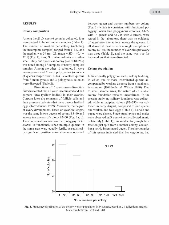
We noted the presence of globular eggs in these ant nests (Fig. 4a, b, arrows; Fig. 5c). These were obviously different in shape and color from those of D. sauteri, which are oblong and grayish in color (Fig. 7b, arrowhead). Al-though no hatchlings emerged in the laboratory from these exogenous eggs, it is likely that they were spider eggs. Indeed, an entire spider egg-sac-like structure was observed in the field when colony 78-141 was discovered inside a dead bamboo internode (Fig. 5a), which contained approximately 250 globular, cream-colored eggs (Table S1; Fig. 5c). This structure also enclosed 33 workers and about 25 eggs of D. sauteri (Ta-ble 1), and those ant eggs were completely inter-
mixed with the mass of prey eggs (Fig. 5b). This habit of jumbling prey eggs and the ant imma-ture brood, including larvae and pupae as well as eggs, was consistently observed in laboratory colonies (Fig. 7b). The size of prey eggs, which ranged in diameter from 0.42 to 0.82 mm, varied slightly from one nest to the next (Table S1), suggesting that these eggs have different origins, perhaps pertaining to different spider species. It should be further noted that prey eggs were found in all colonies collected from mid-April through mid-November (Table S1). The few eggs that were discovered on March 6 in colony 83-49 were most likely left over from the previous season.

Keiichi Masuko6 of 16
Table 2. Reproductive data from queens in monogynous and polygynous colonies of D. sauteri.
Colony code
Date of collection
No. of workers
No. of queens Queen Insemination Corpora
lutea No. of ovarioles
83-49 6 Mar. 1983 64 2 a Yes Present 3+3b Yes Present 3+3
82-60 26 Mar. 1982 76 10 a Yes Present 3+3b Yes Present 3+3c Yes Present 3+3d Yes Present 3+3e Yes Present 3+3f Yes Present 3+3g Yes Present 3+3h Yes Present 3+3i Yes Present 3+4j Yes Present 3+3
82-245 23 July 1982 28 2 a ? ? ?b Yes Present 3+3
82-290 10 Aug. 1982 1 1 Yes Present 3+382-366 19 Sept. 1982 4 1 Yes Present 3+382-419 25 Nov. 1982 51 1 Yes Present 3+3
É
É
É
É
É
É
É
ÉÉÉ
É
ÉÉ
É
É
É
0
50
100
150
0 5 10 15
No.
of w
orke
rs
No. of queens
R2 = 0.49
p < 0.005
Masuko, Fig. 3
Fig. 3. Relationship between queen number and colony worker population, based on 16 queenright, complete, or nearly complete samples of D. sauteri.

Ecology of Discothyrea sauteri 7 of 16
Fig. 4. Inside of a nest of a D. sauteri colony (81-347) in the soil. (a-c) White fibrous material covers the inner surface of nest cavities. Arrows in (a) and (b) indicate prey eggs. Worker ants are about 2 mm long.
Keiichi Masuko8 of 16
Fig. 5. (a) Field sketch of a split-half of a dead bamboo stem in which colony 78-141 was found. Near one end of the stem, the ants existed inside a fibrous structure that was about 25 mm long and seemed to be an egg sac cocoon. (b) Field sketch of a mass of about 250 globular prey eggs mingled with elongate ant eggs. (c) Prey eggs found there. Preserved specimens. Scale bar 0.5 mm.
Silk filaments in D. sauteri nests
In addition to the whole egg-sac-like structure in which a D. sauteri colony sheltered (see above), the inner walls of 12 ant nests were almost en-tirely covered with silk (Fig. 4a–c, Table S1), the origin of which remains to be determined. How-ever, SEM examination of silk structure revealed common characteristics: the filaments formed bundles (Fig. 6c, d, f) and included multiple fi-ber types of different diameters (Fig. 6d, arrow-head). As the bundling indicates that these fibers must have been spun at the same time, the most plausible source of such fibers are the tubuliform and aciniform silk glands of spiders (Hayashi, personal communication) because an individual spider has multiple sets of these glands and is able to simultaneously produce multiple fibers. Also, tubuliform and aciniform glands are known to produce egg sac silks (Foelix 2011). Compared to tubuliform gland silks, aciniform glands con-tribute fibers of smaller diameter (Moon 2003; Correa-Garhwal et al. 2019). Therefore, as fila-ments from D. sauteri nests showed both of these characteristics, they are most likely silks of spi-der egg sacs (Hayashi, personal communication). Moreover, among fibers lining the subterranean cavities of D. sauteri nests, many had cut ends, which were frequently straight (Fig. 6f, arrow-heads). Apparently, the ants severed the filaments with their mandibles (see below).
Predatory behavior in the laboratory
Seven queenright colonies were reared and ob-served in the laboratory. Durations of laboratory maintenance varied, but the longest was only six months because it was difficult to continuously supply ants with spider eggs of appropriate size. Aside from the unidentified arthropod eggs collected from ant field nests, the following organisms that were supplied as prey for labora-tory colonies were retrieved by the workers and consumed by their larvae: eggs and/or hatchlings of the spiders Nephila clavata L. Koch, 1878 (Araneidae), Parasteatoda tepidariorum (L. Koch, 1841) (Theridiidae), and Pardosa astrig-era L. Koch, 1878 (Lycosidae), and eggs of cen-tipedes, undetermined species of Scolopocryp-tops Newport, 1844 (Scolopendromorpha) and Geophilomorpha (genus undetermined). On the other hand, D. sauteri colonies ignored eggs of Polydesmida (genus undetermined) and the bur-rower bug, Macroscytus japonensis (Scott, 1874) (Hemiptera) that were collected from field nests of the ant, Proceratium itoi (see Masuko 2019b). Honey solutions were licked by workers, but modestly, and afterward, no stomodeal trophal-laxis was observed among workers during the study period.

Ecology of Discothyrea sauteri 9 of 16
Fig. 6. Silk filaments found in nests of two D. sauteri colonies collected at Manazuru. (a) Sheet of an egg sac lining the cavity of a dead branch where D. sauteri colony 82-419 was found. The uncovered surface of the dead branch is indicated by an asterisk. (b–d) Enlarged images of the surface of a silk filament sheet shown in (a). Fila-ments form bundles, and in addition to these thick filaments, there is another type of filament, much smaller in diameter (d, arrowhead). (e) Sheet of silk filaments lining the cavity of a D. sauteri nest in the soil (colony 84-7). The soil surface is indicated by an asterisk. (f) Enlarged image of the filaments shown in (e). There are two types of filaments of different diameters, and some filaments have straight cut ends (arrowheads).

Keiichi Masuko10 of 16
Fig. 7. Predation on spider eggs in laboratory colonies of D. sauteri. (a) An egg sac of the spider, Pardosa astrigera, given intact to the ants. It was then cut open and the spiderings inside were taken out by D. sauteri workers. Cut ends of the opening are smooth (arrowhead). (b) Eggs of the spider, Parasteatoda tepidariorum, (spherical and yellowish), mixed with larvae, pupae, and eggs (arrowhead) of D. sauteri. Several D. sauteri queens are seen on these mixed heaps. (c) A D. sauteri worker holding and licking an egg of P. astrigera that is supported by its antennae and abdominal tip. Note that the two antennae and the abdomen form a stable “tripodal” support for the egg. Arrowhead indicates the posterior edge of the 4th abdominal tergite. (d) Two workers of D. sauteri holding and licking eggs of P. astrigera. Arrowheads point the antennal funiculi applied to the egg surface.
When an intact egg sac from the spi-der, P. astrigera, was placed in the terrarium of a laboratory nest, the worker that discovered it immediately started antennation on its surface. Soon afterward the ant returned to the brood chamber, wherein it walked around in excited, rapid gait, which was not observed during its walk to the chamber. Concurrently, the ant that discovered the egg sac frantically palpated other workers with its antennae or gently bit them with its mandibles. After several repetitions of this be-havior, the worker again returned to the egg sac. After being stimulated, other workers dispersed into the terrarium (tandem running and trail fol-lowing were not seen) and a hole was made in
the egg sac at its “equator” during the subsequent 30 min. Egg sacs of P. astrigera are lenticular in shape, being composed of two sheets comprising the upper and lower sides. The seam occurs at the “equator” where the two sheets meet (Fujii 1978). The opening was made at that point because it seemed structurally weak. From this opening, a spidering was removed by a D. sauteri worker (Fig. 7a). Its retrieval into the ant brood chamber occurred 30 min later. The cut ends on the egg sac surface appeared straight and smooth (Fig. 7a, arrowhead), suggesting that D. sauteri work-ers used their mandibles, which have edentate masticatory margins, like scissors (Fig. 8e, f).
Ecology of Discothyrea sauteri 11 of 16
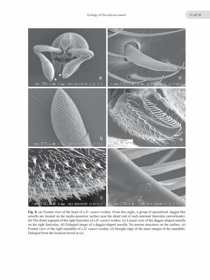
Fig. 8. (a) Frontal view of the head of a D. sauteri worker. From this angle, a group of specialized, dagger-like sensilla are located on the medio-posterior surface near the distal end of each antennal funiculus (arrowheads). (b) The distal segment of the right funiculus of a D. sauteri worker. (c) Lateral view of the dagger-shaped sensilla on the right funiculus. (d) Enlarged image of a dagger-shaped sensilla. No porous structures on the surface. (e) Frontal view of the right mandible of a D. sauteri worker. (f) Straight edge of the inner margin of the mandible. Enlarged from the location boxed in (e).
Keiichi Masuko12 of 16
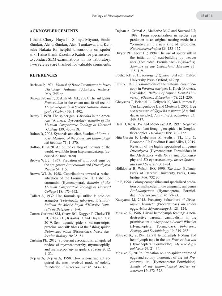
Fig. 10. (a) Posterior view of the 4th to 7th abdominal segments (marked as IV–VII) of a D. sauteri worker. The arrowhead points to the transverse groove on the tergite of the 4th abdominal segment. (b) Transverse groove on the 4th abdominal tergite. Arrowheads point to pores enlarged in (c) and (d).
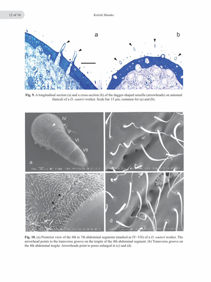
Fig. 9. A longitudinal section (a) and a cross-section (b) of the dagger-shaped sensilla (arrowheads) on antennal funiculi of a D. sauteri worker. Scale bar 15 μm, common for (a) and (b).

Ecology of Discothyrea sauteri 13 of 16
After retrieval, intriguingly, prey eggs and the ant immature brood were mixed together, and adults, especially the queens, remained upon such piles (Fig. 7b). Also, in the field, this place-ment of ant immatures together with prey eggs was usually observed. Worker behavior regarding prey eggs is of further interest. In the laboratory, D. sauteri workers frequently cleaned prey eggs (Fig. 7c, d). During this activity, an ant raised the anterior half of its body, standing on its middle and hind legs, while holding an egg at three points. The posterior end of the ant’s abdomen, curved anteriorly, supported the egg from behind, while its two thick, club-shaped antennal funiculi held it from the front (Fig. 7c, d, arrowheads). By this means, the prey egg was lifted off the sub-strate. The ant did not use its legs to support the egg, but instead it used its forelegs to continu-ously roll the egg while licking its surface with its lower mouthparts (Fig. 7c). After being offered as prey, stored spider eggs changed in appearance, becoming filled with droplets, which indicates changes in embryos inside eggs (see Discussion).
Thanatosis and adult transport
Being disturbed with forceps in the laboratory, D. sauteri workers immediately feign death, or perform thanatosis, by curling up in an immobile, compact shape. Also, when they confront other dead insects, they turn round, directing their gas-ters to the objects motionlessly. In D. oculata, recruitment of nestmates to spider eggs using a scent trail was observed (Dejean et al. 1999), while in D. sauteri, recruit-ment behavior was observed, but scent marking could not be confirmed. On the other hand, in the laboratory, adult transport was observed in D. sauteri, in which a worker (transporter) gaped widely and grasped the sides of the head of anoth-er worker (transportee) with its mandibles while the body of transportee curled over the head of transporter. This body placement by transporter and transportee seems very similar to that re-ported in Rhytidoponera metalica (Smith, 1858) (Möglich & Hölldobler 1974).
SEM and histological studies of worker anten-nae and abdomens
Apparently to facilitate egg handling, the apical segment of each antennal funiculus of a worker has a cluster of short, flat sensilla near its distal end (Fig. 8a, b). These sensilla are located me-dio-posteriorly (Fig. 8a, arrowheads), and firmly contact a prey egg when the ant holds and licks it (Fig. 7c, d). The sensilla are, more precisely, dagger-shaped (Fig. 8c, d), and their number on each side was approximately 50 (46, 47, 50, and 51; n = 4 workers). They are, however, absent from all other areas of the antennal surface (both funiculus and scape). Closer SEM examination at high magnification showed a complete absence of porous structures on the surface (Fig. 8d) and his-tological sections revealed that they are hollow inside (Fig. 9a, b). Still, they may also function as mechanoreceptors, though innervation was not evident in the sections. In contrast to the antennae, no special-ized structures such as setae, nor specialized sur-face structures of the integument, were observed on the dorsal area of the distal abdominal seg-ments that contact a prey egg (Figs. 7c and10a). However, the fourth abdominal segment, count-ing the propodeum as the first abdominal segment (Torre-Bueno 1989), has a transverse groove near the posterior edge of its tergite (Fig. 10a, b, arrowheads).In this groove, several pores align(Fig. 10b–d), and appear to be outlets of exocrine glands located beneath the integument. The pres-ence of such exocrine glands in the vicinity of the posterior margin of the 4th abdominal tergite has already been suggested for ants of the genus Pro-ceratium (Baroni Urbani & de Andrade 2003). Contents of these putative exocrine glands re-main totally unknown. Histological sectioning of D. sauteri worker abdomens revealed that there are tissues inside, which connect to the surface pores, thus being putatively secretory (Masuko, unpublished observation).

Keiichi Masuko14 of 16
DISCUSSION
There are quite a few studies reporting general predator-prey relationships between ants (preda-tors) and spiders (prey) (e.g., Vieira & Höfer 1994; Halaj et al. 1997; Nelson et al. 2004); however, in the Formicidae, predatory special-ization on either adult spiders or their eggs, is only known in Discothyrea (Hölldobler & Wil-son 1990; Dejean & Dejean 1998; Dejean et al. 1999). Recently, Cushing (2012) summarized spider-ant associations, citing many examples of predation on ants by spiders, but none for ant predation on spiders. For ants, spider eggs are undoubtedly excellent food sources because they are weakly chorionated, and thereby easy to con-sume, and they occur in large numbers in a single egg sac. However, spider egg sacs are difficult to cut open, because their silken sheets are firm and tough, and have multiple layers (Gheysens et al. 2005; Foelix 2011), so that special “tools” are needed. For this purpose, the masticatory margins of the mandibles in Discothyrea are edentate and rather straight (Fig. 8e, f), which contrasts with the dentate mandibles of Proceratium (Brown 1958, p.250; Baroni Urbani & de Andrade 2003, Table Ia, d; but see Hita-Garcia et al. (2019) for a few species of Discothyrea having dentate mandibles). Moreover, this masticatory border is “lined with a very even, close rank of minute peg-like bristles” (Brown 1958, p.250) (Fig. 8e), which might serve as a comb to arrange, or thin out, silk filaments. Similar comb-like structures are found on the tibial spurs of the forelegs of D. oculata (Dejean et al. 1999). How Discothyrea ants find egg sacs and retrieve them from the adult female spiders is completely unknown. Discothyrea oculata preys on spider eggs of the family Segestriidae (Dejean & Dejean 1998), which dwell in tubular webs constructed in crevices, within which they place their egg sacs (Preston-Mafham 2011). In the segestriid genus, Ariadna, females accom-pany egg masses and also young spiders for short periods after hatching (Beatty 1970, p. 437; Ya-makawa & Kumada 1973). Therefore, in order to retrieve segestriid eggs, Discothyrea ants must confront the defenses of female spiders in their burrows. Adult Discothyrea have tough, thick in-
teguments. Thanatosis, which had already been observed in other Discothyrea (Brown 1958, p.252; Katayama 2013), and the gaster-directing behavior might be used to deter spiders from at-tacking. In Proceratium, workers and queens have also been observed to turn around, using their vaulted gasters to block the entrance or pas-sageway to a nest (Poldi 1963; Masuko 2019b). The reason why nest cavities of Disco-thyrea are lined with exogenous silk remains un-known. In some cases, such silk linings may be inherited, if the ants simply adopt the shelter of a spider. However, transport of silk separated from egg sacs and its use in lining a glass tube were observed in D. oculata laboratory nests (Dejean et al. 1999); therefore, this elaboration must have certain advantages for nesting by Discothyrea. Beyond using silk as a lining, ants of the genus Polyrhachis Smith, 1857 use spider silk to con-struct arboreal nests (Collart 1932; Dwyer & Eb-ert 1994). Finally, comparisons of Discothyrea and Proceratium might be meaningful. First, although quantitative data are only available for Procerati-um itoi, members of this genus may be generalists for arthropod eggs (Masuko 2019b), whereas Dis-cothyrea specialize in spider eggs. In the cladistic analysis by Baroni Urbani & de Andrade (2003, Table I), important morphological differences be-tween these genera relate to the dentition of man-dibles and shapes of antennal funiculi. Both of these, i.e., edentate mandibles and enlargement of the apical antennal segment in Discothyrea, are related to predation on spider eggs. Second, Katayama (2013) reported changes in appearance of spider eggs stored by D. kamiteta Kubota & Terayama, 1999. Exactly the same phenomenon occurs in D. sauteri. This change may explain why hatchlings have not been obtained from eggs stored in nests of these two Discothyrea species. In contrast, hatchlings did emerge from eggs stored in nests of P. itoi (Masuko 2019b). Third, nutrition of Proceratium queens depends totally on hemolymph of their own larval progeny (Ma-suko 1986, 2019a), whereas this behavior, termed larval hemolymph feeding, has not been observed in Discothyrea; however, other nutritional sourc-es for Discothyrea queens remain unknown.
Ecology of Discothyrea sauteri 15 of 16
ACKNOWLEDGEMENTS
36: 1–492.
Baroni Urbani C, de Andrade ML, 2003. The ant genus Proceratium in the extant and fossil record.
Barbosa P, 1974. Manual of Basic Techniques in Insect Histology. Autumn Publishers, Amherst, MA, 245 pp.
Museo Regionale di Scienze Naturali Mono-grafe (Torino)
Beatty J, 1970. The spider genus Ariadna in the Amer-icas (Araneae, Dysderidae). Bulletin of the Museum Comparative Zoology at Harvard College 139: 433–518.
Bolton B, 2003. Synopsis and classification of Formic-idae. Memoirs of the American Entomologi-cal Institute 71: 1–370.
Bolton, B. 2020. An online catalog of the ants of the world. Available from https://antcat.org. (ac-cessed 27 June 2020)
Brown WL Jr, 1957. Predation of arthropod eggs by the ant genera Proceratium and Discothyrea. Psyche 64: 115.
Brown WL Jr, 1958. Contributions toward a reclas-sification of the Formicidae. II. Tribe Ec-tatommini (Hymenoptera). Bulletin of the Museum Comparative Zoology at Harvard College 118: 173–362.
Collart A, 1932. Une fourmis qui utllise la soie des araignées (Polyrhachis laboriosa F. Smith). Bulletin du Musée Royal d’Histoire Natu-relle de Belgique 8: 1–4.
Correa-Garhwal SM, Chaw RC, Dugger T, Clarke TH III, Chea KH, Kisailus D and Hayashi CY, 2019. Semi-aquatic spider silks: transcripts, proteins, and silk fibres of the fishing spider, Dolomedes triton (Pisauridae). Insect Mo-lecular Biology 28: 35–51.
Cushing PE, 2012. Spider-ant associations: an updated review of myrmecomorphy, myrmecophily, and myrmecophagy in spiders. Psyche 2012: 1–23.
Dejean A, Dejean A, 1998. How a ponerine ant ac-quired the most evolved mode of colony foundation. Insectes Sociaux 45: 343–346.
Masuko K, 2019b. Predation on non-spider arthropod eggs and colony bionomics of the ant Pro-ceratium itoi (Hymenoptera: Formicidae). Annals of the Entomological Society of America 12: 372–378.
Masuko K, 2019a. Larval hemolymph feeding and hemolymph taps in the ant Proceratium itoi (Hymenoptera: Formicidae). Myrmecologi-cal News 29: 21–34.
Masuko K, 1986. Larval hemolymph feeding: a non-destructive parental cannibalism in the primitive ant Amblyopone silvestrii Wheeler (Hymenoptera: Formicidae). BehavioralEcology and Sociobiology 19: 249–255.
Ito F, 1998. Colony composition and specialized preda-tion on millipedes in the enigmatic ant genus Probolomyrmex (Hymenoptera, Formici-dae). Insectes Sociaux 45: 79-83.
Katayama M, 2013. Predatory behaviours of Disco-thyrea kamiteta (Proceratiinae) on spider eggs. Asian Myrmecology 5: 121–124.
Hölldobler B, Wilson EO, 1990. The Ants. Belknap Press of Harvard University Press, Cam-bridge, MA, 732 pp.
Hita-Garcia F, Lieberman Z, Audisio TL, Liu C, Economo EP, Boudinot B and Mikó I, 2019. Revision of the highly specialized ant genus Discothyrea (Hymenoptera: Formicidae) in the Afrotropics with X-ray microtomogra-phy and 3D cybertaxonomy. Insect System-atics and Diversity 3: 1–84.
Halaj J, Ross DW and Moldenke AR, 1997. Negative effects of ant foraging on spiders in Douglas-fir canopies. Oecologia 109: 313–322.
Fujii Y, 1978. Examinations of the maternal care of co-coon in Pardosa astrigera L. Koch (Araneae, Lycosidae). Bulletin of Nippon Dental Uni-versity (General Education) (7): 221–230.
Gheysens T, Beladjal L, Gellynck K, Van Nimmen E, Van Langenhove L and Mertens J, 2005. Egg sac structure of Zygiella x-notata (Arachni-da, Araneidae). Journal of Arachnology 33: 549–557.
Dwyer PD, Ebert DP, 1994. The use of spider silk in the initiation of nest-building by weaver ants (Fomicidae: Formicinae: Polyrhachis). Memoirs of the Queensland Museum 37: 115–119.
Dejean A, Grimal A, Malherbe M-C and Suzzoni J-P, 1999. From specialization in spider egg predation to an original nesting mode in a “primitive ant”: a new kind of lestobiosis. Naturwissenschaften 86: 133–137.
I thank Cheryl Hayashi, Shinya Miyano, Eiichi Shinkai, Akira Shinkai, Akio Tanikawa, and Ken-suke Nakata for helpful discussions on spider silk. I also thank Kazuhiro Katoh for permission to conduct SEM examinations in his laboratory. Two referees are thanked for valuable comments.
REFERENCES Foelix RF, 2011. Biology of Spiders. 3rd edn. Oxford University Press, Oxford, 419 pp.
Keiichi Masuko16 of 16
ASIAN MYRMECOLOGYA Journal of the International Network for the Study of Asian Ants
Communicating Editor: Benoit Guénard
Miyawaki A, Ohba T and Murase N, 1969. Scientifc studies on the Mt. Hakone and PeninsulaManazuru, Kanagawa Prefecture. The Board of Education of the Kanagawa Prefecture, Yokohama, 59 pp. (Japanese)
Möglich M, Hölldobler B, 1974. Social carrying be-havior and division of labor during nest moving in ants. Psyche 81: 105–113.
Nelson XJ, Jackson RR, Pollard SD, Edwards GB and Barrion AT, 2004. Predation by ants on jumping spiders (Araneae: Salticidae) in the Philippines. New Zealand Journal of Zool-ogy 31: 45–56.
Poldi B, 1963. Alcune osservazioni sul Proceratium melinum Rog. e sulla fuzione della partico-lare struttura del gastro. Atti dell’Accademia Nazionale Italiana di Entomologia Rendi-conti 11: 221–229.
Moon MJ, 2003. Fine structural analysis of the cocoon silk production in the garden spider, Argiope aurantia. Korean Journal of Biological Sci-ences 7: 35–41.
Yamakawa M, Kumada K, 1973. Araneae in Tanzawa Mountains. Atypus (60): 31–54. (Japanese)
Vieira RS, Höfer H, 1994. Prey spectrum of two army ant species in central Amazonia, with special attention on their effect on spider popula-tions. Anrias 13: 189–198.
Torre-Bueno JR de la, 1989. The Torre-Bueno Glos-sary of Entomology: Revised Edition of a Glossary of Entomology (Compiled by S. W. Nichols; including Supplement A by G. S. Tulloch). The New York Entomological Society, New York, 840 pp.
Ward PS, Fisher BL, 2016. Tales of dracula ants: the evolutionary history of the ant subfam-ily Amblyoponinae (Hymenoptera: Formici-dae). Systematic Entomology 41: 683–693.
Preston-Mafham K, 2011. World of Animals 29: Arachnids (Edited and translated by J. Aoki, H. Ono and R. Fujimaki). Asakura Shoten, Tokyo, 118 pp. (Japanese)