Colon Screening Program · Colon Screening Program: ... This report was produced by the Colon...
25
Colon Screening Program Standards: Pathology version 7december2015
Transcript of Colon Screening Program · Colon Screening Program: ... This report was produced by the Colon...
Colon Screening ProgramStandards: Pathology
version 7december2015
Colon Screening Program: Pathology Standards
PATHOLOGY STANDARDS 7DECEMBER2015 2
Pathology Standards
Colon Screening Program
……………………………………………………………………
This report was produced by the Colon Screening Program at the BC Cancer Agency
Preferred citation:
BC Cancer Agency: Colon Screening Program Pathology Standards. BC Cancer Agency, 2015
For more information please contact:
Colon Screening Program
801-686 West Broadway
Vancouver, BC
V5Z 1G5
Web: www.screeningbc.ca/colon
Email: [email protected]
Phone: 1-877-70-COLON (26566)
Fax: 1-604-877-6103
Colon Screening Program: Pathology Standards
PATHOLOGY STANDARDS 7DECEMBER2015 3
Acknowledgements
The BC Cancer Agency (BCCA) would like to thank everyone who assisted in the development and refinement of the Colon Screening Program’s Pathology Specimen Handling and Reporting Standards.
Co-authors
Dr. David A. Owen, Vancouver Coastal Health Authority Dr. Chen Zhou, BC Cancer Agency
Contributors
Dr. Amir Rahemtulla, BC Cancer Agency Laura Gentile, BC Cancer Agency Ken Winning, Northern Health Authority Dr. Grant Roden, Northern Health Authority Dr. James Kelly, Vancouver Island Health Authority Dr. Doug Hardy, Interior Health Authority Dr. Peter Zetler, Fraser Health Authority
It has been the innovative and transforming work of the Colon Screening Program’s Pathology Working Group that inspired and informed the development of these standards. We would also like to thank the management of the BCCA Screening Programs, the BCCA executive sponsors of the Colon Screening Program, the Provincial Health Services Authority (PHSA), the Ministry of Health, the BC Society of Gastroenterology, the BC Surgical Society, the Society of General Practitioners of BC, the BCCA Gastrointestinal Tumour Group, and the Colon Screening Program Advisory Committee.
About the BC Cancer Agency
The BC Cancer Agency, an agency of the Provincial Health Services Authority, provides a comprehensive cancer control program for the people of BC in partnership with regional health authorities. This includes prevention, screening and early detection programs, research and education, and care and treatment.
The BC Cancer Agency’s mandate is a three-fold mission:
• To reduce the incidence of cancer
• To reduce the mortality rate of people with cancer
• To improve the quality of life of people living with cancer
This mission drives everything we do, including providing screening, diagnosis and care, setting treatment standards, and conducting research into causes of, and cures for, cancer.
Colon Screening Program: Pathology Standards
PATHOLOGY STANDARDS 7DECEMBER2015 4
Table of Contents
Introduction 1.1 Colon Screening Program 5 1.2 Purpose of the Standards 5 1.3 Sources of Information 5 1.4 General Principles 6
Protocol for 2.1 Clinical Information 7 Handling Specimens
Gross Description 3.0 Gross Description and Dissection 8 and Dissection
Technical Procedures 4.0 Technical Procedures 9
Diagnosis and 5.1 Location 10 Reporting 5.2 Specimen Type 10 5.3 Histologic Classification 10 5.4 Degree of Dysplasia 16 5.5 Completeness of Removal 16
Required Reporting 6.1 Carcinoma Within Polyp 18 for Colorectal Polyps 6.2 Reporting of Resected Colon for 19 Carcinoma 6.3 Diagnosis of Tumour Budding 20
Overview of Key 7.1 Based on Pathology Report 22 Performance 7.2 Based on Pathology Review 22 Indicators
Appendix A Protocol for Handling a Polyp Specimen 23 B Sample Pathology Report 24
Colon Screening Program: Pathology Standards
PATHOLOGY STANDARDS 7DECEMBER2015 5
1. Introduction
1.1 Colon Screening Program
Colorectal cancer (CRC) is the third most common cancer and the second leading cause of cancer death in Canada and in other developed countries. The primary goal of the Colon Screening Program is to detect and remove colonic adenomas that are the precursor lesion of CRC. Detection and resection of asymptomatic CRC at an early clinical stage will be an added benefit. Ultimately, this will reduce the incidence and mortality from CRC.
Pathologists who report colorectal polyps/lesions detected by colonoscopy in patients from a CRC screening program provide not only the pathologic diagnosis but also key information for patient surveillance and management decisions. Standardized identification and classification of colon polyps as well as other precursor lesions of CRC is essential. Similarly, the standardized handling and reporting of colectomies for invasive cancer is essential. By adopting a province-wide uniform specimen handling and reporting protocol, information needed for a multi-site screening program will be provided. The protocols outlined below will permit information gathering and comparisons of results across Canada and internationally. The protocols listed below are not unique or complex. For most pathologists and laboratories, they will be the same as the protocols and diagnoses already in use.
1.2 Purpose of the Standards
The purpose for developing a pathology reporting guideline for colorectal screening program is:
To eliminate variability in diagnosis and nomenclature of colon polyps
To include relevant information needed for patient surveillance
To include key information for patient management if a malignant polyp is diagnosed.
1.3 Sources of Information
This protocol is developed based on the current pathology literature identified from:
Medline
Pathology Breakout Summary for the National Colorectal Screening Program Quality Determinants Workshop, Vancouver, BC, May 22-23, 2008
Quality Determinant Framework for Colorectal Screening in Canada, Toronto, April 2009
The Reporting Lesions in the NHS Bowel Cancer Screening Program: Guidelines from the bowel cancer screening program pathology group, NHS BCSP Publication
Colon Screening Program: Pathology Standards
PATHOLOGY STANDARDS 7DECEMBER2015 6
No.1, Sept, 2007, Surgical Pathology Minimal Reporting Guideline 2008: British Columbia Association of Laboratory Physician (BCALP, April 2008)
Protocol for the Examination of Specimens From Patients With Primary Carcinoma of the Colon and Rectum 2013 from the College of American Pathologists (CAP, Oct 2013).
Pathological reporting of colorectal polyps. Pan-Canadian consensus guidelines. Can J Pathol 2012;4:81-90.
1.4 General Principles
Quality assurance is an essential component of a population-based screening program, and measurements of quality should be applied to all participating laboratories. Uniform provincial standards provide the opportunity to monitor system performance and patient outcomes in a way that supports comparison and learning across jurisdictions.
Consistency in reporting will help to ensure meaningful systems performance and patient outcomes monitoring, and will assist physicians in determining appropriate re-call intervals for screening patients.
A centralized consultation service is available for individual pathologists to refer complex and difficult cases prior to final diagnosis.
Quality assurance is a process of education, consultation and collegiality that will optimize patient outcomes.
Laboratory Standards
All participating laboratories must be DAP Accredited. Laboratories must comply with reporting standards as outlined in this document.
All pathology reports originating from screening patients will be submitted to a central registry that can be assessed by an individual(s) charged with the responsibility for implementing province-wide pathology performance indicators. Selected pathology slides and reports will be made available for forwarding to individuals charged with the responsibility for implementing Pathology Performance Indicators.
Participating Pathologist Standards
It is anticipated that all pathologists participating in the Colon Screening Program will already have experience in the diagnosis of colonic polyps. Pathologists are expected to read the Canadian Guidelines on polyp reporting and to familiarize themselves with uniform diagnostic terminology. The training module with self-assessment is also required.
Colon Screening Program: Pathology Standards
PATHOLOGY STANDARDS 7DECEMBER2015 7
2. Protocol for Handling Pathology Specimens
Colonoscopists will use the Colonoscopy Reporting Form (Appendix X) when documenting the colonoscopy procedure. The Colonoscopy Reporting Form functions as the pathology requisition form. This will provide detailed information to the pathologist regarding each specimen that was removed: size, location, method of removal and whether the specimen was retrieved. The Colonoscopy Reporting Form will also identify patients who are participating in the program to ensure the following pathology reporting is completed for program participants.
The full protocol for handling specimens is outlined in Appendix A.
2.1 Clinical Information
Patient identification required is similar to all other pathology specimen. This must include the patient’s full name, sex, date of birth, personal health number (PHN), name of submitting physician and name of family physician.
In addition, other relevant information must include the location within the colon and the size of the polyp. A clinical note describing the clinical impression of the completeness of removal is desirable. A colonoscopy reporting form which includes this information can be copied and used as a pathology requisition. It is not necessary to fill out a designated pathology requisition.
Multiple polyps should not be placed in a single container. They must be submitted separately and identified as to site. The container should contain adequate (ideally, 10 times the volume of the specimen) 10% formalin to ensure appropriate fixation. Each container should have an outside label containing patient name, date of birth and location of the lesion. Once the specimen is placed in formalin, there is no need to transmit it to the laboratory in an urgent fashion. Nevertheless, reducing the time a specimen is in transit will speed up the delivery of the pathology report. Do not freeze the specimens. Refrigeration is not required.
Colon Screening Program: Pathology Standards
PATHOLOGY STANDARDS 7DECEMBER2015 8
3. Gross Description and Dissection
The specimen should be examined and dissected by a pathologist or a suitably qualified deputy. An anatomical pathology technologist with training and experience in processing pathology specimens is qualified to process the biopsy or polyp specimen. Processing of large polyps or complex resection specimens may be done by a pathologist, pathology resident or Pathologists’ Assistant.
In addition to recording any clinical information, the number, size and gross appearance of the biopsies or polyps should be described. It is of particular importance to note the configuration of a polyp (polypoid or villous) and the presence of a stalk (measure its length and diameter). If the polyp is small or consists of small fragments (< 3-5 mm), it can be submitted in toto without further dissection. If the polyp is larger than 5 mm and smaller than 10 mm, it can be bisected perpendicular to the biopsy margin or base of stalk. Polyps that are larger than 10 mm and have a stalk should be cut longitudinally in 3 mm sections leaving the central section containing an intact stalk. Before dissection of any polyp, the biopsy margin at the base of the stalk should be inked. The whole of a small polyp or each section of a larger polyp should be placed separately in a unique cassette for embedding.
Colon Screening Program: Pathology Standards
PATHOLOGY STANDARDS 7DECEMBER2015 9
4. Technical Procedures
Following description and dissection, the specimen should be processed, embedded and sectioned in the usual manner. Sections that are 6 microns or less in thickness are suggested. A minimum of two levels should be obtained with the deepest level approximating to the mid portion of the tissue block. Note that serial sections from the superficial surface of the block are not considered to be equivalent to levels. Obviously, polyps that present diagnostic difficulty may require deeper or serial sections. All polypectomy specimens showing no abnormality on the initial levels should get deeper levels if the findings would be clinically relevant. Routine hematoxylin and eosin staining procedures that are used in the laboratory will suffice.
Colon Screening Program: Pathology Standards
PATHOLOGY STANDARDS 7DECEMBER2015 10
5. Diagnosis and Reporting
The format of the report should be completed in a consistent way, using standardized terminology. Examples of standardized reports are available in Appendix B. The report should include, in order, the following five key diagnostic features:
Location
Type of specimen (polypectomy or biopsies)
Histologic classification (see descriptions in Section 5.2)
Degree of dysplasia (when any adenoma or sessile serrated adenoma/polyp is diagnosed)
Completeness of removal (when appropriate)
Using standardized terminology does not preclude the pathologist from adding any other type of description in the free text. Rarely, added free text is desirable to clarify or expand on the standardized terminology that is required for the histologic classification.
5.1 Location
This information will have to be obtained from the requisition provided by the referring physician. The following sites within the colon may be identified: cecum, ascending colon, hepatic flexure, transverse colon, splenic flexure, descending colon, sigmoid colon and rectum.
5.2 Specimen Type
Specimens are either biopsies or polypectomies as reported by the endoscopist.
An adenoma measuring 10 mm or larger is considered an “advanced adenoma”. Patients with these lesions need more frequent clinical follow up and 3 year surveillance. Therefore, every effort should be made to determine an accurate measurement in an adenoma that is close to this critical size. If the microscopic measurement differs from the gross measurement the pathologist can include the microscopic measurement in the microscopic diagnostic report when appropriate.
5.3 Histologic Classification
The histologic classification must be selected from one of the possible diagnoses provided in the list below. If the polyp type does not appear on this list, the specimen must be classified as “other”. Further explanation about the histologic classification and explanation of the “other” diagnosis can be described in the free text comment section of the report.
Colon Screening Program: Pathology Standards
PATHOLOGY STANDARDS 7DECEMBER2015 11
Endoscopists may occasionally submit polyps that histologically consist of entirely normal large bowel mucosa. These biopsies should be diagnosed as “normal mucosa” although a comment may be added to the free text portion of the report indicating that they may represent a prominent mucosal fold.
From time to time, polyps will be encountered where the diagnosis is problematic. These may be submitted to a reference centre(s) for consultation.
A. Adenoma
Tubular adenoma (can contain up to 25% of villous component)
Tubulovillous adenoma (contains 25-75% villous component)
Villous adenoma (contains > 75 % villous component)
Flat adenoma (variant of tubular adenoma)
Sessile serrated adenoma/polyp
Traditional serrated adenoma
Mixed hyperplastic-adenomatous polyps
Adenoma with pseudoinvasion / misplaced epithelium/ torsion effect
Hyperplastic polyp
B. Other benign polyps
Juvenile polyp
Peutz-Jeghers polyp
Polypoid mucosal prolapse
Inflammatory polyp
Lymphoid polyp
Mesenchymal polyp (includes lipoma, leiomyoma etc.)
Other polyps
C. Malignant and potentially malignant polyps
Carcinoma within polyp (“malignant polyp”)
Invasive carcinoma
Neuroendocrine tumour (“carcinoid”)
Hyperplastic Polyps
Hyperplastic polyps are typically small and measure less than 5 mm in diameter. They are most common in the distal colon. The colonic crypts are elongated and contain reduced numbers of goblet cells with excess columnar absorptive cells. A variable degree of
Colon Screening Program: Pathology Standards
PATHOLOGY STANDARDS 7DECEMBER2015 12
proliferation may be seen at the crypt base, but the cells are regular with no cytologic dysplasia. The superficial portion of the crypts shows a serrated appearance. The architecture at the base of the crypts is U-shaped.
Polyps that superficially resemble hyperplastic polyps but are greater than 5 mm in diameter and present in the proximal colon may be sessile serrated adenoma/polyps (SSA/SSP) (see Section 2.2.9 below).
The Colon Screening Program does not require that hyperplastic polyps be subtyped into microvesicular, goblet cell rich or mucin poor subtypes. However, this information may be added to the free text portion of the report.
High Risk Polyps
High risk polyps or advanced adenoma are polyps with: a) villous features, b) high grade dysplasia, c) size > 10mm as measured by the colonoscopist at the time of excision, e) sessile serrated adenoma/polyp >10mm in diameter, e) sessile serrated adenoma/polyps with cytologic dysplasia, and f) traditional serrated adenoma. Patients with three or more low risk adenomas are also considered as “high risk”.
Adenoma
By definition all colonic adenomas (with the exception of sessile serrated adenoma/polyp) consist of dysplastic epithelium and are classified as benign neoplasms. Depending on the extent of the villous component, an adenoma may fall into one of three subtypes: tubular, villous or tubulovillous.
The WHO system classifies a tubular adenoma as having less than a 20% villous component; tubulovillous adenoma as having a 20-80% villous component; and villous adenoma as having more than an 80% villous component. This 20/80 rule has been adopted by the British NHS CRC screening program. However, the Colon Screening Program proposes to use a 25/75 rule to separate the three types of adenoma. This rule is widely used in North America and has been endorsed by the National Colorectal Screening Program Pathology Workshop. Using the 25/75 separating rule rather than the 20/80 rule set by WHO will ensure consistent classification of adenoma by pathologists throughout North America and will allow comparison of data across Canadian provinces.
Exactly what constitutes a villus is difficult to define. Three forms are recognized: classical villi, palmate villi and foreshortened villi.
Classical villi are composed of slender up-growths of epithelium on a thin non-branching stromal core. Typically they have parallel sides. When sampled as a complete longitudinal section their base extends down to the muscularis mucosae. The tip may be pointed or bulbous.
Palmate villi resemble the morphology of a palm tree. They are composed of broader branching leaf-like structures. Tubular glands may be present at the base and even within the stromal core.
Foreshortened villi are slender non-branching extensions that protrude from the surface of an otherwise typical tubular adenoma.
Colon Screening Program: Pathology Standards
PATHOLOGY STANDARDS 7DECEMBER2015 13
Flat or depressed adenomas are rare. As the name suggests, they are not really polypoid in configuration but are disc-shaped. They are typically about 10mm in diameter. Histologically, they are a tubular adenoma variant (Soetikno R, Friedland S, Caltenbach T, et al. Gastroenterology 2006;130:566-576). Correctly identifying flat adenoma is desirable but not critical for screening patients as this does not influence patient follow up protocols. Small adenomas containing only a few dysplastic crypts or fragmented small adenoma are sometimes difficult to classify into one of the three morphologic subtypes.
In the past, some pathologists have classified these as “adenomatous polyp”. For the purpose of Colon Screening Program data management, it is necessary to classify these “adenomatous polyps” into one of the three adenoma subtypes. If the dysplastic focus just consists of tubules, it should be diagnosed as tubular adenoma. If the dysplastic epithelium contains villous component greater than 25%, then it should be classified as tubulovillous adenoma. Adenomas with villous features are considered high risk and have a shorter (3 year) surveillance interval.
Adenoma with Misplaced Epithelium (“pseudoinvasion”)
Epithelial misplacement (pseudoinvasion) is relatively common in larger adenoma particularly those that have a long stalk and those present in the sigmoid colon. Misplacement is usually considered to be secondary to polyp torsion and ulceration with re-growth of epithelium below the level of the muscularis mucosae. Obviously this may be confused with early invasive adenocarcinoma.
Histologic features that may help in distinguishing misplaced epithelium from carcinoma include:
the presence of lamina propria surrounding the misplaced crypts,
a degree of dysplasia in the misplaced crypts that is similar to the dysplasia in adjacent non-displaced crypts,
hemosiderin in the polyp stroma
mucin pools in the stroma that may be associated with surviving attenuated epithelial elements.
In cases of diagnostic difficulty, the slides may be referred to a reference centre for consultation.
Traditional Serrated Adenoma (TSA)
Traditional serrated adenomas are polyps with a serrated architecture but with crypts lined by cells with dysplastic nuclei. They are therefore an adenoma variant, not a hyperplastic polyp variant. Generally this type of polyp demonstrates only low-grade dysplasia. Nevertheless, the degree of dysplasia present should be recorded. Previously, these polyps had several other names: serrated adenoma; sessile serrated adenoma with dysplasia and tubular adenoma with overt serrated features. The terminology “traditional serrated adenoma” is now preferred.
Colon Screening Program: Pathology Standards
PATHOLOGY STANDARDS 7DECEMBER2015 14
Sessile Serrated Adenoma/Polyp (SSA/SSP)
These polyps have only recently been described and consequently the full range of their morphologic appearance is as yet, incompletely described and understood. Some evidence suggests that they are the precursors of colon cancers that demonstrate microsatellite instability (MSI). Sessile serrated polyps/adenoma show a unique type of architectural dysplasia but most examples do not contain “classical” cytologic dysplasia. Superficially, they resemble hyperplastic polyps but may be distinguished from them by their architectural abnormalities. These consist of irregular crypts, dilated crypts and serrated crypts present in the bottom half of the mucosa. The proliferative zone at the base of the crypts may extend half way up the crypt length (normally the proliferative zone is one third or less of the crypt length). Crypt irregularity often consists of a lateral proliferation of the crypts along the muscularis mucosae (boot-shaped crypts). These appearances should be contrasted with the smoothly rounded, non-dilated tubular crypt base that characterizes hyperplastic polyps. The superficial portion of the crypts in sessile serrated polyps/adenoma and hyperplastic polyps are similar morphologically.
Clinical features such as large size (≥10 mm) and right-sided location within the colon are more commonly seen in sessile serrated adenoma but these features are not by themselves diagnostic.
At the present time, sessile serrated adenoma/polyp may be sub-classified as demonstrating no dysplasia, low-grade dysplasia or high-grade dysplasia. Sessile serrated adenoma/polyp with dysplasia is considered high risk and has a shorter (3 year) surveillance interval than sessile serrated adenoma/polyp with no dysplasia (5 year surveillance interval).
Mixed Hyperplastic Polyp/Adenoma
These polyps are regarded as being entirely separate but physically adjacent lesions that have grown together. They represent an intermingling of polyp types. The components of mixed polyps should be separately identified and reported in the free text portion of the report. Be careful: do not confuse a mixed polyp with a sessile serrated adenoma with dysplasia. The follow-up of patients with this type of polyp will depend on the microscopic features of the adenomatous component.
Neuroendocrine Tumours
Many of these tumours were previously called carcinoid tumours. The term “carcinoid tumour” is now replaced by “low-grade neuroendocrine tumour”. The majority of these neoplasms will be present in the rectum. High-grade neuroendocrine carcinomas are rare in the colon. They can be subdivided into large cell and small cell types. TNM staging of neuroendocrine tumours is available and should be quoted in pathology reports (TNM Classification of Malignant Tumours, 7th Edition 2009, page 98).
Hamartomas
Most hamartomatous bowel polyps are either Peutz-Jeghers polyps or juvenile polyps (retention polyp). If a Peutz-Jeghers polyp is diagnosed, then a clinical search for other features of the syndrome should be undertaken. It may also be advisable to test the patient
Colon Screening Program: Pathology Standards
PATHOLOGY STANDARDS 7DECEMBER2015 15
for the germ-line mutations that typically characterize this condition. The majority of juvenile polyps are isolated and sporadic. Only rarely is juvenile polyposis present.
Inflammatory Polyps
There are a wide variety of types of inflammatory polyps. These range from pseudopolyps found in inflammatory bowel disease to isolated granulation tissue polyps complicating diverticular disease. Sub-classification of these polyps is not required by the Colon Screening Program. Nevertheless sub-classification should be described in the comment section of the report when it is of clinical relevance.
Mesenchymal Polyps
A wide range of other types of mesenchymal polyp may be encountered. These include lipomas, leiomyomas, ganglioneuromas, gastrointestinal stromal tumour and vascular lesions. The diagnosis of one of these polyps does not generally trigger ongoing patient screening.
Others
There are many other types of colonic polyp although most of these are rare. The usual diagnostic criteria will apply.
Adenoma Containing an Invasive Carcinoma (‘malignant polyp’)
In order for carcinoma to be present within an adenoma, there must be definite evidence of a carcinoma invading into the submucosa. Carcinoma-like glands involving only the epithelium and lamina propria are referred to as high-grade dysplasia, rather than intramucosal carcinoma. As far as colonic polyps are concerned, the terms “carcinoma in situ” and “intramucosal carcinoma” should not be used as they are clinically misleading and may lead to inappropriate surgical over-treatment. Malignant polyps are often removed during colonoscopy without their malignant nature being apparent. Detailed reporting guidelines for this type of polyp are presented in Section 2.6. These are based on five criteria: the depth of invasion (polyp head or stalk), proximity to the resection margin (distance measured in millimeters), presence or absence of lympho-vascular invasion, histologic grade of the carcinoma and presence or absence of high grade tumour budding.
Suspicious for Carcinoma
For cases where high-grade dysplasia is present but unequivocal identification of submucosal invasion is lacking, report the diagnosis as an adenoma with high-grade dysplasia. Further description outlining the suspicion of carcinoma can be described in the free text component of the report.
Colon Screening Program: Pathology Standards
PATHOLOGY STANDARDS 7DECEMBER2015 16
5.4 Degree of Dysplasia
Adenoma may be either low-grade or high-grade. Adenoma with intermediate grades of dysplasia (moderate dysplasia) are now included in the low-grade category. The reason for this is that their clinical behavior and prognostic significance are similar to adenoma with low-grade dysplasia. Therefore, the majority of adenomas detected will have low-grade dysplasia. High-grade dysplasia should only be diagnosed in adenoma that display both high-grade cytologic atypia and high-grade structural complexity. These high-grade changes must involve more than two crypts.
High-grade cytologic dysplasia is characterized by marked elongation and enlargement of nuclei extending to involve more than 65% of the cell cytoplasm, loss of polarity and nuclear stratification, a dispersed chromatin pattern with prominent nucleoli, atypical mitoses and prominent apoptosis that gives the epithelium a “dirty” appearance.
High-grade architectural dysplasia is characterized by complex crypt crowding and irregularity, prominent budding, a cribriform appearance with “back to back” tubules (no lamina propria between crypts) and intraluminal tufting.
It should be recognized that low-grade and high-grade dysplasia are not completely separate entities and that some polyps will be encountered that have features straddling the above descriptions. Architectural high-grade dysplasia is usually obvious on low-power microscopic examination of the slide. Beware of confusing crush artifact with high-grade dysplasia. Crushing by biopsy forceps can produce an artificial apposition of glands as well as induce loss of polarity and nuclear stratification on the surface of an adenoma that otherwise shows only low-grade dysplasia. In cases of diagnostic difficulty, the slides may be referred to the reference centre for consultation.
The National Colorectal Screening Pathology Workshop recommends describing dysplasia as either:
negative for high grade dysplasia; or,
positive for high grade dysplasia.
Adenomas with high grade dysplasia are considered high risk and have a shorter (3 year) surveillance interval.
5.5 Completeness of Removal
Completeness of removal of neoplastic polyps especially those polyps defined as advanced adenoma should be reported if this is possible. This is particularly important in cases where a carcinoma is present within the adenoma (“malignant polyp”). However, assessment of incomplete removal is primarily endoscopic rather than pathologic. Reporting of completeness of excision is only required for high grade risk lesions. The default position is that it cannot be assessed.
Colon Screening Program: Pathology Standards
PATHOLOGY STANDARDS 7DECEMBER2015 17
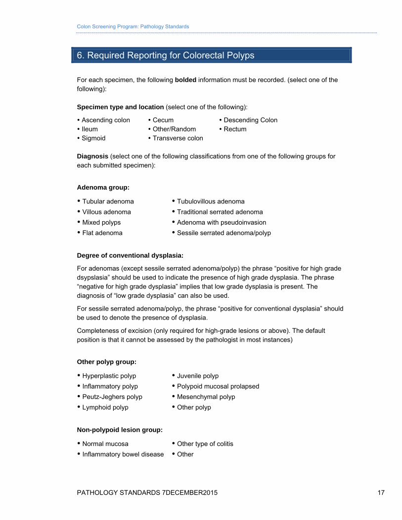
6. Required Reporting for Colorectal Polyps
For each specimen, the following bolded information must be recorded. (select one of the following): Specimen type and location (select one of the following):
Ascending colon Cecum Descending Colon Ileum Other/Random Rectum Sigmoid Transverse colon Diagnosis (select one of the following classifications from one of the following groups for each submitted specimen):
Adenoma group:
Tubular adenoma Tubulovillous adenoma
Villous adenoma Traditional serrated adenoma
Mixed polyps Adenoma with pseudoinvasion
Flat adenoma Sessile serrated adenoma/polyp
Degree of conventional dysplasia:
For adenomas (except sessile serrated adenoma/polyp) the phrase “positive for high grade dsypslasia” should be used to indicate the presence of high grade dysplasia. The phrase “negative for high grade dysplasia” implies that low grade dysplasia is present. The diagnosis of “low grade dysplasia” can also be used.
For sessile serrated adenoma/polyp, the phrase “positive for conventional dysplasia” should be used to denote the presence of dysplasia.
Completeness of excision (only required for high-grade lesions or above). The default position is that it cannot be assessed by the pathologist in most instances)
Other polyp group:
Hyperplastic polyp Juvenile polyp
Inflammatory polyp Polypoid mucosal prolapsed
Peutz-Jeghers polyp Mesenchymal polyp
Lymphoid polyp Other polyp
Non-polypoid lesion group:
Normal mucosa Other type of colitis
Inflammatory bowel disease Other
Colon Screening Program: Pathology Standards
PATHOLOGY STANDARDS 7DECEMBER2015 18
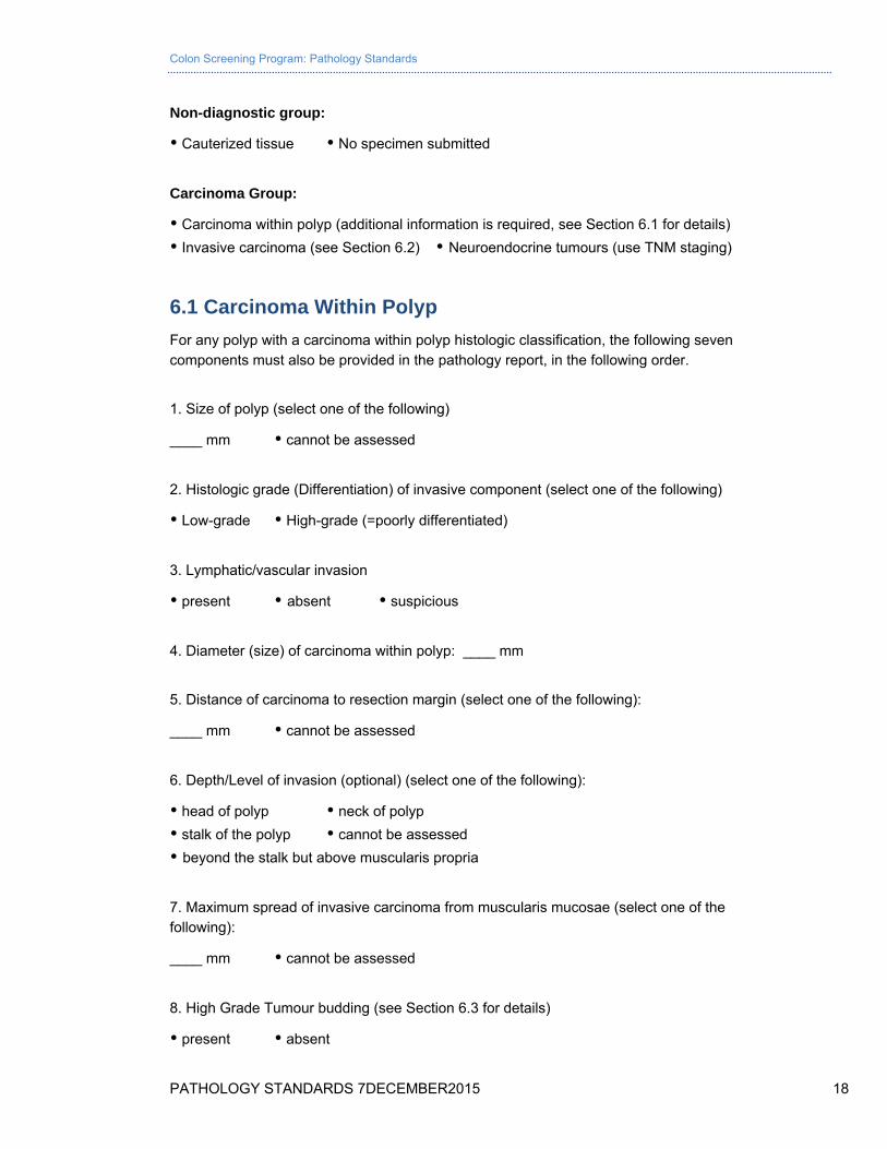
Non-diagnostic group:
Cauterized tissue No specimen submitted
Carcinoma Group:
Carcinoma within polyp (additional information is required, see Section 6.1 for details)
Invasive carcinoma (see Section 6.2) Neuroendocrine tumours (use TNM staging)
6.1 Carcinoma Within Polyp
For any polyp with a carcinoma within polyp histologic classification, the following seven components must also be provided in the pathology report, in the following order.
1. Size of polyp (select one of the following)
____ mm cannot be assessed
2. Histologic grade (Differentiation) of invasive component (select one of the following)
Low-grade High-grade (=poorly differentiated)
3. Lymphatic/vascular invasion
present absent suspicious
4. Diameter (size) of carcinoma within polyp: ____ mm
5. Distance of carcinoma to resection margin (select one of the following):
____ mm cannot be assessed
6. Depth/Level of invasion (optional) (select one of the following):
head of polyp neck of polyp
stalk of the polyp cannot be assessed
beyond the stalk but above muscularis propria
7. Maximum spread of invasive carcinoma from muscularis mucosae (select one of the following):
____ mm cannot be assessed
8. High Grade Tumour budding (see Section 6.3 for details)
present absent
Colon Screening Program: Pathology Standards
PATHOLOGY STANDARDS 7DECEMBER2015 19
6.2 Reporting Of Resected Colon for Carcinoma
Minimum Reporting Guidelines – Colon, Rectum
Microscopic Diagnosis: Colon (specific site), rectum, resection (specify type):
a) Positive for: adenocarcinoma; mucinous (colloid) carcinoma*; signet ring cell carcinoma*; “medullary” carcinoma** * mucinous or signet ring cell components comprise >50% of tumour ** high-grade tumours with a medullary growth pattern and numerous tumour infiltrating lymphocytes occurring in patients under 50 years of age are closely associated with hereditary non polyposis colon cancer syndromes and tumour cell microsatellite instability, with implications for family screening and chemotherapy response. These features should be noted if present
b) Tumour site (for rectal tumours, specify if above or below anterior peritoneal reflection).
c) Tumour grade (low grade [well to moderately differentiated]; high grade (poorly differentiated to undifferentiated)
d) Greatest linear tumour dimension (cm) and fraction of bowel circumference involved by tumour
e) Tumour extension (lamina propria, submucosa, muscularis propria, pericolic subserosal/perirectal adipose tissue, peritonealized serosa, adjacent organs or structures)
f) Surgical margins (proximal, distal, radial). Measure closest approach of tumour to radial margin in mm (direct tumour extension within 1 mm or a positive lymph node at the radial margin are defined as “positive” margin)
g) Completeness of mesorectal excision specimen for rectal tumours (essentially complete with minimal defects/partially complete/ incomplete with exposure of rectal muscularis propria)
h) Lymph-vascular invasion (present/absent, define if intramural or extramural)
i) Perineural invasion (present/absent)
j) Extramural tumour deposits (present/absent) — A tumour nodule in the pericolic/perirectal fat without evidence of residual lymph node tissue is classified as a tumour deposit and is not considered a positive lymph node. Such tumour deposits may represent discontinuous spread, lymphvascular spread with extravascular extension, or totally replaced lymph nodes. In the absence of unequivocal lymph node metastases, tumour deposits are recorded as N1c
k) Lymph node status (x of y lymph nodes positive) ***A minimum of 12 lymph nodes are required to accurately predict pNO stage***
l) Perforation-macroscopic (present/absent)
m) Treatment effect, if applicable (complete, moderate or minimal) response
n) Status of non-carcinomatous mucosa (adenoma, IBD, known hereditary polyposis syndromes) and bowel wall
Colon Screening Program: Pathology Standards
PATHOLOGY STANDARDS 7DECEMBER2015 20
o) pTNM tumour stage (AJCC 7th edition)
Primary Tumour (T)
Tis- Tumour invades lamina propria
T1- Tumour invades submucosa
T2- Tumour invades muscularis propria
T3- Tumour invades colonic subserosa/ nonperitonealized perirectal tissues
T4a- Tumour penetrates visceral peritoneum (tumour present at the serosal surface with inflammatory reaction, mesothelial hyperplasia or erosion; or free tumour cells on the serosal surface with underlying erosion of the visceral peritoneum)
T4b- Tumour directly invades or is adherent to other organs or structures
Regional Lymph Nodes
NX- Regional lymph nodes cannot be assessed
N0- No regional lymph node metastasis
N1a- Metastasis in 1 regional lymph node
N1b- Metastases in 2 or 3 regional lymph nodes
N1c- Tumour deposits in subserosa or pericolic/ perirectal soft tissues without regional lymph node metastasis
N2a- Metastases in 4 to 6 regional lymph nodes
N2b- Metastases in 7 or more regional lymph nodes
Distant Metastasis
M1a- Metastasis to a single organ or site
M1b- Metastases to more than one site or to the peritoneum
6.3 Diagnosis of Tumour Budding
High grade tumour budding has been shown to be a prognostic indicator of lymph node metastasis in pT1 lesions. Tumour budding has been defined by some as an isolated single cancer cell or a cluster composed of fewer than five cancer cells. In the largest study to date examining 292 colorectal carcinomas with submucosal invasion (pT1), 16/38 (42%) tumours in which tumour budding was observed were associated with synchronous lymph node metastases (1). Although in this study only 41 of the tumours were resected by local resection and the average tumour size varied from
1.34 to 2.24 cm 1, 2.
Colon Screening Program: Pathology Standards
PATHOLOGY STANDARDS 7DECEMBER2015 21
At present, we define a tumour bud as up to 5 tumour cells at the invasive front separate from the main tumour mass. High grade tumour budding is present if > 10 buds/ 20 hpf are present at the invasive front. This increases the risk of regional lymph node metastasis to approximately 15-25%.
(1) Ueno H, Mochizuki H, Hashiguchi Y et al. Risk factors for an adverse outcome in early invasive colorectal carcinoma. Gastroenterology 2004; 127; 385–394.
Colon Screening Program: Pathology Standards
PATHOLOGY STANDARDS 7DECEMBER2015 22
(2) Mitrovic B., Schaffer D.F., Riddell R.H., Kirsch R., Tumour budding in colorectal carcinoma: time to take notice. Mod Pathol 2012; 25: 1315-1325
7. Pathology Review
7.1 Pathology Review by the Health Authority
It is expected that pathology review for the purposes of quality assurance is undertaken by the Health Authority on a random sampling of cases as well as specific difficult diagnoses. It is recommended that this be performed externally and include assessment of diagnosis accuracy as well as completeness of the pathology report (see section 6. Required Reporting for Colorectal Polyps).
7.2 Pathology Review by the Colon Screening Program
Annual pathology quality reports will be generated and distributed to pathologists participating in the Colon Screening Program individually and in aggregate form at a Health Authority and provincial level. This audit will include the following:
Total number of Colon Screening Program specimens reported
Number of neoplasms reported
Number of polyps with high grade dysplasia reported
Number of sessile serrated adenoma/polyps reported with/without dysplasia
Number of traditional serrated adenomas reported
Number of hyperplastic polyps arising from the ascending colon or cecum reported
Number of polyps with pseudoinvasion reported
Number of malignant polyps reported
Annual audit of all reports of malignant polyps will be undertaken to assess for compliance with reporting of high-risk features.
Colon Screening Program pathology audits will be reviewed annually by a committee consisting of pathology representatives from each Health Authority, the Health Authority Colonoscopy Leads, and the Colon Screening Program Medical Director.
Colon Screening Program: Pathology Standards
PATHOLOGY STANDARDS 7DECEMBER2015 23
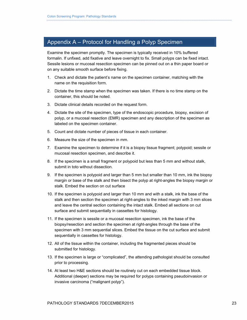
Appendix A – Protocol for Handling a Polyp Specimen
Examine the specimen promptly. The specimen is typically received in 10% buffered formalin. If unfixed, add fixative and leave overnight to fix. Small polyps can be fixed intact. Sessile lesions or mucosal resection specimen can be pinned out on a thin paper board or on any suitable smooth surface before fixing.
1. Check and dictate the patient’s name on the specimen container, matching with the name on the requisition form.
2. Dictate the time stamp when the specimen was taken. If there is no time stamp on the container, this should be noted.
3. Dictate clinical details recorded on the request form.
4. Dictate the site of the specimen, type of the endoscopic procedure, biopsy, excision of polyp, or a mucosal resection (EMR) specimen and any description of the specimen as labeled on the specimen container.
5. Count and dictate number of pieces of tissue in each container.
6. Measure the size of the specimen in mm.
7. Examine the specimen to determine if it is a biopsy tissue fragment; polypoid; sessile or mucosal resection specimen, and describe it.
8. If the specimen is a small fragment or polypoid but less than 5 mm and without stalk, submit in toto without dissection.
9. If the specimen is polypoid and larger than 5 mm but smaller than 10 mm, ink the biopsy margin or base of the stalk and then bisect the polyp at right-angles the biopsy margin or stalk. Embed the section on cut surface
10. If the specimen is polypoid and larger than 10 mm and with a stalk, ink the base of the stalk and then section the specimen at right-angles to the inked margin with 3 mm slices and leave the central section containing the intact stalk. Embed all sections on cut surface and submit sequentially in cassettes for histology.
11. If the specimen is sessile or a mucosal resection specimen, ink the base of the biopsy/resection and section the specimen at right-angles through the base of the specimen with 3 mm sequential slices. Embed the tissue on the cut surface and submit sequentially in cassettes for histology.
12. All of the tissue within the container, including the fragmented pieces should be submitted for histology.
13. If the specimen is large or “complicated”, the attending pathologist should be consulted prior to processing.
14. At least two H&E sections should be routinely cut on each embedded tissue block. Additional (deeper) sections may be required for polyps containing pseudoinvasion or invasive carcinoma (“malignant polyp”).
Colon Screening Program: Pathology Standards
PATHOLOGY STANDARDS 7DECEMBER2015 24
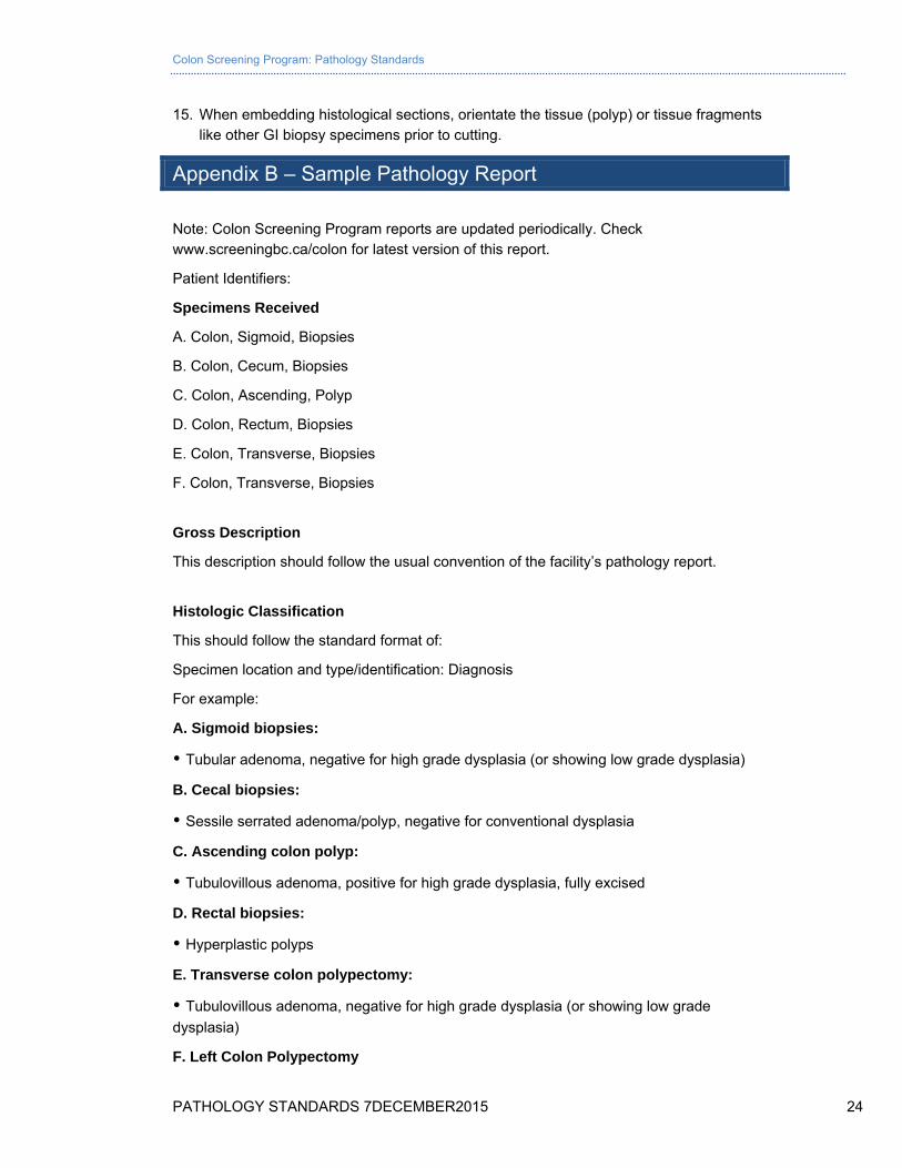
15. When embedding histological sections, orientate the tissue (polyp) or tissue fragments like other GI biopsy specimens prior to cutting.
Appendix B – Sample Pathology Report
Note: Colon Screening Program reports are updated periodically. Check www.screeningbc.ca/colon for latest version of this report.
Patient Identifiers:
Specimens Received
A. Colon, Sigmoid, Biopsies
B. Colon, Cecum, Biopsies
C. Colon, Ascending, Polyp
D. Colon, Rectum, Biopsies
E. Colon, Transverse, Biopsies
F. Colon, Transverse, Biopsies
Gross Description
This description should follow the usual convention of the facility’s pathology report.
Histologic Classification
This should follow the standard format of:
Specimen location and type/identification: Diagnosis
For example:
A. Sigmoid biopsies:
Tubular adenoma, negative for high grade dysplasia (or showing low grade dysplasia)
B. Cecal biopsies:
Sessile serrated adenoma/polyp, negative for conventional dysplasia
C. Ascending colon polyp:
Tubulovillous adenoma, positive for high grade dysplasia, fully excised
D. Rectal biopsies:
Hyperplastic polyps
E. Transverse colon polypectomy:
Tubulovillous adenoma, negative for high grade dysplasia (or showing low grade
dysplasia)
F. Left Colon Polypectomy
Colon Screening Program: Pathology Standards
PATHOLOGY STANDARDS 7DECEMBER2015 25
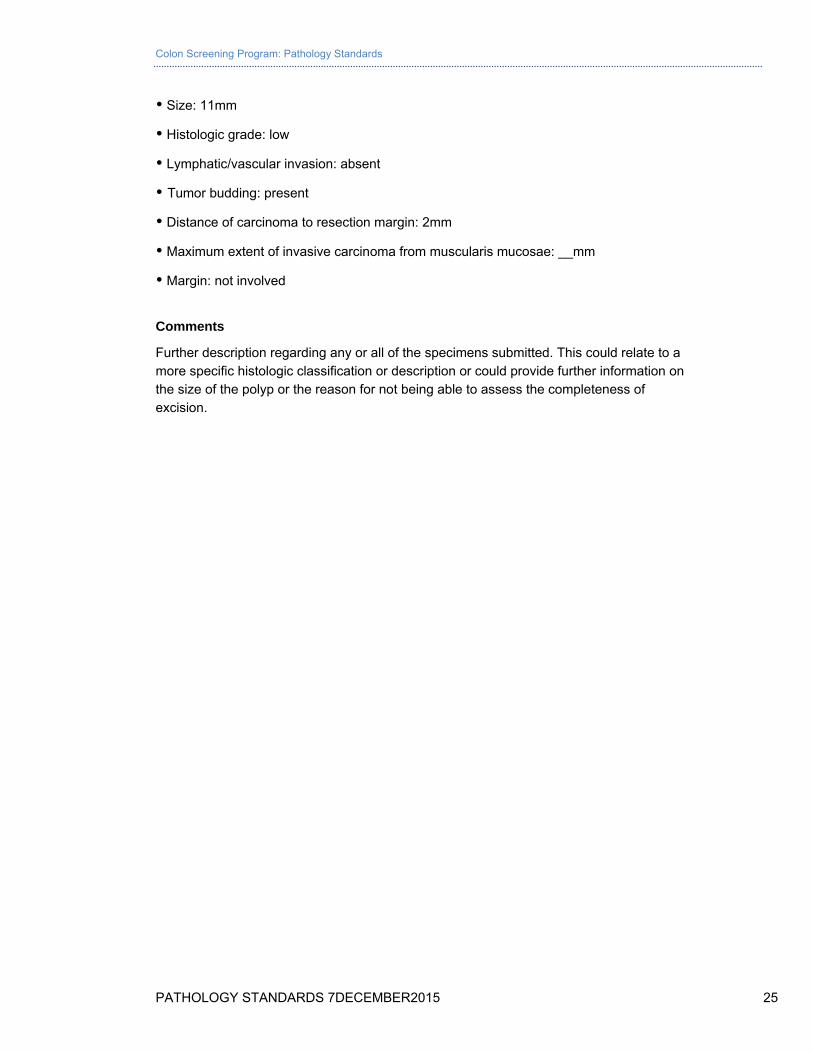
Size: 11mm
Histologic grade: low
Lymphatic/vascular invasion: absent
Tumor budding: present
Distance of carcinoma to resection margin: 2mm
Maximum extent of invasive carcinoma from muscularis mucosae: __mm
Margin: not involved
Comments
Further description regarding any or all of the specimens submitted. This could relate to a more specific histologic classification or description or could provide further information on the size of the polyp or the reason for not being able to assess the completeness of excision.