Collin County Community...
24
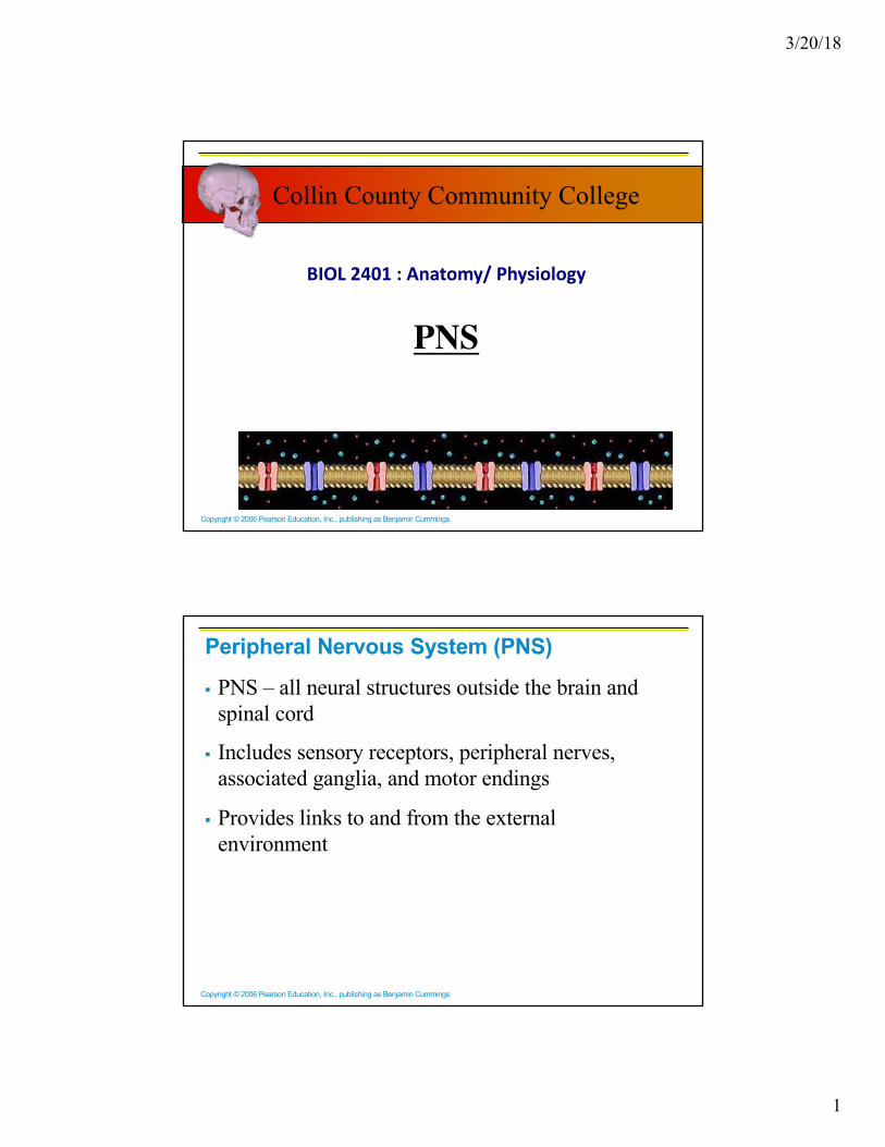
3/20/18 1 Copyright © 2006 Pearson Education, Inc., publishing as Benjamin Cummings BIOL 2401 : Anatomy/ Physiology PNS Collin County Community College Copyright © 2006 Pearson Education, Inc., publishing as Benjamin Cummings Peripheral Nervous System (PNS) § PNS – all neural structures outside the brain and spinal cord § Includes sensory receptors, peripheral nerves, associated ganglia, and motor endings § Provides links to and from the external environment
-
Upload
trinhhuong -
Category
Documents
-
view
221 -
download
1
Transcript of Collin County Community...
3/20/18
1
Copyright © 2006 Pearson Education, Inc., publishing as Benjamin Cummings
BIOL 2401 : Anatomy/ Physiology
PNS
Collin County Community College
Copyright © 2006 Pearson Education, Inc., publishing as Benjamin Cummings
Peripheral Nervous System (PNS)
§ PNS – all neural structures outside the brain and spinal cord
§ Includes sensory receptors, peripheral nerves, associated ganglia, and motor endings
§ Provides links to and from the external environment
3/20/18
2
Copyright © 2006 Pearson Education, Inc., publishing as Benjamin Cummings
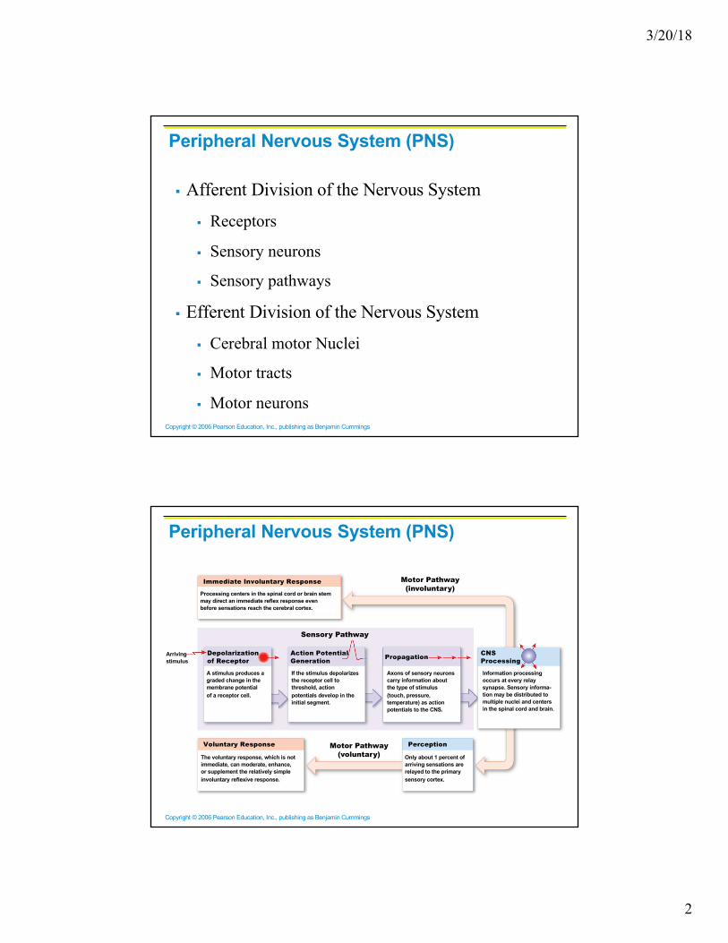
Peripheral Nervous System (PNS)
§ Afferent Division of the Nervous System
§ Receptors
§ Sensory neurons
§ Sensory pathways
§ Efferent Division of the Nervous System
§ Cerebral motor Nuclei
§ Motor tracts
§ Motor neurons
Copyright © 2006 Pearson Education, Inc., publishing as Benjamin Cummings
Arrivingstimulus
Immediate Involuntary Response
Depolarizationof Receptor
Action PotentialGeneration Propagation CNS
Processing
Voluntary Response Perception
Sensory Pathway
Motor Pathway(involuntary)
Motor Pathway(voluntary)
Processing centers in the spinal cord or brain stemmay direct an immediate reflex response evenbefore sensations reach the cerebral cortex.
A stimulus produces agraded change in themembrane potentialof a receptor cell.
If the stimulus depolarizesthe receptor cell tothreshold, actionpotentials develop in theinitial segment.
Axons of sensory neuronscarry information aboutthe type of stimulus(touch, pressure,temperature) as actionpotentials to the CNS.
Information processingoccurs at every relaysynapse. Sensory informa-tion may be distributed tomultiple nuclei and centersin the spinal cord and brain.
The voluntary response, which is notimmediate, can moderate, enhance,or supplement the relatively simpleinvoluntary reflexive response.
Only about 1 percent ofarriving sensations arerelayed to the primarysensory cortex.
Peripheral Nervous System (PNS)

3/20/18
3
Copyright © 2006 Pearson Education, Inc., publishing as Benjamin Cummings
PNS in the Nervous System
Figure 13.1
Copyright © 2006 Pearson Education, Inc., publishing as Benjamin Cummings
Sensory Receptors§ Structures specialized to respond to stimuli
§ Activation of sensory receptors results in depolarizations that trigger impulses to the CNS
§ The realization of these stimuli, sensation and perception, occur in the brain
§ Sensory Receptors can be
§ specialized cells closely associated with peripheral endings of sensory neurons
§ or specialized regions of sensory neurons.

3/20/18
4
Copyright © 2006 Pearson Education, Inc., publishing as Benjamin Cummings
Receptor Classification by Stimulus Type§ Mechanoreceptors – respond to touch, pressure,
vibration, stretch, and itch
§ Thermoreceptors – sensitive to changes in temperature
§ Photoreceptors – respond to light energy (e.g., retina)
§ Chemoreceptors – respond to chemicals (e.g., smell, taste, changes in blood chemistry)
§ Nociceptors – sensitive to pain-causing stimuli
Copyright © 2006 Pearson Education, Inc., publishing as Benjamin Cummings
Receptor Class by Location: Exteroceptors§ Respond to stimuli arising outside the body
§ Found near the body surface
§ Sensitive to touch, pressure, pain, and temperature
§ Include the special sense organs
Receptor Class by Location: Interoceptors§ Respond to stimuli arising within the body
§ Found in internal viscera and blood vessels
§ Sensitive to chemical changes, stretch, and temperature changes
3/20/18
5
Copyright © 2006 Pearson Education, Inc., publishing as Benjamin Cummings
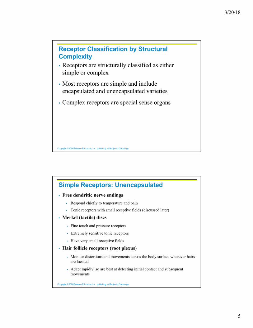
§ Receptors are structurally classified as either simple or complex
§ Most receptors are simple and include encapsulated and unencapsulated varieties
§ Complex receptors are special sense organs
Receptor Classification by Structural Complexity
Copyright © 2006 Pearson Education, Inc., publishing as Benjamin Cummings
Simple Receptors: Unencapsulated§ Free dendritic nerve endings
§ Respond chiefly to temperature and pain
§ Tonic receptors with small receptive fields (discussed later)
§ Merkel (tactile) discs
§ Fine touch and pressure receptors
§ Extremely sensitive tonic receptors
§ Have very small receptive fields
§ Hair follicle receptors (root plexus)
§ Monitor distortions and movements across the body surface wherever hairs are located
§ Adapt rapidly, so are best at detecting initial contact and subsequent movements

3/20/18
6
Copyright © 2006 Pearson Education, Inc., publishing as Benjamin Cummings
Simple Receptors
Merkel cell
Nerveterminal(dendrite)
Tactile disc
Afferent nerve fiber
Free nerve ending
Hair root plexus
Copyright © 2006 Pearson Education, Inc., publishing as Benjamin Cummings
Simple Receptors: Encapsulated§ Meissner’s corpuscles (tactile corpuscles)§ Perceive sensations of fine touch, pressure, and low-frequency vibration
§ Fairly large structures, usually in dermal papillae
§ Adapt to stimulation within 1 second after contact
§ Most abundant in the eyelids, lips, fingertips, nipples, and external genitalia
§ Pacinian corpuscles (lamellated corpuscles)§ Deeper in dermis and sensitive to deep pressure; Fast-adapting receptors
§ Most sensitive to pulsing or high-frequency vibrating stimuli
§ Ruffini’s corpuscles§ Located in the reticular (deep) dermis and sensitive to pressure and distortion of the
skin
§ Tonic receptors that show little if any adaptation

3/20/18
7
Copyright © 2006 Pearson Education, Inc., publishing as Benjamin Cummings
Tactile corpuscleTactile corpuscle
Tactilecorpuscle
Epidermis
Capsule
Dendrites
Dermis
Sensorynerve fiber
LM � 330
Copyright © 2006 Pearson Education, Inc., publishing as Benjamin Cummings
Dermis
Lamellated corpuscle Lamellatedcorpuscle(cross section)
LM � 125
Dendritic process
Acceesory cells(specialized fibroblasts)
Concentric layers(lamellae) of collagen
fibers separatedby fluid

3/20/18
8
Copyright © 2006 Pearson Education, Inc., publishing as Benjamin Cummings
DendritesRuffini corpusclef
CapsuleSensory
nerve fiberCollagen
fibers
Copyright © 2006 Pearson Education, Inc., publishing as Benjamin Cummings
ProprioReceptors§ They monitor position of joints, tension in ligaments and
tendons and state of muscular contraction
§ Joint kinesthetic receptors
§ Golgi tendon organs
§ Muscle spindles
§ Monitor change in pressure
§ Consist of free nerve endings that branch within elastic tissues of the walls of distensible organ (such as a blood vessel
§ Respond immediately to a change in pressure, but adapt rapidly
BaroReceptors

3/20/18
9
Copyright © 2006 Pearson Education, Inc., publishing as Benjamin Cummings
Sensory Receptors§ The simple receptors provide us with information regarding
§ Temperature
§ Pain
§ Touch
§ Pressure
§ Vibration
§ Proprioception
§ The special senses have special within protective structures. They include smell, taste, vision, hearing , equilibrium
General senses
Copyright © 2006 Pearson Education, Inc., publishing as Benjamin Cummings
Receptor Density• Receptors vary in terms of
abundance relative to each other.• For example, there are far more
pain receptors than temperature receptors in the body.
• Receptors also vary in terms of the concentration of their distribution over the surface of the body
• The fingertips having far more touch receptors than the skin of the back of the hand. The figure shows the distribution of temperature receptors in the skin by area.

3/20/18
10
Copyright © 2006 Pearson Education, Inc., publishing as Benjamin Cummings
Thermoreceptors§ As a population, thermoreceptor neurons show two general
response profiles as a function of temperature: Some receptors are cold sensitive, others are warm sensitive.
Copyright © 2006 Pearson Education, Inc., publishing as Benjamin Cummings
From Sensation to Perception§ Survival depends upon sensation and perception
§ Sensation is the awareness of changes in the internal and external environment
§ Perception is the conscious interpretation of those stimuli and of the external world from a pattern of different sensory nerve impulses via the sensory receptors.
§ Some perceptions are indeed integrated compound sensations such as for example “wetness” ( touch, pressure and thermal input…. there is no such thing as a “wet-receptor”)

3/20/18
11
Copyright © 2006 Pearson Education, Inc., publishing as Benjamin Cummings
Organization of the Somatosensory System§ Input comes from exteroceptors,
proprioceptors, and interoceptors
§ The three main levels of neural integration in the somatosensory system are:
§ Receptor level – the sensor receptors
§ Circuit level – ascending pathways
§ Perceptual level – neuronal circuits in the cerebral cortex
Copyright © 2006 Pearson Education, Inc., publishing as Benjamin Cummings
Processing at the Receptor Lever§ The receptor must have specificity for the stimulus energy
( temperature, touch, pressure, light,…)
§ The receptor’s receptive field must be stimulated
§ Stimulus energy must be converted into a graded potential
§ If the receptive field is in the same neuron that generates the action potential, we call it a generator potential.
§ If the receptive field is in a separate cell, it is called a receptor potential. If summed up to reach threshold, hhis will then release neurotransmitters in order to excite the associated sensory neuron.
3/20/18
12
Copyright © 2006 Pearson Education, Inc., publishing as Benjamin Cummings
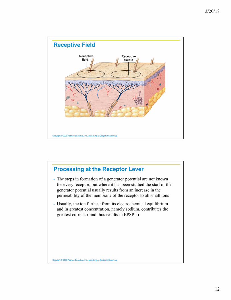
Receptive FieldReceptive
field 1Receptive
field 2
Copyright © 2006 Pearson Education, Inc., publishing as Benjamin Cummings
Processing at the Receptor Lever§ The steps in formation of a generator potential are not known
for every receptor, but where it has been studied the start of the generator potential usually results from an increase in the permeability of the membrane of the receptor to all small ions
§ Usually, the ion furthest from its electrochemical equilibrium and in greatest concentration, namely sodium, contributes the greatest current. ( and thus results in EPSP’s)

3/20/18
13
Copyright © 2006 Pearson Education, Inc., publishing as Benjamin Cummings
Processing at the Receptor LeverStep1 : Stimulus)
Step 2 : Generator Potential
Step 3 : Action Potential
Step 1 : Stimulus
Step 2 : Receptor Potential
Step 3 : Action Potential
N.T. release
Copyright © 2006 Pearson Education, Inc., publishing as Benjamin Cummings
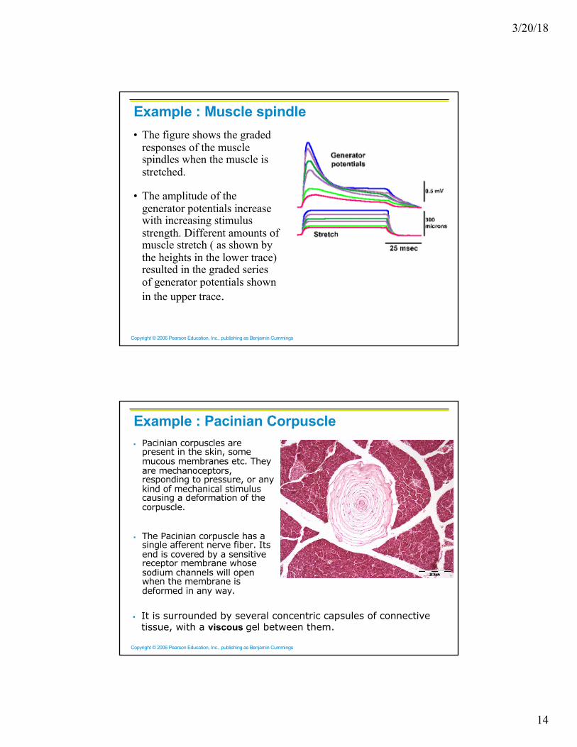
Example : Muscle spindle• Muscle spindles are
composed of 3-10 intrafusal muscle fibers that lack myofilaments in their central regions, are noncontractile, and serve as receptive surfaces
• They inform the body of the muscle tone and length of a muscle. They become activated when stretched and send sensory impulses to the CNS.
3/20/18
14
Copyright © 2006 Pearson Education, Inc., publishing as Benjamin Cummings
Example : Muscle spindle• The figure shows the graded
responses of the muscle spindles when the muscle is stretched.
• The amplitude of the generator potentials increase with increasing stimulus strength. Different amounts of muscle stretch ( as shown by the heights in the lower trace) resulted in the graded series of generator potentials shown in the upper trace.
Copyright © 2006 Pearson Education, Inc., publishing as Benjamin Cummings
Example : Pacinian Corpuscle§ Pacinian corpuscles are
present in the skin, some mucous membranes etc. They are mechanoceptors, responding to pressure, or any kind of mechanical stimulus causing a deformation of the corpuscle.
§ The Pacinian corpuscle has a single afferent nerve fiber. Its end is covered by a sensitive receptor membrane whose sodium channels will open when the membrane is deformed in any way.
§ It is surrounded by several concentric capsules of connective tissue, with a viscous gel between them.

3/20/18
15
Copyright © 2006 Pearson Education, Inc., publishing as Benjamin Cummings
Example : Pacinian Corpuscle§ In the resting state, a cross-
section through the corpuscle looks something like this
§ Now, if the skin over the corpuscle is touched, it will be deformed and make a nuisance of itself:
§ But the viscous gel between the capsules will move and allow the nerve ending to resume its normal shape:
§ If the pressure is now released, the corpuscle as a whole will resume its original shape, but the nerve ending will be deformed in the process:
§ The viscous gel will then flow back, and soon we are back at the beginning.
Copyright © 2006 Pearson Education, Inc., publishing as Benjamin Cummings
Example : Pacinian Corpuscle
§ The result is two generator potentials; one when pressure is applied and one when pressure is released.
§ This system is thus very good for picking up vibrations.

3/20/18
16
Copyright © 2006 Pearson Education, Inc., publishing as Benjamin Cummings
Adaptation of Sensory Receptors§ To a certain extent, the duration of the generator potential
depends upon the duration of the stimulus.
§ However, some receptors have generator potentials that last only a short time, no matter how long the stimulus is maintained.
§ We refer to a decrease in the amplitude of the generator potential or the frequency of discharge of the sensory fiber in the face of a persisting, constant stimulus as adaptation.
Copyright © 2006 Pearson Education, Inc., publishing as Benjamin Cummings
Adaptation of Sensory Receptors
§ Those that adapt slow or not at all are called tonic receptors.
§ Receptors responding slowly include Merkel’s discs, Ruffini’s corpuscles, and interoceptors that respond to chemical levels in the blood
§ Pain receptors and proprioceptors do not exhibit adaptation (why not ?)

3/20/18
17
Copyright © 2006 Pearson Education, Inc., publishing as Benjamin Cummings
Stimulus
a
Normal NormalIncreased
Time
Frequencyof action
potentials
Tonic receptors are always active and generate actionpotentials at a frequency that reflects the background level of stimulation. When the stimulus increases or decreases, therate of action potential generation changes accordingly.
Copyright © 2006 Pearson Education, Inc., publishing as Benjamin Cummings
Adaptation of Sensory Receptors
§ Some receptors are fast adapting. Those that adapt fast are called phasic receptors.
§ Receptors responding to pressure, touch, and smell adapt quickly
3/20/18
18
Copyright © 2006 Pearson Education, Inc., publishing as Benjamin Cummings
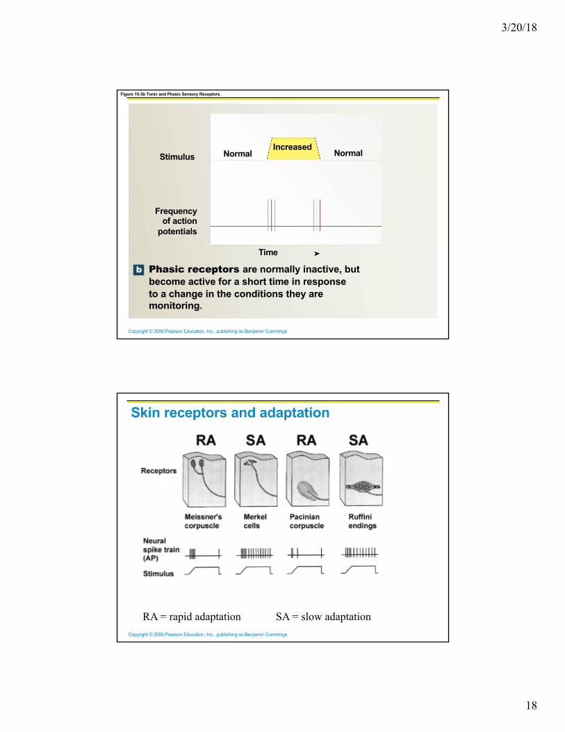
Figure 15-3b Tonic and Phasic Sensory Receptors.
b
Stimulus Normal NormalIncreased
Time
Frequencyof action
potentials
Phasic receptors are normally inactive, butbecome active for a short time in responseto a change in the conditions they aremonitoring.
Copyright © 2006 Pearson Education, Inc., publishing as Benjamin Cummings
Skin receptors and adaptation
RA = rapid adaptation SA = slow adaptation
3/20/18
19
Copyright © 2006 Pearson Education, Inc., publishing as Benjamin Cummings
Tonic and Phasic Receptors
There is purpose to these differences.
Tonic (slowly-adapting) receptors are important in situations where constant information about a stimulus is important ( they thus send information about ongoing stimulation)
Example : internal blood pressure, muscle spindles, injuries (pain)
Phasic (rapidly-adapting) receptors send information related to changing stimuli. They stop responding to a maintained stimulus, but when the stimulus is removed, they respond again
Example : Hair follicles, pacinian corpuscles,..
Copyright © 2006 Pearson Education, Inc., publishing as Benjamin Cummings
Information Coding
Any stimulus contains within it certain features that are of interest to the body. Stimuli have
• intensities or strengths• locations or sites of application• frequencies of application• rates of application• modalities
Modality, broadly speaking, is a class of sensations that are referred to a single type of receptor. Vision, hearing, touch, smell, and taste are all modalities ( energy forms).
Sensory receptors may be sensitive to different kind of energies. For example, putting pressure on the eye cause you to see light flashes, although the function of the eye receptors is to detect light.

3/20/18
20
Copyright © 2006 Pearson Education, Inc., publishing as Benjamin Cummings
Information Coding
• Doctrine of Specific Nerve Energies, as formulated by Johannus Müller, says that, although a sense organ may be sensitive to many forms of stimulus energy other than its real stimulus (called the adequate stimulus), the sensation evoked is always like that associated with the adequate stimulus, no matter what kind of energy was applied.
• For example : electrical stimulation of the optic nerve, does not result in an electric shock; the sensation evoked is one of seeing light.
• The doctrine of specific nerve energies implies that the modality or submodality of a sensation is determined not by the stimulus, but by what specific receptor or nerve fiber is stimulated. The doctrine also implies that the subjective qualities of a modality are determined, not in the receptors themselves, but in the central nervous system. (in this case for the optic nerve, it is determined by the visual cortex).
Copyright © 2006 Pearson Education, Inc., publishing as Benjamin Cummings
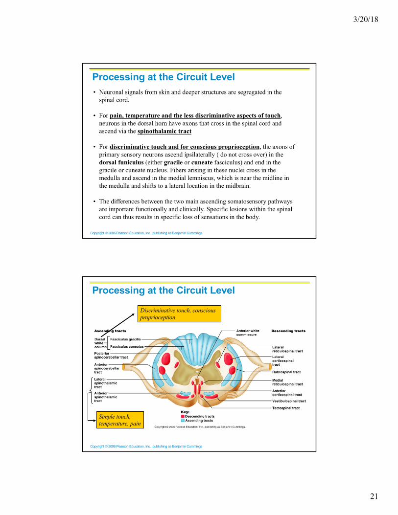
Processing at the Circuit Level
§ Chains of three (3) neurons conduct sensory impulses upward to the brain
§ First-order neurons – soma reside in dorsal root or cranial ganglia, and conduct impulses from the skin to the spinal cord or brain stem
§ Second-order neurons – soma reside in the dorsal horn of the spinal cord or medullary nuclei and transmit impulses to the thalamus or cerebellum
§ Third-order neurons – located in the thalamus and conduct impulses to the somatosensory cortex of the cerebrum
3/20/18
21
Copyright © 2006 Pearson Education, Inc., publishing as Benjamin Cummings
Processing at the Circuit Level• Neuronal signals from skin and deeper structures are segregated in the
spinal cord.
• For pain, temperature and the less discriminative aspects of touch, neurons in the dorsal horn have axons that cross in the spinal cord and ascend via the spinothalamic tract
• For discriminative touch and for conscious proprioception, the axons of primary sensory neurons ascend ipsilaterally ( do not cross over) in the dorsal funiculus (either gracile or cuneate fasciculus) and end in the gracile or cuneate nucleus. Fibers arising in these nuclei cross in the medulla and ascend in the medial lemniscus, which is near the midline in the medulla and shifts to a lateral location in the midbrain.
• The differences between the two main ascending somatosensory pathways are important functionally and clinically. Specific lesions within the spinal cord can thus results in specific loss of sensations in the body.
Copyright © 2006 Pearson Education, Inc., publishing as Benjamin Cummings
Processing at the Circuit Level
Simple touch, temperature, pain
Discriminative touch, conscious proprioception

3/20/18
22
Copyright © 2006 Pearson Education, Inc., publishing as Benjamin Cummings
Processing at the Perceptual Level§ Both spinothalamic tract and the medial lemniscus
terminate in the ventral posterior nucleus of the thalamus.
§ The thalamus projects fibers to:
§ The somatosensory cortex of postcentral gyrus
§ Sensory association areas
§ First one modality is sent, then those considering more than one
§ The result is an internal, conscious image of the stimulus
Copyright © 2006 Pearson Education, Inc., publishing as Benjamin Cummings
Main Aspects of Sensory Perception§ Perceptual detection – detecting that a stimulus has
occurred and requires summation
§ Magnitude estimation – how much of a stimulus is acting
§ Spatial discrimination – identifying the site or pattern of the stimulus
§ Feature abstraction – used to identify a substance that has specific texture or shape
§ Quality discrimination – the ability to identify submodalities of a sensation (e.g., sweet or sour tastes)
§ Pattern recognition – ability to recognize patterns in stimuli (e.g., melody, familiar face)

3/20/18
23
Copyright © 2006 Pearson Education, Inc., publishing as Benjamin Cummings
Structure of a Nerve§ Nerve – cordlike organ of the
PNS consisting of peripheral axons enclosed by connective tissue
§ Connective tissue coverings include:
§ Endoneurium – loose connective tissue that surrounds axons
§ Perineurium – coarse connective tissue that bundles fibers into fascicles
§ Epineurium – tough fibrous sheath around a nerve
Copyright © 2006 Pearson Education, Inc., publishing as Benjamin Cummings
Classification of Nerves
§ Sensory and motor divisions
§ Sensory (afferent) – carry impulse to the CNS
§ Motor (efferent) – carry impulses from CNS
§ Mixed – sensory and motor fibers carry impulses to and from CNS; most common type of nerve

3/20/18
24
Copyright © 2006 Pearson Education, Inc., publishing as Benjamin Cummings
Peripheral Nerves
§ Mixed nerves – carry somatic and autonomic (visceral) impulses
§ The four types of mixed nerves are:
§ Somatic afferent and somatic efferent
§ Visceral afferent and visceral efferent
§ Peripheral nerves originate from the brain or spinal column