Collection, transport & Processing Of Mycology · PDF fileProcessing Of Mycology Samples...
62
Collection, transport & Processing Of Mycology Samples Eman Elmansoury
Transcript of Collection, transport & Processing Of Mycology · PDF fileProcessing Of Mycology Samples...
Collection, transport & Processing Of Mycology
Samples
EmanElmansoury

Laboratory Diagnosis
1. Mycological Diagnosis
a. Sampling
b. Direct microscopical examination.
c. Cluture.
d. Identification.
2. Histopathological DiagnosisSpecial fungal stains i.e. PAS and GMS.
3. Serological Diagnosis.
4. Molecular diagnosis.

When to Suspect Fungal Infections?
Persistent illness, despite appropriate antibacterial therapy.
Premature infants, oncology patients who become neutro-penic and other intensive care patients with indwelling central catheters.


For successful isolation consider:
Patient requestPatient information (labeling)Proper collectionProper transportCorrect processingInoculation of sample on proper culture media

Criteria for specimen rejection
1. Absence of patient identification or discrepancybetween the information on request form andsample labelAction: Return for resoltion
2. Sputum with squamous cell >25 cell/ low power fieldAction:
Pathogenic fungi maybe recovered in presence oforal contamination if antibiotic containingselective media is used
Final report should indicate presence off oralcontamination

3. Sample with inadequate volume orAction: Order 2nd sample
4. Dried out swab (if wet)Action:(Swaps not suitable for recovery of fungi &should be rejected…deep aspirtion or biobsy better)
Criteria for specimen rejection

5. sample in improper container or unsuitable conditions (drying, leakage, lack of sterility)Action: If 2nd specimen couldn’t be obtained process final report: specimen was compromised& result interpreted in light of clinicalpresentation
6. 24 hour urine or sputum for fungal culture chance of contmination with bacteria & invironmental moldsAction: Another sample
Criteria for specimen rejection

Generally Samples examined as soon as possible
Specimens not processed immediately held at room temerature, refrigerated, freezing unacceptble.
If there is delay in processing,To prevent growh of commensal bacteria in nonsterile specimens add:
◦ 20 IU/ml penicillin,◦ 10 mg/ml streptomycin or◦ 0.2 mg/ml chloramphenicol

1. Sputum
Collection & Transport:◦ 1st early morning sample before breakfast
◦ rinse mouth with water
◦ 5 ml in screw capped sterile container
◦ If inadequate → use aerosol saline suspension by heating

Lower Respiratory Tract Samples:
Lung biopsyBrushingBronchoalveolar lavage→ in sterile sealed container
1. Sputum

Processing:select most purulent or bloody part of sample
if viscid → homogenized by adding N-acetyl L-cystein & Avoid NaOHwhich give alkaline pH for culture media
1. Sputum

microscopic examination KOH mount
culture: inoculate 0.5 ml to each culture media which contain antibiotic to inhibit bacteria flora in sputum e.g. IMA & BHIwith chloramphenicol + cycloheximide
1. Sputum

2. C.S.F.
Collection & Transport:◦By lumbar puncture in sterile container (number 3 tube) to avoid skin flora
◦ if delay is suspected → do not refrigerate
◦ left in room temperature as CSF itself is considered as adequate culture media for fungal elements

processing: < 2 ml → direct culture◦3-5 drops on tube media or ◦2-3 drops …… ½ ml on plate culture media > 2 ml → concentration
◦ Centrifuge for 5 minutes at 2000 rpm
◦ Remove supernatant fluid by sterile pipette for cryptococcal antigen testing
2. C.S.F.

◦Direct examinationuse 1 drop of the sediment to make an
India ink mount if Cryptococcus is suspected
◦Culture: Re-suspend sediment in 1-2 ml of CSF &
inoculate onto IMA or SABHI
2. C.S.F.

2. C.S.F.

3. Urine
Collection & Transport: Volume: 2 - 5 ml
1st early morning sample on 3 successive days (fungi tend to be shed at intermittent intervals)
Random sample is accepted
Sample is collected aseptically in sterile screw capped container and sent immediately to lab.
If delay → refrigerated at 4° C to inhibit growth of rapidly growing bacteria

Processing:◦ centrifuge at 2000 rpm for 5 minutessediment.
3. Urine

Direct examination◦ Prepare a direct smear of the sediment in KOH
for direct microscopy
Culture:◦ inoculate ½ ml sediment to both IMA & BHI with
chloramphenicol + Cycloheximide
3. Urine


4. Prostatic secretion
1st empty the bladder
Do prostatic massage
Inoculate secretion directly on proper fungal media
Collect 5 – 10 ml (1st voided) urine in separate container after prostatic massage

5. Exudates
Collection & Transport: 1st disinfect the skin by iodine & ethyl alcohol 70 % Aspirate by sterile syringe which serve as transport
container
Processing: if > 2 ml → concentrate → sediment → direct examination &
1/2 ml for culture
if > 2 ml with clot → inoculate clot onto culture media
if < 2 ml with clot → mince with scalpel → inoculate whole sample on culture media
6. Biopsy
Collection & Transport:
Biopsy taken if aspiration fail
Taken from inflamed site (from edge & from center)
Transported in sterile moistened gauze with sterile saline in screw capped container
Specimen Should not allowed to dry or to be frozen
If delay → refrigerate at 4°C for 8 – 10 hours maximum , (no formaline)

Processing: ( surface area of specimen to exposethe micro-organism)
placed in sterile Petri dish
add few drops of sterile water
by sterile scalpel cut the sample into 1 mm piecesinoculate on surface of culture media as IMA &SABHI
Avoid using tissue grinder (destruction of hyphalform especially zygomycetes)
6. Biopsy

7. Swabs
Swabs is inadequate for recovery of molds and suboptimal for yeast even in oral thrush (scraped with sterile tongue depressor)
So aspiration or biopsy are better
High vaginal swab
On transport media
If delay → refrigerate at 4°C maximum overnight
Vortex in 1/2 ml sterile water → direct examination & culture

8. Blood
Collection & Transport:
1st disinfect the skin
Collect 8-10 cc blood with heparinanticoagulant

1. Direct culture method:
centrifugationbuffy coat Inoculate 0.5-1.0 ml onto the
surface of the media.
8. Blood

2. Biphasic culture bottle: Slant of BHI agar and 60-100 ml of BHI
broth
Enhance recovery of fungi from blood
1:10 (blood to broth)
Tilted daily broth flows over agar.
Checked daily for growth gram stain bottle contents to detect fungal elements.
Cultures incubated at 30°C for 4 weeks.
8. Blood

3. Lysis centrifugation isolator system:(Wampole Isolator system) lyse leukocytes & erythrocytes (releasing microorganisms)
&inactivate plasma, complement &certain antibiotics
Centrifugation step concentrate the organisms in the blood sample. Concentrate inoculated onto culture media.
Improves recovery of fungi from blood e.g. Candida (C. krusei & C. glaborata) & Histoplasma capsulatum take longer time (discarded as negative)
8. Blood

9. Bone marrow aspiration
Collection & Transport:
3 – 5 ml bone marrow aspirate in sterile container with heparin
Inoculate on pediatric blood culture

Primary isolation media for blood & bone marrow culture
1) SDA with chloramphenicol and gentamicin and incubate duplicate cultures at 26°C and 35°C.
2) BHI supplemented with 5% sheep blood and incubate at 35°C. Maintain cultures for 4 weeks.

10. Skin, Nail &hairCollection
1. Skin scrapings Swab skin area with 70 % alcohol to remove surface
bacterial contaminant & ointments
Scrape the lesion at the advancing border using a blunt scalpel, or a bone curette, side of glass microscopic slide.
In vesicular tinea pedis, the tops of any fresh vesiclesshould be removed (as the fungus is often plentiful in the roof of the vesicle).
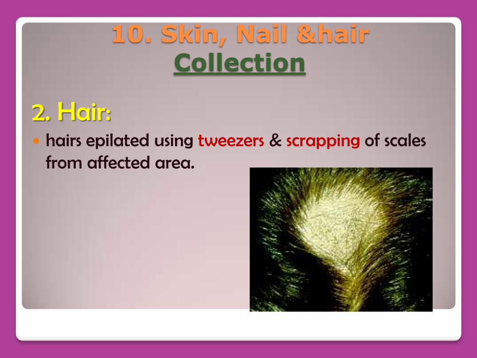
2. Hair: hairs epilated using tweezers & scrapping of scales
from affected area.
10. Skin, Nail &hairCollection

3. Nails: Cleaned with 70% ethanol
Scraped & clipped
Scraped using a blunt scalpel
Scrapings are taken from the proximal to the distal end of the nail. The first 4-5 scrapings are discarded, nail sampled near nail plate until the crumbling white degenerating portion is reached
10. Skin, Nail &hairCollection

Transport: in dry envelop or glass Petri dish (avoid plastic container)
Special black cards (dermapak)
◦ Easier to see how much material has been collected
◦ Ideal conditions for transportation.
Stored at room temperature, as dermatophytesmay be killed at 4 - 6° C
10. Skin, Nail &hairTransport

Direct examintion 10% KOH
Put the sample 1-2 drops of 10-20% KOH Cover slide Gentle heating or add DMSO to dissolve keratin Skin scraping: left for 20 minutes Nail clipping: left for 12 to 24 hours Hair: examined after addition of KOH (being fragile)
Parker ink lactophenol cotton blue
calcofluor white mounts.
10. Skin, Nail &hairProcessing

dermatophyte hyphae breaking up into arthroconidia
10. Skin, Nail &hair

Culture:1. SDA with & without Actidione incubate at 26
&35°C2. BHI with chloramphenicol + cycloheximide or3. dermatophyte test media in heavily contaminated
samples for minimum 30 days before reporting negative result
10. Skin, Nail &hairProcessing

Examination of hair
Nodules presenta. black & hard → black piedra (Piedra hortae)b. soft & white → white piedra (Trichosporonbeigelli)
Nodules absent a. Arthrocondia
Ectothrix → Microsporium Endothrix →Trichophyton
b. No Arthroconidia → Trichophyton

Examination of hair

Ectothrix invasion byM. canis
Endothrix invasion caused by T. tonsurans.


Direct Microscopic Examinations
Stains: KOH 10 % calcoflour India ink Geimsa stain mucicarmin stain Fontana masson

Advantage: coast effective method in diagnosis of fungal
infection
Detect fungal element in clinical samples in <1 hours
Specifically identify the organism at species level e.g. Cryptococcus neoformans, Aspergillus, Candida, dermatophytes, Zygomycetes, hyalo-hyphomycosis&dematecious fungi
Direct Microscopic Examinations

Assess amount of fungi in sample
commensal fungi considered pathogenic if detected in large amount in sample
Differentiate between contamination (spores only) and infection (Aspergillus head) in pulmonary Aspergillosis
Direct Microscopic Examinations

I- yeast cell : 1. globose
a. multiple budding: Paracoccidioidesb. single blastoconidia
- broad attachment :Blastomycetes- narrow attachment: Histoplasma
or Cryptococcus2. ovoid : Candida or Histoplasma or Sporothrix
Direct Microscopic Examinations

Direct Microscopic Examinations 2. Hyphae1. Hyaline:
A. Septate: 1. Dichotomously branched (at 45 degree)
septate Aspergillus2. Regular hyphae with arthrospores
dermatophytesB. Aseptate:
1. Irregular ribbon like zygomycetes2. Pigmented: dematecious fungi

Telephone reports are to be issued for:
a. True hyphae seen in a direct exam. b. Fungi seen in any normally sterile body specimen.
Direct Microscopic Examinations


Common fungal recovery culture media
BHI → 1ry recovery of saprophytic and dimorphic fungi
if cycloheximide and chloramphenicolare added → recovery of dimorphic fungi
IMA → enriched media + gentamycin& chloramphenicol → recovery of dimorphic fungi

SA BHI → 1ry recovery of saprophytic, dimorphic fungi and fastidious fungi
mycosel = mycobiotic = dermasel →contain cycloheximide and chloramphenicol → for recovery of dermatophytes
SDA → 2nd work up of cultures
Common fungal recovery culture media

On cover of plate → name sample date
Labeling culture

◦ 2 sets at 30 C & 35 C (for recovery of yeast form of dimorphic fungi)
◦ Cultures kept minimum 3o days before discharging as negative even if plate appear contaminated by bacteria or other fungi
◦ Place flat open pan on bottom shelf in incubator to provide moisture and precaution must to be done to avoid growth of environmental molds in water
Incubation of fungal culture

Special request to dimorphic fungi → 6 weeks(check daily in 1st 2weeks & 3 times /week in remaining 4 weeks)
If Histoplasma suspected → 12 weeks
Special request for Malassizia → 1-2 weeks
Blood cultures & body fluids & respiratory tract → 4 weeks (1 W & 3 W)
environmental samples → 5 days
Incubation of fungal culture

Identificaion of fungal growth
Macroscopic Examination:1. rate of growth2. Colonial morphology3. Surface pigment4. Reverse pigment5. Growth on cycloheximide containing agarMicroscopic Examination: tease mount transparency tape preparation microslide culture technique

Pathogenic fungi resistant to Cycloheximide:Blastomyces dermatitidisHistoplasma capsulatumCoccidioides immitisSporothrix schenckiiParacoccidioides brasiliensisTrichophyton sp.Microsporum sp.Epidermophyton floccosum
Identificaion of fungal growth

Germ tube test Rapid test for the presumptive identification of C.
albicans.
Procedure Put 3 drops of serum into a small glass tube
touch a colony of yeast by Pasteur pipetteaemulsify in serum (pipette can be left in the tube) Incubate at 35oC to 37oC for up to 3 hours but no longer Transfer a drop of the serum to a slideCoverslip examine microscopically using x 40 objective.

InterpretationPositive test: short lateral filaments
(germ tubes) one piece structure (no constriction between the yeast cell and the germination tube).
C. albicans C. dubleninses
Negative test: yeast cells only (or with pseudohyphae) always two pieces
Non-albicans yeast
Germ tube test

Common nosocomial fungi
1. Candida species2. Aspergillus species3. Zygomycetes as Mucor 4. Fusarium species 5. Acremonium species6. M. furfur

Antifungal susceptibility tests
Recently antifungal antibiogram was developed especially for fungi in yeast forms.
Disc diffusion method micro dilution methods