Collateral Ligament Injuries of the Knee -...
12
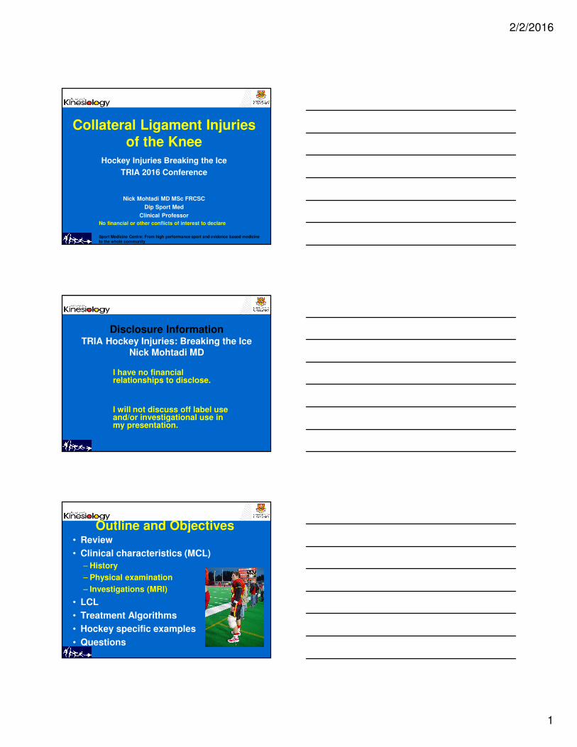
2/2/2016 1 Collateral Ligament Injuries of the Knee Hockey Injuries Breaking the Ice TRIA 2016 Conference Nick Mohtadi MD MSc FRCSC Dip Sport Med Clinical Professor Sport Medicine Centre: From high performance sport and evidence based medicine to the whole community No financial or other conflicts of interest to declare Disclosure Information TRIA Hockey Injuries: Breaking the Ice Nick Mohtadi MD I have no financial relationships to disclose. I will not discuss off label use and/or investigational use in my presentation. Outline and Objectives • Review • Clinical characteristics (MCL) – History – Physical examination – Investigations (MRI) • LCL • Treatment Algorithms • Hockey specific examples • Questions
Transcript of Collateral Ligament Injuries of the Knee -...
2/2/2016
1
Collateral Ligament Injuries of the Knee
Hockey Injuries Breaking the Ice
TRIA 2016 Conference
Nick Mohtadi MD MSc FRCSC
Dip Sport Med
Clinical Professor
Sport Medicine Centre: From high performance sport and evidence based medicine to the whole community
No financial or other conflicts of interest to declare
Disclosure InformationTRIA Hockey Injuries: Breaking the Ice
Nick Mohtadi MD
I have no financial relationships to disclose.
I will not discuss off label use and/or investigational use in my presentation.
Outline and Objectives• Review
• Clinical characteristics (MCL)
– History
– Physical examination
– Investigations (MRI)
• LCL
• Treatment Algorithms
• Hockey specific examples
• Questions

2/2/2016
2
Review• Sprain
– Grade 1 or First degree sprain
• Stretching of the ligament but not loose
– Grade 2 or Second degree sprain
• Partial damage to the ligament abnormal laxity
– Grade 3 or Third degree sprain
• Complete disruption of the ligament
• Not to be confused with “strain”
• Not to be confused with the physical
examination
Review• MCL
– Typically refers to the superficial ligament that originates on the medial femoral
epicondyle and attaches to the metaphysis of the tibia deep and distal to the pes
• LCL (Fibular collateral Ligament)
– Lateral femoral epicondyle to the conjoined insertion, with biceps tendon on
the fibular head
Review
• Extra-articular
structures
• Therefore have
the potential to heal
• Tightest in full extension
2/2/2016
3
Review: Ligament Healing• Three overlapping and sequential
stages
– Injury; Bleeding and inflammation
• Hours to days to a few weeks
– Cell proliferation and matrix production
• Days to weeks to a few months
– Matrix remodelling
• Weeks to months to a few years
SCAR TISSUE
Clinical Characteristics• Did they feel something “rip,” “pop,” or
“tear”?
– If the answer is yes!
• Hockey three mechanisms
– Goalie down in butterfly position
– Direct blow to the knee with valgus force
– Hit against the boards while bracing for the hit with the inside limb/skate
– Land on inside edge…other
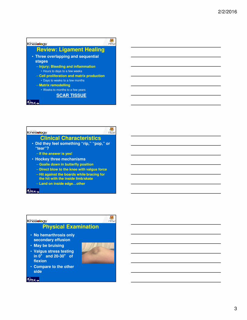
Physical Examination
• No hemarthrosis only
secondary effusion
• May be bruising
• Valgus stress testing
in 0°°°° and 20-30°°°° of
flexion
• Compare to the other
side
2/2/2016
4
Physical Examination
• Point of maximal
Tenderness (PMT)
indicates where the
injury has occurred
Physical Examination
• Alignment
– Varus
– Valgus
– neutral
Physical Examination
• Valgus stress
testing in 0°°°°
and 20-30°°°° of flexion
• Compare to the
other side

2/2/2016
5
Physical examination
• Grading of Laxity
– Grade 1
• 0 mm to < 5mm ssd
– Grade 2
• 5 mm to < 10 mm ssd
– Grade 3
• 10 mm or greater ssd
• End point assessment
Valgus Stress• knee is unstable to valgus stress in the
fully extended position (0°°°°)
– you must look for an associated cruciate ligament injury!
• knee is stable at 0°°°° and opens at 20-30°°°° <
5 mm ssd
– the superficial MCL is damaged but not likely complete
• If opens > 5 mm ssd
– then likely complete superficial MCL tear
Valgus Stress
• What if the knee opens in flexion > 10
mm ssd?
– Superficial MCL completely torn
– Likely the deep MCL fibres (meniscotibialand meniscofemoral)
– Possibly the posterior oblique ligament i.e. the posteromedial corner
– Likely would open in full extension

2/2/2016
6
MRI
• Knee injury clinic in Calgary (AKIC)
– Last year we saw 2340 new knee injuries
– MRI utilization was 10%
• Calgary Flames (1995-2015)
– 100-200 new knee injuries
– MRI utilization was 100%
MRI
MRI
2/2/2016
7
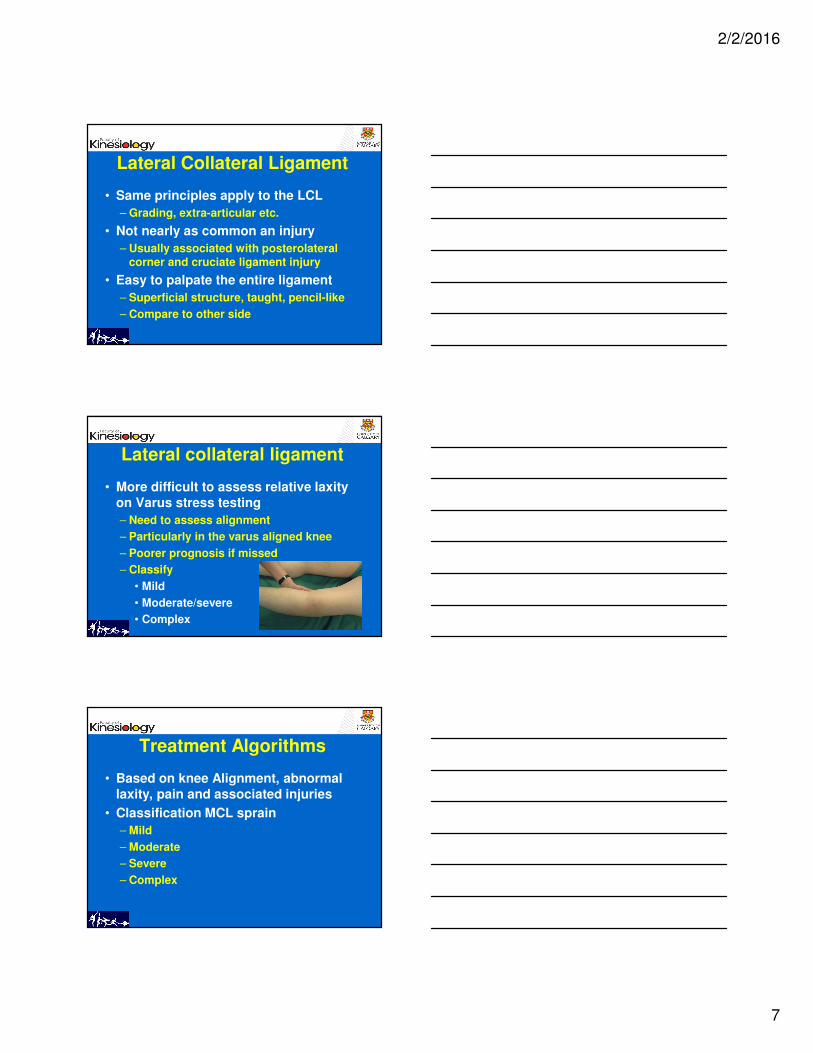
Lateral Collateral Ligament
• Same principles apply to the LCL
– Grading, extra-articular etc.
• Not nearly as common an injury
– Usually associated with posterolateralcorner and cruciate ligament injury
• Easy to palpate the entire ligament
– Superficial structure, taught, pencil-like
– Compare to other side
Lateral collateral ligament
• More difficult to assess relative laxity
on Varus stress testing
– Need to assess alignment
– Particularly in the varus aligned knee
– Poorer prognosis if missed
– Classify
• Mild
• Moderate/severe
• Complex
Treatment Algorithms
• Based on knee Alignment, abnormal
laxity, pain and associated injuries
• Classification MCL sprain
– Mild
– Moderate
– Severe
– Complex
2/2/2016
8
Mild MCL Injury
• Pain only
• Stable knee
• Symptomatic treatment
• Protection and prevention of re-injury
Moderate MCL Injury• Description: The knee is stable with valgus
stress in full extension but opens in flexion.
• Management:
– Follow initial treatment protocol
– Crutches protected weight bearing
– Knee strengthening exercises
– Knee (proprioception) balance exercises
– Bracing optional depending on symptoms and activity
– Follow-up: 2-week intervals to re-examine the knee
Moderate MCL Injury *
• * Follow severe MCL sprain bracing
protocol if patient described a pop, rip or tear with their mechanism of injury
or if they have valgus alignment.

2/2/2016
9
Severe MCL Injury
• Description: The knee opens with valgus stress
in full extension and greater in flexion in a neutral or varus aligned
knee OR the knee is stable in extension but opens in flexion in a valgus aligned
knee.
Severe MCL Injury• Management:
– Follow initial treatment protocol
– Range of Motion (ROM) brace with 30°°°° to 90°°°° of motion worn 23.5 hours a day
• Follow-up: 2 week intervals to assess range of motion and MCL healing.
• Management: Based on quality of healing of MCL, adherence (compliance) with bracing
and range of motion.
• Return to sport
Bracing Protocol• 23.5 hours per day at 30-90°°°°
• Initial goal is to get the knee
stiff
– Assess adherence with ROM
– If they can straighten out their knee they have not been wearing the brace
• Physical examination
– at 2 week intervals

2/2/2016
10
Bracing Protocol
• Minimum Goal
– knee stable to valgus stress in full extension
– Adjust brace accordingly
• 3-8 weeks return to play
• Functional knee brace for remainder of
the season
• Think of future injury prevention
Complex MCL injury
• MCL + ACL
– Bracing protocol for MCL
– Then reconstruct ACL
• MCL + PCL
– Brace in extension to ensure
that the knee is reduced
– Allow MCL to heal
– Non-op Rx for PCL
LCL Treatment Algorithms
• Parallel the “MCL” algorithms; Adjust
for Varus knee
– Mild LCL no Laxity
• Symptomatic Rx
• Protection from re-injury
– Moderate/severe LCL
• Bracing protocol 30° to 90°
– Complex LCL
• Consider surgery

2/2/2016
11
Treatment
• Role of Surgical treatment
– Rare
– Avulsions
– Complex injuries
– Patient who had delayed treatment of their collateral ligament injuries
– Reconstruction usually with postero-lateral corner
Hockey Examples
• Two players (Centre
and Goalie)
• Injured 2 days apart
• Both had severe
MCL injuries
– MRI confirmed
Forward and Goalie
• How should they be
treated?
– Bracing protocol
– Added treatment (PRP; Stem cells)
• When should they return to play?
• Protect with taping or bracing?

2/2/2016
12
Summary
• MCL common injury in hockey
• Clinical
– Mechanism of injury and physical exam
– MRI not necessary for majority of patients
• Consider limb alignment
– MCL in Valgus knee = severe
– LCL in Varus knee = severe
Summary
• Adherence to Bracing protocol
– Almost universally successful
• Adjuvant treatment with PRP or Stem
cells not proven
• Surgery rarely required
– Only in multiple ligament injured knees
– Repair early
– Reconstruct late