爀圀椀氀氀 搀椀猀挀甀猀猀 眀栀攀渀 戀甀琀 渀漀琀 …...Michelle Jo Semins, MD...
15
Michelle Jo Semins, MD University of Pittsburgh Medical Center
Transcript of 爀圀椀氀氀 搀椀猀挀甀猀猀 眀栀攀渀 戀甀琀 渀漀琀 …...Michelle Jo Semins, MD...

Michelle Jo Semins, MD University of Pittsburgh Medical Center
Presenter
Presentation Notes
Not discussing residual postop stones bc sara discussion this correct? Will discuss when but not how to bc not enough time…right? Lipkin paper if want me to discuss

Increased imaging 2007 - 72 million CT scans
Increased diagnosis of asymptomatic stones Large screening studies – Prevalence 8-10%
Brenner et al. 2007 Berrington de Gonzalez et al. 2009
Presenter
Presentation Notes
Lipkin review for References 2 screening studies with CT for colon cancer and potential kidney donors in asx patients - 5000 and 2000 patients – stones diag in 7.8 and 9.7% will increase

Technological advances, Minimally invasive Well tolerated, low morbidity BUT inherent risk in surgical intervention
Natural history? Poorly defined Progression risk? Unclear

2010: > 7mm active removal - low rate of spontaneous passage
2013: option - active surveillance with annual evaluation of stone and symptoms for 2-3 years Intervention should be considered after this period Observation might be associated with greater risk of
necessitating more invasive procedures Patients should be adequately informed
Presenter
Presentation Notes
Grade C – 2012 Made despite the absence of directly applicable clinical studies of good quality

Growth High risk for stone
formation Obstruction Infection Symptomatic
(Pain/hematuria)
Stones>15mm <15mm if choice of
treatment Patient preference Comorbidity Social situation
(profession, travel) “Natural history of small, non-obstructing asymptomatic lower
pole calculi is not well defined, and the risk of progression is unclear. There is still no consensus on the follow-up duration, timing and type of intervention.”

Prospective Randomized Controlled Trials (3) Keely et al BJU 2001 SWL v OBS Yuruk et al 2010 PNL v SWL v OBS Sener et al 2015 F-URS v SWL v OBS
Prospective (2) – Inci 2007, Krambeck 2015 Retrospective (5) – Hubner & Porpaczy 1990,
Glowacki 1992, Burgher 2004 , Koh 2012, Kang 2013
Presenter
Presentation Notes
TO ANSWER THESE QUESTIONS OF NATURAL HISTORY AND RISK OF PROGRESSION MANY GROUPS HAVE STUDIED THE ASYMPTOMATIC STONE 10 studies Sener – low intervention rate in obs group 12% - n =150, 50 per group – swl/urs equivalent but swl required >1 session (prior studies show superiority of urs over swl for small lower pole stones) Keely et al BJU 2001 n= 228 OBS v SWL SF 17 v 28% (no sig diff). 72% LP stones. Of OBS grp 21% req additional intervention. Fu = 2.2y. Obs associated with a greater risk that patient will eventually require more invasive procedures PRCT Asx LP stones Turkey 2010 yuruk N=94 swl obs, pnl (31/31/32) 1.37cm, 19 mo f/u Obs – 3.1%passage, 18.7% intervention, Progression = obstruction, growth, recurrent uti, hematuria referred for intervention CT 3 and 12 mo post intervention DMSA done 6 and 12 weeks post intervention PNL 100% SF – scar 3.2% SWL – 54.8% SF – scar 16% - Neg- no URS group Br J Urol. 1990 Jul;66(1):9-11. Treatment of caliceal calculi. Hübner W1, Porpaczy P. J Urol. 2005 Jun;173(6):2005-9. Prospective, randomized trial comparing shock wave lithotripsy and ureteroscopy for lower pole caliceal calculi 1 cm or less. Pearle MS1, Lingeman JE, Leveillee R, Kuo R, Preminger GM, Nadler RB, Macaluso J, Monga M, Kumar U, Dushinski J, Albala DM, Wolf JS Jr, Assimos D, Fabrizio M, Munch LC, Nakada SY, Auge B, Honey J, Ogan K, Pattaras J, McDougall EM, Averch TD, Turk T, Pietrow P, Watkins S. Pearle 2005 LP stone PRCT <1cm swl v urs but no observation group ( no diff in SF rate – greater pt acceptance and short convalenscence for SWL) Sener 2014 - 140 patients – PRCT – no OBS grp. swl needed 2.7 sessions – sf rate 90%, for urs 1 session and sfr for urs = 100%

Most - lower pole stones Follow-up - 19-53 months Stone size – 4.4mm-10.8mm N – 24-550

Spontaneous passage rate – 3-29% Progression rate – 33-77% Intervention rate – 7-40%
50% will progress over 5y but most will not
require surgery
Presenter
Presentation Notes
(only 1 was 40% - and that was study from 1990 – the others were 7-26%) intervention rate of 7-40% only …meaning up 60-93% of the time nothing is done! Glowacki - 5y prob of symptomatic stone event = 48.5% H&P - Propose 83% intervention w/in 5y, 45% growth over 7.4y, and 40% were removed surgically, Only 11% of patients with caliceal calculi remain symptom-free after 10 years Given low SF rate - SWL does not improve sf rates or qol compared to observation for asx calyceal stones <15mm Kang - 3 and 5-year event probability of 41% and 47% respectively – all very similar!!!
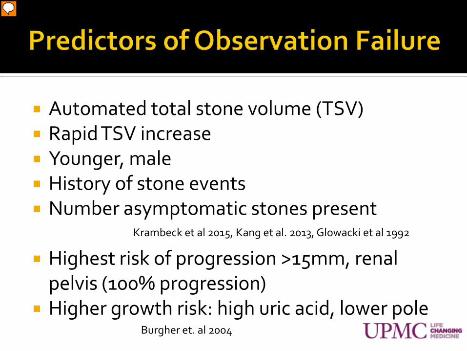
Automated total stone volume (TSV) Rapid TSV increase Younger, male History of stone events Number asymptomatic stones present
Highest risk of progression >15mm, renal
pelvis (100% progression) Higher growth risk: high uric acid, lower pole
Krambeck et al 2015, Kang et al. 2013, Glowacki et al 1992
Burgher et. al 2004
Presenter
Presentation Notes
Krambeck – prospective – survey and med record review stone clinic – 550 patient – 43% had stone event for median of 4.7y (did not count growth as a clinical event) – Automated TSV was the only independent predictor of symptomatic events on multivariate analysis (they looked at largest stone diameter, number of stones, and bilateral stones) – this was strongest in first 2 years. TSV >1000mm3 3y risk of events in younger formers of 58% and older of 43%. TSV < 79mm3 3y risk in younger of 20% and older 8%. Those with interim stone events and repeat CT – if TSV increases >570mm3 they have 84% chance of stone event in 3 years versus 46% w/o increased TSV of 570. (more comprehensive characterization of stone burden than other radiographic measures, also more accurate bc based on automated image processing algorithm as opposed to manual review of CT images by individuals). Also rapid increase In stone volume appears to be predictive of future stone events in patients who had an interim stone event (flaw – very large stones i.e. staghorn or very large were removed and excluded from study so skews results a bit) Krambeck – other – predictors – younger age, cystine, high sscaox, low urine pot and sulfate. (likely new stones and growth and not detachment) self-reported, only total volume not individual stone volume, location not examined Glowacki – events associated with number of asx stones Kang – 1/2 have progression (passage, pain, growth, intervention) Inci – growth in 1/3 patients Burgher/monga – large stone diameter predictive of progression (pain, growth, intervention) 300 males followed for 3.26y avg age 62.8 VA clinic (not typical population) Disease progression – larger stones High uric acid associated with stone growth as was lower pole stones (targeted medical therapy given to 28%) – also do not know what indication for treatment was given i.e. ? Symptoms? Total - 77% progressed with 26% requiring intervention 100% stones >15mm either grew, developed pain or needed intervention Kaplan Meier analysis suggests a 50% risk of intervention at 7.25 years Those faring best – small <4mm upper pole stones Ref 2,6 – about ½ (48.5%) of patients with stones <4mm will have symptoms, require intervention or both within 5 years – those patients with a stone history of multiple stones more likely to become symptomatic (2) Glowacki 1992 107pt Asymptomatic stones that do pass spontaneously do so early 30% passing in 2 years and progressively fewer during subsequent years Small <4mm upper pole stones fair the best Koh - Quotes – 9.5% risk of complications for stones >5mm and 14.3% risk of complications for stones >10mm – and ? Should be offered intervention

Increased passage, decreased intervention younger patients, smaller stones, no stone growth
in followup
Small <4mm upper pole stones fare best
Kang et al. 2013
Burgher et al. 2004
Presenter
Presentation Notes
Sener 2015 - Asx lower pole stones <1cm PRCT – �swl, urs, obs, evliyaoglu Turkey 2015 150 patients URS – 92% stone free SWL – 90% stone free with 1.48 treatments Obs – 3 passed w/o sx, 3 pain, 2 passed with response to analgesics – 6 required surgery – 2 year f/u 88% noneventlful Complications higher grade in swl Obs reasonable f/u 21 +/- 3.65 month Ref 4 – need for intervention 10% per year >50% in 5 years Ref 5 – kang – 50% of cases require intervention within 19 months time (?type of stone?) – 30% spont passage rate at 31 months. 45% progression Koh – 45% progression – 50 patients EAU recommends observation for nonsymptomatic calyceal stones – guidelines 2013! – recommend stone removal if infections, growth, obstructions, >15mm, patient preference Ref 8 inci prospective asx stones 2007 – 24 patients 52.3 months, 11% intervention, 1/3 progression

½ Retrospective = selection bias Definitions differ between studies stone growth = increase in stone size greater than
50% versus absolute stone size increase Progression – symptoms, growth, +/- intervention Some intervention was for growth, not symptoms
Medical therapy - some include, most do not Natural history different with aggressive medical
management?
Presenter
Presentation Notes
explains likely why some numbers are so different. Kang –intervention rate 24.5% but included prophylactic intervention bc of stone growth (i.e. progression)

Scenario - 8mm asymptomatic lower pole stone OBS, annual KUB – within 4 years – 40% growth, 30% passage/migration, 30% stabile
56.4% preferred to defer decision to physician 22.8% OBS, 29.7% URS, 47.5% SWL
Experience dictates decisions Decision based on prior treatment type Prior larger stone passage less likely to choose OBS 11 patients had no prior stone passage or surgery 82% SWL, 18% URS
No prior procedure but prior stone passage 39% OBS, 52% SWL, 9% URS
Sarkissian et al. 2013
Presenter
Presentation Notes
With that knowledge – what do patients think???? OBS with annual KUB – within 4 years - 40% chance of growth, 30% chance of passage/migration, 30% chance of stability Monga decision making paper 12 patients no prior procedure but prior stone passage

Social Fertile women High risk occupation – Pilots,
travelers, truckers, military Poor access or compliance Children
High risk patient Solitary kidney ? Renal insufficiency Transplant kidney – do not
get classic pain ?SCI patients Urinary tract reconstruction Immunodeficiency
Stone burden, location Stone > 15mm Large stone burden Renal pelvis Significant stone growth
Concomitant symptomatic stone requiring treatment
Patient preference
Symptom development
METABOLIC EVALUATION RECOMMENDED FIRST
Presenter
Presentation Notes
Hydronephrosis, ureteral (obstruction) – listed somewhere but not really asymptomatic definition When treating symptomatic ipsilateral and/or contralateral stone ? Prior history of symptomatic stone events – unclear if this is a risk Frequent travel to 3rd world countries Staghorn calculus

Monitoring/surveillance guidelines Do not exist Annual KUB, urinalysis (non uric-acid) Ultrasound (uric acid) If symptoms develop – CT scan
Cost effectiveness studies
Presenter
Presentation Notes
Many options in literature including creatinine, urinalysis, culture, ultrasound xray and ct

Questions?