Coaxial electrospun fibers: applications in drug delivery ...
24
Overview Coaxial electrospun fibers: applications in drug delivery and tissue engineering Yang Lu, 1 Jiangnan Huang, 2 Guoqiang Yu, 1 Romel Cardenas, 1 Suying Wei, 3 * Evan K. Wujcik 1 * and Zhanhu Guo 2 * Coelectrospinning and emulsion electrospinning are two main methods for pre- paring core–sheath electrospun nanofibers in a cost-effective and efficient man- ner. Here, physical phenomena and the effects of solution and processing parameters on the coaxial fibers are introduced. Coaxial fibers with specific drugs encapsulated in the core can exhibit a sustained and controlled release. Their exhibited high surface area and three-dimensional nanofibrous network allows the electrospun fibers to resemble native extracellular matrices. These fea- tures of the nanofibers show that they have great potential in drug delivery and tissue engineering applications. Proteins, growth factors, antibiotics, and many other agents have been successfully encapsulated into coaxial fibers for drug delivery. A main advantage of the core–sheath design is that after the process of electrospinning and release, these drugs remain bioactive due to the protection of the sheath. Applications of coaxial fibers as scaffolds for tissue engineering include bone, cartilage, cardiac tissue, skin, blood vessels and nervous tissue, among others. A synopsis of novel coaxial electrospun fibers, discussing their applications in drug delivery and tissue engineering, is covered pertaining to proteins, growth factors, antibiotics, and other drugs and applications in the fields of bone, cartilage, cardiac, skin, blood vessel, and nervous tissue engineer- ing, respectively. © 2016 Wiley Periodicals, Inc. How to cite this article: WIREs Nanomed Nanobiotechnol 2016, 8:654–677. doi: 10.1002/wnan.1391 INTRODUCTION T he process of electrospinning was first patented by Gooley in 1900 and 1902. 1 In the 1990s, this technique was revived by Reneker et al. 2,3 for fabri- cation of various One-dimensional nanostructures. Electrospinning is of great interest due to its simple method, low cost, varied electrospinnable materials, and fabrication control of micro to nanostructures with inherently high surface to volume ratio. Appli- cations of electrospun fibers include drug delivery, 4,5 tissue engineering, 6 filtration membranes, 7 self- cleaning materials, 1 reinforcement fillers, 8 solar cells, 9 sensors, 10 supercapacitors, 11 lithium-ion batteries, 12 among others. The electrospinning process utilizes a strong electrostatic force produced by a high voltage source to draw micro- or nanoscale fibers from the charged liquid. Compared with single component round- shaped fibers, the coaxial fibers have many advan- tages on biomedical applications: (1) The bioactive drugs can be safely escorted by the sheath phase from the harsh environment. (2) With the control of thick- ness of core phase and sheath phase, a sustained and *Correspondence to: [email protected]; Evan.Wujcik@lamar. edu; [email protected] 1 Materials Engineering and Nanosensor (MEAN) Laboratory, Dan F. Smith Department of Chemical Engineering, Lamar University, Beaumont, TX, USA 2 Integrated Composites Laboratory (ICL), Department of Chemi- cal and Biomolecular Engineering, The University of Tennessee, Knoxville, TN, USA 3 Department of Chemistry and Biochemistry, Lamar University, Beaumont, TX, USA Conflict of interest: The authors have declared no conflicts of inter- est for this article. 654 © 2016Wiley Periodicals, Inc. Volume 8, September/October 2016
Transcript of Coaxial electrospun fibers: applications in drug delivery ...

Overview
Coaxial electrospun fibers:applications in drug deliveryand tissue engineeringYang Lu,1 Jiangnan Huang,2 Guoqiang Yu,1 Romel Cardenas,1 Suying Wei,3*Evan K. Wujcik1* and Zhanhu Guo2*
Coelectrospinning and emulsion electrospinning are two main methods for pre-paring core–sheath electrospun nanofibers in a cost-effective and efficient man-ner. Here, physical phenomena and the effects of solution and processingparameters on the coaxial fibers are introduced. Coaxial fibers with specificdrugs encapsulated in the core can exhibit a sustained and controlled release.Their exhibited high surface area and three-dimensional nanofibrous networkallows the electrospun fibers to resemble native extracellular matrices. These fea-tures of the nanofibers show that they have great potential in drug delivery andtissue engineering applications. Proteins, growth factors, antibiotics, and manyother agents have been successfully encapsulated into coaxial fibers for drugdelivery. A main advantage of the core–sheath design is that after the process ofelectrospinning and release, these drugs remain bioactive due to the protectionof the sheath. Applications of coaxial fibers as scaffolds for tissue engineeringinclude bone, cartilage, cardiac tissue, skin, blood vessels and nervous tissue,among others. A synopsis of novel coaxial electrospun fibers, discussing theirapplications in drug delivery and tissue engineering, is covered pertaining toproteins, growth factors, antibiotics, and other drugs and applications in thefields of bone, cartilage, cardiac, skin, blood vessel, and nervous tissue engineer-ing, respectively. © 2016 Wiley Periodicals, Inc.
How to cite this article:WIREs Nanomed Nanobiotechnol 2016, 8:654–677. doi: 10.1002/wnan.1391
INTRODUCTION
The process of electrospinning was first patentedby Gooley in 1900 and 1902.1 In the 1990s, this
technique was revived by Reneker et al.2,3 for fabri-cation of various One-dimensional nanostructures.
Electrospinning is of great interest due to its simplemethod, low cost, varied electrospinnable materials,and fabrication control of micro to nanostructureswith inherently high surface to volume ratio. Appli-cations of electrospun fibers include drug delivery,4,5
tissue engineering,6 filtration membranes,7 self-cleaning materials,1 reinforcement fillers,8 solarcells,9 sensors,10 supercapacitors,11 lithium-ionbatteries,12 among others.
The electrospinning process utilizes a strongelectrostatic force produced by a high voltage sourceto draw micro- or nanoscale fibers from the chargedliquid. Compared with single component round-shaped fibers, the coaxial fibers have many advan-tages on biomedical applications: (1) The bioactivedrugs can be safely escorted by the sheath phase fromthe harsh environment. (2) With the control of thick-ness of core phase and sheath phase, a sustained and
*Correspondence to: [email protected]; [email protected]; [email protected] Engineering and Nanosensor (MEAN) Laboratory, DanF. Smith Department of Chemical Engineering, Lamar University,Beaumont, TX, USA2Integrated Composites Laboratory (ICL), Department of Chemi-cal and Biomolecular Engineering, The University of Tennessee,Knoxville, TN, USA3Department of Chemistry and Biochemistry, Lamar University,Beaumont, TX, USA
Conflict of interest: The authors have declared no conflicts of inter-est for this article.
654 © 2016 Wiley Per iodicals , Inc. Volume 8, September/October 2016

controlled release of a model drug can be achieved.(3) By choosing two suitable components, the coaxialfibers can exhibit desirable mechanical properties.(4) The structure of coaxial fibers resembles extracel-lular matrix (ECM). (5) For scaffolds, a more bio-compatible polymer can be selected as a shell while aless biocompatible polymer is selected as a core.
COAXIAL ELECTROSPUNNANOFIBERS
MethodsThere are several approaches for preparing core–sheath electrospun nanofibers including coelectros-pinning,13,14 emulsion electrospinning,15–17 templatedeposition method,18,19 electrostatic force inductionmethod,20 phase separation method,21,22 and otherunique set ups.23–25
The coelectrospinning method is a modificationof the traditional single spinneret electrospinning setup. The single spinneret is replaced by two coaxialcapillaries in which two channels are connected totwo reservoirs. The coaxial configuration of nozzlescan provide different pathways for inner and outersolutions. The basic coelectrospinning set up is illus-trated in Figure 1. This innovative method was firstreported by Loscertales et al.13 in 2002 and devel-oped by Sun et al.14 in 2003. Loscertales et al.13 pro-duced the capsules with diameters ranging from150 nm to 10 μm in an electrospray manner. Sunet al.14 first used this set up to prepare nanofiberswith core–sheath structure and named this technique‘coelectrospinning.’ Xu et al.15 first produced coaxialnanofibers by emulsion electrospinning using the
conventional single spinneret in 2006. The water-in-oil (W/O) emulsion consists of amphiphilicpoly(ethylene glycol)-poly(L-lactic acid) block copol-ymer in chloroform as the organic phase andpoly(ethylene oxide) in water as the water phase.Owing to unstable stretching and evaporation of thelow boiling point organic solvent, the emulsion ispartially de-emulsified and the viscosity of oil phaseincreases more rapidly than the water phase (chloro-form evaporates faster). The resulting viscosity gradi-ent causes the water phase to move into the innerspace leading to core–sheath nanofibers (Figure 2).Additional emulsifiers are often needed to improvethe electrospinnability and kinetic stability of theemulsion.27 This method is simple and particularlysuitable for drug delivery of water-soluble drugsencapsulated by sheath polymer. However, thismethod is often difficult to prepare homogeneous,stable, and electrospinnable emulsions and usuallyleaves defects in the core–sheath structure. In manycases, the resulting fibers have a dispersed waterphase as small blobs without core–sheath structure.28
The template deposition method uses a traditionalelectrospun fiber as the template. In combinationwith other techniques including chemical vapor depo-sition (CVD),29 sol–gel method30 and atomic layerdeposition (ALD),18,19 the sheath materials likemetals, metal oxides, or polymers can be coated ontothe electrospun fiber template forming a core–sheathstructure. However, each of the deposition methodshas its own limitations. For example, the uniformcoating is hard to achieve due to frequently depletionof the precursor in CVD method. In addition, tem-plate deposition is a two-step method. Coaxial nano-fibers can be obtained by electrostatic force inductionas well. Li et al.20 dispersed copper nanoparticles inpoly(vinyl alcohol) (PVA) solution with dioctyl sulfo-succinate sodium (AOT) as surfactant. As the solu-tion is highly polarized, the copper/PVA nanofibersare formed based on the high mobility, short dis-tance, and electrostatic absorption. Phase separationis another simple way for fabrication of coaxialfibers. Under a high electrical field, the homogenoussolution of different components tends to be unstableand phase separation results due to viscosity andcharge density difference.21 There are some othermethods reported which are mainly focused on devel-oping set ups of electrospinning. Fakhrali et al.23
used two opposite asymmetric nozzles to obtaincoaxial electrospinning fibers. Liu et al.24 combinedthe conventional electrospinning and PCA (precipita-tion with a compressed fluid antisolvent) process toprepare coaxial fibers with hollow structure. Leeet al.25 developed a core-cut nozzle system which can
High voltage supply
Sheath solution
SheathCo-axialcapillary
Co-axial cone
Whipping
co-axial jet
Core
Grounded
collector
Core solution
FIGURE 1 | A schematic of coaxial electrospinning. (Reprintedwith permission from Ref 26. Copyright 2008 Taylor & Francis (www.tandfonline.com))
WIREs Nanomedicine and Nanobiotechnology Biomedical coaxial electrospun fibers
Volume 8, September/October 2016 © 2016 Wiley Per iodica ls , Inc. 655
effectively reduce the instability of fluid jet. Viryet al.31 combined the emulsion electrospinning withcoelectrospinning. The emulsion as the core phasewas coelectrospun with another polymer solution asthe shell. The internal structure can be tailored bythe ratio of the dispersed phase to the continuousphase.
Among these different methods, coelectrospin-ning is used most extensively. It is a one-step methodwhich is simple to adopt. Coelectrospinning allows anumber of core materials which are not capable offorming electrospun fibers alone, to be electrospuninto a 1-D nanostructure via the protection and guid-ance of a sheath solution. Many unstable com-pounds, such as growth factors or antibiotics, can beisolated by a shell from harsh environment and elec-tric charges. Even living cells have been directly elec-trospun as core and were found viable afterwards.32
After removing the sheath layer, nonelectrospinnablefibers can be obtained. The size of coaxial fibers iseasy to control by adjusting the solution and proces-sing parameters. It is for these reasons that makingcoelectrospinning stands out among variousmethods.
Physical Theories and PhenomenaTo prepare and control the desired coaxial electro-spun fibers, it is best to understand the coaxial elec-trospinning phenomena and the underlying physics.In many cases, electrospraying, formation of mono-lithic fiber or clogging of the spinneret may happenwhile using coelectrospinning. Unlike electrospray-ing, the solution of electrospinning stays in a continu-ous jet which is dominated by viscoelastisity andbending instability. At the exit of coaxial nozzles,core solutions are trapped in sheath solution forming
a droplet with a similar prolate shape to the Taylorcone (Figure 3). The droplet and fluid jet are formedby the strong pulling stress of electrostatic forcebetween spinneret and collector. The jet is linearlystraight at first and becomes unstable due to bendinginstability after traveling a short distance. Numericalsimulations34 showed that electric charges move tothe surface of the jet rapidly during electrospinning.For this reason, the movement of core solution ispossibly entrained only by viscous traction. Accord-ing to Earnshaw’s theorem of electrostatics, it isimpossible to maintain a stable stationary equilib-rium configuration for point charges. Charges on thesurface induce the jet subject to complex spiralingtrajectories to minimize the repulsive Coulomb inter-actions. This bending instability along with the highelectrostatic force results in the rapid evaporation ofsolvent and dramatically stretches the jet into core-shell nanofibers. Tracer particle tracking system35
reveals that the acceleration of fluid jet can be as highas 600 m/second2 and the deformation of the jet ison the order of 103. Sometimes the instability cancause perturbation in core–sheath interphase. Coax-ial fibers can be prepared even by the identical poly-mer and solvent with the same concentration. Sunet al. observed the coaxial morphology of same
Original emulsion
Partially de-emulsified
Final fiber
Stretching and evaporation
Stretching and evaporation
StretchingStretching
Moving inward
and
merged
FIGURE 2 | Proposed mechanism of emulsion electrospinning.(Reprinted with permission from Ref 15. Copyright 2006 JohnWiley & Sons)
Outer tube
Inner tube
a
b
500 nm
3.18 mm
(a)
(b)
FIGURE 3 | (a) Coaxial fluid jet formed by coelectrospinning.(b) Transmission electron microscopy (TEM) image of coaxialPANI/poly(vinyl alcohol) (PVA) fibers. (Reprinted with permission fromRef 33. Copyright 2004 John Wiley & Sons)
Overview wires.wiley.com/nanomed
656 © 2016 Wiley Per iodicals , Inc. Volume 8, September/October 2016

polymer by using Bromophenol as the colormarker.14 With suitable processing parameters, thecharacteristic time of the coaxial electrospinningshould be on the order of τ1 = 1 ms. Meanwhile, thediffusion time of core or sheath solution is τ2 = d2/Da
(d: diameter of fiber. Da: diffusion coefficient) whichis normally on the order of 10 ms. With shorter char-acteristic time, boundaries of two solutions should beable to survive in electrospinning. On the collector,nonwoven coaxial fibers are randomly depositedforming a mat. Detailed theoretical and experimentalworks of coelectrospinning have been addressedpreviously.25,34,36,37
Solution ParametersNumerous materials, including organic and inorganicmaterials, have been successfully electrospun intoultrafine coaxial fibers. Selection of the appropriatematerials and solvent is extremely important forsteady generation of coaxial fibers especially thosefor biomedical applications.
The viscosity of sheath solution is required tobe sufficient to produce enough viscous traction forcore solution. High viscosity can overcome the inter-facial tension and allow a stable Taylor cone to beformed. Only when the sheath solution has sufficientviscoelasticity, the fluid jet can be stabilized to pro-duce coaxial fibers instead of core-shell particles.With low viscosity, the jet will breakup due to Ray-leigh instability. When the viscosity is higher but stillbelow a critical value, the Rayleigh instability is notsuppressed completely, in which case beads-on-stringfibers will be produced. The core solution can have alower viscosity. However, a minimum viscosity isrequired by the core solution to prevent the breakupof jet solution in the bending instability stage. Poly-L-lactic acid (PLLA)38, polyethylene glycol (PEG),39
and polyvinyl alcohol (PVA)40, for example, areoften added into the core solution to increase the vis-cosity. However, recently Luo et al.41 demonstratedthat a polymer sheath solution with low viscosity canbe electrospun into coaxial fibers when using a suita-ble liquid solvent as the core. The core liquid isrequired to have high surface tension and high inter-facial tension to suppress the Rayleigh instability. Weknow from previous studies that the sheath solutionneeds to be electrospinnable to form the fiber struc-ture whereas the core solution could be nonelectros-pinnable. McCann et al.42 used nonelectrospinnableC16–C20 hydrocarbons as a core and an organic–inorganic blend solution (Polyvinylpyrrolidone(PVP)/TiO2) as a shell to prepare coaxial fibers successfully.Polymer chain entanglements to some extent
determine the viscosity of the solution. Ways ofincreasing viscosity include decreasing operating tem-perature, using high molecular weight polymer orincreasing polymer concentration.43 However, whenthe concentration or viscosity is too high, the solu-tion frequently clogs—not allowing fibers to bedrawn.
The choice of the inner and outer solventsdetermines the miscibility of two solutions. Previousworks showed that core and sheath solutions withmiscible or even the same solvent are capable of pre-paring coaxial fibers. Sun et al.14 demonstrated thatthe coelectrospinning period from the exit of the noz-zle to the collector is shorter than the characteristictime of diffusion. He used the same polymer and sol-vent with a same concentration and successfully pro-duced core–sheath nanofibers. Yu et al.33 stated thatusing the same solvent can decrease the interfacialtension, which is beneficial for developing a uniformcore–sheath fiber. The use of a volatile solvent canresult in porous nanofibers, which is sometimesfavorable for constant release of drugs. However, ifthe volatility is too high, the rapid evaporation cancause clogs of the spinneret or multiple jets; in bothcases, no coaxial fibers will be drawn.
The concentration of polymer solution controlsthe viscosity. With an increase of polymer concentra-tion, the viscosity of the solution increases. Zhanget al.44 investigated the influence of polymer concen-tration. His works showed that a higher concentra-tion of both core and sheath solution can producecoaxial nanofibers with higher core and sheath dia-meters. By controlling the concentration, the dia-meters of the core or of the sheath can becontinuously tuned. In drug delivery systems, thethickness of the sheath can determine the rate of drugrelease and the thickness of the core can determinethe amount of available drugs.
Another solution parameter is conductivity.The conductivity of the core is not necessary. Studieshave shown that a nonconductive core solution canbe used in coelectrospinning as long as the sheathsolution is conductive. In addition, a higher conduc-tivity of core solution can cause the breakup of acore due to a strong pulling force.33 A higher con-ductivity of a sheath solution favors thinner fibersdue to strong electrostatic force and bending instabil-ity. Baumgarten45 showed that the diameter of elec-trospun fiber is inversely related to the cube root ofthe solution conductivity. Additionally, the conduc-tivity of the core or sheath solution can be achievedby adding salts such as NaCl, Fe(NO3)3, etc.
46 Whenthe conductivity of the sheath solution is too high,the abundant surface charges can affect the
WIREs Nanomedicine and Nanobiotechnology Biomedical coaxial electrospun fibers
Volume 8, September/October 2016 © 2016 Wiley Per iodica ls , Inc. 657

uniformity of coaxial fibers due to strong bendinginstability. Therefore, only within an optimal rangeof the core and the sheath conductivity, smooth sur-faces and interphases can be formed.
Overall, the suitable core and sheath solutionsshould have the following features: (1) Electrospin-nable sheath material. (2) A relatively high viscosityof sheath solution. (3) Viscosity of core solutionabove a critical value (can be not as high as sheathsolution). (4) Low surface tension of core solution.(4) Sheath solution should be conductive. These fea-tures are general but not necessary. To prepare uni-form and steady coaxial fibers, experiments areneeded to determine the optimal solution parameters.
Processing ParametersProcessing parameters can greatly affect the forma-tion and structure of coaxial fibers as well. Normally,there are three main processing parameters:(1) Applied high voltage. (2) Flow rate of core andsheath solution. (3) Tip-to-collector distance.
If the applied voltage is too low, the gravity canstill play an important role, in which case pendentdrops will be formed at the exit of nozzles. Increasingthe applied voltage to a critical value is a requirementfor a stable Taylor cone to be formed. However,excess of high voltage will start to pull the fluid jetinside of the capillary and often clog the capillaries.The Taylor cone tends to recede and two fluid jetscorresponding to core and sheath solution can beobserved.26,47 Further high voltage may result incorona discharge. Within an optimal range of appliedvoltage, a higher voltage will yield similar effects as ahigher conductivity of solution, in which cases smal-ler diameter fibers are formed.
Generally, the tip-to-collector distance isdirectly related to the flight time of fluid jet and elec-tric field strength.48 If the distance is too short thenthere is not enough time for the solvent of the sheathto evaporate completely. Sometimes, excess solventcan dissolve the incompletely solidified fiber to givean interconnected fiber mesh49 (Figure 4). A longerdistance favors the stretch of coaxial fibers, thus asmooth and thin nanofiber can be formed. However,if the distance is too long, beaded fibers can be cre-ated in some cases.35 With a further increase of dis-tance, due to the lack of electrostatic force, no fibercan be deposited on the collector.
Flow rate determines the amount of solutionfor coelectrospinning. Compared with the flow rateof the sheath, the flow rate of the core plays a moreimportant role for preparing the coaxial fibers. Gen-erally, if the flow rate of the core is too low, the core
phase will not be continuous and breakup of corewill happen. If the flow rate is too high, pendent dro-plets can be formed. Within an optimal range, Wanget al.50 demonstrated that the diameter of the corecan be readily controlled by the flow rate of the core.They discovered that the range of optimal appliedvoltage is rather small when preparing poly(DL-lacticacid)/poly(3-hydroxy butyrate) (PDLLA/PHB) core–sheath fibers. In contrast, the flow rate of the coresolution is related to the diameter of the core by scal-ing laws of df ~ Qc
0.18 (df: inner fiber diameter, Qc:flow rate of the core solution) while the diameter ofthe sheath remains unchanged. We gather from pre-vious studies that the flow rate of core solution isusually lower than that of the sheath solution.
In addition to the above parameters, there areothers such as the diameters of the inner and outernozzles and atmosphere conditions such as tempera-ture, humidity, etc. From previous studies, theseparameters have minor influences on the formationand uniformity of coaxial fibers.
Morphology of Coaxial NanofibersWith the core–sheath structure of fibers, some furthermodifications have to be adopted to change themorphologies. Researchers can modify the collectorto control the direction or orientation of electrospunfibers. Generally, the flat-grounded collector made upby conductive materials such as aluminum foils isused to keep the electric field stable and dissipate theresidual charges. Owing to the bending instabilitydescribed in previous sections, the flight path of thejet is very complex, and a nonwoven mat of coaxialfibers can be deposited on the flat collector. Thereare numerous studies about nonwoven coaxial elec-trospun fibers been reported.41,50
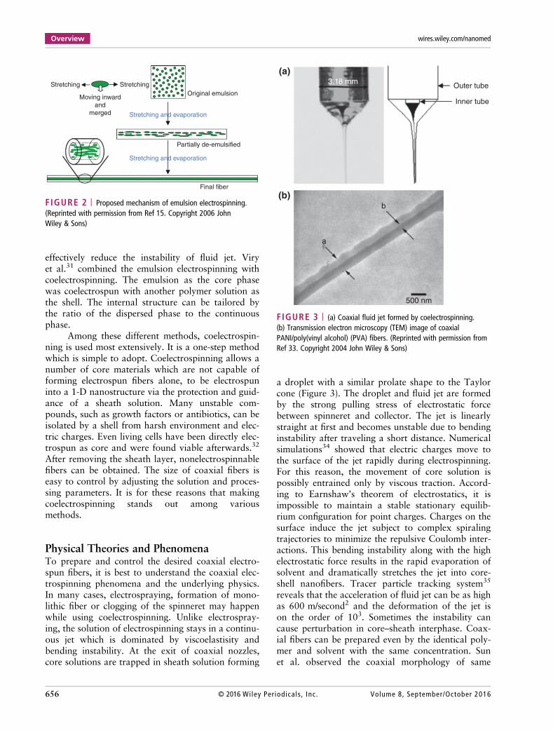
By replacing the flat collector with a rotatingdrum collector, highly aligned coaxial fibers can beobtained. Mathew et al.51 investigated the influenceof rotating speed on aligned fibers. When the rotatingspeed is low, the fibers deposited on the collector arestill unwoven. As the rotating speed increases to anoptimal range around 4000 rpm, the degree of align-ment increases significantly. However, if the speed istoo high, the resulting fibers can be broken due tothe pulling force. Aligned nanofibers are desirable inmany applications especially in tissue engineering.Aligned nanofibers resemble ECM, smooth musclecells, neurons, etc., and have been shown to offernanotopographical cues for direct migration of cells.Wang et al.52 fabricated highly aligned coaxial nano-fibers containing nerve growth factor (NGF) and usethem as nerve growth conduits (NGC). The in vivo
Overview wires.wiley.com/nanomed
658 © 2016 Wiley Per iodicals , Inc. Volume 8, September/October 2016

experiment showed that highly aligned coaxial fiberscombine the advantages of both bimolecular signalsand physical guidance cues and significantly pro-moted the nerve regeneration (Figure 5). Liao et al.53
used the same technique to prepare coaxial nanofi-bers in which poly-ε-caprolactone (PCL) as a shellwhile platelet-derived growth factor (PDFG)-bb orbovine serum albumin (BSA) as a core. The resultshowed that the loading level is high. The releasedgrowth factor maintained its bioactivity and sus-tained release was achieved.
Many researchers have interests in porousnanomaterials due to its large surface area. Porousfibers can allow large drug loading, outstanding cellattachment, sufficient nutrients transport, and quickwaste removal.54 There are various approaches forproducing porous electrospun fibers. Using a lowboiling point solvent such as dichloromethane (boil-ing point of 39.6�C) is a common method.55,56 Thepores on the surface are generated due to the fastevaporation of the solvent and the rapid solidifica-tion. Other ways include increasing humidity57 andusing a water-immersed collector.58 Gulfam et al.58
fabricated a highly porous core–sheath fiber networkby coelectrospinning and placing the collector underwater (Figure 6). The core phase was gelatin whilethe shell was PCL. The mechanism proposed is thatthe phase separation and solvent evaporation enablethe pores to form. They used this porous coaxialfiber as a scaffold to culture embryonic stem cells,breast cancer cells, and fibroblast cells. After severaldays, the cells were still viable and attached to thefibers due to covalent bonds.
Coelectrospinning is able to prepare hollownanofibers as well. Tubular nanofibers are believedto be useful in numerous applications including drugdelivery and tissue engineering. Compared to othermethods, coelectrospinning is preferred in preparinglong continuous nanoscale hollow fibers, astemplate-directed approaches only work for shortlength fibers. The fiber diameters of hollow fibersprepared by mechanical drawing method are oftenlimited to the microscale as well. Dan et al.59 firstfabricated hollow nanofibers by coelectrospinning.The researchers chose mineral oil as the core solutionand Ti(OiPr)4/PVP in ethanol as the sheath solution.The PVP was used to increase the viscoelasticity dur-ing the electrospinning process. The mineral oil wasextracted by immersing coaxial fibers in octane for anumber of hours. The hollow pure Titania fiberswere obtained by calcining in air at 500�C. TEMimages showed uniform and circular cross sections inthese hollow nanofibers. Based on this method,numerous ceramic hollow nanofibers can be fabri-cated by simply replacing Ti(OiPr)4 with other sol–gel precursors. Gu et al.30 successfully prepared hol-low LiNi0.8Co0.1O2-MgO coaxial fibers using a simi-lar method. The key in preparing hollow electrospunfibers is the selective removal of the cores. Commonways are either by thermal decomposition or throughuse of a selective solvent. The core and sheath sol-vents must be immiscible. In addition, the sheathmaterial is required to endure the annealing tempera-ture or the selective solvent conditions. The hollowmorphology can also be observed after the release ofcore phase in some drug-encapsulated coaxial
(a) (b)
5.0 µm 5.0 µm
FIGURE 4 | (a) Nylon-6,6 electrospun fibers prepared at a tip-to-collector distance of 2 cm. (b) Interconnected fiber mesh prepared at a tip-to-collector distance of 0.5 cm. (Reprinted with permission from Ref 49. Copyright 1999 Elsevier)
WIREs Nanomedicine and Nanobiotechnology Biomedical coaxial electrospun fibers
Volume 8, September/October 2016 © 2016 Wiley Per iodica ls , Inc. 659
fibers.60 With a water-soluble drug phase as the core,during the sustained release period, the core phasestarts to diffuse out. After a complete release, thesolid coaxial nanofiber becomes hollow. The corephase is required to be water soluble or has a muchhigher degradation rate than the sheath phase.
In addition to these morphologies, there areother unique structures like three-dimensional struc-tures61 and fiber-in-tube structures.62 However, bio-medical applications are rarely reported based onfibers with these structures.
COAXIAL ELECTROSPUNNANOFIBERS FOR DRUG DELIVERY
Various drug delivery systems including polymermicelles, liposomes, gels, complexes, or coaxial nano-fibers have been extensively investigated.63 Coaxial
nanofibers have the advantages of flexibility in select-ing materials and drugs, high encapsulation efficacy,high resistance of harsh environment for bioactivedrugs, low cost, ease to operate, and sustained con-trol of drug release making it an appealing system fordrug delivery.
MaterialsThe materials used for drug delivery must be biocom-patible, meaning no or minor toxicity and immuneresponses are induced. Biocompatible materials canbe further classified into biodegradable and nonbio-degradable materials. By far, most of studies are con-cerned with commercially synthetic polymers ornatural polymers. This section will focus on the sup-porting or protective matrix of coaxial fibers. Theencapsulated drugs will be discussed in later sections.Some material and solvent selections of coaxial fibers
Core
Shell
SEM of aligned nanofibers TEM of core-shell nanofibers
1 mm5 mm
Removed nerve
Nerve guidance conduit
Regenerated nerve
Nerve guidance conduit
FIGURE 5 | (a) Scanning electron microscopy (SEM) and TEM images of the aligned coaxial nanofibers. (b) Surgical implantation of thealigned coaxial fibers for nerve regeneration in a rat. The fibers were used to bridge a 13-mm nerve defect. (c) The contour of nerve after12 weeks. (d) Regenerated nerve after removing the aligned coaxial fibers. (Reprinted with permission from Ref 52. Copyright 2012 Taylor &Francis (www.tandfonline.com))
Overview wires.wiley.com/nanomed
660 © 2016 Wiley Per iodicals , Inc. Volume 8, September/October 2016

used for biomedical applications are listed inTable 1.
Biodegradable materials are more popular dueto elimination of a second surgery to remove theimplanted fibers.84 Common biodegradable syntheticpolymers including PCL, PEG, poly-L(or D or DL)-lactide (PLA, PLLA, PDLA, PDLLA), PVA,poly(lactic-co-glycolic acid) (PLGA), poly(L-lactide-co-caprolactone) (PLCL), etc., are widely used. Poly-saccharides such as chitosan, cellulose, alginate, chi-tin, and proteins such as collagen, gelatin, zein, andsilk fibroin (SF) are commonly used natural poly-mers. Sridhar et al.85 have summarized the naturalmaterials in the field of electrospun nanofibers forbiomedical applications. Additionally, Lee et al.86
summarized the polysaccharides based electrospunfibers.
One important issue regarding material selec-tion is the rate of degradation. When using nonbio-degradable materials as a drug delivery carrier, therelease of the drug is primarily through slow diffu-sion. However, the release behavior is more compli-cated when using biodegradable materials. Generally,a sustained and controlled release is preferred. In abiodegradable system, as the supporting materialdegrades, the release of the drug can become veryrapid. Sometimes the local drug concentration can be
high enough to be toxic. Altering the compositionand crystallinity of a polymer blend or copolymer isa straightforward way to control the rate of degrada-tion. For example, PLGA is a copolymer widely usedin therapeutic devices. Depending on the ratio of lac-tide to glycolide, the mechanical properties, degrada-tion rate, hydrophilicity, and crystallinity can becontinuously controlled. Specifically, the presence ofthe methyl group in PLA makes it more hydrophobicthan PGA and thus degrades slower. A detailedreview of PLGA can be found in Ref 87. Viry et al.31
and Wang et al.52 have used the PLGA as a shell tocontrol the degradability of drug delivery carriers.The addition of some proteins can regulate the bio-degradation rate as well. Xiao et al.88 encapsulatedproteinase K in PEG matrix as a core while PLA wasused as a shell. The proteinase K is a fungal serineprotease which is able to accelerate the hydrolysisof PLA.
In most cases, a hybrid blend of natural poly-mer and synthetic polymer is desirable. The syntheticpolymers can provide sufficient mechanical propertiesand improve electrospinnability. The natural poly-mers can improve the cellular attachment for a bettermimicking of the environment of ECM. In addition,the enhanced hydrophilicity provided by naturalpolymers is beneficial when carrying hydrophilic
Power Power
Syringe pump
Water-immersed collector
Core-shell nanofiber Porous core-shell fiber network
0.1 µm
Shell (PCL)
Core (Gelatin)
(a) (b)(c)
FIGURE 6 | (a) Set up of coelectrospinning for core–sheath nanofibers. (b) Set up of coelectrospinning in a water-immersed collector forporous core–sheath nanofibers. (c) TEM image of core–sheath gelatin/poly-ε-caprolactone (PCL) nanofibers. (Reprinted with permission from Ref58. Copyright 2011 American Chemical Society)
WIREs Nanomedicine and Nanobiotechnology Biomedical coaxial electrospun fibers
Volume 8, September/October 2016 © 2016 Wiley Per iodica ls , Inc. 661

TABLE
1|Recent
Works
onCo
axialN
anofibersforD
rugDe
liveryandTissue
Engineering
Core
Core
solvent
Shell
ShellSolvent
Drug
Tissue
Reference
Endogenous
basic
fibroblast
grow
thfactor
(bFG
F)in
PVA
Water
PCL-co-PEG
andPC
LMethanoland
chloroform
Endogenous
bFGFand
epidermalgrow
thfactor
(EGF)
Skin
64
Amodeldrug
inPLLA
DMF/Acetone
PVPwith
nanosilverp
articles
(NSPs)
Water
Modeldrug
38
PGS
HFP
Fibrinogen
HFP
Cardiactissue
65
PCL
HFP
Gelatin
HFP
Bone
66
Epidermalinductionfactors
[EGF,insulin,
hydrocortisone,and
retinoicacid(RA)]
5%BSA
GelatinandPLLCL
HFP
EGF,insulin,h
ydrocortisone,
andRA
Skin
67
Nylon-6
Form
icacid
andacetic
acid
Lacticacid(LA),then
mineralized
tocalcium
lactate(CL)
Form
icacidand
aceticacid
Bone
68
PLLA
DCM
ChitosanandPEO
Form
icacid
Vascular
gasket
69
Dexamethasone
(DEX)
Plantsoil
Silkfibroin(SF)/PEO
Water
DEX
Endothelium
70
Ketoprofen
(KET)inethyl
cellulose
Ethanol
PVP
Ethanoland
DMAc
KET
71
Salicylicacid(SA)
inPEG
DCM
and
DMAc
PLA
DCM
andDM
AcSA
39
AmpicillininPM
MA
Chloroform
and
methanol
Nylon-6
Form
icacid
Ampicillin
72
Levetiracetam
in75:25PLGA
Water
85:15PLGA
DCM
Levetiracetam
31
KETinzein
Ethanol
KETinPV
PDM
Acandethanol
KET
73
Dimethyloxalylglycine
(DMOG)inPD
LLA
DMF
PHB
DMFand
chloroform
DMOG
50
β-tricalcium
phosphate(TCP
)inPLA
DMFand
chloroform
PLA
DMFand
chloroform
β-TCP
Bone
55
Gelatin
Aceticacid
PCL
Aceticacid
Embryonicstem
cells,b
reast
cancer
cells,and
fibroblast
cells
58
BSA
Water
PSL-lim
onene
BSA
74
bFGF-2inPEO
Water
PCL
DCM
andDM
FbFGF-2
Connectivetissue
75
BSAor
lysozymeindextran
Water
PCLandPEG
BSAor
lysozyme
76
Overview wires.wiley.com/nanomed
662 © 2016 Wiley Per iodicals , Inc. Volume 8, September/October 2016

TABLE
1|Co
ntinued
Core
Core
solvent
Shell
ShellSolvent
Drug
Tissue
Reference
DMFand
chloroform
Metoclopram
ide
hydrochlorideinPV
AWater
PCL,PLLA
or80:20PLGA
Chloroform
and
DMF
Metoclopram
ide
hydrochloride
40
Lysozymeinmethylcellulose
PBS
PDLLA
Chloroform
lysozyme
16
Nylon-6
Form
icacid
andacetic
acid
LAor
CLForm
icacidand
aceticacid
Bone
21
PLA
Triflouro-
aceticacid
Chitosan
Triflouro-acetic
acidandacetic
acid
77
Sodium
alginate
Water
PEO
Water
Connectivetissue
78
bFGFincyclodextrin
PBA
PELA
Chloroform
bFGF
Skin
17
PCL
TFE
Collagen
TFE
Skin
79
Gentamicinincollagen
Water
PLA
HFPand
chloroform
Gentamicin
80
Flurbiprofen
axetil(FA)
inPV
PEthanoland
DMF
PLGA
DCM
andDM
FFA
81
Bone
morphogeneticprotein
2(BMP-2)
inBSA
PBS
DEXinPLLACL–collagen
HFP
BMP-2anddexamethasone
(DEX)
Bone
82
Vascular
endothelialgrowth
factor
(VEG
F)indextranor
BSA
Water
PLLCL
Chloroform
VEGF
CardiacTissue
27
Ibuprofen(IB
U)inzein
DMF
DMF
DMF
IBU
83
Recombinant
ratβ
-NGFin
BSAandPEG
Water
PLGA85:15
HFP
Recombinant
ratβ
-nerve
grow
thfactor
(β-NGF)
Peripheralnerve
52
Platelet-derived
grow
thfactor
(PDFG)-b
bor
BSA
Water
PCL
DCM
andethanol
(PDFG)-b
bor
BSA
53
BSA
,bo
vine
serum
albu
min;DCM,dichlorometha
ne;DMAc,
dimethy
lacetamide;
DMF,
dimethy
lformam
ide;
HFP
,1,1,1,3,3,3-hexa
fluo
ro-2-propa
nol;PA
-6,ny
lon-6;
PBS,
phosph
atebu
ffered
salin
e;PC
L,po
ly-
ε-caprolactone;PE
G,po
lyethy
lene
glycol;PE
LA,po
ly(ethyleneglycol)-po
ly(D
L-lactide);PE
O,po
lyethy
lene
oxide;
PGS,
poly(glycerolseba
cate);PH
B,po
ly(3-hyd
roxy
butyrate);PL
GA,po
ly(lactic-co
-glycolic
acid);
PLLA,p
oly-L-lactide;PL
LCL,p
oly(L-lacticacid)-co-poly-(ε-cap
rolacton
e);P
MMA,p
oly(methy
lmetha
crylate);P
S,po
lystyrene;
PVA,p
oly(viny
lalcoh
ol);PV
P,po
ly(vinyl
pyrrolidon
e);T
FE,2
,2,2-trifluo
roetha
nol.
WIREs Nanomedicine and Nanobiotechnology Biomedical coaxial electrospun fibers
Volume 8, September/October 2016 © 2016 Wiley Per iodica ls , Inc. 663

drugs. Giner et al.80 developed PLA–collagen coaxialfibers for controlled delivery of gentamicin antibio-tics. He chose PLA as a shell and collagen as a core.Collagen is supposed to hold gentamicin and increasethe affinity of gentamicin to PLA in fibers.
Drug Release MechanismThe drug release mechanism is dominated by diffu-sion when nonbiodegradable polymers are chosen asa matrix. For water-soluble drugs, the water mole-cules diffuse into the polymer matrix first. Then thedrugs are dissolved in water and finally diffuse out ofthe polymer networks.89 For biodegradable coaxialfibers, the release mechanism becomes more compli-cated. Three main factors including diffusion, degra-dation, and shell thickness should be consideredtogether. The basic mechanisms of drug release areshown in Figure 7.
The typical release profile for a coaxial drugdelivery system is an initial burst release followed bya long period of sustained release. This initial releaseis through the quick diffusion of drugs deposited onthe fiber surfaces. The highest drug concentration islocated in the core phase. The sustained release ismainly controlled by diffusion through the polymermatrix and slow degradation of the polymer. Tianet al.27 dissolved the vascular endothelial growth fac-tor (VEGF) in BSA water solution and dispersedwater phase with oil phase (PLCL in chloroform).After emulsion electrospinning, the resulting nanofi-bers were found to have a coaxial morphology. Invitro release study showed a sustained release for upto 672 h (28 days). The entire release profile can besplit into two stages, the burst stage (first 24 h) andthe sustained stage (later 648 h). By changing theprotective agent from BSA to dextran, the burstrelease can be largely suppressed from 9.6% forPLCL–BSA to 1.0% for PLCL–Dextran. The
difference is due to the changed drug distribution incoaxial fibers. An x-ray photoelectron spectroscopy(XPS) study that showed more VEGF was located onthe surface of PLCL–BSA. After 28 days, the averagefiber diameter became smaller compared with thefibers before the release. In addition, breakages anderosion of coaxial fibers were found in SEM imageswhich further substantiated the proposed mechanism.The results indicated that the release pattern isgreatly determined by the drug quantity loaded onthe surface or sheath phase. A higher loading on thesurface will result in a stronger burst release. Wanget al.52 and Yan et al.82 reported similar biphasicdrug release patterns and mechanisms.
The amount of initial released drugs can be tai-lored by adjusting the flow rate of the sheath solutionas well. Yu et al.71 used PVP as the sheath, and Keto-profen (KET) in ethyl cellulose as the core. Two dif-ferent sheath fluid flow rates (0.5 and 0.8 mL/h) arechosen to investigate the influence of the sheath solu-tion flow rate. The initial burst is 30.7 and 41.2%for 0.5 and 0.8 mL/h, respectively.
Nguyen et al.39 compared porous coaxial fiberswith nonporous coaxial fibers. They fabricated theporous coaxial composite fibers with salicylic acid(SA) in PEG as a core and PLA as a sheath. The non-porous fibers released a much smaller amount of SAin the first day. Over a period of 5 days, the amountof drugs released from the porous fibers is two timeshigher than nonporous samples. The porous mor-phology has a higher surface area that allows easypenetration of water molecules into the core phase.
Binary or multiple releases of drugs are capableof releasing two or more kinds of drugs simultane-ously. In most cases of the binary release coaxialfiber system, two drugs are dispersed separatelyinto two different phases. Choi et al.64 prepared thedual release coaxial fibers by encapsulating basicfibroblast growth factor (bFGF) in the core phase
(a) (b) (c) (d)
FIGURE 7 | Basic release mechanisms: (a) Diffusion through pores. (b) Diffusion through polymer networks. (c) Osmotic pumping. (d) Erosionof a polymer. (Reprinted with permission from Ref 89. Copyright 2011 Elsevier)
Overview wires.wiley.com/nanomed
664 © 2016 Wiley Per iodicals , Inc. Volume 8, September/October 2016

and immobilized epidermal growth factor (EGF)on the surface of the shell. Figure 8 is the schematicdiagram. Surface immobilization was accomplishedthrough the reaction between surface exposed aminegroup and carboxyl group of EGF. The release pro-files showed a clear binary release of each growthfactor. The bFGF showed a high initial burst in 24 h,while the EGF showed negligible release in first7 days. This physically loaded bFGF released muchfaster than chemically loaded EGF. A similar studyshowed the same release profiles for this type of sys-tem.90 Multiple drugs can also be incorporated incore phase together. Jin et al.67 encapsulated multipleepidermal induction factors (EIFs) including EGF,insulin, hydrocortisone, and retinoic acid (RA) in thecore by dissolving them in a BSA solution. Thesheath is gelatin and poly-L-lactide (PLLCL). Noburst release was observed, instead a stable and sus-tained release for more than 15 days was shown bythe release profile. A sheath phase can protect thecore phase and effectively suppress the initial burst.The EIF encapsulated coaxial nanofibers could beserved as a promising graft for skin regeneration.
ProteinsMore and more bioactive drugs have been incorpo-rated into nanofibers. One important advantage ofcoelectrospinning compared with the conventionalmethod is that the core phase can be protected by thesheath phase from direct exposure to the harsh envi-ronment. By using the blend-electrospinning method,the bioactive molecules can lose their bioactivitycompletely or at least partially. The protecting agentsare of importance. Special care must be taken toensure that drugs remain bioactive.
Fibrinogen (factor I) is a glycoprotein thataids in the formation of blood clots. One of thebenefits of fibrinogen is that it can be produced froma patient’s own blood so no inflammatory responseswill be induced. Ravichandran et al. 65 fabricatedpoly(glycerol sebacate)/fibrinogen (PGS/fibrinogen)coaxial fibers. The core phase of elastomeric PGS canprovide mechanical properties comparable to nativetissue and the shell phase of fibrinogen is designed topromote cell–fibers interactions.
Wang et al.74 and Jiang et al.76 encapsulatedwater-soluble proteins, such as BSA and lysozyme, in
In aqueous phase
EG
F
conju
gatio
n
Released bFGF
bFGF/EGF NF
PEG-NH2 linker
NH2
EDC, HOBt
HOOC
FGF
EGF
Exposed-amine groups on
surface of nanofiber
bFGF nanofiberby coaxial electrospinning
Proliferation of epidermal cells by releasedbFGF and conjugated EGF from bFGF/EGF NF
FIGURE 8 | Schematic of preparation of coaxial nanofibers with basic fibroblast growth factor (bFGF) encapsulated in the core and epidermalgrowth factor (EGF) immobilized on the surface. The fibers showed a dual release profile. (Reprinted with permission from Ref 64. Copyright 2011Royal Society of Chemistry)
WIREs Nanomedicine and Nanobiotechnology Biomedical coaxial electrospun fibers
Volume 8, September/October 2016 © 2016 Wiley Per iodica ls , Inc. 665

the core phase and investigated their release profiles.Wang used emulsion electrospinning and chose poly-styrene (PS) as the sheath while model protein–BSAas the core. His research suggested that the releaserate can be tuned by tailoring the molecular weightof PS. With a higher molecular weight polymer, anincreased release rate was observed. Higher molecu-lar weight polymer has a higher viscosity whichslows the inward movement of water droplets duringthe emulsion electrospinning. So, more water dro-plets containing proteins are located on or near thesurface. Jiang controlled the release rate by varyingthe feed rate of the core solution. For a higher feedrate, there is an associated increase in loading ofmodel protein and accelerated the release of BSA. Bymeasuring the turbidity change at 450 nm in aMicrococcus lysodeikticus bacterial cell solution, therelative activity of lysozyme was determined. No sig-nificant decrease was observed suggesting that lyso-zyme remains bioactive after the electrospinning andrelease process.
Growth FactorsAs a special type of protein, growth factors play animportant role in drug delivery and tissue regenera-tion. Typically, growth factors are capable ofinstructing specific cellular responses. The signalingcan result in cell actions including cell growth, prolif-eration, migration, healing, and cellular differentia-tion.91 The instruction is completed through bindingto specific transmembrane receptors on the targetcells.
Bone morphogenetic protein 2 (BMP-2) hasbeen shown to stimulate the production of bone tis-sue. Signal transduction studies reveal that Smad1,5, and 8 are the immediate downstream molecules ofBMP receptors.92 The BMP can potently induce oste-oblast differentiation in a variety of cells. Dexameth-asone (DEX) is a steroid which has been adopted tosupport the osteogenic differentiation in vitro,together with β-glycerophosphate and ascorbic acid.Su et al.82 successfully incorporated BMP-2 and DEXinto the poly(l-lactide-co-ε-caprolactone) (PLLACL)/collagen coaxial fibers and controlled/altered therelease profile. The release result showed a similarprofile to BMP-2 and DEX. After 504 h, the releasedamount of BMP-2 and DEX reached 73.7 and60.5%, respectively. Diffusion and degradation ofcoaxial fibers were thought to control the releaserate. Increased alkaline phosphatase activity, mineral-ization, and osteoblast marker expression wereobserved on coaxial fibers showing that the drugs
remained bioactive after electrospinning and releas-ing. Srouji et al.93 used PCL/polyethylene oxide(PEO) fibers as the carrier for BMP-2 and the scaf-fold showed a sustained release. These results demon-strate that coaxial electrospun nanofibers have agreat potential for bone tissue regeneration.
Poor innate healing capability of fibroblastictissues (e.g., pelvic floor fascia) can be attributed tothe scarcity of fibroblastic to produce collagen.Rubert et al.75 preserved bFGF in PCL/PEO ultrafinefibers (which is biocompatible and anti-inflamma-tory). The diameter of the fibers was well tuned bychanging the electrospinning parameters. No toxiceffects were found by lactate dehydrogenase activityassay. The drug delivery system showed a controlledrelease and enhanced connective tissue regenerationdemonstrating that PCL/PEO coaxial nanofiber is apractical delivery vehicle to preserve the functionalityof growth factors. Yang et al.17 imbedded bFGF intocoaxial fibers using emulsion electrospinning tech-nique. An initial burst release of bFGF as low as14.0 � 2.2% was observed, followed by sustainedrelease for around 4 weeks. Owing to a high affinityof polymeric matrix with bFGF, the protein detach-ment from the core phase is slow which leads to alow initial burst release. Liao et al.53 encapsulatedPDGF in aligned core–sheath nanofibers. The PDGFcould be released completely in 35 days with nearzero-order kinetics and preserved bioactivity. Tianet al.27 encapsulated VEGF in PLCL/BSA and PLCL/dextran. Choi et al.64 investigated bFGF- and EGF-binary release system.
AntibioticsAfter surgery, infection is a big concern. Severe infec-tions can require another surgical intervention whichis costly and can also endanger health.94 Antibioticsare extensively used as biocides to kill or inhibit thegrowth of bacteria. The coaxial fibers containingantibiotics are promising in applications of wounddressings. Ignatova et al.95 summarized some antibio-tics such as levofloxacin, tetracycline hydrochloride,ciprofloxacin, moxifloxacin, Fusidic acid, and silvernanoparticles (AgNPs) encapsulated nanofibers forwound dressing.
Giner et al.80 examined the encapsulation ofgentamicin antibiotic into PLA/collagen coaxialfibers. As for comparison, pure PLA fibers and blendfibers of PLA–collagen containing gentamicin wereprepared. The gentamicin was almost completelyreleased (98%) within the first 50 h for the blend ofPLA–collagen fibers. Regarding PLA fibers, because
Overview wires.wiley.com/nanomed
666 © 2016 Wiley Per iodicals , Inc. Volume 8, September/October 2016

of the hydrophobic nature of PLA, only 33% of gen-tamicin was released in the first 50 h. From 250 h to500 h, almost no release was observed and theretained amount was as high as 58%. However, thecoaxial fibers partially suppressed the burst releaseand showed a sustained release instead. The resultantfibers displayed a high antibacterial performanceagainst Staphylococcus epidermis, Pseudomonas aer-uginosa, and Escherichia coli along with a high cellproliferation capacity. The results demonstrate thatdrug-encapsulated coaxial fibers are promising drugdelivery carrier with strong and timing controllableantibacterial properties.
Sohrabi et al.72 incorporated ampicillin intopoly(methyl methacrylate) (PMMA)/nylon-6 coaxialfibers. As described before, adding salt will increasethe conductivity of solution which results in astronger electric force. Owing to the ionic nature ofthe ampicillin sodium salt, the diameter of fibersdecreases by increasing the drug content. The investi-gation of release kinetics indicated that the burstrelease stage (Stage I) followed non-Fickian diffusionwhile Stage II and III obeyed Fickian diffusion. StageIII has a lower diffusion coefficient due to the crystal-lization as a consequence of the long time incubationin potassium buffer solution. Coaxial fibers with ahigh concentration of drugs (20%) showed the high-est antibacterial activity against Gram-positive Liste-ria innocua (Figure 9).
The AgNPs have been recognized as the opti-mal candidates among recently nontraditional anti-bacterial agents.96 Zhang et al. reported a drugdelivery system in which PVP and AgNPs wereused as the sheath and PLLA and a model drug wereused as the core. The binary release profile and anti-microbial properties were studied.38 For chitosan-based coaxial fibers, chitosan itself can be used asantibacterial agent and help deliver the modeldrug.77
Other DrugsLevetiracetam (Lev, Keppra) is an antiepileptic drugused to treat epilepsy in adults.97 Viry et al.31 exam-ined the release of Lev in PLGA coaxial fibers. Anovel technique which combined emulsion and coe-lectrospinning was presented. Figure 10 is the mor-phology of coaxial and emulsion/coaxial fibers.Water phase containing Lev are emulsified in a PLGAoil phase which is then coelectrospun with purePLGA solution. After the evaporation of the solvent,the core architecture was composed of microcavitiessurrounded by fiber bulk. The microcavities wereregarded as drug reservoirs and showed distinctiverelease kinetics from conventional coaxial fibers. Asmall disperse-to-continuous phase ratio in the coresolution allows the formation of small cavities and along diffusive length, whereas a bigger ratio forms alarger and shorter diffusive path. The presence ofdrug reservoirs allows a nearly linear and sustainedrelease of Lev for over 21 days. This PLGA/Levnanofiber is aimed to be implanted into the brain fortreatment of epilepsy. In addition, the technique ofemulsion coelectrospinning provides a novel way toprepare nanofibers with drug microreserviors. Thisarchitecture is ideal for decreasing the diffusion rateof drugs, especially for those with low molecularweight.
DEX is a widely used steroid that treats variousdiseases with its anti-inflammatory and immunosup-pressant effects. Chen et al.70 prepared a core/shellnanofiber with DEX as the core and SF/poly(ethyleneoxide) as the sheath by emulsion electrospinning.‘Green electrospinning’ without an organic solventensured the nontoxicity of the fibers. In vitrostudy showed that the released drug could reduce thelipopolysaccharide (LPS)-induced porcine hipartery endothelial cells (PIECs) inflammatorydamage. The release profiles of nonsteroidal anti-inflammatory drugs such as ketprofen,71,73 flurbipro-fen axetil,81 SA,39 ibuprofen,83 etc. were also wellstudied.
1.2
1
0.8
0.6
0.4
0.2
0
Op
ticl
a d
ensi
ty
Bla
nk
NF
NF
-AC
1
AC
1
NF
-AC
2
AC
2
NF
-AC
3
AC
3
NF
-AC
4
AC
4
NF
-AC
5A
C5
Blank NF-AC AC
FIGURE 9 | Antibacterial activity of ampicillin incorporated fibersby optical density measurement. The gradual increase of antibacterialeffectiveness for NF-AC1, NF-AC2, NF-AC3, NF-AC4, and NF-AC5(corresponding to the concentration of 1, 2, 5, 15, and 20% ampicillinrespectively). (Reprinted with permission from Ref 72. Copyright 2013Elsevier)
WIREs Nanomedicine and Nanobiotechnology Biomedical coaxial electrospun fibers
Volume 8, September/October 2016 © 2016 Wiley Per iodica ls , Inc. 667

COAXIAL ELECTROSPUNNANOFIBERS FOR TISSUEENGINEERING
Tissue engineering has attracted significant researchinterests for decades. Biological substitutes can bedeveloped from an individual’s own cells with excel-lent biocompatibility and the ability to repair, regen-erate and remodel.98 The electrospinning techniquehas been found to have great potential for regenera-tion of tissues such as bones, cartilage, nerves, bloodvessels, cardiac muscles, skins, etc. The mechanical
strength, bioactive components, hydrophilicity, sur-face conditions, and compatibility are main concernswhen using electrospun fibers for tissue engineering.The electrospun fibers can be used as a tissue engi-neering scaffold due to a similar morphology toECM. The inherently high surface to volume ratio ofelectrospun fibers can enhance drug loading, masstransfer, cell attachment, proliferation, and differenti-ation. With a superior performance in drug deliveryas described above, the coaxial fibers with controlledrelease are found to have better performance thanmonolithic fibers on tissue engineering.
(a) (b)
(c) (d)
(e) (f)
FIGURE 10 | SEM images of coaxial fibers (a and c) and emulsion coaxial fibers (b, d, e, and f ). (e) A bigger disperse-to-continuous phaseratio (1/5) in core. (F) A smaller ratio (1/55).31
Overview wires.wiley.com/nanomed
668 © 2016 Wiley Per iodicals , Inc. Volume 8, September/October 2016

MaterialsLike drug delivery, the materials used for tissue engi-neering must be biocompatible. Also, biodegradableor hybrid materials are preferred due to the elimina-tion of a second surgery to remove the implantedscaffold.84 The rate of degradation can be controlledby hydrophilicity, polymer molecular weight, crystal-linity, and polymer blends to coincide with the rateof tissue regeneration. If the degradation rate is tooslow, the tissue regeneration will be impeded. How-ever, if the degradation rate is too fast, the mechani-cal stability and the release of encapsulated drugswill be greatly compromised.
The coaxial fiber scaffold should have amechanical strength close to the anatomical sitewhere the scaffold is to be implanted. It must bestrong enough and structurally stable in order toallow surgical handling during the process of implan-tation.99 The natural materials such as chitosan, gela-tin, collagen, and chitin usually have weak physicalproperties. Nguyen et al.77 investigated the mechani-cal properties of PLA/chitosan (PLA/CS) nanofiberswith different compositions. The pure chitosan fibershave poor mechanical properties (0.5 MPa tensilestrength and 5.2% strain at break) while the PLA/CScoaxial fibers can reach 1.4 MPa and 10.9%, respec-tively. As for comparison, pure PLA fibers had a hightensile strength of 3.3 MPa. With the introduction ofPLA component, the mechanical properties ofPLA/CS coaxial nanofibers can be significantlyimproved. Therefore, an ideal mechanical strengthcan be tuned by changing the composition of materi-als. Some material and solvent selections of coaxialfibers used for tissue engineering are listed inTable 1.
BoneBone tissue is the major structural and supportiveconnective tissue of the body formed by cells, organicmatrices (type I collagen fibers), and inorganic salts(hydroxyapatite, HA). Bone tissue engineering oftenuses the osteoblasts seeded fibrous scaffold as a tem-plate for bone tissue regeneration. Numerous studiesincorporated mineral components such as HA, demi-neralized bone powders, calcium carbonate, andβ-tricalcium phosphate (TCP) into coaxial fibers topermit and promote the proliferation and ossificationof bone cells.55 McCullen et al.100 found that theoptimal concentration of calcium ions for enhancinghuman adipose-derived stem cells (hASCs) osteogenicdifferentiation in culture medium is 8 × 10⁻³ M. Thecoaxial fibers with a sustained release can deliver cal-cium ions at a nearly constant optimal concentration,which is impossible for single component fibers. Asliet al.55 doped TCP into PLA fibers with three differ-ent morphologies (single component, porous fibers,and core/sheath fibers) to investigate the release pro-files and bone tissue engineering performances. Dif-ferent from the other two morphologies, no burstrelease was observed for coaxial fibers and the rateof release remained fairly constant for a long periodof 36 days. Desorption and degradation werethought to be primary mechanisms for the release ofTCP nanoparticles. The scaffold degradation analysisindicated that porous fibers exhibited the highest rateof weight loss (30% degraded in 36 days, Figure 11).Live/dead analysis showed that all three fibers main-tained high hASC viability after 21 days.
Pant et al.21 fabricated nylon-6/lactic acid(LA)coaxial nanofibers by simply blending the LA andnylon-6 solution. Unlike the conventional single
1 2 4 8 12 16 20 24 28 32 36
30
25
20
15
10
5
0
Weig
ht lo
ss (
%)
Porous Single component Core sheath
FIGURE 11 | Weight loss as a function of time for porous, single component, and coaxial fiber scaffolds. (Reprinted with permission from Ref55. Copyright 2012 John Wiley & Sons)
WIREs Nanomedicine and Nanobiotechnology Biomedical coaxial electrospun fibers
Volume 8, September/October 2016 © 2016 Wiley Per iodica ls , Inc. 669

nozzle electrospinning, the resulting fibers showed acoaxial morphology instead of a monolithic mor-phology. The complex phase separation was thoughtto be responsible for the core–sheath structure. Withfurther neutralization by calcium hydroxide, thesheath phase of LA converted into calcium lactate(CL) which can offer nucleation sites for calciumphosphate during bone regeneration. The cell viabil-ity test showed the Nylon-6/CL scaffold had a higherviability of cells than pristine nylon-6 scaffold. A bet-ter cell attachment and proliferation were alsoobserved in Nylon-6/CL fibers. The results indicatedthat the formation of CL on the fiber surface canincrease the biocompatibility and facilitate the nucle-ation of HA.
Some researchers used coaxial fibers as reinfor-cing fillers as a part of scaffold. Kai et al.66 presenteda hydrogel-based tissue scaffold by incorporatingPCL/gelatin coaxial electrospun nanofibers into gela-tin hydrogel matrix. This electrospun fiber of rein-forced hydrogel nanocomposite can provide thenecessary mechanical support as a scaffold andenhance the proliferation of bone marrow mesenchy-mal stem cells (BM-MSCs). By increasing the amountof fibers, the Young’s modulus increased more than6 times from 3.29 to 20.30 kPa. With interactionsbetween nanofibers and hydrogel, the swelling ratiodecreased from 600 to 450–500%.
Cardiac TissueHeart failure is a major cause of death worldwide,claiming 17 million people each year.101 The myocar-dial infarction, which is well known as ‘heart attack,’can kill cardiomyocyte, remodel myocardial, anddamage cardiac function, finally resulting in heartfailure.102 Cardiac tissue engineering aims to create afunctional tissue construct that is able to repair orreform the structure and function of the damagedmyocardium.103
Fibrin is widely used in tissue engineeringresearch. However, low mechanical stiffnesshas restricted its applications. Ravichandran et al.65
designed poly(glycerol sebacate)/fibrinogen (PSG/fibrinogen) coaxial fibers as a cardiac patch. The corephase of PSG is elastomeric and deformable therebymaking the patch has similar mechanical propertieswith native cardiac tissue. The patch is extremelyhydrophilic with the water contact angle as low as10.4 � 1.6�. By immunocytochemical analysis, thecardiac-specific marker proteins showed the charac-teristic striations and muscle filaments. This provedthat the isolated cells were functional cardiomyo-cytes. Moreover, the cells cultured on the coaxial
fiber patch could form gap junctions with the hostcardiomyocytes and facilitated the myocardial regen-eration. The PGS/fibrinogen coaxial fibers have suita-ble mechanical properties comparable to that ofnative tissue and excellent biocompatibility, makingthem an ideal cardiac patch for the regeneration ofinfarcted myocardium.
Regenerative medicine such as growth factorscan assist in the process of tissue engineering. Tianet al.27 encapsulated BSA or dextran protected VEGFinto the PLCL fibers with a core/shell structure. Witha prolonged release of bioactive growth factor, excel-lent attachment and proliferation of human bonemarrow-derived mesenchymal stem cells (MSCs)were observed on the VEGF/PLCL coaxial scaffoldsin vitro test. They could be potential substrates forcardiac tissue regeneration.
SkinA recent study showed that over 6 million people suf-fer from a severe burn every year, and among these300,000 persons die.104 It has been proven that coax-ial fibers used as a scaffold help wound healing fromhemostasis to re-epithelialization. Various growthfactors can stimulate the migration and differentia-tion of cells suh as fibroblasts, endothelial cells, kera-tinocytes, etc in skin tissues. Yang et al.17 imbeddedbFGF into poly(ethylene glycol)-poly(DL-lactide)(PELA) nanofibers by emulsion electrospinning. Invitro study on a mouse embryo fibroblast showedthat bFGF/PELA scaffold can help cell adhesion, pro-liferation, and secretion of ECM. In vivo experimentwas conducted for the dorsal wounds of diabeticrats. The bFGF-loaded scaffold revealed a signifi-cantly higher wound healing rate with a complete re-epithelialization and regeneration of skin. Theseresults suggest the potential use of PELA/bFGF coax-ial fiber mats to rapidly heal of wounded skin forpatients with diabetic mellitus.
Apart from bFGF, there are various mediatorsthat can stimulate receptors facilitating wound heal-ing. The EGF has been shown to stimulate the prolif-eration and migration of keratinocytes and acceleratewound closure in vivo.105 Insulin can improvewound matrix formation.105 Hydrocortisone and RAhave been reported to promote proliferation of kera-tinocyte and guide epithelial differentiation of stemcells.106 Jin et al.67 incorporated EGF, insulin, hydro-cortisone, and RA as multiple EIFs with gelatininto PLLCL fibers. Blend and coelectrospinning wereperformed for comparison. They optimized therelease profiles of multiple mediators for a desiredperiod of time. The in vitro cell proliferation of
Overview wires.wiley.com/nanomed
670 © 2016 Wiley Per iodicals , Inc. Volume 8, September/October 2016

adipose-derived stem cells (ADSCs) showed that thecell proliferation on Gel/PLLCL/EIF (core/shell) was43.6% higher than Gel/PLLCL/EIF (blend) after15 days due to a sustained release of EIF. The epider-mal differentiation study indicated that the percent-age of keratinized epithelial cells on the Gel/PLLCL/EIF (core/shell) and Gel/PLLCL/EIF (blend) were62.2 and 43.0%, respectively. ADSCs on a scaffoldof Gelatin/PLLCL, PLLCL, and TCP without EIFremained undifferentiated with a fibroblastic pheno-type. This study can sheds light on the design of asite-specific niche-like microenvironment for epider-mal differentiation and present insights on the designthe delivery carrier containing multiple drugs forwound healing.
Zhang et al.79 investigated the influence of sur-face coating for proliferation of human dermal fibro-blasts (HDFs). It has been previously proven thatcollagen on the surface can facilitate the proliferationof HDF. They prepared coaxial electrospun nanofi-bers of PCL/collagen with collagen as the shell bycoelectrospinning. Rough coaxial PCL/collagennanofibers were also prepared by soaking the PCLfibers in a collagen solution overnight. Their experi-ment results showed that the density of HDF on acoaxial PCL/collagen scaffold is almost twice as highas a rough coaxial PCL/collagen scaffold. And SEMimages showed that the coaxial PCL/collagen scaffoldencouraged cell migration within the scaffolds.Therefore, cells are more prone to interact with coax-ial fibers than rough-coated fibers. Chen et al.70
encapsulated DEX into SF/PEO nanofibers by emul-sion electrospinning. The DEX-loaded scaffold canreduce the PIECs inflammatory damage and LPS-induced apoptosis.
CartilageCartilage is a type of connective tissue which consist-ing of 68–85% water, 10–20% collagen, and 5–10%are proteoglycans.107 The specialized cells in cartilageare called chondrocytes. They can maintain andremodel the cartilage tissue. However, the self-repairability of cartilage is poor due to the avascularity andthe low mitotic ability of chondrocytes.108 Currently,no appropriate clinical method is able to fully regen-erate the functional cartilage. Despite the variousmethods, scaffolds, seed cells, and bioactive factorsare the three basic components for cartilage regenera-tion. Bone marrow-derived stem cells (BMSCs) andrecombinant human transforming growth factor-b1(rhTGF-b1) are seed cells and growth factors exten-sively used in cartilage tissue engineering research.Man et al. 60 designed a codelivery system using
coaxial electrospun fibers as the scaffold. The core iscomposed of PVP/BSA/rhTGF-b1 composite, whereasthe shell is composed of PCL. Owing to the hydro-phobic nature of PCL, there are no recognition sitesfor cell attachment which means the cellularresponses and interactions between cells and scaf-folds are poor. A BMSC-affinity peptide (E7) whichis discovered by Man’s group is conjugated to thecoaxial fibers by covalent bonds to improve theBMSC adhesion to the scaffold. Owing to the lowdegradation rate of shell PCL, a sustained rhTGF-b1growth factor release was observed. TEM imagesshowed hollow tube morphology after the release ofdrug. More cartilage-specific genes were expressed inthe coaxial scaffolds than the control samples after a14-day incubation period, in vitro, meaning the scaf-fold can effectively promote BMSC chondrogenic dif-ferentiation. This codelivery scaffold was found tosynchronously improve all of the three basic compo-nents (scaffold, seed cells, and bioactive factors)needed for cartilage tissue engineering which is rarelyseen in other electrospun scaffolds. Platelet deriva-tives are known to be an efficient source of naturalgrowth factors. Buzgo et al.109 incorporated plateletα-granules into the coaxial fiber to exam its perfor-mance in cartilage regeneration. The bioactivity ofplatelet α-granules was well preserved. The scaffoldwas observed to stimulate the cell viability and chon-drogenic differentiation of mesenchymal stem cells.These results indicated that coaxial electrospun fibersare promising scaffolds for cartilage regeneration.However, further animal tests are necessary to fullyexam the scaffolds.
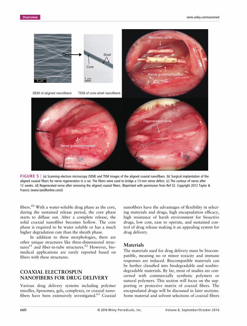
Blood VesselsVascular grafts, especially small diameter vein orartery (less than 5 mm), are important therapeuticmaterials for the treatment of coronary artery dis-ease.110 Wang et al.69 prepared PLA/CS coaxialfibers as a vascular gasket and compared it with apure PLA fiber graft and a commercial vascularpatch. CS served as a sheath due to its higher bio-compatibility. The surface of the coaxial fiber waschemically cross linked by genipin and modified byheparin to improve the mechanical properties. Bytwisting the flat fiber mat around a rod, the shape ofthe vascular graft was well developed. For in vitroexperiments, a high viability of 92.4% was observedfor a PLA/CS graft in 7 days, showing a good bio-compatibility. Blood tests of the activated partialthromboplastin time (aPTT) and prothrombin time(PT) showed that the coagulation time of blood
WIREs Nanomedicine and Nanobiotechnology Biomedical coaxial electrospun fibers
Volume 8, September/October 2016 © 2016 Wiley Per iodica ls , Inc. 671
(32 s, 9.2 s, respectively) of coaxial PLA/CS graftwas longer than a PLA mat and similar to a commer-cial patch (Figure 12). The anticoagulant propertyof coaxial fibers was superior to PLA and the com-mercial patch. In vitro blood flow tests showedthat PLA/CS graft had nearly no blood cells adheredonto the graft after 4 h. Thus, the chemically modi-fied PLA/CS coaxial fibers can be used as apotential anticoagulant biomaterials and intravascu-lar stent.
Nervous TissueSurgical treatment for peripheral nerve defects is aserious clinical task for surgeons. Recently, a lot ofresearch has focused on nerve guidance conduits(NGC) for treatment of nerve injuries.111 The NGChas a tubular structure that bridges the gap betweena proximal and a distal nerve. The NGF is known tobe responsible for axonal growth. It has beenreported that NGF can regulate the differentiationof Schwann cells and remyelination of the axon.112
Wang et al.52 prepared coaxial nanofibers withNGF as the core and PLGA as the sheath in analigned fiber morphology (Figure 5). The effectof NGF/PLGA coaxial NGC on nerve regenerationwas evaluated in a rat with a 13-mm sciatic nervedefect model. In vivo study showed that this NGCcan provide the physical guidance and bimolecularsignals for promoting nerve regeneration. The elec-trophysiological study was conducted on the nerve
defected rats after 12 weeks. The performance ofnerve conduction velocity (NCV) and compoundmotor action potential (CMAP) for NGF/PLGAmodel were almost the same as the autograftmodel and significantly better than the PLGA model.The morphological and functional analysis provedthat NGF/PLGA coaxial NGC could greatly promotethe peripheral nerve regeneration. Comparedwith many other NGC, the aligned coaxial fiberscontaining NGF combine the benefits of physicalguidance cues and biomolecular signals to promotenerve generation. Owing to these features, thealigned drug-doped coaxial fibers are believed tohave a potent NGC for the treatment of peripheralnerve injuries.
CONCLUSION
The presence of a two coaxial-nozzle configurationand emulsion electrospinning has made it easy todevelop electrospun nanofibers with a unique core/shell morphology. Recently, due to widespread inter-est of coaxial electrospun fibers in drug delivery andtissue engineering, significant advances have beenmade. By coelectrospinning, some nonelectrospin-nable materials can form a fibrous structure due tothe protection and guidance of the sheath phasewhich largely broadens the choice of materials. Withcareful selection of raw materials and optimal para-meters, various drugs have been encapsulated intocoaxial fibers. A bioactive drug can be isolated andremain bioactive after the release. The drug-loadedcoaxial fibers typically exhibit a sustained release bymitigating the burst release. Meanwhile, the nanofi-brous structure resembles the topological features ofECM, promoting the cell adhesion, migration, prolif-eration, and differentiation. The methods of coelec-trospinning and emulsion electrospinning allow astraightforward way to precisely manipulate the mor-phology, biocompatibility, degradation, wettability,and mechanical property of the resulting fibers as wellas the biological response to the coaxial scaffold.
Still many challenges surround this area ofresearch. First of all, although the physical theoriesabout the formation of coaxial nanofibers are wellunderstood, a fully theoretical prediction of the finalmorphology remains impossible. It is because rheo-logical and electric properties differ for each system.To get a satisfactory coaxial morphology, manyexperimental trials with different parameters arerequired. Compared to the conventional method,some special requirements should be met. Second, thedimension of the coaxial fiber is larger than that of a
45
40
35
30
25
20
15
0
5
0
Co
ag
ula
tio
n t
ime
(se
co
nd
)
APTT
PT
∗
∗∗∗∗
PLA PLA/CS Commercialized Blank
FIGURE 12 | The activated partial thromboplastin time (aPTT)and prothrombin time (PT) for different meshes. *P < 0.05.**P > 0.05. (Reprinted with permission from American ChemicalSociety)69
Overview wires.wiley.com/nanomed
672 © 2016 Wiley Per iodicals , Inc. Volume 8, September/October 2016

single component fiber. It is probably due to theimpeded evaporation of the solvent and the elonga-tion of the jet caused by the interphase. Up to now,the diameter of fine coaxial fibers is about 60 nmwhile single component fibers with several nan-ometers have been reported. It remains impossibleto create a uniform and massive production of coax-ial electrospun nanofibers. Presently, most studies ofcoaxial fibers regarding drug delivery and tissue
engineering are conducted in vitro. In vivo studiesare rare but continue to increase in number. Otherissues include precisely controlling the rate of degra-dation and drug release, needing a deeper under-standing of interaction between coaxial fibers andcells, etc. Further investigation and cooperation ofmaterial scientists, biologists, chemists, physicists,and biomedical scientists are needed to overcomethese challenges.
FURTHER READINGChen Y, Lu Z, Zhou L, Mai Y-W, Huang H. Triple-coaxial electrospun amorphous carbon nanotubes with hollow gra-phitic carbon nanospheres for high-performance Li ion batteries. Energy Environ Sci 2012, 5:7898.
REFERENCES1. Cooley JF. Apparatus for electrically dispersing fluids.
1902. Available at: http://www.google.com/patents/US692631. (Accessed October 13, 2015).
2. Doshi J, Reneker DH. Electrospinning process andapplications of electrospun fibers. J Electrostat 1995,35:151–160.
3. Reneker DH, Chun I. Nanometre diameter fibres ofpolymer, produced by electrospinning. Nanotechnol-ogy 1996, 7:216.
4. Hou Z, Li X, Li C, Dai Y, Ma P, Zhang X, Kang X,Cheng Z, Lin J. Electrospun upconversion compositefibers as dual drugs delivery system with individualrelease properties. Langmuir 2013, 29:9473–9482.
5. Song B, Wu C, Chang J. Dual drug release from elec-trospun poly(lactic-co-glycolic acid)/mesoporous silicananoparticles composite mats with distinct release pro-files. Acta Biomater 2012, 8:1901–1907.
6. Yoshimoto H, Shin Y, Terai H, Vacanti J. A biode-gradable nanofiber scaffold by electrospinning and itspotential for bone tissue engineering. Biomaterials2003, 24:2077–2082.
7. Yoon K, Kim K, Wang X, Fang D, Hsiao BS, Chu B.High flux ultrafiltration membranes based on electro-spun nanofibrous PAN scaffolds and chitosan coating.Polymer 2006, 47:2434–2441.
8. Neppalli R, Causin V, Marigo A, Meincken M,Hartmann P, van Reenen AJ. Effect of electrospun eth-ylene vinyl alcohol copolymer (EVOH) fibres on thestructure, morphology, and properties of poly (lacticacid)(PLA). Polymer 2013, 54:5909–5919.
9. Dharani S, Mulmudi HK, Yantara N, Trang PTT,Park NG, Graetzel M, Mhaisalkar S, Mathews N,Boix PP. High efficiency electrospun TiO2 nanofiberbased hybrid organic–inorganic perovskite solar cell.Nanoscale 2014, 6:1675–1679.
10. Yang DJ, Kamienchick I, Youn DY, Rothschild A,Kim ID. Ultrasensitive and highly selective gas sensorsbased on electrospun SnO2 nanofibers modified by Pdloading. Adv Funct Mater 2010, 20:4258–4264.
11. Kim C, Ngoc BTN, Yang KS, Kojima M, Kim YA,Kim YJ, Endo M, Yang SC. Self-sustained thin websconsisting of porous carbon nanofibers for supercapa-citors via the electrospinning of polyacrylonitrile solu-tions containing zinc chloride. Adv Mater 2007,19:2341–2346.
12. Luo L, Cui R, Qiao H, Chen K, Fei Y, Li D, Pang Z,Liu K, Wei Q. High lithium electroactivity of electro-spun CuFe2O4 nanofibers as anode material forlithium-ion batteries. Electrochim Acta 2014,144:85–91.
13. Loscertales IG, Barrero A, Guerrero I, Cortijo R,Marquez M, Ganan-Calvo AM. Micro/nano encapsu-lation via electrified coaxial liquid jets. Science 2002,295:1695–1698.
14. Sun Z, Zussman E, Yarin AL, Wendorff JH,Greiner A. Compound core–shell polymer nanofibersby co-electrospinning. Adv Mater 2003,15:1929–1932.
15. Xu X, Zhuang X, Chen X, Wang X, Yang L, Jing X.Preparation of core-sheath composite nanofibers byemulsion electrospinning. Macromol Rapid Commun2006, 27:1637–1642.
16. Yang Y, Li X, Qi M, Zhou S, Weng J. Release patternand structural integrity of lysozyme encapsulated incore-sheath structured poly(DL-lactide) ultrafine fibersprepared by emulsion electrospinning. Eur J PharmBiopharm 2008, 69:106–116.
17. Yang Y, Xia T, Zhi W, Wei L, Weng J, Zhang C,Li X. Promotion of skin regeneration in diabetic ratsby electrospun core-sheath fibers loaded with basic
WIREs Nanomedicine and Nanobiotechnology Biomedical coaxial electrospun fibers
Volume 8, September/October 2016 © 2016 Wiley Per iodica ls , Inc. 673

fibroblast growth factor. Biomaterials 2011,32:4243–4254.
18. Kayaci F, Ozgit-Akgun C, Donmez I, Biyikli N,Uyar T. Polymer-inorganic core-shell nanofibers byelectrospinning and atomic layer deposition: flexiblenylon-ZnO core-shell nanofiber mats and their photo-catalytic activity. ACS Appl Mater Interfaces 2012,4:6185–6194.
19. Peng Q, Sun XY, Spagnola JC, Hyde GK, Spontak RJ,Parsons GN. Atomic layer deposition on electrospunpolymer fibers as a direct route to AL2O3 microtubeswith precise wall thickness control. Nano Lett 2007,7:719–722.
20. Li Z, Huang H, Wang C. Electrostatic forces inducepoly(vinyl alcohol)-protected copper nanoparticles toform copper/poly(vinyl alcohol) nanocables via electro-spinning. Macromol Rapid Commun 2006,27:152–155.
21. Pant HR, Risal P, Park CH, Tijing LD, Jeong YJ,Kim CS. Core-shell structured electrospun biomimeticcomposite nanofibers of calcium lactate/nylon-6 for tis-sue engineering. Chem Eng J 2013, 221:90–98.
22. Li XH, Shao CL, Liu YC. A simple method for con-trollable preparation of polymer nanotubes via a singlecapillary electrospinning. Langmuir 2007,23:10920–10923.
23. Fakhrali A, Ebadi SV, Gharehaghaji AA. Productionof core-sheath nanofiber yarn using two oppositeasymmetric nozzles. Fiber Polym 2015, 15:2535–2540.
24. Liu J, Shen ZH, Lee SH, Marquez M, McHugh MA.Electrospinning in compressed carbon dioxide: hollowor open-cell fiber formation with a single nozzle config-uration. J Supercrit Fluids 2010, 53:142–150.
25. Lee BS, Jeon SY, Park H, Lee G, Yang HS, Yu WR.New electrospinning nozzle to reduce jet instabilityand its application to manufacture of multi-layerednanofibers. Sci Rep 2014, 4:6758.
26. Moghe AK, Gupta BS. Co-axial electrospinning fornanofiber structures: preparation and applications.Polym Rev 2008, 48:353–377.
27. Tian L, Prabhakaran MP, Ding X, Kai D,Ramakrishna S. Emulsion electrospun vascularendothelial growth factor encapsulated poly(l-lacticacid-co-ε-caprolactone) nanofibers for sustained releasein cardiac tissue engineering. J Mater Sci 2011,47:3272–3281.
28. Yarin AL. Coaxial electrospinning and emulsion elec-trospinning of core-shell fibers. Polym Adv Technol2011, 22:310–317.
29. áLo Nigro R. Core-shell Zn-doped TiO 2–ZnO nanofi-bers fabricated via a combination of electrospinningand metal–organic chemical vapour deposition. Crys-tEngComm 2010, 12:3858–3865.
30. Gu Y, Jian F. Hollow LiNi0.8Co0.1Mn0.1O2 − MgOcoaxial fibers: sol–gel method combined with co-
electrospun preparation and electrochemical proper-ties. J Phys Chem A 2008, 112:20176–20180.
31. Viry L, Moulton SE, Romeo T, Suhr C, Mawad D,Cook M, Wallace GG. Emulsion-coaxial electrospin-ning: designing novel architectures for sustained releaseof highly soluble low molecular weight drugs. J MaterChem 2012, 22:11347.
32. Townsend-Nicholson A, Jayasinghe SN. Cell electro-spinning: a unique biotechnique for encapsulating liv-ing organisms for generating active biologicalmicrothreads/scaffolds. Biomacromolecules 2006,7:3364–3369.
33. Yu JH, Fridrikh SV, Rutledge GC. Production of sub-micrometer diameter fibers by two-fluid electrospin-ning. Adv Mater 2004, 16:1562–1566.
34. Reznik SN, Yarin AL, Zussman E, Bercovici L. Evolu-tion of a compound droplet attached to a core-shellnozzle under the action of a strong electric field. PhysFluids 2006, 18:062101.
35. Agarwal S, Greiner A, Wendorff JH. Functional mate-rials by electrospinning of polymers. Prog Polym Sci2013, 38:963–991.
36. Dror Y, Salalha W, Avrahami R, Zussman E,Yarin AL, Dersch R, Greiner A, Wendorff JH. One-step production of polymeric microtubes by co-electro-spinning. Small 2007, 3:1064–1073.
37. Reneker DH, Yarin AL, Fong H, Koombhongse S.Bending instability of electrically charged liquid jets ofpolymer solutions in electrospinning. J Appl Phys2000, 87:4531.
38. Zhang H, Lou S-F, Nie H-L, Quan J, Zhu L-M. Prepa-ration of core–shell structured PVP-NSPs/PLLAbinary-drug loaded complex fibermats by electrospin-ning; in vitro release and antimicrobial properties. JControl Release 2013, 172:e37.
39. Nguyen TT, Ghosh C, Hwang SG, Chanunpanich N,Park JS. Porous core/sheath composite nanofibers fab-ricated by coaxial electrospinning as a potential matfor drug release system. Int J Pharm 2012,439:296–306.
40. Tiwari SK, Tzezana R, Zussman E, Venkatraman SS.Optimizing partition-controlled drug release from elec-trospun core-shell fibers. Int J Pharm 2010,392:209–217.
41. Luo CJ, Edirisinghe M. Core-liquid-induced transitionfrom coaxial electrospray to electrospinning of low-viscosity poly(lactide-co-glycolide) sheath solution.Macromolecules 2014, 47:7930–7938.
42. McCann JT, Marquez M, Xia Y. Melt coaxial electro-spinning: a versatile method for the encapsulation ofsolid materials and fabrication of phase change nanofi-bers. Nano Lett 2006, 6:2868–2872.
43. Shenoy SL, Bates WD, Frisch HL, Wnek GE. Role ofchain entanglements on fiber formation during electro-spinning of polymer solutions: good solvent, non-
Overview wires.wiley.com/nanomed
674 © 2016 Wiley Per iodicals , Inc. Volume 8, September/October 2016

specific polymer–polymer interaction limit. Polymer2005, 46:3372–3384.
44. Zhang Y, Huang Z-M, Xu X, Lim CT,Ramakrishna S. Preparation of core-shell structuredPCL-r-gelatin bi-component nanofibers by coaxial elec-trospinning. Chem Mater 2004, 16:3406–3409.
45. Baumgarten PK. Electrostatic spinning of acrylicmicrofibers. J Colloid Interface Sci 1971, 36:71–79.
46. Ding W, Wei S, Zhu J, Chen X, Rutman D, Guo Z.Manipulated electrospun PVA nanofibers with inex-pensive salts. Macromol Mater Eng 2010,295:958–965.
47. Deitzel JM, Kleinmeyer J, Harris D, Beck Tan NC.The effect of processing variables on the morphologyof electrospun nanofibers and textiles. Polymer 2001,42:261–272.
48. Ramakrishna S, Fujihara K, Teo W-E, Lim T-C,Ma Z. An Introduction to Electrospinning and Nanofi-bers, vol. 90. Singapore: World Scientific; 2005.
49. Buchko CJ, Chen LC, Shen Y, Martin DC. Processingand microstructural characterization of porous bio-compatible protein polymer thin films. Polymer 1999,40:7397–7407.
50. Wang C, Yan K-W, Lin Y-D, Hsieh PCH. Biodegrada-ble core/shell fibers by coaxial electrospinning: proces-sing, fiber characterization, and its application insustained drug release. Macromolecules 2010,43:6389–6397.
51. Mathew G, Hong JP, Rhee JM, Leo DJ, Nah C. Prepa-ration and anisotropic mechanical behavior of highly-oriented electrospun poly(butylene terephthalate)fibers. J Appl Polym Sci 2006, 101:2017–2021.
52. Wang CY, Liu JJ, Fan CY, Mo XM, Ruan HJ, Li FF.The effect of aligned core-shell nanofibres deliveringNGF on the promotion of sciatic nerve regeneration. JBiomater Sci Polym Ed 2012, 23:167–184.
53. Liao IC, Chew SY, Leong KW. Aligned core-shellnanofibers delivering bioactive proteins. Nanomedicine(Lond) 2006, 1:465–471.
54. Sill TJ, von Recum HA. Electrospinning: applicationsin drug delivery and tissue engineering. Biomaterials2008, 29:1989–2006.
55. Asli MM, Pourdeyhimi B, Loboa EG. Release profilesof tricalcium phosphate nanoparticles from poly(L-lactic acid) electrospun scaffolds with single compo-nent, core-sheath, or porous fiber morphologies: effectson hASC viability and osteogenic differentiation.Macromol Biosci 2012, 12:893–900.
56. Bognitzki M, Czado W, Frese T, Schaper A,Hellwig M, Steinhart M, Greiner A, Wendorff JH.Nanostructured fibers via electrospinning. Adv Mater2001, 13:70–72.
57. Megelski S, Stephens JS, Chase DB, Rabolt JF. Micro-and nanostructured surface morphology on
electrospun polymer fibers. Macromolecules 2002,35:8456–8466.
58. Gulfam M, Lee JM, Kim JE, Lim DW, Lee EK,Chung BG. Highly porous core-shell polymeric fibernetwork. Langmuir 2011, 27:10993–10999.
59. Li D, Xia Y. Direct fabrication of composite andceramic hollow nanofibers by electrospinning. NanoLett 2004, 4:933–938.
60. Man Z, Yin L, Shao Z, Zhang X, Hu X, Zhu J, Dai L,Huang H, Yuan L, Zhou C, et al. The effects of co-delivery of BMSC-affinity peptide and rhTGF-β1 fromcoaxial electrospun scaffolds on chondrogenic differen-tiation. Biomaterials 2014, 35:5250–5260.
61. Bonino CA, Efimenko K, Jeong SI, Krebs MD,Alsberg E, Khan SA. Three-dimensional electrospunalginate nanofiber mats via tailored charge repulsions.Small 2012, 8:1928–1936.
62. Chen H, Wang N, Di J, Zhao Y, Song Y, Jiang L.Nanowire-in-microtube structured core/shell fibers viamultifluidic coaxial electrospinning. Langmuir 2010,26:11291–11296.
63. Tiwari G, Tiwari R, Sriwastawa B, Bhati L, Pandey S,Pandey P, Bannerjee SK. Drug delivery systems: anupdated review. Int J Pharm Investig 2012, 2:2.
64. Choi JS, Choi SH, Yoo HS. Coaxial electrospun nano-fibers for treatment of diabetic ulcers with binaryrelease of multiple growth factors. J Mater Chem2011, 21:5258.
65. Ravichandran R, Venugopal JR, Sundarrajan S,Mukherjee S, Sridhar R, Ramakrishna S. Expression ofcardiac proteins in neonatal cardiomyocytes on PGS/fi-brinogen core/shell substrate for cardiac tissue engi-neering. Int J Cardiol 2013, 167:1461–1468.
66. Kai D, Prabhakaran MP, Stahl B, Eblenkamp M,Wintermantel E, Ramakrishna S. Mechanical proper-ties and in vitro behavior of nanofiber-hydrogel com-posites for tissue engineering applications.Nanotechnology 2012, 23:095705.
67. Jin G, Prabhakaran MP, Kai D, Ramakrishna S. Con-trolled release of multiple epidermal induction factorsthrough core-shell nanofibers for skin regeneration.Eur J Pharm Biopharm 2013, 85:689–698.
68. Pant HR, Risal P, Park CH, Tijing LD, Jeong YJ,Kim CS. Synthesis, characterization, and mineraliza-tion of polyamide-6/calcium lactate composite nanofi-bers for bone tissue engineering. Colloids Surf BBiointerfaces 2013, 102:152–157.
69. Wang T, Ji X, Jin L, Feng Z, Wu J, Zheng J, Wang H,Xu ZW, Guo L, He N. Fabrication and characteriza-tion of heparin-grafted poly-L-lactic acid-chitosancore-shell nanofibers scaffold for vascular gasket. ACSAppl Mater Interfaces 2013, 5:3757–3763.
70. Chen W, Li D, Ei-Shanshory A, El-Newehy M,Ei-Hamshary HA, Al-Deyab SS, He C, Mo X. Dexa-methasone loaded core-shell SF/PEO nanofibers via
WIREs Nanomedicine and Nanobiotechnology Biomedical coaxial electrospun fibers
Volume 8, September/October 2016 © 2016 Wiley Per iodica ls , Inc. 675

green electrospinning reduced endothelial cells inflam-matory damage. Colloids Surf B Biointerfaces 2014,126:561–568.
71. Yu DG, Wang X, Li XY, Chian W, Li Y, Liao YZ.Electrospun biphasic drug release polyvinylpyrroli-done/ethyl cellulose core/sheath nanofibers. Acta Bio-mater 2013, 9:5665–5672.
72. Sohrabi A, Shaibani PM, Etayash H, Kaur K,Thundat T. Sustained drug release and antibacterialactivity of ampicillin incorporated poly(methyl methac-rylate)–nylon-6 core/shell nanofibers. Polymer 2013,54:2699–2705.
73. Jiang YN, Mo HY, Yu DG. Electrospun drug-loadedcore-sheath PVP/zein nanofibers for biphasic drugrelease. Int J Pharm 2012, 438:232–239.
74. Wang X, Yuan Y, Huang X, Yue T. Controlled releaseof protein from core-shell nanofibers prepared byemulsion electrospinning based on green chemical.J Appl Polym Sci 2014, 132:41811.
75. Rubert M, Dehli J, Li Y-F, Taskin MB, Xu R,Besenbacher F, Chen M. Electrospun PCL/PEO coaxialfibers for basic fibroblast growth factor delivery.J Mater Chem B 2014, 2:8538–8546.
76. Jiang H, Hu Y, Zhao P, Li Y, Zhu K. Modulation ofprotein release from biodegradable core-shell struc-tured fibers prepared by coaxial electrospinning.J Biomed Mater Res B Appl Biomater 2006,79:50–57.
77. Nguyen TTT, Chung OH, Park JS. Coaxial electro-spun poly(lactic acid)/chitosan (core/shell) compositenanofibers and their antibacterial activity. CarbohydrPolym 2011, 86:1799–1806.
78. Ma G, Fang D, Liu Y, Zhu X, Nie J. Electrospunsodium alginate/poly(ethylene oxide) core–shell nanofi-bers scaffolds potential for tissue engineering applica-tions. Carbohydr Polym 2012, 87:737–743.
79. Zhang YZ, Venugopal J, Huang ZM, Lim CT,Ramakrishna S. Characterization of the surface bio-compatibility of the electrospun PCL-collagen nanofi-bers using fibroblasts. Biomacromolecules 2005,6:2583–2589.
80. Torres-Giner S, Martinez-Abad A, Gimeno-Alcañiz JV,Ocio MJ, Lagaron JM. Controlled delivery of gentami-cin antibiotic from bioactive electrospun polylactide-based ultrathin fibers. Adv Eng Mater 2012, 14:B112–B122.
81. Zhu T, Chen S, Li W, Lou J, Wang J. Flurbiprofenaxetil loaded coaxial electrospun poly(vinyl pyrroli-done)-nanopoly(lactic-co-glycolic acid) core-shell com-posite nanofibers: preparation, characterization, andanti-adhesion activity. J Appl Polym Sci 2015,132:41982.
82. Su Y, Su Q, Liu W, Lim M, Venugopal JR, Mo X,Ramakrishna S, Al-Deyab SS, El-Newehy M. Con-trolled release of bone morphogenetic protein 2 anddexamethasone loaded in core-shell PLLACL-collagen
fibers for use in bone tissue engineering. Acta Biomater2012, 8:763–771.
83. Huang W, Zou T, Li S, Jing J, Xia X, Liu X. Drug-loaded zein nanofibers prepared using a modified coax-ial electrospinning process. AAPS PharmSciTech 2013,14:675–681.
84. Zhao P, Jiang H, Pan H, Zhu K, Chen W. Biodegrada-ble fibrous scaffolds composed of gelatin coatedpoly(epsilon-caprolactone) prepared by coaxial electro-spinning. J Biomed Mater Res A 2007, 83:372–382.
85. Sridhar R, Lakshminarayanan R, Madhaiyan K,Amutha Barathi V, Lim KH, Ramakrishna S. Electro-sprayed nanoparticles and electrospun nanofibersbased on natural materials: applications in tissueregeneration, drug delivery and pharmaceuticals.Chem Soc Rev 2015, 44:790–814.
86. Lee KY, Jeong L, Kang YO, Lee SJ, Park WH. Electro-spinning of polysaccharides for regenerative medicine.Adv Drug Deliv Rev 2009, 61:1020–1032.
87. Makadia HK, Siegel SJ. Poly lactic-co-glycolic acid(PLGA) as biodegradable controlled drug delivery car-rier. Polymers (Basel) 2011, 3:1377–1397.
88. Li X, Zhang H, Li H, Yuan X. Encapsulation of pro-teinase K in PELA ultrafine fibers by emulsion electro-spinning: preparation and in vitro evaluation. ColloidPolym Sci 2010, 288:1113–1119.
89. Fredenberg S, Wahlgren M, Reslow M, Axelsson A.The mechanisms of drug release in poly(lactic-co-glycolic acid)-based drug delivery systems—a review.Int J Pharm 2011, 415:34–52.
90. Choi JS, Yoo HS. Nanoinspired fibrous matrix withbi-phasic release of proteins. J Nanosci Nanotechnol2010, 10:3038–3045.
91. Lee K, Silva EA, Mooney DJ. Growth factor delivery-based tissue engineering: general approaches and areview of recent developments. J R Soc Interface 2011,8:153–170.
92. Chen D, Zhao M, Mundy GR. Bone morphogeneticproteins. Growth Factors 2004, 22:233–241.
93. Srouji S, Ben-David D, Lotan R, Livne E, Avrahami R,Zussman E. Slow-release human recombinant bonemorphogenetic protein-2 embedded within electrospunscaffolds for regeneration of bone defect: in vitro andin vivo evaluation. Tissue Eng Part A 2010,17:269–277.
94. Jürgens C, Schulz AP, Porté T, Faschingbauer M,Seide K. Biodegradable films in trauma and orthopedicsurgery. Eur J Trauma 2006, 32:160–171.
95. Ignatova М, Rashkov I, Manolova N. Drug-loaded elec-trospun materials in wound-dressing applications andin local cancer treatment. Expert Opin Drug Deliv2013, 10:469–483.
96. Rizzello L, Pompa PP. Nanosilver-based antibacterialdrugs and devices: mechanisms, methodological
Overview wires.wiley.com/nanomed
676 © 2016 Wiley Per iodicals , Inc. Volume 8, September/October 2016

drawbacks, and guidelines. Chem Soc Rev 2014,43:1501–1518.
97. Lynch BA, Lambeng N, Nocka K, Kensel-Hammes P,Bajjalieh SM, Matagne A, Fuks B. The synaptic vesicleprotein SV2A is the binding site for the antiepilepticdrug levetiracetam. Proc Natl Acad Sci USA 2004,101:9861–9866.
98. Shinoka T. Development of a tissue-engineering vascu-lar graft for use in congenital heart surgery. EBioMedi-cine 2014, 1:12–13.
99. O’Brien FJ. Biomaterials and scaffolds for tissue engi-neering. Materialstoday 2011, 14:88–95.
100. McCullen SD, Zhan J, Onorato ML, Bernacki SH,Loboa EG. Effect of varied ionic calcium on humanadipose-derived stem cell mineralization. Tissue EngPart A 2010, 16:1971–1981.
101. Bouten C, Dankers P, Driessen-Mol A, Pedron S,Brizard A, Baaijens F. Substrates for cardiovasculartissue engineering. Adv Drug Deliv Rev 2011,63:221–241.
102. Smits AM, Vliet P, Hassink RJ, Goumans MJ,Doevendans PA. The role of stem cells in cardiacregeneration. J Cell Mol Med 2005, 9:25–36.
103. Vunjak-Novakovic G, Tandon N, Godier A,Maidhof R, Marsano A, Martens TP, Radisic M.Challenges in cardiac tissue engineering. Tissue EngPart B Rev 2009, 16:169–187.
104. Torpy JM, Lynm C, Glass RM. JAMA patient page.Burn injuries. JAMA 2009, 302:1828–1828.
105. Barrandon Y, Green H. Cell migration is essential forsustained growth of keratinocyte colonies: the roles
of transforming growth factor-α and epidermalgrowth factor. Cell 1987, 50:1131–1137.
106. Brzoska M, Geiger H, Gauer S, Baer P. Epithelial dif-ferentiation of human adipose tissue-derived adultstem cells. Biochem Biophys Res Commun 2005,330:142–150.
107. Garrigues NW II. Electrospun Scaffolds for CartilageTissue Engineering: Methods to Affect Anisotropy,Material, and Cellular Infiltration. PhD Dissertation,Duke University, 2011.
108. Mow VC, Ratcliffe A, Robin PA. Cartilage and dia-rthrodial joints as paradigms for hierarchical materi-als and structures. Biomaterials 1992, 13:67–97.
109. Buzgo M, Jakubova R, Mickova A, Rampichova M,Prosecka E, Kochova P, Lukas D, Amler E. Time-regulated drug delivery system based on coaxiallyincorporated platelet α-granules for biomedical use.Nanomedicine 2013, 8:1137–1154.
110. Du F, Wang H, Zhao W, Li D, Kong D, Yang J,Zhang Y. Gradient nanofibrous chitosan/poly-caprolactone scaffolds as extracellular microenviron-ments for vascular tissue engineering. Biomaterials2012, 33:762–770.
111. Kehoe S, Zhang XF, Boyd D. FDA approved guid-ance conduits and wraps for peripheral nerve injury:a review of materials and efficacy. Injury 2012,43:553–572.
112. Huang EJ, Reichardt LF. Neurotrophins: roles in neu-ronal development and function. Annu Rev Neurosci2001, 24:677.
WIREs Nanomedicine and Nanobiotechnology Biomedical coaxial electrospun fibers
Volume 8, September/October 2016 © 2016 Wiley Per iodica ls , Inc. 677