Coating techniques for biomaterials: A review
12
CMU.J.Nat.Sci Special Issue on Manufacturing Technology (2011) Vol.10(1) ➔ 39 Coating techniques for biomaterials: A review Anirut Chaijaruwanich Department of Industrial Engineering, Chiang Mai University, Chiang Mai 50200, Thailand. Materials Science Research Center, Faculty of Science, Chiang Mai University, Chiang Mai 50200, Thailand. *Corresponding author: E-mail: [email protected] ABSTRACT Biomaterials have been extensively studied for many decades. Surface modifications play a major role in determining the biological response. The purpose of this paper is to present an up-to-date progress in the coating techniques currently applied for biomaterials, including chemical treatment, anodic oxidation, micro-arc oxidation, sol-gel, plasma spray, electrostatic spray deposition, electrophoretic deposition and pulsed laser deposition. In the pres- ent paper, an overview of the basic principles behind the techniques is briefly given. A large number of research studies have shown a great thrust towards development of surface modification techniques for biomaterials. Coatings and films with nanostructures on bio-implants using various techniques have become a subject of interest. Keywords: Coating, Biomaterials, Nanostructure, Hydroxyapatite INTRODUCTION Biomaterials have been extensively used for many decades. It is well known that the bulk and surface properties of biomaterials determine their long-term performance and stability in the biological environment. The bulk properties of a biomaterial are characterized by its mechanical behaviour and chemical stability under in vivo condition. On the other hand, the surface properties are evaluated by surface morphology, surface wettability and surface chemistry (Paital and Dahotre, 2009). Metals generally possess superior mechanical properties and find applica- tions in hip joint prosthesis and dental implants. However, as a result of their lack of bioactivity, they cannot serve long-term implantations. This limitation can be overcome by applying bioactive ceramic coating on the metal surface. Hence, the bioactivity of metallic biomaterials is improved. Consequently, various surface coating/modification techniques have exten- sively applied for biomedical applications. Typical techniques include chemical treatment, anodic oxidation, micro-arc oxidation (MAO), sol-gel, plasma spray-
Transcript of Coating techniques for biomaterials: A review
CMU.J.Nat.Sci Special Issue on Manufacturing Technology (2011) Vol.10(1)➔ 39
Coating techniques for biomaterials: A review
Anirut Chaijaruwanich
Department of Industrial Engineering, Chiang Mai University, Chiang Mai 50200, Thailand.
Materials Science Research Center, Faculty of Science, Chiang Mai University, Chiang Mai 50200, Thailand.
*Corresponding author: E-mail: [email protected]
ABSTRACT
Biomaterials have been extensively studied for many decades. Surface modifications play a major role in determining the biological response. The purpose of this paper is to present an up-to-date progress in the coating techniques currently applied for biomaterials, including chemical treatment, anodic oxidation, micro-arc oxidation, sol-gel, plasma spray, electrostatic spray deposition, electrophoretic deposition and pulsed laser deposition. In the pres-ent paper, an overview of the basic principles behind the techniques is briefly given. A large number of research studies have shown a great thrust towards development of surface modification techniques for biomaterials. Coatings and films with nanostructures on bio-implants using various techniques have become a subject of interest.
Keywords: Coating, Biomaterials, Nanostructure, Hydroxyapatite
INTRODUCTION
Biomaterials have been extensively used for many decades. It is well known that the bulk and surface properties of biomaterials determine their long-term performance and stability in the biological environment. The bulk properties of a biomaterial are characterized by its mechanical behaviour and chemical stability under in vivo condition. On the other hand, the surface properties are evaluated by surface morphology, surface wettability and surface chemistry (Paital and Dahotre, 2009). Metalsgenerallypossesssuperiormechanicalpropertiesandfindapplica-tions in hip joint prosthesis and dental implants. However, as a result of their lack of bioactivity, they cannot serve long-term implantations. This limitation can be overcome by applying bioactive ceramic coating on the metal surface. Hence, the bioactivity of metallic biomaterials is improved. Consequently,varioussurfacecoating/modificationtechniqueshaveexten-sively applied for biomedical applications. Typical techniques include chemical treatment, anodic oxidation, micro-arc oxidation (MAO), sol-gel, plasma spray-

CMU.J.Nat.Sci Special Issue on Manufacturing Technology (2011) Vol.10(1)➔40
ing, electrostatic spray deposition (ESD), electrophoretic deposition (EPD), and pulsed-laser deposition (PLD). The basic principle of each technique is firstbrieflygiven and then its applications are reviewed.
CLASSIFICATION OF BIOMATERIALS
Biomaterialscanbebroadlyclassifiedas(i)metals,(ii)ceramicsand(iii)polymers. In this paper, it is mainly focused on the metallic bio-implants as a substrate and the bioactive ceramics as a coating.
A. Metals Metallic biomaterials are commonly used for load bearing implants due to their superior mechanical properties. They include stainless steels (316L), Co-Cr based alloys and commercially pure titanium and its alloys. However, elements such as Ni Co and Cr are found to be released from the stainless steel and Co-Cr alloys due to the corrosion in the body environment (Geetha et al., 2009). Thus, the surface coatings of these materials have been applied and developed. B. Ceramics It is well known that calcium phosphates (CaP) are biocompatible and bioactive ceramics, which can induce bone regeneration and bone in growth at tissue-implant interface. Therefore, the CaP can be used in order to increase a bioactivity of metallic implants without compromising its mechanical behavior. Hydroxyapatite (HA), for example, has widely been used for coating of metallic implants. C. Polymers Polymers are widely used in surgery, prosthetic systems and controlled drug delivery. Currently, both synthetic and natural polymers have been extensively studied as biodegradable polymeric biomaterials.
COATING TECHNIQUES
In thispart, thebasicprinciplesbehind thecoating techniquesarebrieflydescribed and followed by its current applications for biomaterials. As mention earlier, only metallic biomaterials as a substrate are considered in the review. A. Chemical treatment Chemical treatments involve chemical reactions at the interface between a substrate and solution using acids, alkalis, and hydrogen peroxide (H2O2). They havebeenwidely studies for surfacemodificationof biomaterials especiallyTiand its alloys due to their simplicity andflexibility. Nanostructures such as titania nanorods nanoflowers and nanowires canbe formed by chemically treating using H2O2 at relatively low temperature. Wu

CMU.J.Nat.Sci Special Issue on Manufacturing Technology (2011) Vol.10(1)➔ 41
(2004) prepared titania nanorods by oxidizing pure titanium in hydrogen peroxide solution at 80°C for 3 days and found that the addition of F- and SO4
2- helped the formation of pure anatase; while the addition of Cl- favoured rutile structure. Wu et al., (2005) deposited large-scale titania nanorods on Ti using the previ-ous method followed by heating in air at 450°C for 1 h. Most of the nanorods were mixtures of anatase and rutile structures. The post heat treatment kept the morphology but improved the crystallinity. The same author and co-workers (Wuetal.,2006)alsosynthesizedtitaniananoflowers(Figure1)bythepreviousmethod, followed by heating at 400°C for 1 h. Varying concentration of nitric acid resulted in difference in phase composition. Shi et al. (2009) prepared sandblasted, dual acid-treated (HF/HNO3) and H2O2/HCl heat-treated Ti-6Al-4V. The authors suggested that the treated Ti alloy surface had the ability to deposit apatite and enhanced proliferation.
B. Anodic oxidation Anodic oxidation or electrochemical deposition involves electrode reac-tions inanelectrolyte, resulting in the formationofanoxidefilmon theanodesurface. Different dilute acids are used as electrolytes in the process. Anodic oxidation is generally a simple and low-cost technique to produce nanostructures on the surface of titanium. Wang et al., (2008) formed a well oriented and uniform TiO2 nanotube array on the surface of titanium substrate using anodic oxidation. The ano-dized Ti substrate was electrochemically deposited in an electrolyte containing calcium and phosphate ions. It was suggested that the surface morphology of TiO2 nanotube promoted mechanical interlocking between HA and TiO2.
Figure 1.SEMmicrographs of titania nanoflowers synthesized by oxidation ofTi with 30wt% H2O2 solution containing 0.014 M hexamethylene-tetramine and 0.4 M nitric acid at a temperature of 80°C, followed byheatingat400°C for1h: (a) low (b)highmagnifications. (Wuetal., 2006).

CMU.J.Nat.Sci Special Issue on Manufacturing Technology (2011) Vol.10(1)➔42
Figure 2.SEMtop-viewimagesof thefirstnanotube layer(A),and thesecondnanotube layer (inset of A), cross-sectional image for the double nano-tube layers (B), and cross-sectional images of the interface between the upper and the lower TiO2 nanotube layers (C) and (D). (Cui et al., 2009).
Kunz et al., (2008) fabricated the nanotubular layers on titanium by electrochemicalanodization influoride-containingelectrolytes, followedbyHAcoating. It was observed that the nanotube oxide layer enhanced the formation of a thick apatite layer. Song et al., (2009) fabricated amphiphilic TiO2 nanotube layers (see Figure 2) using a double anodization method combined with organic monolayer grafting. The amphiphilic characteristics with the hydrophobic outside and the hydrophilic were obtained. Cuietal.,(2009)preparedtitaniumoxidefilmsusinganodicoxidationonthe surface of commercially pure titanium in four different electrolytes: sulphuric acid, acetic acid, phosphoric acid, and sodium sulphate solutions. Amorphous titania observed in acetic and phosphoric acids could not induce apatite formation whereas rutile structure prepared from sulphuric and sodium sulphate solutions favoured apatite deposition. Park et al., (2010) formed amorphous titania nanotubes on titanium sur-face by an anodic oxidation process. The crystallization and corrosion resistance improved were induced by a post-heat treatment at 500°C for 2 hrs. Nanotube arrays can also be formed in an F- containing electrolyte by the competing two electric field-assisted processes (Crawford and Crawla, 2009).Wangetal.,(2009)depositedHAandfluoridatedHA(FHA)coatingsontitaniumsubstrates using an electrochemical technique in electrolytes containing Ca(NO3)2, NH4H2PO4, NaNO3 and H2O2 (NaF added for FHA coating). The FHA coating showed higher bond strength and lower dissolution rate than HA coating.
C. Micro-arc oxidation Micro-arc oxidation (MAO) is also known as anodic spark oxidation. In this process, the component to be coated is connected to a high-voltage power supply

CMU.J.Nat.Sci Special Issue on Manufacturing Technology (2011) Vol.10(1)➔ 43
and immersed in an aqueous electrolytic stainless steel vessel, which serves as the counter-electrode. As an asymmetric alternating voltage is then applied and exceeds a critical value, micro-plasma discharge occurs on the surface of the component. The process can be performed at room temperature for components with complex geometries, therefore it is considered as a simple and economical coating technique. Several researchers have studied the possibility Ca-P coatings on metallic biomaterials using MAO technique (Kim et al., (2007), Sun et al., (2007), Han et al., (2008)).Nano-crystallineHA filmswere formed at the surface ofTi bya single-step MAO using Ca2+ and P5+ ion-containing electrolytes (Kim et al., 2007). Strong crystallinity dependense on the CaCl2 concentration in the elec-trolytes was found. The mechanism for HA coating was proposed based on the formation of amorphous CaTiO3 and its dissolution to TiO(OH)2 by H+ ions. Sun et al. (2007) proposed a novel method to directly coat HA on Ti-6Al-4V using MAO in Ca- and P-containing electrolyte. The coatings obtained were composed of a bi-layer structure of HA/TiO2 (Figure 3). The HA peaks increased when the applied voltage increased from 430 to 480 V or increased treated time at 480 V.
Figure 3. SEM micrographs showing the surface morphologies of the MAO coatings formed at 480 V for (A) 1.5 (B) 3 (C) 10 (D) 20 min. (Sun et al., 2007).
Han et al., (2008) synthesized HA-based coatings on Ti alloy by MAO in an electrolyte containing calcium acetate (Ca(CH3COO)2) and β-glycerophosphate (β-GP) disodium at high applied voltage. It was suggested that hydrolysis of β-GP and decomposition of (CH3COO)- were promoted by the intense micro-arc discharge generated at high voltage. Wei et al., (2009) formed TiO2-based coating on Ti alloy by MAO in electrolytes containing nano-HA, calcium and phosphate salts. A post-heat treat-ment resulted in the increase of surface roughness, decreasing in cell adhesion and proliferation ability.

CMU.J.Nat.Sci Special Issue on Manufacturing Technology (2011) Vol.10(1)➔44
Song et al., (2009) fabricated porous oxide films on pure titanium usingan anodic spark oxidation technique with different electrolytes: H2SO4, H3PO4 and CH3COOH. A TiP2O7filmwasmainlyobservedonthespecimensanodizedin the H3PO4 electrolyte whereas TiO2 in the acetic and sulphuric acids. Micro-arc oxidation has been also applied in other Ti alloys. Chen et al., (2009) studied the structure and osteoblast cell compatibility of the TiO2 layer coated on β-Ti alloy (Ti-13Cr-3Al-1Fe) using MAO in NaH2PO4 solution. Sur-face morphologies of coating were a function of voltage and treatment time. Tao et al., (2009) synthesized a porous oxide layer on Ti-24Nb-4Zr-7.9Sn alloy (in wt%) by MAO treatment using calcium acetate electrolytic solution. A post-heat treatment at 600°C for 60 min resulted in the oxide films consisting of TiO2, CaO, Nb2O5 and SnO2 and improved the apatite forming ability.
D. Sol-gel Sol-gel processing can be described by a serried of steps as followed: (i) The preparation of a sol formed by mixing of particles in a liquid vehicle. The hydrolysis and condensation reactions occur when a metal alkoxide is mixed with water and a mutual solvent in a presence of acid or base catalyst. (ii) Ageing of the sol is carried out at suitable temperature. (iii) The aged sol is then subjected to coating, dipping or spraying for obtaining a gel form through sol-gel transition. (iv)Atthefinalstepthesurfacemodifiedspecimenissubjectedtodrying,sometimes followed by sintering. Nanocrystalline carbonate HA coatings deposited on Ti-6Al-4V by sol-gel technique using different precursors containing different water/ethanol content were performed (Hijon et al., 2006). It was revealed that all the sols formed uniform andhomogeneousfilms,increasingtheuniformityinpresenceofethanol.Garciaet al., (2006) developed a double layer coating obtained by sol-gel technique containing bioactive glass, glass-ceramics or HA particles on Ti-6Al-4V alloy. Ballarre et al. (2007) investigated an organic-inorganic (hybrid) sol-gel coating containing glass-ceramic particles in order to improve the bioactivity of AISI 316L stainless steel. HA deposited on the samples after 30 days of immersion in simulated bodyfluid (SBF)was detected. Harle et al., (2006) prepared both pure and composite HA and TiO2 on commercially pure Ti using sol-gel technique. It was reported that human osteo-blast cells showed good attachment and proliferation on all coatings. The pure HA and HA + 10% TiO2 composite coating enhanced proliferation at 4 days. Ochsenbein et al. (2008) coated pure titanium with different oxides: TiO2, SiO2, Nb2O5 and SiO2-TiO2 using the sol-gel process. A nanoporous structure in the TiO2 and Nb2O5 layers was revealed, whereas the SiO2 and SiO2-TiO2 layers appeared almost smooth. Arnould et al., (2010) improved the highly purity titanium surface by formingbilayercoatingusingbothsol-gelanddip-coating techniques.Thefirstlayer was tantalum oxide and the second was formed over the first by threedifferent organophosphonic acid molecules. The chemical treatment using the

CMU.J.Nat.Sci Special Issue on Manufacturing Technology (2011) Vol.10(1)➔ 45
acids led to an increase in the hydrophilic character of the surface. Among the organic acid used, the ethylene diamine-tetra-methylene phosphonic acid and amino-tris-methylene phosphonic acid appear to be the best candidates for HA nucleation.
E. Plasma spraying In plasma spraying process, materials in the form of powder is injected into a high-temperature plasma flame, where it is rapidly heated and acceler-ated at a high velocity towards a substrate. It employs an electric arc to ionize the gas and creates high-pressure plasma. This technique is currently used in commercial for HA coating in biomedical application. However, the interface between the HA and the metal substrate may be defective, as a result of high degree of porosity and poor bond strength. Inagaki and Karmeyama (2007) developed highly oriented HA coatings on titanium using a combination of heat and hydrothermal treatments, following a radio-frequency thermal plasma spraying method. The orientation degree of the coating was slightly affected by the post-heat treatment. Liu et al., (2008) prepared titania nanostructural coatings on titanium alloy using plasma spraying to investigate the bioactivity of the coating with UV-irradiation. The ability of apatite formation on the nano- TiO2 surface was improved with the increase of UV-irradiation time. Chen et al., (2008) deposited a dense titania coating on stainless steel from an ethanol-based solution containing titanium isopropoxide using the solution precursor plasma spray process. A post chemical treatment in 5 M NaOH solu-tion at 80°C for 2 days was required for apatite formation. Lu et al., (2008) revealed the surface crystallization of plasma-sprayed HA coating was revealed during post heat treatment and therefore improved osteoblastic cell compatibility. F. Electrostatic spray deposition Electrostatics spray deposition (ESD), also known as electrospraying, is a process of liquid atomization by means of electrical forces. In this process, the liquidflowingoutofacapillarynozzlemaintainedathighpotentialisforcedbythe electric field to be dispersed into fine droplets.These droplets are directedtowards a grounded and heated substrate. Siebers et al., (2004) noticed that the observed increase in cell proliferation can be a result of the carbonate apatite formed in the ESD coatings compared to the apatite in the RF magnetron sputtered coating. Leeuwenburgh et al., (2005) studied the effect of processing parameters on the morphology in ESD. From the results, it was pointed that the ESD had a potential for synthesis of CaP with controlled morphology. The same authors (2006) further investigated the morphology of CaP coating by in situ measure-ments of droplet sizes and velocities using Phase Doppler Anemometry (PDA). It was indicated that the chemical composition of the precursor solutions and themixingcharacteristicsof theCaPprecursorcomponents strongly influenced
CMU.J.Nat.Sci Special Issue on Manufacturing Technology (2011) Vol.10(1)➔46
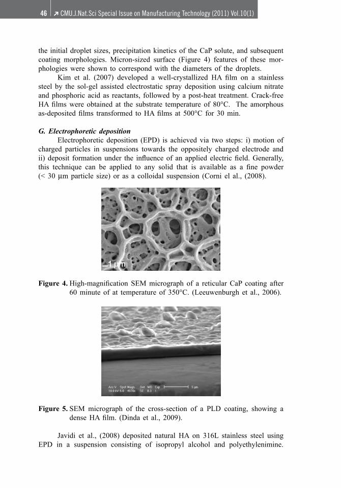
the initial droplet sizes, precipitation kinetics of the CaP solute, and subsequent coating morphologies. Micron-sized surface (Figure 4) features of these mor-phologies were shown to correspond with the diameters of the droplets. Kim et al. (2007) developed a well-crystallized HA film on a stainlesssteel by the sol-gel assisted electrostatic spray deposition using calcium nitrate and phosphoric acid as reactants, followed by a post-heat treatment. Crack-free HAfilmswere obtained at the substrate temperature of 80°C. The amorphousas-depositedfilms transformed toHAfilms at 500°C for 30min.
G. Electrophoretic deposition Electrophoretic deposition (EPD) is achieved via two steps: i) motion of charged particles in suspensions towards the oppositely charged electrode and ii) deposit formationunder the influenceof an applied electricfield.Generally,this technique can be applied to any solid that is available as a fine powder (< 30 µm particle size) or as a colloidal suspension (Corni el al., (2008).
Figure 4.High-magnification SEMmicrograph of a reticularCaP coating after60 minute of at temperature of 350°C. (Leeuwenburgh et al., 2006).
Figure 5. SEM micrograph of the cross-section of a PLD coating, showing a denseHAfilm. (Dinda et al., 2009).
Javidi et al., (2008) deposited natural HA on 316L stainless steel using EPD in a suspension consisting of isopropyl alcohol and polyethylenimine.

CMU.J.Nat.Sci Special Issue on Manufacturing Technology (2011) Vol.10(1)➔ 47
The deposited substrates were sintered in a vacuum furnace at 800°C for 1 h. Thecoating efficiencyand surface roughness increasedwith increasing applied voltage and deposition time but the deposition rate decreased. Kwok et al., (2009) fabricated submicron HA coatings with and with-out multi-walled carbon nanotube powder on Ti-6Al-4V by EPD, followed by vacuum sintering at 800°C for 1 h. It was reported that the adhesion strength of all coatings studied was higher than values commonly reported for plasma sprayed HA coating. Sun et al., (2009) developed an electrophoretic deposition method for a composite HA-chitosan coating on 304 stainless steel. Crack-free composite coating with needle-shaped crystals in a chitosan matrix was obtained. H. Pulsed laser deposition Basically, the pulsed laser deposition (PLD) technique employs laser to ablate a target material and condense it on the surface of a substrate. The principle of this technique is a very complex physical phenomenon; readers can refer to Bao et al., (2005). Blind et al., (2005) produced HA thin film coating by PLD on Ti and Ti-6Al-4V substrate. The authors stated that these HA films were adherent tothe substrate and a high degree of crystallinity was obtained. Yanget al., (2009)depositedHAfilmon laser gasnitriding (LGN)NiTisubstrate using PLD technique. It was revealed that TiN dendrites provided a higher number of nucleation sites for HA deposition, thus a lot HA particles were deposited on LGN NiTi substrates. Dinda et al., (2009) deposited HA coating on Ti-6Al-4V by PLD. The as-deposited HA films were amorphous, granular and 2.5 µm in thickness (Figure 5). It was suggested that post-deposition annealing for 4 hrs at 300°C had a potential to produce pure, adherent and crystalline HA coatings without dissolution in a SBF.
CONCLUSION
Various coating techniques have been extensively studied for biomedical applications.Thedevelopmentofsurfacemodificationsofmetallicbiomaterialshas been received a great deal of attention and a wealth of knowledge has been accumulated. However, better and simpler techniques need to be developed in order to achieve optimal and more reliable surface coating characteristics. In addition, it is essential to evaluate the safety of the functionalized coatings before clinical uses.
ACKNOWLEDGEMENTS
The author wish to thank the National Research University (NRU) Pro-ject underThailand’sOffice of theHigherEducationCommission forfinancial support.

CMU.J.Nat.Sci Special Issue on Manufacturing Technology (2011) Vol.10(1)➔48
REFERENCES
Arnould, C., J. Denayer, M. Planckaert, J. Delhalle and Z. Mekhalif. 2010. Bilayer coatings on titanium surface: The impact on the hydroxyapatite initiation. J. Colloid Inter. Sci. 341:75-82.
Ballarre, J., D.A. Lopez, W.H. Schreiner, A. Duran and S.M. Cere. 2007. Pro-tective hybrid sol-gel coatings containing bioactive particles on surgical grade stainless steel: Surface characterization. Appl. Surf. Sci. 253: 7260-7264.
Bao, Q., C. Chen, D. Wang, Q. Ji and T. Lei. 2005. Pulsed laser deposition and its current research status in preparing hydroxyapatite thin films. Appl.Surf. Sci. 252: 1538-1544.
Blind, O., L.H. Klein, B. Dailey and L. Jordan. 2005. Characterization of hy-droxyapatitefilmsobtainedbypulsedlaserdepositiononTiandTi-6Al-4Vsubstrates. Dental Mater. 21: 1017-1024.
Chen, D., E.H. Jordan, M. Gell and M. Wei. 2008. Apatite formation on alkaline-treated dense TiO2 coatings deposited using the solution precursor plasma spray process. Acta Biomater. 4: 553-559.
Chen, H. T., C.H. Hsiao, H.Y. Long, C.J. Chung, C.H. Tang, K.C. Chen and J.L. He. 2009. Micro-arc oxidation of β-titanium alloy: Structural characteriza-tion and osteoblast compatibility. Surf. Coat. Techol. 204: 1126-1131.
Corni, I., M.P. Ryan and A.R. Boccaccini. 2008. Electrophoretic deposition: From traditional ceramics to nanotechnology. J. Eur. Ceram. Soc. 28: 1353-1367.
Crawford, G.A. and N. Chawla. 2009. Pourous hierarchical TiO2 nano-structures: Processing and microstructure relationship. Acta Mater. 57: 854-867.
Cui, X., H.M. Kim, M. Kawashita, L. Wang, T. Xiong, T. Kokubo and T. Nakamura. 2009.Preparationof bioactive titaniafilmson titaniummetalvia anodic oxidation. Dental Mater. 25: 80-86.
Dinda, G.P., J. Shin and J. Mazumder. 2009. Pulsed laser deposition of hydroxy-apatite thinfilmsonTi-6Al-4V:Effectofheat treatmentonstructureandproperties. Acta Biomater. 5: 1821-1830.
Garcia, C., S. Cere and A. Duran. 2006. Bioactive coatings deposited on titanium alloys. J. Non-Cryst. Solids 352: 3488-3495.
Geetha, M., A.K. Singh, R. Asokamani and A.K. Gogia. 2009 Ti based bioma-terials, the ultimate choice for orthopaedic implants - A review. Progress Mater.Sci. 54: 397-425.
Han, Y., J. Sun and X. Huang. 2008. Formation mechanism of HA-based coat-ings by micro-arc oxidation. Electrochem. Commun. 10: 510-513.
Harle, J., H.W. Kim, N. Mordan, J.C. Knowles and V. Salih. 2006. Initial responses ofhumanosteoblaststosol-gelmodifiedtitaniumwithhydroxyapatiteandtitania composition. Acta Biomater. 2: 547-556.
Hijon, N., M.V. Cabanas, I.I. Barba, M.A. Garcia and M.V. Regi. 2006. Nano-crystalline bioactive apatite coatings. Solid State Sci. 8: 685-691.

CMU.J.Nat.Sci Special Issue on Manufacturing Technology (2011) Vol.10(1)➔ 49
Inagaki, M. and T. Kameyama. 2007. Phase transformation of plasma-sprayed hydroxyapatite coating with preferred crystalline orientation. Biomaterials 28: 2923-2931.
Javidi, M., S. Javadpour, M.E. Bahrololoom and J. Ma. 2008. Electrophoretic deposition of natural hydroxyapatite on medical grade 316L stainless steel. Mater. Sci. Eng. C 28: 1509-1515.
Kim, B.H., J.H. Jeong, Y.S. Jeon, K.O. Jeon and K.S. Hwang. 2007. Hydroxy-apatite layers prepared by sol-gel assisted electrostatic spray deposition. Ceram. Inter. 33: 119-122.
Kim, M.S., J.J. Ryu and Y.M. Sung. 2007. One-step approach for nano-crystalline hydroxyapatite coating on titanium via micro-arc oxidation. Electrochem. Commun. 9: 1886-1891.
Kwok, C.T., P.K. Wong, F.T. Cheng and H.C. Man. 2009. Characterization and corrosion behaviour of hydroxyapatite coating on Ti6Al4V fabricated by electrophoretic deposition. Appl. Sufr. Sci. 255: 6736-6744.
Kunze, J., L. Muller, J.M. Macak, P. Greil, P. Schmuki and F.A. Muller. 2008. Time-dependent growth of biomimetic apatite on anodic TiO2 nanotubes. Electrochim. Acta 53: 6995-7003.
Leeuwenburgh,S.C.G.,J.G.C.Wolke,J.SchoonmanandJ.A.Jansen.2005.Influ-ence of deposition parameters on morphological properties of biomedical calcium phosphate coatings prepared using electrostatic spray deposition. Thin Solid Films 472: 105-113.
Leeuwenburgh, S.C.G., M.C. Heine, J.G.C. Wolke, S.E. Pratsinis, J. Schoonman and J.A. Jansen. 2006. Morphology of calcium phosphate coatings for biomaterials applications deposited using electrostatic spray deposition. Thin Solid Films 503: 69-78.
Liu, X., X. Zhao, B. Li, C. Cao, Y. Dong, C. Ding and P.K. Chu. 2008. UV-irradiation-induced bioactivity on TiO2 coatings with nanostructural surface. Acta Biomater. 4: 544-552.
Lu, Y.P., Y.M. Chen, S.T. Li and J.H. Wang. 2008. Surface nanocrystallization of hydroxyapatite coating. Acta Biomater. 4: 1865-1872.
Ochsenbein, A., F. Chai, S. Winter, M. Traisnel, J. Breme and H.F. Hildebrand. 2008. Osteoblast responses to different oxide coatings produced by the sol-gel process on titanium substrates. Acta Biomater. 4: 1506-1517.
Paital, S.R. and N.B. Dahotre. 2009. Calcium phosphate coating for bio-implant applications: Materials, performance, factors, and methodologies. Mater. Sci. and Eng. R 66:1-70.
Park, H.H., I.S. Park, K.S. Kim, W.Y. Jeon, B. K. Park, H.S. Kim, T.S. Bae and M.H. Lee. 2010. Bioactive and electrochemical characterization of TiO2 nanotubes on titanium via anodic oxidation. Electrochem. Acta 55: 6109-6114.
Siebers, M.C., X.F. Walboomers, S.C.G. Leeuwenburgh, J.G.C. Wolke and J.A. Jansen. 2004. Electrostatic spray deposition (ESD) of calcium phosphate coatings, an in vitro study with osteoblast-like cells. Biomaterials 25: 2019-2027.

CMU.J.Nat.Sci Special Issue on Manufacturing Technology (2011) Vol.10(1)➔50
Shi, G.S., L.F. Ren, L.Z. Wang, H.S. Lin, S.B. Wang and Y.Q. Tong. 2009. H2O2/HCl and heat treated Ti-6Al-4V stimulates pre-osteoblast proliferation and differentiation. OOOOE 108: 368-375.
Song, Y.Y., F.S. Stein, S. Bauer and P. Schmuki. 2009. Amphiphilic TiO2 nano-tube arrays: An activity controllable drug delivery system. J. Am. Ceram. Soc. 131: 4230-4232.
Song, H.J., S.H. Park, S.H. Jeong and Y.J. Park. 2009. Surface characteristics and bioactivity of oxide films formed by anodic spark oxidation on titaniumin different electrolytes. J. Mater. Proc. Tech. 209: 864-870.
Sun, F., X. Pang and I. Zhitomirsky. 2009. Electrophoretic deposition of com-posite hydroxyapatite-chitosan-heparin coatings. J. Mater. Proc. Techol. 209: 1597-1606.
Sun, J., Y. Han and X. Huang. 2007. Hydroxyapatite coatings prepared by micro-arc oxidation in Ca- and P-containing electrolyte. Surf. Coat. Techol. 201: 5655-5658.
Tao, X.J., S.J. Li, C.Y. Zheng, J. Fu, Z. Guo, Y.L. Hao, R. Yang and Z. X. Guo. 2009. Synthesis of a porous oxide layer on a multifunctional biomedical titanium by micro-arc oxidation. Mater. Sci. Eng. C 29: 1923-1934.
Wang, Y.Q., J. Tao, L. Wang, P.T. He and T. Wang. 2008. HA coating on titanium with nanotubular anodized TiO2 intermediate layer via electrochemical deposition. Tran. Nonferrous Met. Soc. China 18: 631-635.
Wang, J., Y. Chao, Q. Wan, Z. Zhu and H. Yu. 2009. Fluoridated hydroxyapatite coatings on titanium obtained by electrochemical deposition. Acta Biom-ater. 5: 1798-1807.
Wei, D. and Y. Zhou. 2009. Characteristic and biocompatibility of the TiO2-based coatings containing amorphous calcium phosphate before and after heat treatment. Appl. Surf. Sci. 255: 6232-6239.
Wu, J.M. 2004. Low-temperature preparation of titania nanorods through direct oxidation of titanium with hydrogen peroxide. J. Crys. Growth 269: 347-355.
Wu, J.M., T.W. Zhang, Y.W. Zeng, S. Hayakawa, K. Tsuru and A. Osaka. 2005 Large-scale preparation of ordered titania nanorods enhanced photocatalytic activity. Langmuir 21: 6995-7002.
Wu, J.M., B. Huang,M.Wang andA. Osaka. 2006. Titania nanoflowers withhigh photocatalytic activity. J. Am. Ceram. Soc. 89: 2660-2663.
Yang, S., W. Xing and H.C. Man. 2009. Pulsed laser deposition of hydroxy-apatite film on laser gas nitriding NiTi substrate. Appl. Surf. Sci. 255:9889-9892.
![TO based oxide nanomaterials with · 2016. 9. 13. · coating techniques (spin-coating, deep-coating, spraying) [12]. The benefits of sol-gel method are numerous, as: obtaining of](https://static.fdocuments.us/doc/165x107/5ff0ffec30076d51aa44c5ac/to-based-oxide-nanomaterials-2016-9-13-coating-techniques-spin-coating-deep-coating.jpg)