Abbott's financial ratio analysis(Shraddha Bhatt & Jincey Jose),SVKM'S NMiMS

COARCTATION OF THE AORTA WITH PATENTDUCTUS ARTERIOSUS
BY
J. GIBSON GRAHAM AND J. D. OLAV KERRReceived February 28, 1941
While serving in the R.A.M.C. at a military hospital, it has been our goodfortune to see a considerable number of patients with congenital heart disease.One of these with coarctation of the aorta presented certain unusual features,notably patency of the ductus arteriosus. In themselves these two defects arenot amongst the rarer developmental abnormalities of the cardiovascular system,but their combination in a diagnosis made during life appears to be rare enoughto justify detailed consideration.
Evans (1933) stated that coarctation of the aorta was found in approximatelyone in every 1000 necropsies, while Blackford (1928) estimated the incidenceas one in every 1550 necropsies.
Patency of the ductus arteriosus occurred in 262 of Abbott's (1936) seriesof 1000 cases of congenital cardiac defects; in 40 it was combined with pul-monary atresia or stenosis, in each instance being associated with a septaldefect. In the same series of 1000 cases she described 178 examples of coarcta-tion of the aorta, of which 105 belonged to the adult type; 70 were analysedin detail, and in only 6 of these was the ductus arteriosus patent.
DESCRIPTION OF THE CASEOur patient, aged 25, was the wife of a corporal. There was nothing of
note in the family history. Both parents, her sisters, and two brothers werealive and well; her husband was in good health. She herself could rememberno previous illness, apart from diphtheria at twelve years, when she was illfor six weeks. As a child she had made no complaint of breathlessness, andtook part in all games when at school with enjoyment. Later she was employedas a shop assistant without incident. She was married at nineteen years andwas evidently a most active housewife. A year later she became pregnant,and at the seventh month began to suffer for the first time from headache,nausea, and vomiting that was difficult to control. While no cedema wasobserved, albumin was present in the urine, and on the fifth day of severe*symptoms the pregnancy was terminated. It should be noted that there wasno history of convulsions. All symptoms disappeared in three days, but shewas told she had " kidney trouble " and that another pregnancy would bestrongly contra-indicated. This unfavourable prognosis apparently induced
121
on Decem
ber 25, 2019 by guest. Protected by copyright.
http://heart.bmj.com
/B
r Heart J: first published as 10.1136/hrt.3.2.121 on 1 A
pril 1941. Dow
nloaded from

J. G. GRAHAM AND J. D. 0. KERR
an element of introspection and doubt as to her own well-being which led thepatient to present herself at various hospitals for examination; however, littleamiss seems to have been found. In September 1940 she was six weeks inhospital with a diagnosis of pyelitis. At this time there was some dysuriawhich continued to a lesser extent, but at no time had polyuria been noted.
She came under the care of one of us (J. G. G.) some four months later witha provisional diagnosis of chronic nephritis. She made no specific complaintapart from slight discomfort on micturition and lumbar pain when tired. How-ever, she volunteered that she became breathless on exertion and that her lipsoccasionally turned blue, her nails becoming blue easily in cold weather. Shealso stated that her feet were always cold, although there was no history ofchilblains or of intermittent claudication.
The patient was of small build and stature (height, 4 feet, 11 inches; weight,91 pounds), and was somewhat pallid and wax-like in complexion. On admis-sion to hospital there was no cyanosis or clubbing of the fingers, nor was thereany evidence of dilated vessels in the thorax; there was, however, marked arterialpulsation at the root of the neck. Neither dyspncea nor orthopncea was noted.Physical examination of lungs, abdomen, and nervous system did not show anyabnormality.
Cardiovascular System. The pulse rate was 70 per minute, the rhythmbeing regular; the force was good, but the pressure was higher at the left wristthan at the right. The vessels were not hardened to palpation. Pulsationcould not be detected either in the abdominal aorta or in the great vessels of thelower limbs. Palpation showed the apex beat to be in the fifth left interspace,four inches from the mid-line. There was marked pulsation of both carotids,and also of the aorta in the episternal notch, but no tracheal tugging. Per-cussion did not elicit any obvious change in cardiac dulness. On auscultationat the apex three heart sounds were heard, the third following at a short intervalafter an apparent second sound. One inch internal to the apex beat systolicand diastolic murmurs were present, the heart sounds themselves being wellheard and of good tone. At this point the third sound was not audible andthe diastolic murmur partly obscured and followed the second sound. Therewas nothing to suggest the presence of a presystolic murmur. At the aorticcartilage and down the sternum to the xiphoid there was a systolic murmur,but the second sound was pure. Over the aorta and great vessels a systolicbruit was heard, and also in both interscapular areas, especially at the thirdand fourth left interspaces. In the second left interspace in front, about oneand a half inches from the mid-line, a continuous machinery murmur of thewater-wheel type was heard, being crescendo during systole and continuingthroughout diastole. This murmur was also conducted up to the left clavicle.Throughout the period of observation there were considerable variations in thesystolic blood pressure from day to day, but at first there was a constant differ-ence of some 50 millimetres of mercury between the readings obtained in thetwo arms, the left being the higher. The readings in both upper limbs weregreatly in excess of those in the legs, typical figures being: right arm, 175/90;left arm, 225/1 10; legs, 130 systolic.
122
on Decem
ber 25, 2019 by guest. Protected by copyright.
http://heart.bmj.com
/B
r Heart J: first published as 10.1136/hrt.3.2.121 on 1 A
pril 1941. Dow
nloaded from

COARCTA TION OFAORTA WITH PATENT DUCTUS ARTERIOSUS 123
Fic
On admission the urine showed a trace of albumin and contained a fewpolymorphs and coliform organisms. No casts were found. A urea concen-tration test gave a figure of 2 65 per cent at the second hour. The blood ureawas 39 mg. per 100 c.c. Later the urine became normal. A gynecologicalexamination did not reveal any abnormality.
Blood examination: Hemoglobin 82 per cent, red blood cells 4,700,000 perc.mm., white blood cells 11,200 per c.mm. Examination of blood films,including a differential count, did not reveal any abnormality. The Kahn testwas negative. The blood sedimentation rate was well within normal limits.
Ophthalmic examination: The pupils were always moderately dilated andunequal, the degree of inequality varying from day to day; sometimes the oneand sometimes the other was the more dilated. The reactions both to lightand to convergence were sluggish. The fundi showed no evidence of arterialor venous pulsation or any other abnormality.
Radiological examination of the chest showed enlargement of the rightventricle and a prominent and pulsatile conus; the aortic knuckle was some-what small (Fig. 1). Fig. 2, a similar view from one of our uncomplicated
I.1.-Antero-posterior view of our patient, at FIG. 2.-Antero-posterior view, at 2 metres, of2 metres, showing right-sided cardiac enJarge- uncomplicated case of patent ductus artement, and erosion of ribs. OSUS.
cases of patent ductus arteriosus, a female aged 28 years, is included for com-parison. Although oblique views of the subject of this paper revealed anapparently normal ascending and transverse aorta, the remainder of the archcould not be visualized. In the left (II) oblique position the aorta and itsbranches, as Lewis (1931) has described, seemed to rise as a column from theheart shadow up into the root of the neck.K
aneri-
on Decem
ber 25, 2019 by guest. Protected by copyright.
http://heart.bmj.com
/B
r Heart J: first published as 10.1136/hrt.3.2.121 on 1 A
pril 1941. Dow
nloaded from

J. G. GRAHAM AND J. D. 0. KERR
Rosler's sign-erosion of ribs-was present. This is clearly seen in Figs. 1,an antero-posterior view taken at two metres, and in greater detail in Fig. 3. In
FIG. 3.-Left upper quadrant, showing erosionof ribs in detail.
FIG. 4.-Left (II) oblique view of ourpatient.
Fig. 4, a left (II) oblique view, the prominent pulmonary conus is well seen,but the descending arch of the aorta is not visualized.
An electrocardiogram, Fig. 5, shows changes in the ventricular complexescomparable with those described by Evans and Turnbull (1937), following thework of Wilson et al. (1934), as denoting right bundle branch block. Thisnewer curve, which is said to be much commoner than the one standard for rightbundle branch block, is characterized by a deep S wave as a component of awide QRS complex in lead I (measuring 0-12 sec. in this case), and also in lead JI,by the QRS complex in lead III being directed downwards; and by the T wavebeing upright in leads I and II and inverted in lead III. The changes seen inthe sternal lead (Fig. 5) would seem to be in conformity with this diagnosis.
After three weeks' rest in bed the patient stated that she felt fitter in allrespects. She had been afebrile throughout this period. The differencebetween the two radial pulses and the two brachial blood pressures becamemuch less marked. The systolic pressure in the arms, however, remainedconsiderably in excess of that in the legs, typical findings being: right arm,205/120; left arm, 215/120; legs, 130 systolic.
DIscUSSION
This case presents many unusual features, both in the history and clinicalfindings. There had been no evidence of cardiac insufficiency until the patient'stwentieth year, when she was a seven months primagravida. At that time the
124
on Decem
ber 25, 2019 by guest. Protected by copyright.
http://heart.bmj.com
/B
r Heart J: first published as 10.1136/hrt.3.2.121 on 1 A
pril 1941. Dow
nloaded from

COARCTATION OF AORTA WITH PATENT DUCTUS ARTERIOSUS 125
4'i:QpNA F
iL ~ I-
-+7~~~~~~~~~~~~~~~~~~~~~~~~~~~~~~~~~~~~~~~~~~~~~~~~~~~~~~~~~~~~~~~~~~~~~~~~~~~~~~~~~~~~~~~~~~~~~~~~~~~~~~~~~~~~~~~~~~~~~~~~~~~~~------
FIG.5.-Electrocardiogramof the case described, showing a conduction defect comparable~~~~~~~~~~~~~~~~~~~~~~~~~~~~~~~~~~~~~~~~~~~~~~~~~~~~~~~~~~~~~~~..withthe "newer" type of right bundle branch block~
iFecIon lethoatresponded tof thecsimplestrbdtheraeuicg mascnures)itn sefetcmsareason-ableto sugge thaathernative explanatrion. Hudebransymptomsmybeitrpee
adutotedevelopment ofchroheaueanic vemtnous congesione wthruh the latsencmotsofplumnreganc ovenerpaxing ascicuaiong already heampdiersed.b anconenitalcardadubefuectnthistr anow suggresutedoththconsiertivsiationofthwpyical fningsrindicatesho a copexidefe of whpichd thenasevralctomputonentsfar outlnd.uinr
(1)ectoarcthatirsondof the Aota. X-rayes filmsrakentimaturtwo metre showsedasn-ascendingugaortaaofanormaltsize,xwithaairatHer unesizedmsmakncke, wiltrpethedreainduertofthe archlwasenot visualoized Moenuovger,Rsteiosin wasug present.Ionadtisonprganlou stolieruaitg was hcuardo atlteaorti camprtilage acndgwasicondctedintodeethe necks andotsgethedinatecapulaerareanposteiryThebhsclooidigprdaessr in eithler armecwas markedl raised,randiopngeatsaexs ofuthatnnethloerlibsCortheio goss difeArencai p-ressurembtween ath two upetre lmshnoteda
oscnadmsiongaotaofhospial, przesumablyawitheareduedcrszdianckreere, whien theleftbaciale exeeethe righwant byisomlied50 millimetre ofsmercurysignwseplaienablbycthed cnoartationecbengdiutdjutdsato theitrcplraentrpofstherirypThen ductustoestheaoreta,rfrom whic thearkeatyvessels maydtaein unutxcsua origin.ith
(2) Patency of the Ductus Arteriosus, as instanced by the characteristicmachinery murmur heard in the second left space and conducted up to theleft clavicle. In this case the shunt was predominantly arterio-venous, thepatient being free from cyanosis.
on Decem
ber 25, 2019 by guest. Protected by copyright.
http://heart.bmj.com
/B
r Heart J: first published as 10.1136/hrt.3.2.121 on 1 A
pril 1941. Dow
nloaded from
J. G. GRAHAM AND J. D. 0. KERR
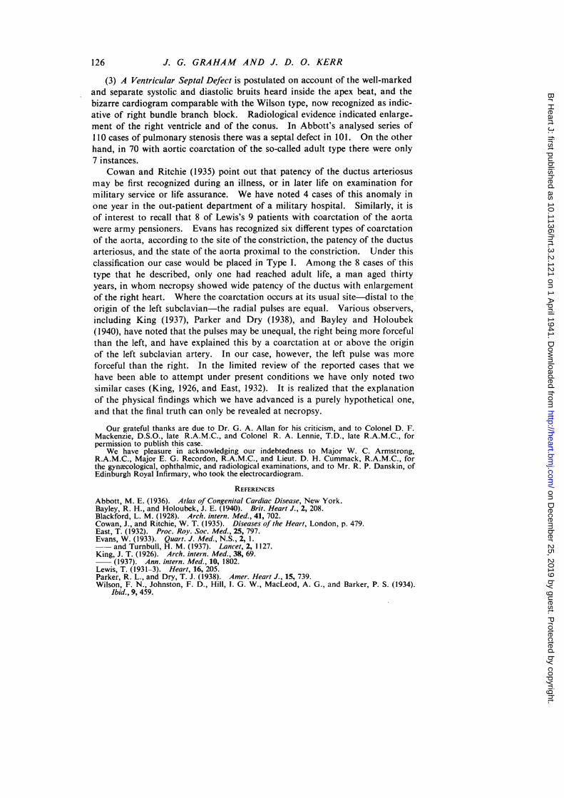
(3) A Ventricular Septal Defect is postulated on account of the well-markedand separate systolic and diastolic bruits heard inside the apex beat, and thebizarre cardiogram comparable with the Wilson type, now recognized as indic-ative of right bundle branch block. Radiological evidence indicated enlarge-ment of the right ventricle and of the conus. In Abbott's analysed series of110 cases of pulmonary stenosis there was a septal defect in 101. On the otherhand, in 70 with aortic coarctation of the so-called adult type there were only7 instances.
Cowan and Ritchie (1935) point out that patency of the ductus arteriosusmay be first recognized during an illness, or in later life on examination formilitary service or life assurance. We have noted 4 cases of this anomaly inone year in the out-patient department of a military hospital. Similarly, it isof interest to recall that 8 of Lewis's 9 patients with coarctation of the aortawere army pensioners. Evans has recognized six different types of coarctationof the aorta, according to the site of the constriction, the patency of the ductusarteriosus, and the state of the aorta proximal to the constriction. Under thisclassification our case would be placed in Type I. Among the 8 cases of thistype that he described, only one had reached adult life, a man aged thirtyyears, in whom necropsy showed wide patency of the ductus with enlargementof the right heart. Where the coarctation occurs at its usual site-distal to theorigin of the left subclavian-the radial pulses are equal. Various observers,including King (1937), Parker and Dry (1938), and Bayley and Holoubek(1940), have noted that the pulses may be unequal, the right being more forcefulthan the left, and have explained this by a coarctation at or above the originof the left subclavian artery. In our case, however, the left pulse was moreforceful than the right. In the limited review of the reported cases that wehave been able to attempt under present conditions we have only noted twosimilar cases (King, 1926, and East, 1932). It is realized that the explanationof the physical findings which we have advanced is a purely hypothetical one,and that the final truth can only be revealed at necropsy.
Our grateful thanks are due to Dr. G. A. Allan for his criticism, and to Colonel D. F.Mackenzie, D.S.O., late R.A.M.C., and Colonel R. A. Lennie, T.D., late R.A.M.C., forpermission to publish this case.
We have pleasure in acknowledging our indebtedness to Major W. C. Armstrong,R.A.M.C., Major E. G. Recordon, R.A.M.C., and Lieut. D. H. Cummack, R.A.M.C., forthe gynecological, ophthalmic, and radiological examinations, and to Mr. R. P. Danskin, ofEdinburgh Royal Infirmary, who took the electrocardiogram.
REFERENCES
Abbott, M. E. (1936). Atlas of Congenital Cardiac Disease, New York.Bayley, R. H., and Holoubek, J. E. (1940). Brit. Heart J., 2, 208.Blackford, L. M. (1928). Arch. intern. Med., 41, 702.Cowan, J., and Ritchie, W. T. (1935). Diseases of the Heart, London, p. 479.East, T. (1932). Proc. Roy. Soc. Med., 25, 797.Evans, W. (1933). Quart. J. Med., N.S., 2, 1.- and Turnbull, H. M. (1937). Lancet, 2, 1127.King, J. T. (1926). Arch. intern. Med., 38, 69.
(1937). Ann. intern. Med., 10, 1802.Lewis, T. (1931-3). Heart, 16, 205.Parker, R. L., and Dry, T. J. (1938). Amer. Heart J., 15, 739.Wilson, F. N., Johnston, F. D., Hill, 1. G. W., MacLeod, A. G., and Barker, P. S. (1934).
Ibid., 9, 459.
126
on Decem
ber 25, 2019 by guest. Protected by copyright.
http://heart.bmj.com
/B
r Heart J: first published as 10.1136/hrt.3.2.121 on 1 A
pril 1941. Dow
nloaded from