CO6 1 Study of DNA DSB end structure induced by ionizing ... · Agarose gel electrophoresis of...
17
30001 Study of DNA DSB end structure induced by ionizing radiation K. Akamatsu 1 , N. Shikazono 1 and T. Saito 2 1 Radiation DNA Damage Research Group, Kansai Pho- ton Science Institute, National Institutes for Quantum and Radiological Science and Technology (QST) 2 KURNS, Kyoto University INTRODUCTION: Clustered damage site, that is a DNA region with multiple lesions within a few helical turns, is believed to hardly be repaired. However, chemical and spatial details of them are not known. We are developing a methodolo- gy for studying structural feature of DNA DSB end and affinity between the DSB end and a repair protein. We have tried to irradiate gamma-rays and iron ion beam with LET of ~0.2 and ~200 keV/m, respectively, to pUC19 in a cell-mimetic buffered solution. As a result, we found that these ions tend to produce direct DSB (ex- cept for a DSB by sequentially-produced opposed close SSBs) compared with 60 Co -rays. We will isolate linear pUC19 with DSB ends and study how a repair protein recognize and process a DSB end. EXPERIMENTS: ●Sample preparation and irradiation The plasmid DNA (pUC19, 2686bp) was used. The DNA was dissolved to be 0.1 g/L in 0.2 M Tris-HCl buffer (pH 7.5) which is a cell-mimetic condition in rela- tion to radical scavenging capacity. Twenty microliters of the DNA solution was transferred to a micro chamber (20-L size), and was irradiated with Fe ion beam (LET: ~200 keV/m; HIMAC QST) and 60 Co -rays (LET: ~0.2 keV/m; KURNS Kyoto University) as a standard radia- tion source. The irradiated DNA sample was purified by ethanol precipitation and was dried in vacuum, followed by being kept at -20 o C until use. ●Agarose gel electrophoresis of irradiated DNA The dry irradiated DNA samples were dissolved to TE buffer (~2g/L). The electrophoresis was performed for 4h, 70V at 4 o C. The gel was stained by ethidium bromide (EtBr). The different DNA forms separated (supercoiled (sc), open circular (oc), and linear (L) form) were quantified by a gel imager (Pharos FX, BioRad). ●Large-amount Isolation of lin fraction using agarose electrophoresis We have constructed a protocol for collecting large amount of lin fraction using general agarose electropho- resis (1% agarose in 0.5xTBE, 25V, 18h) pUC19/SmaI was used as a marker. EtBr staining was performed only to the marker lane. L fraction was collected from L-containing gel fragments by electroelution (D-Tube Dialyzer, Maxi, Novagen), followed by buffer exchange to TE (Amicon Ultra-4, Millipore). The collected L frac- tion will be used for several biochemical and physico- chemical analysis. RESULTS: Fig. 1. Relationship between absorbed dose and DNA fractions (●: sc, ▲: oc, ■: L) for 60 Co -rays (upper panel) and Fe ion beam (lower panel). The fraction maximum of L for 60 Co -rays is ~0.8, whereas that for Fe beam dose is ~0.6, suggesting that Fe beam is likely to induce direct dsb formation from sc form compared with the -rays. The yield ratios of dsb to total strand breaks for and Fe are calculated to be 0.067 and 0.17, respectively, according to Cowan’s theory [1] . What is a direct dsb? How difference are shown be- tween a direct dsb and successive dsb (by ssb + ssb)? How is a direct dsb repaired? These are our issues to be solved. REFERENCES: [1] R.Cowan, et al., J.Theor.Biol. 81 (1987) 229-245. CO6-1
Transcript of CO6 1 Study of DNA DSB end structure induced by ionizing ... · Agarose gel electrophoresis of...

30001
Study of DNA DSB end structure induced by ionizing radiation
K. Akamatsu1, N. Shikazono1 and T. Saito2
1Radiation DNA Damage Research Group, Kansai Pho-ton Science Institute, National Institutes for Quantum and
Radiological Science and Technology (QST)
2KURNS, Kyoto University
INTRODUCTION:
Clustered damage site, that is a DNA region with
multiple lesions within a few helical turns, is believed to
hardly be repaired. However, chemical and spatial details
of them are not known. We are developing a methodolo-
gy for studying structural feature of DNA DSB end and
affinity between the DSB end and a repair protein. We
have tried to irradiate gamma-rays and iron ion beam
with LET of ~0.2 and ~200 keV/m, respectively, to
pUC19 in a cell-mimetic buffered solution. As a result,
we found that these ions tend to produce direct DSB (ex-
cept for a DSB by sequentially-produced opposed close
SSBs) compared with 60Co -rays. We will isolate linear
pUC19 with DSB ends and study how a repair protein
recognize and process a DSB end.
EXPERIMENTS:
●Sample preparation and irradiation
The plasmid DNA (pUC19, 2686bp) was used. The
DNA was dissolved to be 0.1 g/L in 0.2 M Tris-HCl
buffer (pH 7.5) which is a cell-mimetic condition in rela-
tion to radical scavenging capacity. Twenty microliters of
the DNA solution was transferred to a micro chamber
(20-L size), and was irradiated with Fe ion beam (LET:
~200 keV/m; HIMAC QST) and 60Co -rays (LET: ~0.2
keV/m; KURNS Kyoto University) as a standard radia-
tion source. The irradiated DNA sample was purified by
ethanol precipitation and was dried in vacuum, followed
by being kept at -20oC until use.
●Agarose gel electrophoresis of irradiated DNA
The dry irradiated DNA samples were dissolved to
TE buffer (~2g/L). The electrophoresis was performed
for 4h, 70V at 4oC. The gel was stained by ethidium
bromide (EtBr). The different DNA forms separated
(supercoiled (sc), open circular (oc), and linear (L) form)
were quantified by a gel imager (Pharos FX, BioRad).
●Large-amount Isolation of lin fraction using agarose
electrophoresis
We have constructed a protocol for collecting large
amount of lin fraction using general agarose electropho-
resis (1% agarose in 0.5xTBE, 25V, 18h) pUC19/SmaI
was used as a marker. EtBr staining was performed only
to the marker lane. L fraction was collected from
L-containing gel fragments by electroelution (D-Tube
Dialyzer, Maxi, Novagen), followed by buffer exchange
to TE (Amicon Ultra-4, Millipore). The collected L frac-
tion will be used for several biochemical and physico-
chemical analysis.
RESULTS:
Fig. 1. Relationship between absorbed dose and DNA fractions (●: sc,
▲: oc, ■: L) for 60Co -rays (upper panel) and Fe ion beam (lower
panel).
The fraction maximum of L for 60Co -rays is ~0.8,
whereas that for Fe beam dose is ~0.6, suggesting that Fe
beam is likely to induce direct dsb formation from sc
form compared with the -rays. The yield ratios of dsb to
total strand breaks for and Fe are calculated to be 0.067
and 0.17, respectively, according to Cowan’s theory [1].
What is a direct dsb? How difference are shown be-
tween a direct dsb and successive dsb (by ssb + ssb)?
How is a direct dsb repaired? These are our issues to be
solved.
REFERENCES:
[1] R.Cowan, et al., J.Theor.Biol. 81 (1987)229-245.
CO6-1
30002
Oligomeric structures of HspB1 from CHO cell
M. Yohda, K. Ankai, R. Inoue1, K. Morishima1, N. Sato1,M. Sugiyama1
Department of Biotechnology and Life Science, Tokyo University of Agriculture and Technology 1Institute for Integrated Radiation and Nuclear Science, Kyoto University
INTRODUCTION: Small heat shock proteins (sHsps) endow cells with stress tolerance [1]. They bind to par-tially folded or denatured proteins, thereby preventing ir-reversible aggregation or promoting correct substrate fold-ing. Various species of sHsps are present in mammals [2]. HspB1, also known as Hsp27, is a ubiquitous sHsp [3]. The sHsps of mammals, which reflect their status as ho-meotherms, are regulated by phosphorylation. To examine the structure and function of HspB1, we expressed, puri-fied and characterized HspB1 from Chinese hamster (Cri-cetulus griseus) ovary cells (CgHspB1) (Manuscript in preparation). CgHspB1 forms a large oligomeric structure. In dilute conditions, CgHspB1 dissociates into small oli-gomers at the elevated temperatures. In contrast, dissocia-tion of the oligomer was not observed at the relatively high protein concentrations. The phosphorylation mimic mu-tant of CgHspB1 with the replacement of Ser15 to Asp (CgHspB1_S15D) exhibited relatively lower oligomer sta-bility and higher ability for protecting thermal aggregation than the wild-type protein. The result clearly showed the correlation between oligomer dissociation with chaperone activity. In this study, we compared the oligomeric struc-tures of CgHspB1 wild type and CgHspB1_S15D by vari-ous methods.
EXPERIMENTS: CgHSPB1 variants (CgHspB1_WT, CgHspB1_S15D) were expressed in E. coli and purified by anion exchange chromatography with DEAE-TOYOPEARL, anion exchange chromatography with RE-SOURCE Q and size-exclusion chromatography with Su-perdex 200. The molecular weights of the oligomers of CgHspB1 variants were analyzed by Size-exclusion chro-matography - multiangle light scattering (SEC–MALS) using a multiangle light-scattering detector (MINI DAWN, Wyatt Technology). The oligomeric conformation of CgH-spB1 variants were analyzed by small angle X-ray scatter-ing (SAXS) using NANOPIX (Rigaku) and analytical ul-tracentrifugation (AUC) using XL-1 (Beckman Coulter Diagnostics).
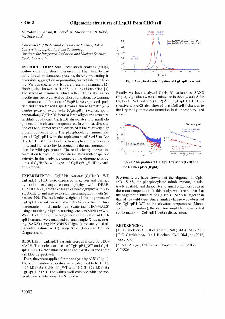
RESULTS: CgHspB1 variants were analyzed by SEC–MALS. The molecular mass of CgHspB1_WT and CgH-spB1_S15D were estimated to be about 470 kDa and about 780 kDa, respectively. Then, they were applied for the analysis by AUC (Fig. 1). The sedimentation velocities were calculated to be 15.1 S (403 kDa) for CgHspB1_WT and 18.2 S (829 kDa) for CgHspB1_S15D. The values well coincide with the mo-lecular mass determined by SEC-MALS.
Finally, we have analyzed CgHspB1 variants by SAXS (Fig. 2). Rg values were calculated to be 58.4 (± 0.6) Å for CgHspB1_WT and 66.9 (± 1.2) Å for CgHspB1_S15D, re-spectively. SAXS also showed that CgHspB1 changes to the larger oligomeric conformation in the phosphorylated state.
Previously, we have shown that the oligomer of CgH-spB1_S15S, the phosphorylated mimic mutant, is rela-tively unstable and dissociates to small oligomers even at the room temperature. In this study, we have shown that the oligomeric structure of CgHspB1_S15S is larger than that of the wild type. Since similar change was observed for CgHspB1_WT at the elevated temperature (Manu-script in preparation), the structure might be the activated conformation of CgHspB1 before dissociation.
REFERENCES: [1] U. Jakob et al., J. Biol. Chem., 268 (1993) 1517-1520.[2] C. Garrido et al., Int. J. Biochem. Cell. Biol., 44 (2012) 1588-1592.[3] A.P. Arrigo., Cell Stress Chaperones., 22 (2017) 517-529.
Fig. 1 Analytical centrifugation of CgHspB1 variants
Fig. 2 SAXS profiles of CgHspB1 variants (Left) and the Guinier plots (Right)
CO6-2

30008
Effect of ligand Binding on Solution Structure of Multi-domain protein, MurD
H. Nakagawa, T. Saio1,2, M. Sugiyama3, R. Inoue3
Materials Science Research Center, Japan Atomic Energy Agency 1Graduate School of Chemical Sciences and Engineering, Hokkaido University 2Department of Chemistry, Faculty of Science, Hokkaido University 3KURNS, Kyoto University
INTRODUCTION: In structural biology, precise de-termination of three-dimensional structures of proteins has been focused, and the structures with an atom-ic-resolution have given solid platforms to understand their biological functions. Recently, the idea of structural biology has extended beyond the static structural infor-mation with atomic resolution, in order to cover more complex and dynamical structures at different levels of space and time resolution. SAXS measurement can ob-serve the solution structure of flexible protein under the physiological conditions [1].
MurD (UDP-N-acetylmuramoylalanine--D-glutamate ligase) is a typical multi-domain protein (Fig.1), which is one of the ATP-driven Mur ligases that are responsible for peptidoglycan biosynthesis. The crysital structure of MurD has been determined, but the ATP-bound form is not detemied.
EXPERIMENTS: MurD were expressed in E. coli strain BL21 (DE3). Small angle X-ray scattering (SAXS) were measured for MurD in ATP-unbound and bound states at the concentration of 5, 10, 20 and 50 mg/ml. The buffer conditions were 20 mM Tris-HCl at pH=7.2. For
the sample of ATP-bound state, AMP-PNP and MgCl2 were added at the concentration of 2 mM and 5 mM, re-spectively. For the sample of Compound1-bound state, Compound1 was added at 3 eq.
RESULTS: SAXS profiles of MurD were successfully obtained for Apo, ATP-bound and Compound1-bound states at 5 mg/mL, where the inter-particle interaction effects were not observed in the profiles. As shown in Fig. 2, SAXS profiles are different among the three samples, indicating the structural change by ATP or Compound1 binding. The previous NMR measurement predicted that the domain orientation of MurD is changed into semi-closed conformation by ATP binding, and closed conformation by Compound1 binding [2]. And our pre-liminary MD simulation shows that the conformation and fluctuation of MurD domains are changed by ATP- or Compond1-binding, and that these are consistent with the NMR results. The comparison of the SAXS profiles with the results of the NMR and MD simulation are in pro-gress.
REFERENCES: [1] J. Trewhella et al., Acta Cryst. D73 (2017) 710-728.[2] T. Saio et al., Sci. Rep., 5 (2015) 16685.
Fig. 2. SAXS profiles in Apo, ATP-bound and Compound1-bound states of MurD at the concentra-tion of 5 mg/ml.
Fig. 1. Multi-domain protein, MurD, which are composed of three domains, D1, D2 and D3.
CO6-3
30010
Structural characterization of circadian clock potein complexes
H. Yagi, Y. Yunoki, K. Morishima1, N. Sato1, R. Inoue1,and M. Sugiyama1
Graduate School of Pharmaceutical Sciences, Nagoya City University, 1Institute for Integrated Radiation and Nuclear Science, Kyoto University
INTRODUCTION: The central oscillator that gen-erates the circadian rhythm in the cyanobacterium comprises only three proteins—KaiA, KaiB, and KaiC. Through interactions among these proteins in the presence of ATP, KaiC undergoes phosphorylation and dephosphorylation cycles with the period of 24 h, which proceeds in vitro without daylight oscillation, indicating that the internal clock mechanism can be autonomous irrespective of transcriptional and transla-tional feedback systems. Although the formation of several complexes, such as KaiA-KaiC, KaiB-KaiC, and KaiA-KaiB-KaiC oscillated in a circadian manner, their stoichiometry or the detailed structures remains to be elucidated. Herein, in order to understand the oscilla-tion mechanism mediated by the clock protein complex, we characterized the complexes by using analytical ul-tracentrifugation (AUC) and small angle X-ray scattering (SAXS) analyses. Especially, in this study, we focused on the characterization of KaiA-KaiB-KaiC complex.
EXPERIMENTS: The expression and purification of clock proteins, KaiA, KaiB and KaiC were performed according to methods previously described [1]. SAXS pattern was collected with NANOPIX (Rigaku Corpora-tion, Japan) equipped with HyPix-6000. A Cu Kα line (MicroMAX-007HF) was used as a beam source, which was further focused and collimated with a confocal mul-tilayer mirror (OptiSAXS). The camera length was set to 1.326 m and the range of the scattering vector q was from 0.007 to 0.24 A-1. The AUC experiments were performed using an ProteomeLab XL-I analytical ultra-centrifuge (Beckman-Coulter) at 25 °C and an angular velocity of 60,000 rpm. Data were recorded with Ray-leigh interference optical system, followed by the analy-sis with a c(s) distribution of the Lamm equation solu-tions calculated by the SEDFIT v15.01. The sedimenta-tion coefficient s were converted to the value in water at 20 ºC.
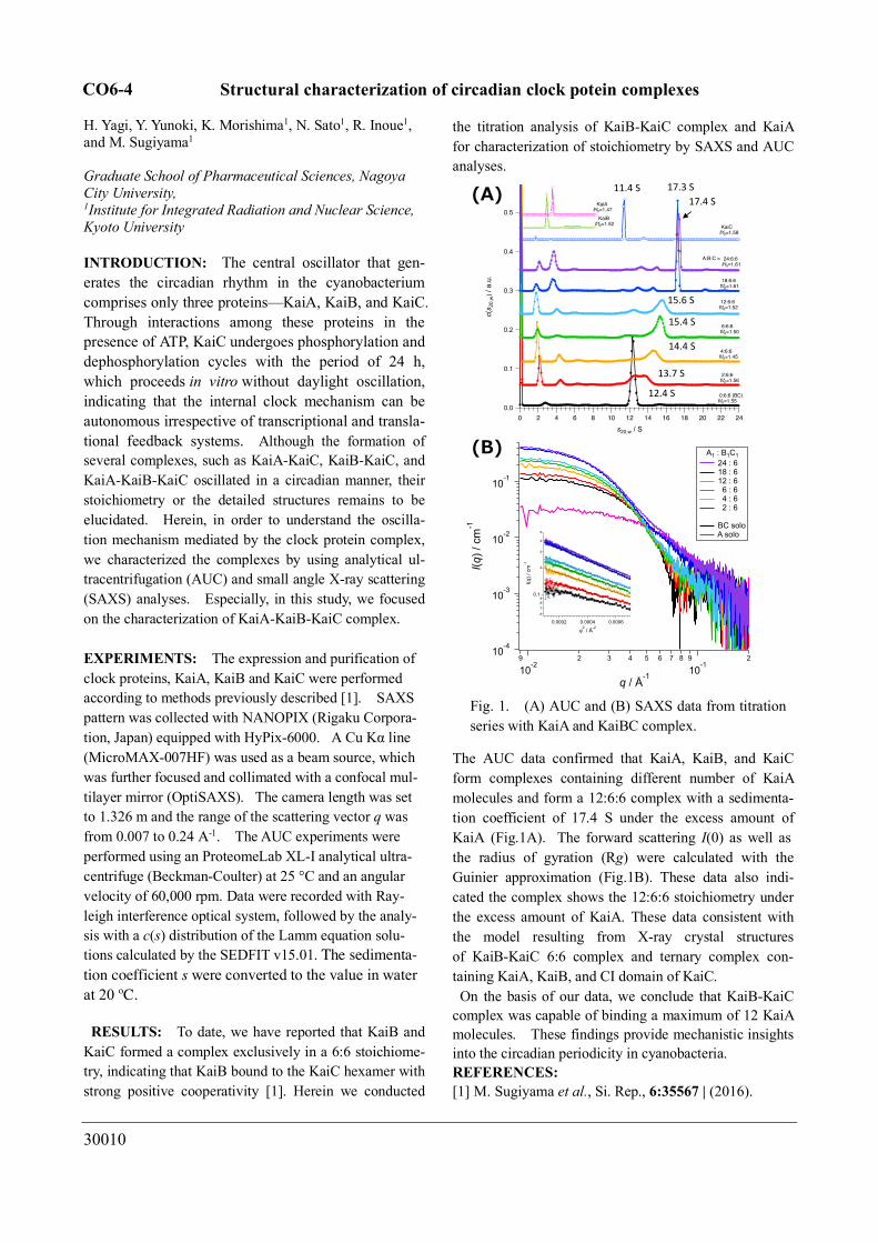
RESULTS: To date, we have reported that KaiB and KaiC formed a complex exclusively in a 6:6 stoichiome-try, indicating that KaiB bound to the KaiC hexamer with strong positive cooperativity [1]. Herein we conducted
the titration analysis of KaiB-KaiC complex and KaiA for characterization of stoichiometry by SAXS and AUC analyses.
The AUC data confirmed that KaiA, KaiB, and KaiC form complexes containing different number of KaiA molecules and form a 12:6:6 complex with a sedimenta-tion coefficient of 17.4 S under the excess amount of KaiA (Fig.1A). The forward scattering I(0) as well as the radius of gyration (Rg) were calculated with the Guinier approximation (Fig.1B). These data also indi-cated the complex shows the 12:6:6 stoichiometry under the excess amount of KaiA. These data consistent with the model resulting from X-ray crystal structures of KaiB-KaiC 6:6 complex and ternary complex con-taining KaiA, KaiB, and CI domain of KaiC. On the basis of our data, we conclude that KaiB-KaiC
complex was capable of binding a maximum of 12 KaiA molecules. These findings provide mechanistic insights into the circadian periodicity in cyanobacteria. REFERENCES: [1] M. Sugiyama et al., Si. Rep., 6:35567 | (2016).
���
10-4
10-3
10-2
10-1
I(q) /
cm
-1
9
10-2
2 3 4 5 6 7 8 9
10-1
2
q / Å-1
A1 : B1C1 24 : 6 18 : 6 12 : 6 6 : 6 4 : 6 2 : 6
BC solo A solo
6
789
0.1
2
3
4
5I(q
) / c
m-1
0.00060.00040.0002
q2 / Å-2
���
0.5
0.4
0.3
0.2
0.1
0.0
c(s 2
0,w
) / a
.u.
242220181614121086420
s20,w / S
24:6:6f/f0=1.61
18:6:6f/f0=1.61
12:6:6f/f0=1.52
6:6:6f/f0=1.50
4:6:6f/f0=1.45
2:6:6f/f0=1.56
0:6:6 (BC)f/f0=1.55
KaiAf/f0=1.47
KaiBf/f0=1.52 KaiC
f/f0=1.58
A:B:C =
17.3 S17.4 S
15.6 S
15.4 S
14.4 S
13.7 S
12.4 S
11.4 S
Fig. 1. (A) AUC and (B) SAXS data from titration series with KaiA and KaiBC complex.
CO6-4

30019
Sustainable Photoproduction of Medical 18
F and 99m
Tc Isotopes
T. Takeda, M. Fujiwara1,2
, M. Kurosawa1, N. Takahashi
1,
M. Tamura1, T. Kawabata, Y. Fujikawa, K.N. Suzuki,
N. Abe3, T. Kubota
3 and T. Takahashi
3
Department of Physics, Kyoto University 1Research Center for Nuclear Physics, Osaka University
2National Institutes for Quantum and Radiological Science
and Technology 3Institute for Integrated Radiation and Nuclear Science,
Kyoto University
INTRODUCTION:
We examined two sustainable methods for producing
the medical 18
F and 99m
Tc isotopes. A new method is
proposed for producing medical 18
F isotopes as an alter-
native to the 18
F cyclotron production. 18
F isotopes are
produced via the photoreaction on 20
Ne. Natural neon gas
is used as a target in the gas recycling flow system. The 18
F-FDG radio-pharmaceuticals are obtained by blowing
small bubbles of irradiated neon gas in the FDG liquid.
Another method is the production of 99m
Tc isotopes via
the photoreaction on natural MoO3. In these two methods,
both 99m
Tc isotopes for the SPECT inspections and 18
F
isotopes for the PET inspections are sustainably produced
using the photoreaction on natural MoO3 and Ne targets.
We have demonstrated the feasibilities of practical usages
of these two new methods.
18F EXPERIMENTS:
We used an electron beam from the linear accelerator
facility at Institute for Integrated Radiation and Nuclear
Science of Kyoto University (KURNS-LINAC) for pro-
ducing RI from photoreactions. A 40 MeV electron
beams with an intensity of 3-9 A is used to bombard a
platinum target with a thickness of 4 mm to generate
bremsstrahlung photons for producing 18
F via the photo-
reaction on natural Ne gas.
The Ne-gas circulation system has been newly
constructed for the present experiment. The irra-
diation time of bremsstrahlung photons was one
hour. The produced 18F isotopes are expected to
circulate with Ne gas. When the Ne gas is blow
with 18F isotopes in the FDG liquid, 18F isotopes
are trapped by FDG, resulting 18F-FDG. The first
purpose of the experiment was to examine this
principle. Instead of FDG, we used a sodium hy-
droxide (NaOH) aqueous solution to trap 18F in
the first trial.
Figure shows the -ray spectrum measured with a
CdZnTe detector as a function of the elapsed time in
step of 100 second. The decay curve of the 511 keV
-ray peak is found to be well fitted with a half-life
of 110 minutes, demonstrating that the ra-
dio-activities trapped in the NaOH water solution
are only due to the 18F isotopes. On base of this
initial experiment, we have obtained a patent [1].
There are the remaining problems to be solved: One is
the trapping efficiency of produced 18
F radio-activities in
the used gas flow system. We now improve the system to
avoid the surface absorption of 18
F activities at the inner
pipes of the gas flow system. In order to drastically in-
crease the trapping efficiency, we have introduced the
SiC ceramic chamber with Teflon coating at inner sur-
face. Another problem is the small yield of 11
C with a
short half-life of 20.3 minutes, which should be reduced
to be as small as possible by replacing the Teflon cham-
ber to SiC chamber. This reduction is also possible by
properly collimating the bremsstrahlung photons at the
Ne gas target. At present, we are preparing a new
paper concerning the 18F production on Ne gas [3].
99mTc EXPERIMENTS:
Our new paper [2] on the 99m
Tc production published in
August 2018 has attracted experts of attention. The
download amounts to 320 in a half year. In order to refine
the project furthermore, a technology to easily extract the 99m
Tc isotopes from the radioactivities produced via the
(γ,n) reaction on natural Mo are being developed in con-
nection with the noble technologies in materials science.
REFERENCES: [1] N. Takahashi, Patent Number 6274689 (Japan).
[2] T. Takeda et al., 99m
Tc production via the (γ,n)
reaction on natural Mo, Journal of Radioanalytical
and Nuclear Chemistry (2018) 318: 811-821.
[3] M. Kurosawa, M. Fujiwara, M. Tamura, N. Takahashi
et al., to be submitted to JRNC.
CO6-5

30023
Conformational Characterization of Archaeral Homolog of
Proteasome-assembly Chaperone PbaA
M. Yagi-Utsumi, R. Inoue1, N. Sato1, M. Sugiyama1 and
K. Kato
Institute for Molecular Science, National Institutes of
Natural Sciences 1Institute for Integrated Radiation and Nuclear Science,
Kyoto University
INTRODUCTION: Recent bioinformatic analyses
identified proteasome assembly chaperone-like proteins,
PbaA and PbaB, in archaea. PbaB forms a homotetramer
and functions as a proteasome activator, whereas PbaA
does not interact with the proteasome despite the pres-
ence of an apparent C-terminal proteasome activation
motif [1, 2]. The C-terminal α6 helices of the PbaB te-
tramer show tentacle-like structures that project from the
core domain, whereas the corresponding C-terminal heli-
cal segments of a PbaA pentamer are packed against the
core. These structural features may explain the distinct
proteasome-binding capabilities of PbaA and PbaB, alt-
hough the conformational difference may be due to dif-
ferent modes of crystal packing [3]. Interestingly, previ-
ous proteome and SAXS-based structural proteomics
analyses revealed that PbaA forms a stable complex with
an unknown function protein PF0014 [4]. Existence of
the putative binding partner protein raised possibilities
that it might have some specific role in the PbaA struc-
tural design. However, there are no detailed structural
information about the protein complex. Thus, we at-
tempted to characterize the structural features of PbaA
together with the PF0014 protein by an integrative struc-
tural analysis including small-angle X-ray scattering
(SAXS).
EXPERIMENTS: The expression and purification of P.
furiosus PbaA, PbaB and the 20 S proteasome were per-
formed according to methods previously described [1, 2].
SAXS experiments were performed with NANOPIX
(Rigaku) at 20°C. X-rays from a high-brilliance
point-focused X-ray source (MicroMAX-007HF) were
focused and collimated with a confocal multilayer mirror
(OptiSAXS) and low parasitic scattering pinhole slits
(ClearPinhole). The scattered X-rays were detected using
a two-dimensional semiconductor detector (HyPix-6000).
The SAXS pattern was converted to a one-dimensional
scattering profile, and then standard corrections were
applied for initial beam intensity, background scattering
and buffer scattering. HS-AFM and EM analyses were
also performed.
RESULTS: To obtain information on the overall struc-
ture of PbaA/PF0014 complex, we performed HS-AFM
and SAXS experiments of PbaA/PF0014 in solution. The
HS-AFM data revealed that PbaA/PF0014 complex
makes dumbbell-shaped structure in solution and the
central pore of PbaA was closed upon complex formation
of PF0014. The Ab initio shape modelling from SAXS
data (Fig. 1) also demonstrated the dumbbell-shaped
structure, in which the estimated radius of gyration (Rg)
and the maximum dimension (Dmax) were 55.3±0.1 Å
and 178 Å, respectively. Furthermore, using EM and
small-angle neutron scattering, we determined the spatial
arrangement of PbaA and PF0014 in their dumb-
bell-shaped complex. Our study underscores the idea that the functional bind-
ing partner of PbaA in archaeal cells is not the pro-
teasome but the PF0014 protein. Although the function of
this protein tholos remains unexplored, the structural ar-
chitecture suggests its capability for molecular encapsu-
lation in archaeal cells.
The unique assembly state of PbaA with PF0014 can
provide a new direction to think why this complexity
does exist or whether it has some sophisticated novel
functional roles in the living system. For example, be-
cause of its conformational versatility, PbaA may form
different oligomeric structures in response to environ-
mental changes surrounding the organism.
In summary, this study revealed unique structural archi-
tectures involving the archaeal homologs of proteasome
assembly chaperones, giving new insights into the struc-
tural design underlying the dynamic ordering of biomol-
ecules that have internal complexities for the creation of
integrated functions.
Fig. 1. The SAXS profile of PbaA/PF0014 complex.
REFERENCES:
[1] K. Kumoi et al., PLoS ONE, 8 (2013) e60294.
[2] A. Sikdar et al., Biochem. Biophys. Res. Commun., 453 (2014) 493-497.
[3] M. Yagi-Utsumi et al., Protein Eng. Des. Sel., 31 (2018) 29-36.
[4] Hura, G. L. et al. Nat Methods 6 (2009) 606-612.
CO6-6
30028
Time-Resolved Small Angle X-ray Scattering (SAXS) Measurements
on the Formation of Insulin B Chain Prefibrillar Intermediates
N. Yamamoto, E. Chatani, R. Inoue1, K. Morishima1, and
M. Sugiyama1
Graduate School of Science, Kobe University 1KURNS,Kyoto University
INTRODUCTION: Amyloid fibrils are a kind of pro-
tein aggregates associated with a numerous number of dis-
eases. Amyloid fibrils typically exhibit fibrous morphol-
ogy and β-sheet-rich structure, and the formation of amy-
loid fibrils typically follows a nucleation-dependent
polymerization mechanism. Although a one-step nucle-
ation has widely been accepted as the simplest scheme, a
variety of oligomers have been identified in early stages of
fibrillation. The oligomers have recently been focused
on as an intermediate species involved in the nucleation
process as well as molecular species responsible for cyto-
toxicity [1].
We recently found that prefibrillar intermediates were
formed in the early phases of amyloid fibril formation of
an insulin-derived peptide (insulin B chain) [2]. In this
study, to elucidate the formation mechanisms of the pre-
fibrillar intermediates, we performed time-resolved SAXS
measurements of the fibrillation reaction of insulin B
chain. Among the wide variety of established analytical
techniques stated above, SAXS is one of the most useful
approaches to monitor the early events that direct the for-
mation of fibril nuclei [3,4].
EXPERIMENTS: Human insulin was dissolved in 50
mM Tris-HCl, pH8.7. Dithiothreitol was then added to
start the reduction of insulin. The solution was kept un-
der 25 °C overnight and a precipitate formed was separated
by ultracentrifugation. After rinsed with cold water, the
precipitate was dissolved in 10 mM NaOH to 2.5–3.5
mg/ml. The purity of the B chain was confirmed to be >
95 % by the 1H signals of ε protons in tyrosine residues
obtained using the NMR spectrometer. The purified in-
sulin B chain was stored at -80 °C before use.
The stock insulin B chain in 10 mM NaOH was diluted
with 50 mM Tris-HCl buffer at a concentration of 1.4
mg/ml, which was then put in a 1-mm path-length quartz
cell. The small angle X-ray scattering (SAXS) pattern
was collected at 25 °C with NANOPIX equipped with
HyPix-6000 (Rigaku Corporation, Japan). A Cu K-α line
(MicroMAX-007HF) was used as a beam source, which
was further focused and collimated with a confocal multi-
layer mirror (OptiSAXS). The camera length was set to
1.33 m and the range of the scattering vector q was from
0.006 to 2.35 Å-1. Scattering data were collected with an
exposure time of 30 min at an interval of 30 min.
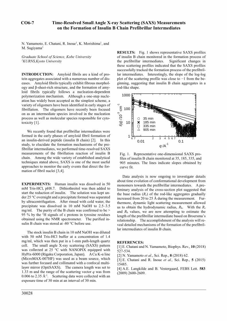
RESULTS: Fig. 1 shows representative SAXS profiles
of insulin B chain monitored in the formation process of
the prefibrillar intermediates. Significant changes in
these scattering profiles indicated that the SAXS profiles
successfully tracked the formation process of the prefibril-
lar intermediates. Interestingly, the slope of the log-log
plot of the scattering profile was close to −1 from the be-
ginning, suggesting that insulin B chain aggregates in a
rod-like shape.
Data analysis is now ongoing to investigate details
about time evolution of conformational development from
monomers towards the prefibrillar intermediates. A pre-
liminary analysis of the cross-section plot suggested that
the base radius (Rc) of the rod-like aggregates gradually
increased from 20 to 25 Å during the measurement. Fur-
thermore, dynamic light scattering measurement allowed
us to obtain the hydrodynamic radius, Rh. With the Rc
and Rh values, we are now attempting to estimate the
length of the prefibrillar intermediate based on Broersma’s
relationship. The accomplishment of the analysis will re-
veal detailed mechanisms of the formation of the prefibril-
lar intermediates of insulin B chain.
REFERENCES: [1] E. Chatani and N. Yamamoto, Biophys. Rev., 10 (2018)
527-534.
[2] N. Yamamoto et al., Sci. Rep., 8 (2018) 62.
[3] E. Chatani and R. Inoue et al., Sci. Rep., 5 (2015)
15485.
[4] A.E. Langkilde and B. Vestergaard, FEBS Lett. 583
(2009) 2600-2609.
1
10
100
1000
I(q)
/10
-3·c
m-1
6 7
0.012 3 4 5 6 7
0.12
q /A-1
35 min 185 min 335 min 905 min
Fig. 1. Representative one-dimensional SAXS pro-
files of insulin B chain monitored at 35, 185, 335, and
905 minutes. The lines indicate slopes obtained by
curve fit.
CO6-7
30029
Analysis of the initial step of fibrillation by amyloid-β peptide
T. Nakagawa, D. Tsuri, I. Nishii1, K. Morishima2, R.Inoue2, M. Sugiyama2 and M. Hoshino
Graduate School of Pharmaceutical Sciences, Kyoto University 12Faculty of Science, Nara Women’s University KURNS, Kyoto University
INTRODUCTION: Alzheimer’s disease is a progres-sive neurodegenerative disorder. One of its pathological hallmarks is the extracellular deposition of senile plaques in the brain. The major component of these plaques is fibrillar aggregates (amyloid fibrils) of amyloid β-peptide (Aβ). Although the conversion of soluble Aβ monomers to insoluble amyloid fibrils is considered to be a key step to understand the development of Alzheimer’s disease, little is known about the molecular mechanism of this process.
The difficulty to analyze the initial step of fibril formation is mainly originated from the extraordinarily high cooperativity of the reaction. While the solution of monomer peptides seems quiescent before the formation of the “nucleus” of amyloid fibrils, a numerous number of molecules rapidly and almost irreversibly bind to the “nucleus” once it formed. As a result, the most important intermediate, “nucleus” or “soluble oligomer”, exists only rarely and temporarily.
Here, we examine the conformation of a covalently linked Aβ dimer peptide as a model compound for the initial intermediate of the amyloid fibril formation. We found that the aggregation reaction was enhanced re-markably by the covalent link of two molecules. How-ever, the conformation of tethered molecule was, by itself, very similar to that of random coil structure. We also found that morphology of the aggregates depended sig-nificantly on the peptide concentrations. From these ob-servations, we propose that formation of amyloid fibrils are governed by a delicate balance between association and dissociation rate constants.
EXPERIMENTS: The wild type and mutant Aβ(1–40) peptides were constructed as fusion proteins with ubiq-uitin. The proteins were expressed and purified as de-scribed previously [1].
Transmission electron microscopy observations were performed on a JEOL JEM JEM-2100F electron microscope with an acceleration voltage of 100 kV. Samples were applied to Formvar-coated grids and nega-tively stained with 2% (w/v) uranyl acetate.
NMR experiments were performed on a Bruker Avance 600 spectrometer with triple-resonance probe. A typical 1H-15N HSQC experiments were performed at protein concentration of 50 µM. The solvent conditions used were 20 mM sodium acetate (pH 5.0), and 10% D2O. The chemical shift value was referenced to DSS.
The kinetics of the aggregation of Aβ peptides was monitored as the increase in fluorescence of thioflavin T (ThT) measured at 490 nm with an excitation wavelength of 446 nm on a Shimadzu RF-5300 spectrofluorometer.
RESULTS: In order to prepare a covalently linked-dimer Aβ peptide, we introduced a cysteine resi-due by substituting alanine residue at the second N-terminal position. By oxidizing two ceysteine residues,
Aβ molecules were covalently linked in the parallel ori-entation, which was called as A2C-dimer. In contrast to the wild type Aβ peptide, in which fibril formation is preceded by a long lag-period (typically 12~24 hrs), in-cubation of A2C-dimer resulted in a rapid increase in ThT fluorescence. Dynamic light scattering experiments also suggested rapid formation of aggregates with hy-drodynamic radius of several hundred nm. A detailed analysis by TEM observation revealed, however, the morphology of rapidly formed aggregates by A2C-dimers was different from those by wild type Aβ.
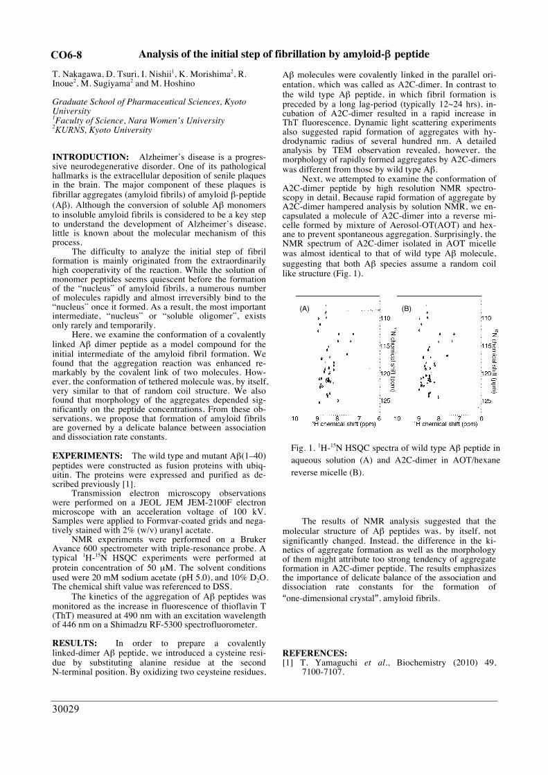
Next, we attempted to examine the conformation of A2C-dimer peptide by high resolution NMR spectro-scopy in detail. Because rapid formation of aggregate by A2C-dimer hampered analysis by solution NMR, we en-capsulated a molecule of A2C-dimer into a reverse mi-celle formed by mixture of Aerosol-OT(AOT) and hex-ane to prevent spontaneous aggregation. Surprisingly, the NMR spectrum of A2C-dimer isolated in AOT micelle was almost identical to that of wild type Aβ molecule, suggesting that both Aβ species assume a random coil like structure (Fig. 1).
The results of NMR analysis suggested that the molecular structure of Aβ peptides was, by itself, not significantly changed. Instead, the difference in the ki-netics of aggregate formation as well as the morphology of them might attribute too strong tendency of aggregate formation in A2C-dimer peptide. The results emphasizes the importance of delicate balance of the association and dissociation rate constants for the formation of “one-dimensional crystal”, amyloid fibrils.
REFERENCES: [1] T. Yamaguchi et al., Biochemistry (2010) 49,
7100-7107.
(A) (B)
Fig. 1. 1H-15N HSQC spectra of wild type Aβ peptide in aqueous solution (A) and A2C-dimer in AOT/hexane reverse micelle (B).
CO6-8

30032
Generation of radioresistant Escherichia coli by adaptive evolution using gamma rays as selection pressure
T. Saito
Institute for Integrated Radiation and Nuclear Science, Kyoto University
INTRODUCTION: In nature, organisms have evolved diversely by adapting themselves to various environmental conditions. Some organisms have been found to survive in environments that can be easily perceived as extremely se-vere. Elucidating the adaptive mechanisms of organisms to severe environmental conditions can provide meaning-ful information regarding evolution and diversity overall. Some bacteria, known as radioresistant bacteria, demon-strate extreme resistance to ionizing radiations. The ex-treme resistance mechanism of these bacteria to ionizing radiation is an interesting area of research from the stand-point of adaptive mechanisms employed by organisms in nature. In order to elucidate the mechanisms of radiore-sistance in these organisms, it is important to investigate their biological defense mechanisms against external stresses at the molecular level. However, studies con-ducted on radioresistant organisms existing in nature are likely to encounter many challenges due to limited knowledge of their genetic and biochemical properties. Therefore, in this study, the generation of radioresistant cells of Escherichia coli was attempted by experimenting on their ability to undergo adaptive evolution when ex-posed to gamma rays The following operations were re-peated in this experiment: gamma-ray irradiation of E. coli cells whose genetic and biochemical properties are suffi-ciently known, growth of the surviving cells, and irradia-tion of the grown cells. EXPERIMENTS: Evaluation of the sensitivity of E. coli cells to gamma irradiation: E. coli K-12 cells were grown to the early log phase in LB medium at 37°C at 200 rpm. One milliliter of the culture was centrifuged at 4000 ´ g at 20°C for 10 min. The supernatant was discarded and the pellet was suspended in 1 mL of PBS (–). The cell sus-pension was irradiated with gamma rays at a dose rate of 22 Gy/min at 20 ± 3°C. Gamma irradiation was carried out at the Co-60 Gamma-ray Irradiation Facility of the Insti-tute for Integrated Radiation and Nuclear Science, Kyoto University. The gamma-irradiated cell suspension was di-luted appropriately with PBS (–), plated on LB agar, and incubated at 37°C for 12 hr. After incubation, the colonies were counted, colony forming units were determined, and survival rates were calculated.
Selection with gamma rays: E. coli K-12 clone cells ob-tained by single colony pick-up were cultured to the early log phase in LB medium at 37°C at 200 rpm. The cell sus-pension was prepared as described above and irradiated with the 1% survival dose of gamma rays at a dose rate of 22 Gy/min at 20 ± 3°C. One milliliter of the gamma-irra-diated cell suspension was inoculated in 100 mL of LB medium and cultured at 37°C at 200 rpm till the cells reached their early stationary phase. The glycerol stock was prepared and stored at –80°C. This procedure was re-peated after culturing the glycerol stock cells to the early log phase. RESULTS: In order to generate radioresistant E. coli cells by adaptive evolution to gamma rays as the selection pressure, E. coli cells were irradiated with a 1% survival dose of gamma rays, and the surviving cells were grown. The 1% survival dose of the surviving cell population to gamma rays was evaluated, and the cell population was once again irradiated with the 1% survival dose. The 1% survival dose to gamma rays of the wild type E. coli clone without selection was 242 Gy, whereas that of the E. coli population obtained after 20 selection cycles was 1872 Gy; the resistance of E. coli to gamma rays was increased 7.7 fold in this adaptive evolution experiment (Fig. 1). Fur-thermore, the resistance gradually increased in the process of the adaptive evolution. These results indicate that mutations and changes in the expres-sion levels of many genes are likely to be involved in this increase of resistance. In the future, further studies, namely that the gene expression levels of radioresistant E. coli cells obtained by the adaptive evolution experiment are analyzed and compared with those of wild type E. coli cells, will be required.
REFERENCES: [1] T. Saito, Viva Origino, 30 (2007) 85–92.
Fig. 1. Increase in resistance of E coli cell populations to gamma rays during adaptive evolution.
200400600800
1000120014001600180020002200
0 1 2 3 4 5 6 7 8 9 10 11 12 13 14 15 16 17 18 19 20
1% s
urvi
val d
ose
(Gy)
Number of selection cycle
CO6-9

30033
Physicochemical study on ILEI suppressing amyloid-β generation
E. Hibino1, K. Morishima2, R Inoue2, M Sugiyama2, M Nakano1, N Watanabe1, T Sugi1 and M Nishimura1
1Molecular Neuroscience Research Center, Shiga Uni-versity of Medical Science 2KURNS, Kyoto University
INTRODUCTION: The number of patients with Alzheimer’s disease (AD)
is increasing in Japan. However, any effective AD modi-fying drug is still undeveloped. The pathogenic processes of AD are thought to be triggered by accumulation of amyloid-β protein (Aβ) in brain. Previously, we have identified a secretory protein named interleukin-like epi-thelial-mesenchymal transition inducer (ILEI, also known as FAM3 superfamily member C) as a negative regulator of Aβ production by the unique activity1. ILEI is secreted from cell to bind to the extracellular region of Prese-nilin-1, a component of the γ-secretase complex.
The interaction of ILEI with Presenilin-1 enhances nonspecific degradation of Aβ precursor protein. Howev-er, the molecular mechanism based on the structural findings is still to be clarified. Although the monomeric and dimeric forms of ILEI have been reported recently2, the structure-function relationships are also still unclear.
The objective of this study is to elucidate whether the dimerization of ILEI is indispensable for the function on Aβ generation.
EXPERIMENTS: Human ILEI (residues 55-227) tagged with His was
over-expressed in Rosetta-gamiB (DE3) pLysS stain and purified using Ni-NTA resin and size exclusion chroma-tography (SEC). CBB-stained SDS-PAGE gels and SEC profile on non-redox condition showed a single band and peak, respectively. Subsequently, around 1.5 mg/mL ILEI protein solution was diluted with buffer at each pH con-dition (6.0, 7.3 and 8.0).
To confirm the monomer-dimer transition of ILEI at 25˚C, ILEI solution was analyzed by analytical ultracen-trifugation at 60,000 r.p.m. or 20,000 r.p.m.
RESULTS: The profiles measured at 60,000 r.p.m. of the rotating
speed by Rayleigh interferometer are shown in Fig. 1. The peak derived from around 40 kDa was identified although the main peak corresponding to 20 kDa ac-counted for the greater part. Most ILEI molecules existed as monomeric state since the molecular weight of ILEI monomer was predicted to be 20.0 kDa. In addition, the abundance ratios of multimer, which were 4.5%, 3.7% and 15.2% at pH 6.0, pH 7.3 and pH 8.0, respectively, were increased in correlation with an increase in pH val-ue.
Furthermore, to identify whether ILEI proteins were dis-sociated or not, we also measured the profiles at 20,000 r.p.m, so that, the peaks were put into one peak (data notshown). This suggested that ILEI was in mono-mer-multimer equilibrium state. Moreover, the dissocia-tion constants calculated from the populations were 4.3×10⁻⁴ M (25℃, pH 7.3) and 1.0×10⁻⁴ M (25℃, pH 8.0).
In conclusion, although ILEI can form homodimers,most ILEI molecules are thought not to dimerize, taking it consideration that population of dimer is low at pH 7.3 and that ILEI is a secretory protein. We speculate that ILEI functions in monomeric state, although a possibility that ILEI dimers have functions dissimilar from the monomer cannot be ruled out.
REFERENCES:
[1] H. Hasegawa et al., Nat. Commun., 5:3917 (2014).[2] A.M. Jansson et al., J. Biol. Chem., 297 (2017)15501-15511.
Fig. 1. (A) The solid line, dashed line and dotted line show distributions of sedimentation coeffi-cient derived from ILEI at pH 6.0, pH 7.3 and pH 8.0, respectively. (B) The expanded of the lower range of A.
(A)
(B)
CO6-10

30041
Structural characterization of the S. pombe nucleosome containing histone variant H2A.Z
M. Koyama1, R. Inoue2, K. Morishima2, W. Nagakura3, M. Sugiyama2, H. Kurumizaka1,3
1 Institute for Quantitative Biosciences, The University of Tokyo 2KURNS, Kyoto University 3 Division of Advanced Science and Engineering, Waseda University
INTRODUCTION: In the eukaryotic cells, genomic DNA binds histone
proteins to form the chromatin structure, and stored in the nucleus. The structural unit of the chromatin is the nu-cleosome, in which about 145-base-pairs of DNA are wrapped around the histone octamer, containing two molecules of each of four core histones (H2A, H2B, H3, and H4) [1, 2].
The fission yeast S. pombe is a single cell eukaryote that shares many characteristics of the chromatin struc-ture and function with higher eukaryotes. S. pombe has only one H2A variant, H2A.Z, which is preferentially associated with actively transcribed chromatin. In this study, to analyze the structural property of the S. pombe H2A.Z nucleosome, we reconstituted the nucleosome in vitro, and measured its small angle X-ray scattering pro-file.
EXPERIMENTS: The S. pombe canonical (H2A) or H2A.Z nucleo-
somes were reconstituted in vitro, by mixing a 146-base-pair DNA fragment with S. pombe four core histones (SpH2A or SpH2A.Z, SpH2B, SpH3, and SpH4). The reconstituted nucleosomes were further purified by polyacrylamide gel electrophoresis using the Prep Cell apparatus (BioRad). The purified samples were concen-trated using a Millipore concentrator (Mw cutoff of 30,000). After filtration, the samples were used for the SAXS measurement.
RESULTS: SAXS profiles of the S. pombe canonical nucleo-
some, and the S. pombe H2A.Z nucleosome are shown in Fig. 1. Guinier plots of the data are shown in Fig. 2. The straight line represents the least-square fitting for the data. This result revealed that the gyration radius (Rg) of the S. pombe H2A.Z nucleosome is 46.2 ± 0.3 Å, which is larger than that of the S. pombe canonical nucleosome (Rg = 45.0 ± 0.2 Å). The distance distribution functions of the nucleosomes are shown in Fig. 3. The longest dis-tances, Dmax values, are 150 Å for the canonical H2A nu-cleosome, and 157 Å for the H2A.Z nucleosome. These findings suggest that the H2A.Z nucleosome has more stretched structure, as compared to the canonical nucleo-some in S. pombe.
Fig. 1. SAXS profiles of the S. pombe canonical nucleosome (green), the S. pombe H2A.Z nucleo-some (red), and the human canonical nucleosome (blue).
Fig. 2. Guinier plots of the nucleosomes. The color coding is the same as in Fig. 1.
Fig. 3 Distance distribution functions of the S. pombe H2A.Z nucleosome (blue) and S. pombe canon-ical nucleosome (red).
REFERENCES: [1] K. Luger et al., Nature, 389, 251-260 (1997).[2] M. Koyama and H. Kurumizaka, J. Biochem., 163,85-95 (2018).
0.05 0.10 0.15 0.20 0.25
10-3
10-2
10-1
I(Q) /
cm
-1 /
(mg/
mL)
Q / Å-1
HC PC PZ
0.0000 0.0005 0.0010
0.1I(Q) /
cm
-1
Q2 / Å-2
HC PC PZ
CO6-11

30048
Production of medical radioisotopes using electron linear accelerator
S. Sekimoto, T. Ohtsuki
Institute for Integrated Radiation and Nuclear Science,
Kyoto University
INTRODUCTION: A shortage in the supply of 99Mo
resulting from the shutdown of reactors used for its pro-
duction is a global problem. Because 99Mo is an indis-
pensable source of 99mTc, which is used in nuclear medi-
cine to make diagnoses using techniques such as scintig-
raphy and single photon emission computed tomography
(SPECT), a stable supply of 99Mo is vital. Therefore,
production of 99Mo by using neutrons or protons gener-
ated in accelerators has been investigated [1–3]. To sepa-
rate 99mTc from 99Mo produced by an accelerator, meth-
ods based on sublimation, solvent extraction, and
ion-exchange column chromatography have been exam-
ined and developed [2,4–6]. In addition, Gopalakrishna et
al. have reported the preparation of 99Mo by the 100Mo(,n) reaction using bremsstrahlung photons [6],
followed by conventional solvent extraction using methyl
ethyl ketone (MEK) and zirconium (Zr) molybdate gel to
separate 99mTc. According to the regulations of the Japa-
nese pharmacopeia, the extraction using organic materials
and the gel method using heavy metal elements such as
Zr are not approved for the 99mTc-separation methods.
Additionally, it is also difficult and impractical to use the
sublimation method, which requires complicated and/or
large scale devices for the mass-production of pure 99mTc.
In the previous work, we carried out the production of 99Mo by the 100Mo(,n) reaction using bremsstrahlung
photons generated in an electron linear accelerator
(LINAC), a technique that has not been investigated sig-
nificantly in Japan. The amounts of 99Mo produced at
several electron energies (Ee) were examined. In this re-
port, the amounts are compared with those predicted by
calculation. The experimental condition and results are
described in ref. [7].
RESULTS: The 99Mo activities in the pellets were cal-
culated by using the particles and heavy ion transport
code system (PHITS) [8], and the calculated values are
shown in Fig. 1 for comparison with the experimental
values. Although the experimental value at an Ee of
32 MeV agrees with the calculated value, the experi-
mental values are lower than those calculated at Ee’s of
21 and 25.5 MeV and higher than those calculated at Ee’s
of 35 and 41 MeV. These differences between experi-
mental and calculated values can be explained by the
dispersion of the photon beams. At higher Ee, in general,
the intensity of electrons in the longitudal direction is
greater than that of electrons in the transverse direction;
i.e., the dispersion of higher energy electron beams
(higher Ee) tends to be smaller than those at lower Ee due
to the space charge density of electrons. Therefore, the
dispersion of photon beams generated by electrons at
higher Ee becomes smaller. This tendency can be con-
firmed by simulation using the Geometry and Tracking
(GEANT) code. Because the dispersion of the photon
beams in the PHITS calculation performed in this work is
assumed to be identical at Ee’s between 21 and 41 MeV, a
disagreement was found between the experimental and
calculated values. However, the validity of the experi-
mental 99Mo is supported by those found for 196Au pro-
duced via the 197Au(,n) reaction, e.g., the Ee dependence
of the rate of 99Mo-production is similar to that of 196Au-production. This trend depends on the fact that the
shape of the excitation functions for the photon energies
in the 100Mo(,n) and the 197Au(,n) reactions are similar,
although the absolute values are different by about one
order of magnitude [9].
0 10 20 30 40 50
100
200
300
400
500
Ee (MeV)
Acti
vit
y o
f 99M
o (
kB
q) Experiment (This work)
PHITS
Fig. 1. Activity of 99Mo produced using electron linear
accelerator (The irradiation time, beam current of elec-
trons, and amount of target material (100MoO3) were
normalized to 5 min, 100 micro-A, and 10 mg, respec-
tively.)
REFERENCES: [1] Y. Nagai et al., J. Phys. Soc. Jpn. , 82 (2013) 064201.[2] Y. Nagai et al., J. Phys. Soc. Jpn. , 83 (2014) 083201.[3] K. Nakai et al., Proc. Jpn. Acad. Ser. B 90 (2014)
413–421.[4] M. Kawabata et al., J. Phys. Soc. Jpn. , 84 (2015)
023201.[5] K. Mang’era et al., J. Radioanal. Nucl. Chem., 305
(2015) 79-85.[6] A. Gopalakrishna et al., J. Radioanal. Nucl. Chem.,
308 (2016) 431-438.[7] S. Sekimoto and T. Ohtsuki, KUR-progress report
(2017) CO6-14.[8] Niita K et al., (2010) PHITS: Version 2.23,
JAEA-Data/Code 2010-022, JAEA.
[9] Dietrich SS, Berman BL (1988) Atlas of photoneu-
tron cross sections obtained with monoenergetic
photons. Atomic Data Nucl Data Tables.
38:199–338.
CO6-12

30058
Determination of degree of deutteartion level of deuterated protein through small-angle neutron scattering
R. Inoue, K. Morishima, N. Sato, M. Sugiyama
Institute for Integrated Radiation and Nuclear Science, Kyoto University
INTRODUCTION: Small-angle Neutron (SANS) technique gives the overwhelming opportunities for structural analyzes on target samples in solution. Espe-cially, contrast-variation SANS (CV-SANS) method [1], which utilizes the modulation of scattering length density (scattering contrast) between solute and solvent, offers the fascinating opportunity for studying the partial struc-ture of complex biological samples. It is known that the scattering length density of normal (or hydrogenated) protein is nearly equal to that of 40% D2O. Namely, hy-drogenated protein is invisible in 40% D2O in terms of CV-SANS. With this characteristic feature, theCV-SANS method has been applied for various hydro-genated proteins. On the other hand, on of the big disad-vantages associated with above CV-SANS method ishigh incoherent signal from 60% H2O, hindering the de-tailed structural analyzes on target biological samples. Inorder to overcome such a situation, the inverse contractmatching SANS (ICM-SANS) method [1] has been paidattention. The essence of this technique is that the scat-tering length density of protein is tuned to be matchedwith that of 100% D2O by modulating the deuterationlevel of protein. The most stressing point in ICM-SANSis the realization of high S/N SANS data attained by thesuppression of incoherent scattering from solvent. However, the procedure for preparation of partially deu-terated protein that is perfectly contrast-matched with100% D2O has not been well established up to now. Forthis purpose, we have to grasp the proper deuterationcondition and the protocol for the determination of de-gree of deuteration of prepared partially deuterated pro-tein. It is considered that SANS and mass spectrometryare usable for the determination of degree of deuterationof prepared partially deuterated protein. Prior to usage ofSANS meausremnets, we try to determine the degree ofdeuteartion of partially deuterated protein with massspectrometry.
EXPERIMENTS: In addition to the preparation of hydrogenated αB-crystallin, we prepared partially deu-terated αB-crystallin by mixing ratio of hydrogenated and deutearted glucose 3:1 in 75% D2O. Mass spec-trometry measurements were performed with microflex, BRUKER.
RESULTS: Fig. 1 shows mass spectrum from hydro-genated αB-crystallin and partially deuterated αB-crystallin, respectively. It can be clearly seen that main peak shifted to high m/z value by deuteration, im-plying the exact increase of mass during cultivation.
Considering the amino acid sequence of αB-crystallin, we have calculated the degree of deuteration of partially deuterated αB-crystallin. The degree of deuteartion of partially deuterated αB-crystallin was found to be 70.0%. As written at the experimental section, we have cultivated partially deuterated αB-crystallin in 75% deuterated glu-cose and 75% D2O. Hence, the resulting degree of deu-teartion of prepared αB-crystallin is much lower than 75%. It is expected that further control of cultivation condition such as mixing ratio of deuterated glucose must be taken into account. As a next step for this project, we are now trying to evaluate the degree of deuteartion of αB-crystallin by changing the mixing ratio of deuterated glucose and D2O. It will contribute to realizing the perfectly con-trast-matched partially deuterared protein in 100% D2O. It will further reinforce the powerfulness of ICM-SANS.
REFERENCES: [1] M. Sugiyama et al., BBA, 1862 (2018) 253-274.
Fig. 1 (a) Mass spectrum from hydrogenated αB-crystallin. (b) Mass spectrum from partially deu-terated αB-crystallin.
CO6-13
30079
Measurement of Trancemitance Spectra of a Humann Calcificated Aorta Tissue
in the Sub-Terahertz Region, which Related with a SEM-EDX Elements Imaging (II)
N. Miyoshi and T. Takahashi1
Department of Molecular Chemistry, Kyoto Institute of
Technology 1Institute for Integrated Radiation and Nuclear Science,
Kyoto University
INTRODUCTION: The LINAC (Electron linear accel-erator) technology in the millimeter- and terahertz- waves had been unique and had been used as a coherent synchrotron light source in the Institute for Integrated Radiation and Nuclear Science of Kyoto university (KURNS) to observe the transmittance spectra of a hu-man calcified aorta tissue as a collaborate study. The ab-sorption spectra in the sub-terahertz region had been not so clear for the raw tumor tissue although Ashworth-PC. et al. [1] had reported for the excised human breast can-cer by a terahertz pulsed spectroscopy observed at 320 GHz, which was estimated a longer relaxation time component of the induced electricity for water molecules [2-3] in the raw tumor tissue for three years at the linear analysis.
We had started to measure of new biological sample of a calcified human aorta dried tissue sampling from the pathological autopsy in the last year. Furthermore, the getting spectral information was estimated with the SEM-EDX elements images of the calcified aorta tissue with a biological meaning in this year. It was reported the relation of the biological information between both of the element images and sub-THz spectral components in this report.
EXPERIMENTS: (1) Instrument of Near-field in Te-
rahertz Region: The photograph of the instrument was
shown in Fig. 1. Mark-A: Pre-probe Wiston cone;
50-10mm diameter, Length=60mm; the irradiate diame-
ter=0.775mm; Mark-B: The concentrate light probe (di-
ameter=3mm). The instrument was developed by Dr. T.
Takahashi [4] for the transmittance measurements.
Fig. 1. The near field area of sample holder position.
(2) Sample Preparation for SEM-EDX Elements (P, and
Ca) Images of Calcified Aorta: A calcified aorta tissue
was cryo-sectioned in vertical of the blood wall at -20 ℃
at 8 m depth. The sample tissues was measured by a
Scanning Electron Microscope (JEOL) combined with
Energy Dispersion X-ray (EDX) analyzer.
RESULTS and DISCUTION: (1) The morphological
images were observed in side of the calcified aorta tissue
by the SEM-EDX analysis as shown inf Fig. 2.
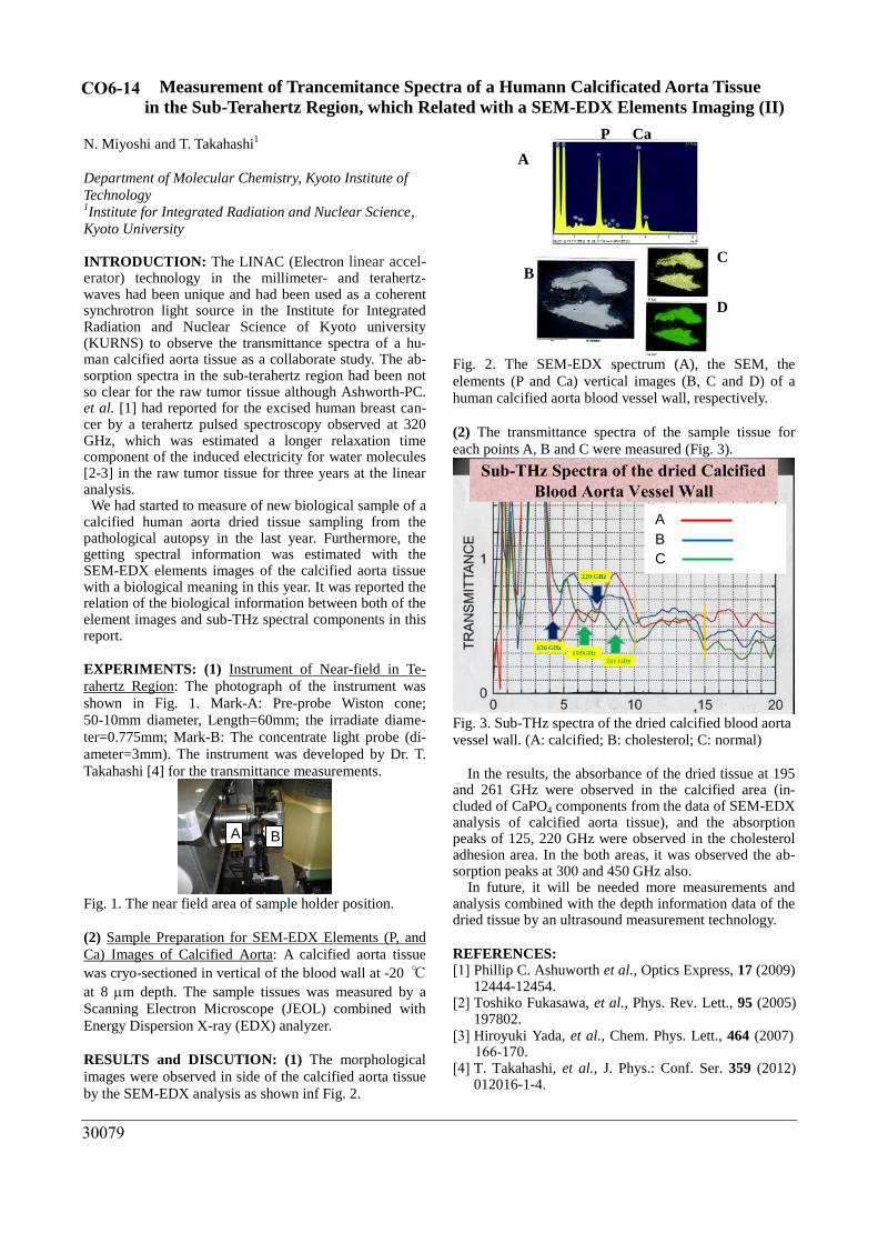
Fig. 2. The SEM-EDX spectrum (A), the SEM, the
elements (P and Ca) vertical images (B, C and D) of a
human calcified aorta blood vessel wall, respectively.
(2) The transmittance spectra of the sample tissue for
each points A, B and C were measured (Fig. 3).
Fig. 3. Sub-THz spectra of the dried calcified blood aorta
vessel wall. (A: calcified; B: cholesterol; C: normal)
In the results, the absorbance of the dried tissue at 195 and 261 GHz were observed in the calcified area (in-cluded of CaPO4 components from the data of SEM-EDX analysis of calcified aorta tissue), and the absorption peaks of 125, 220 GHz were observed in the cholesterol adhesion area. In the both areas, it was observed the ab-sorption peaks at 300 and 450 GHz also. In future, it will be needed more measurements and analysis combined with the depth information data of the dried tissue by an ultrasound measurement technology.
REFERENCES: [1] Phillip C. Ashuworth et al., Optics Express, 17 (2009)
12444-12454.[2] Toshiko Fukasawa, et al., Phys. Rev. Lett., 95 (2005)
197802.[3] Hiroyuki Yada, et al., Chem. Phys. Lett., 464 (2007)
166-170.[4] T. Takahashi, et al., J. Phys.: Conf. Ser. 359 (2012)
012016-1-4.
A B
A
B
C
A
B C
D
P Ca
CO6-14

30088
Preparation of bovine intact FoF1ATP synthase for a cryo-EM structural analysis
C. Jiko1, C. Gerle2 and Y. Morimoto1
1Institute for Integrated Radiation and Nuclear Science, Kyoto University 2Institute for Protein Research, Osaka University
INTRODUCTION: Atomic resolution structure analysis of biomolecules using cryo-electron microscopy (cryo-EM) has been developed (Henderson et al., Nobel Prize, 2017) and the analysis is currently overwhelming for study of membrane proteins that are extremely diffi-cult to crystallize. Although it is powerful, the key to success in structural analysis by a cryo-EM is the ability to purify large quantities of stable and intact samples as well as crystallization.
In this study, we aim to analyze the full length of mammalian mitochondrial FoF1 ATP synthase with atomic resolution. An FoF1 ATP synthesis is an im-portant energy conversion mechanism for maintaining mammalian life activities, and is driven by proton transport in the mitochondrial inner membrane. The FoF1 ATP synthase responsible for this is a membrane protein complex with a molecular weight of 600,000 consisting of 29 subunits. The enzyme not only produces 90% of ATP but also plays an important role in the for-mation of the inner mitochondrial membrane. It is also suggested that dysfunction of this enzyme leads to apop-tosis (cell death). Threfore elucidation of the precise three-dimensional structure of this enzyme is considered to be an important basis for drug molecule design in the future, and is widely studied. Because of this biological and medical importance, ef-
forts to elucidate the structure of the FoF1 ATP synthase have been made for over 50 years, but the understanding of the function of the FoF1 ATP synthesis enzyme is delayed by the fact that the entire structure has not been elucidated at the atomic level. The enzyme is a membrane-bound protein of the mac-
romolecule complex and is highly flexible and unstable because of its rotation, which makes it difficult to obtain a purified preparation having a uniform three-dimensional structure throughout the enzyme.
EXPERIMENTS: Bovine heart was obtained immedi-ately after slaughter, and mitochondrial inner membrane was separated followed by solubilization of FoF1 ATP synthase. At this time, purification was required in order to solubilize other proteins. As a purification method of this enzyme, one using an affinity column such as ion exchange chromatography is generally used, and we have also purified by the same method.
However, it was found that this method led to low uni-formity of the purified sample. This is because the en-zyme has extremely high flexibility as it rotates, and the
subunit of this enzyme, which is composed of as many as 29 subunits per monomer, starts dissociation from the intermembrane domain during the purification process Although this ion exchange chromatography is effective for improving the purification purity, it is considered that the interaction with the ion exchange resin places a bur-den on the original structure of the enzyme and the deg-radation of this enzyme is caused. Therefore, it is neces-sary to establish a purification method that can replace ion exchange chromatography. Dealing with this issue, we developed a purification method by density gradient centrifugation without loading the protein (column-free purification) and succeeded in purifying the enzyme in a very stable and intact state.
RESULTS and DISCUSSION: Using this stable en-zyme with full subunits, prepared by the column-free purification developed by the applicant, it was possible to produce a cryogrid suitable for high-resolution single particle analysis by cryo-EM (Fig. 1).
Fig.1 Recent images of mammalian FoF1 dimers by cryo-electron microscopy.
Although the greatest obstacle to collecting high quality data of membrane proteins is the detergent (Rubinstein, Methods, 2007), usually, when the detergent is removed from solution, membrane proteins aggregate and dena-tures instantaneously. On the other hand, the method developed by Gerle et al. (GraDeR; Hauer et al., Struc-ture, 2015) makes it possible to remove the detergent in the solution before the instant freezing of grid prepara-tion. Currently, grid adjustment is carried out using this method, and observation by cryo-EM is being conducted.
ACKNOWLEDGEMENT: This work is partly sup-ported by the grant of Naito foundation 2018, the grant-in-aid for the young researchers of the JSPS 2019 (C.J.) and the TERUMO foundation 2018, the grant for an advanced research of the KURRI 2018 (Y.M.) .
CO6-15
30105
Preclinical study of boron neutron capture therapy for bone metastasis using human lung cancer cell lines
T. Andoh1, T. Fujimoto2, Y. Nagasaki1, M. Suzuki3, H. Tanaka4, T. Takata4, Y. Sakurai4, and H. Ichikawa1.
1Faculty of Pharmaceutical Sciences, Kobe Gakuin Uni-versity, Japan. 2Department of Orthopaedic Surgery, Hyogo Cancer Center, Japan. 3Particle Radiation Oncology Research Center, Institute for Integrated Radiation and Nuclear Science, Kyoto University, Japan. 4Division of Radiation Life Science, Institute for Inte-grated Radiation and Nuclear Science, Kyoto University, Japan
INTRODUCTION: Lung cancer is the leading cause of cancer-related death globally [1]. Rapid metastasis to the bone can occur in 25-40% of lung cancer patients, which has a poorer prognosis [2]. When systematic pharmacotherapy is not effective, the disease is difficult to control. Boron neutron capture therapy (BNCT) may be the sole option in such cases. We have previously demonstrated the effectiveness of BNCT with the use of p-borono-L-phenyl-alanine (L-BPA) on tumors in the limbs of human clear cell sarcoma-bearing nude mouse models [3]. In the present study, we established a bone metastasis model for lung cancer and investigated in vivo biodistribution of L-BPA and antitumor effects after BNCT in the bone metastasis model.
EXPERIMENTS: Cells of A549-luc, a lung cancer cell line of human origin, were suspended in Matrigel® and injected into the tibia of the left hind leg of nude mice [4]. After 3-4 weeks, a tumor mass was observed in tibia of the mice using a computed tomography scan and luminescence imaging. The biodistribution of 10B was explored by the intravenous administration of BPA-fructose complex (BPA-Fr, 24 mg 10B/kg) to a bone metastasis mouse model of lung cancer. At a predeter-mined time after administration, the mice were sacrificed and blood and tissue samples were immediately collected. The concentration of 10B in the samples was then meas-ured by inductively coupled plasma atomic emission spectroscopy. In the BNCT trial, the model mice were allocated into a BNCT and control groups. The tumors in the left hind legs were exposed to thermal neutron irradi-ation at the Institute for Integrated Radiation and Nuclear Science, Kyoto University.
RESULTS: Bone metastasis was successfully produced in the human lung cancer-bearing mouse model. The
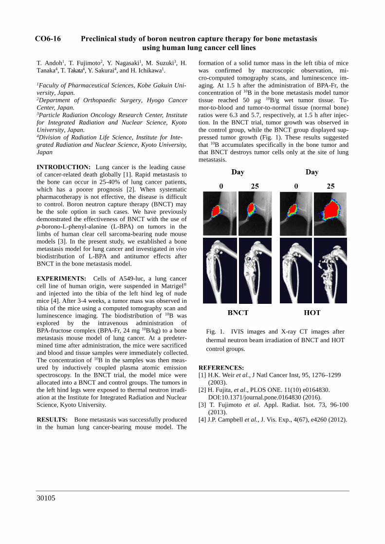
formation of a solid tumor mass in the left tibia of mice was confirmed by macroscopic observation, mi-cro-computed tomography scans, and luminescence im-aging. At 1.5 h after the administration of BPA-Fr, the concentration of 10B in the bone metastasis model tumor tissue reached 50 μg 10B/g wet tumor tissue. Tu-mor-to-blood and tumor-to-normal tissue (normal bone) ratios were 6.3 and 5.7, respectively, at 1.5 h after injec-tion. In the BNCT trial, tumor growth was observed in the control group, while the BNCT group displayed sup-pressed tumor growth (Fig. 1). These results suggested that 10B accumulates specifically in the bone tumor and that BNCT destroys tumor cells only at the site of lung metastasis.
REFERENCES: [1] H.K. Weir et al., J Natl Cancer Inst, 95, 1276–1299
(2003).[2] H. Fujita, et al., PLOS ONE. 11(10) e0164830.
DOI:10.1371/journal.pone.0164830 (2016).[3] T. Fujimoto et al. Appl. Radiat. Isot. 73, 96-100
(2013).[4] J.P. Campbell et al., J. Vis. Exp., 4(67), e4260 (2012).
Fig. 1. IVIS images and X-ray CT images after thermal neutron beam irradiation of BNCT and HOT control groups.
CO6-16

T
Fig. 1. Schematic diagram showing the configurations for absorption
spectroscopy and irradiation using CTR.
Coherent Transition Radiation mm-Wave Light Source with an Electron Linac for Absorption Spectroscopy and Irradiation
S. Okuda1, Y. Tanaka1 and T. Takahashi2
1Organization for Research Promotion, Osaka Prefecture
University, Sakai, Osaka, Japan
2Institute for Integrated Radiation and Nuclear Science, Kyoto
University, Kumatori, Osaka, Japan
Abstract—The coherent transition radiation from bunched electron beams of a linear accelerator has continuous spectra in a sub-mm to mm range and has extremely high peak intensity. In Kyoto University the coherent transition light source was developed by using a 45 MeV electron linear accelerator. In this work the characteristics of the light source have been measured and the system has been modified for the applications to absorption spectroscopy and irradiation.
I. INTRODUCTION
HE coherent transition radiation (CTR) from high-
energy bunched electron beams of a linear accelerator
(linac) has continuous spectra in a sub-mm to mm
wavelength range. The CTR light source has been established
[1, 2] by using the electron beams of the 45 MeV L-band
electron linac in Kyoto University. It has extremely high peak-
intensities in a picosecond light pulse compared with the other THz light sources. The light source has been applied to
interferogram obtained by the interferometer to be about 3 ps.
Such a relatively short pulse length is due to the special
bunching process in the optimized operational conditions of the
linac. These results indicated that the peak light intensity in the
micropulse is about 3x10⁶ W. In order to increase the peak
intensity the electron-gun pulser has been developed to generate
the single-bunch electron beam. By using this system it is
expected to be a few tens of MW. While the averaged CTR
power is sufficiently low not to induce thermal effects in the
samples, the comparatively high peak power might cause any
nonlinear effects.
Some results for the absorption spectroscopy for liquid
samples and the irradiation to investigate the biological effects
especially on the gene of fruit fly, Drosophila melanogaster,
are ongoing to be analyzed.
Shielding wall Linac
absorption spectroscopy for various kind of matters. In the present work the detailed properties of the light source have
been measured. The light source system has been changed for
Mirror system Collimator Mirror system
the applications to absorption spectroscopy and irradiation to
investigate the nonlinear effects or biological effects.
II. EXPERIMENTAL
The configurations of the light source system changed
for the absorption spectroscopy and the irradiation with CTR
are schematically shown in Fig. 1. The electron linac in Kyoto
Sample
Detector
Lock-in Amp.
Linac trigger
Sample
Interferometer
Light Source
University was used in the experiments. In most experiments
the beam energy, macropulse length and the repetition rate are
42 MeV, 47 ns and 60 Hz, respectively. The output CTR light
from a light source chamber was transported out from the
accelerator room. The spectrum of the CTR light was measured
with a Martin-Puplett type interferometer and a liquid-He-
cooled silicon bolometer. In the absorption spectroscopy the
sample was located on the light path between the interferometer
and the detector. The wavenumber resolution was 0.1 cm -1 in
most experiments, which can be changed. In the interferometer
the incident light is divided into two parts by a polarizer: one
for spectroscopy and another for irradiation experiments.
III. RESULTS
The light source spectrum has a peak at a wavenumber of
about 7 cm-1. The wavenumber range used for spectroscopy was
evaluated to be 2-35 cm-1, in which the highest number is
limited by the sensitivity of the detector. The intensity of light
was estimated to be about 10-7 W/0.1%b.w. about the spectral
peak and was found to be sufficiently high even if it reduces by
6 orders from the initial one after transmission through the
sample with relatively strong absorption. The pulse structure of
the CTR corresponds to that of the electron beam from the linac.
The micropulse length of the CTR was evaluated by
the
Experimental room Accelerator room
In conclusion, the characteristics of the CTR light source
in Kyoto University have been measured and the system has
been changed for absorption spectroscopy and irradiation
experiments. The light source will be applied to investigating
some nonlinear effects and biological effects.
This work has been carried out in part under the Visiting
Researcher’s Program of the Research Reactor Institute, Kyoto
University, and supported by JSPS KAKENHI Grant Number
JP15K04733.
REFERENCES
[1]. T. Takahashi, T. Matsuyama, K. Kobayashi, Y. Fujita, Y.
Shibata, K. Ishi and M. Ikezawa, “Utilization of coherent
transition radiation from a linear accelerator as a source of
millimeter-wave spectroscopy”, Review of Scientific Instruments,
vol. 69, pp. 3770-3775, 1998.
[2]. S. Okuda and T. Takahashi, “Absorption spectroscopy using
a coherent transition radiation mm wave light source”, Infrared
Physics and Technology, vol. 51, pp. 410-412, 2008.
30127
CO6-17