Cloning of a novel collagenase gene from Gram -negative
30
1 Cloning of a novel collagenase gene from Gram-negative 1 bacterium Grimontia (Vibrio) hollisae 1706B and its efficient 2 expression in Brevibacillus choshinensis 3 4 Naoko Teramura 1, a , Keisuke Tanaka 1, a, *, Katsumasa Iijima 1 , Osamu Hayashida 1 , Koki 5 Suzuki 2 , Shunji Hattori 1, 2 and Shinkichi Irie 1, 2 6 1 Nippi Research Institute of Biomatrix, Toride, Ibaraki 302-0017 Japan 7 2 Japan Institute of Leather Research, Adachi, Tokyo 120-8601, Japan 8 a These two authors contributed equally to this work 9 10 Running title: Collagenase gene of G. hollisae 11 12 *Address correspondence to: Keisuke Tanaka, Nippi Research Institute of Biomatrix, 13 520-11, Kuwabara, Toride, Ibaraki 302-0017, Japan, Phone: +81-297-71-3045, Fax: 14 +81-297-71-3041, E-mail: [email protected] 15 Copyright © 2011, American Society for Microbiology and/or the Listed Authors/Institutions. All Rights Reserved. J. Bacteriol. doi:10.1128/JB.01528-10 JB Accepts, published online ahead of print on 22 April 2011 on April 9, 2019 by guest http://jb.asm.org/ Downloaded from
Transcript of Cloning of a novel collagenase gene from Gram -negative

1
Cloning of a novel collagenase gene from Gram-negative 1
bacterium Grimontia (Vibrio) hollisae 1706B and its efficient 2
expression in Brevibacillus choshinensis 3
4
Naoko Teramura1, a
, Keisuke Tanaka1, a,
*, Katsumasa Iijima1, Osamu Hayashida
1, Koki 5
Suzuki2, Shunji Hattori
1, 2 and Shinkichi Irie
1, 2 6
1Nippi Research Institute of Biomatrix, Toride, Ibaraki 302-0017 Japan 7
2Japan Institute of Leather Research, Adachi, Tokyo 120-8601, Japan 8
a These two authors contributed equally to this work 9
10
Running title: Collagenase gene of G. hollisae 11
12
*Address correspondence to: Keisuke Tanaka, Nippi Research Institute of Biomatrix, 13
520-11, Kuwabara, Toride, Ibaraki 302-0017, Japan, Phone: +81-297-71-3045, Fax: 14
+81-297-71-3041, E-mail: [email protected] 15
Copyright © 2011, American Society for Microbiology and/or the Listed Authors/Institutions. All Rights Reserved.J. Bacteriol. doi:10.1128/JB.01528-10 JB Accepts, published online ahead of print on 22 April 2011
on April 9, 2019 by guest
http://jb.asm.org/
Dow
nloaded from
2
Abstract. 1
The collagenase gene was cloned from Grimontia (Vibrio) hollisae 1706B, 2
and its complete nucleotide sequence was determined. Nucleotide sequencing 3
showed that the open reading frame was 2,301 bp in length and encoded an 84 kDa 4
protein of 767 amino acid residues. The deduced amino acid sequence contains a 5
putative signal sequence and a zinc metalloprotease consensus sequence, the HEXXH 6
motif. G. hollisae collagenase showed 60% and 59% amino acid sequence identity 7
to Vibrio parahaemolyticus and Vibrio alginolyticus collagenase, respectively. In 8
contrast, this enzyme showed less than 20% sequence identity with Clostridium 9
histolyticum collagenase. When the recombinant mature collagenase, which 10
consisted of 680 amino acids with a calculated molecular mass of 74 kDa, was 11
produced by the Brevibacillus expression system, a major gelatinolytic protein band 12
of approximately 60 kDa was determined by zymographic analysis. This result 13
suggested that cloned collagenase might undergo processing after secretion. 14
Moreover, the purified recombinant enzyme was shown to possess a specific activity 15
of 5,314 units/mg, an approximately 4-fold greater activity than that of C. 16
histolyticum collagenase. 17
on April 9, 2019 by guest
http://jb.asm.org/
Dow
nloaded from

3
Introduction 1
Bacterial collagenases are metalloproteases containing a consensus motif for 2
zinc proteases, the HEXXH sequence, and are capable of digesting both native and 3
denatured collagen. They make multiple cleavages at the Y-Gly bond in repeating 4
X-Y-Gly sequences within triple helical regions, where proline and hydroxyproline 5
residues are most common in the X and Y positions, respectively (17). Because of 6
their characteristics, bacterial collagenases have been widely used in biological 7
experiment as tissue-dispersing enzymes, as well as in medical procedures such as the 8
isolation of pancreatic islet cells for transplantation (14) and the treatment for 9
Dupuytren’s disease (6). 10
Much of our knowledge of bacterial collagenases has come from studies of the 11
enzymes produced by Clostridium histolyticum (13, 15-17, 34). Analysis of the 12
primary structure of the gene product from C. histolyticum has revealed that 13
clostridial collagenases consist of three domains (catalytic domain, polycystic kidney 14
disease (PKD) domain and collagen-binding domain (CBD)) in their molecules. 15
Moreover, CBD has utilized for anchoring molecule that growth factors fused to CBD 16
can be functional to bind to collagen fibrils and maintain biological activities (21). 17
On the other hand, one of the other well-investigated bacterial collagenases is Vibrio 18
alginolyticus collagenase (7, 10, 11, 28). The collagenase activity of V. alginolyticus 19
collagenase is higher than that of any other bacterial collagenase, and it was found 20
highly efficient in debridement of necrotic burns, ulcers and decubitus. To date, 21
bacterial collagenases have been purified from various species, and their genes have 22
been cloned and sequenced (8, 12, 18, 24, 35). However, many collagenases have 23
not yet been both enzymatically and structurally characterized. 24
Vibrio hollisae is a Gram-negative bacterium first described in 1982 (4) and 25
on April 9, 2019 by guest
http://jb.asm.org/
Dow
nloaded from

4
recently reclassified as the novel genus Grimontia (29). Grimontia (Vibrio) hollisae 1
has been reported as a toxic bacterium, whose toxin was clarified as thermostable 2
direct hemolysin (22), and is primarily known to cause moderate to severe cases of 3
gastroenteritis in healthy people (6). G. hollisae strain 1706B was isolated from 4
seashore sand collected from the Shin-Kiba coast in Tokyo (27). This organism 5
produces a collagenase with a very high specific activity in the presence of gelatin, 6
and this enzyme even degrades the tanned leather (26). The characteristics of this 7
organism and purified collagenase have been described in a series of papers (25-27). 8
The properties of this collagenase are as follows: (i) it has a molecular weight of 60 9
kDa, (ii) degrades insoluble collagen, soluble collagen, Z-GPLGP peptide and 10
Pz-PLGPR peptide, but not casein, (iii) has an optimum pH of 7.0-8.0 for insoluble 11
collagen hydrolysis, (iv) is stable in the range between pH 4.5 and 11 (25). In order 12
to clarify its enzymic characteristics and to utilize it for biological applications, the 13
primary structure of the collagenase needs to be elucidate. 14
In the present study, we cloned and sequenced a novel collagenase gene from 15
G. hollisae 1706B to elucidate its primary structure, and demonstrated the expression 16
and characterization of recombinant mature collagenase using the Brevibacillus 17
expression system. Moreover, we discussed the characteristics of the corresponding 18
amino acid sequence of this enzyme and its similarity to those of other bacterial 19
collagenases. 20
on April 9, 2019 by guest
http://jb.asm.org/
Dow
nloaded from

5
Materials and Methods 1
Bacterial strains and plasmids 2
G. hollisae strain 1706B was obtained around the shore of Shin-Kiba, Tokyo, 3
Japan and used throughout this study (27). The plasmid pGEM-T Easy and the E. 4
coli competent cells JM109 (Promega, Madison, WI) were used as subcloning vector 5
and host, respectively. The expression plasmid vector pCC1BAC and the host 6
TransforMaxTM
EPI300TM
E. coli (EPICENTRE Biotechnologies, Madison, WI) were 7
used to make a BAC clone library of G. hollisae. Bacillus brevis expression vector 8
pNY326 and Brevibacillus choshinensis S5 (Takara Bio, Shiga, Japan) were used for 9
expression of recombinant proteins. 10
11
Construction of a genomic library from G. hollisae 1706B 12
The genomic DNA of G. hollisae 1706B was purified with a QIA genomic 13
DNA extraction kit (QIAGEN, Hilden, Germany). The purified DNA was digested 14
with the restriction enzyme EcoRI and separated on a 0.6%(w/v) agarose gel. Only 15
DNA fragments over 2kb were ligated into the E. coli expression vector, pCC1BAC 16
(EPICENTRE Biotechnologies). Then, pCC1BAC was transformed into 17
TransforMaxTM
EPI300TM
E. coli by electroporation, and the transformants were 18
plated onto LB agar plates containing 50 µg/ml ampicillin. 19
20
DNA probe preparation 21
Degenerate primers were designed based on the internal peptide sequence of 22
original collagenase (Fig. 2, No. 6) for F1 and the consensus sequence of catalytic site 23
from V. alginolyticus and V. parahaemolyticus collagenase (Fig.2, underlined) for R1. 24
The primer sets F1 (5’-GAGGCNATCTTTAGCTCCAATCATATGTAYAAY-3’) 25
on April 9, 2019 by guest
http://jb.asm.org/
Dow
nloaded from

6
and R1 (5’-ATCTAAGTAATGCACGTATTCATGYTCNAGRTT-3’) were used for 1
amplification of probe sequence. Y, R and N represent C/T, A/G and A/C/T/G, 2
respectively. Amplification was performed in 50 cycles of 0.5 min at 95°C, 0.5 min 3
at 45°C, and 1 min at 72°C. The amplified 1,080bp PCR product was electro-eluted 4
from 1% agarose gel, ligated into pGEM-T Easy vector (Promega), and then 5
transformed into E.coli strain JM109. The purified plasmid was used as a template 6
to create DIG-labeled DNA probes using DIG-high prime labeling reagent (Roche, 7
Basel, Switzerland) according to the manufacturer’s instructions. 8
9
Screening of the genomic library with DNA probe 10
The genomic library was screened by colony hybridization according to the 11
manufacturer’s instructions (Roche). Briefly, the ampicillin-resistant transformants 12
were blotted onto a nylon membrane (Roche), and lysed with 0.5 M NaOH. The 13
denatured DNA was then immobilized, followed by a protease K treatment. The 14
DIG-labeled DNA probe was used for hybridization. Positive clones were picked 15
from cultured LB agar plates, and subcultured in LB liquid medium with ampicillin at 16
37°C. BAC DNA of collagenase-positive colonies was prepared using the QIA 17
genomic DNA extraction kit (QIAGEN). 18
19
DNA sequencing and alignment of deduced amino acid sequence 20
Purified BAC DNA was amplified with cycle sequencing using a thermal 21
cycler (Takara Bio), and sequenced with a DNA auto sequencer (ABI PRISM 310, 22
ABI). Appropriate oligonucleotide primers (Sigma-Aldrich, St. Louis, MO) and 23
Platinum Taq DNA Polymerase High Fidelity (Invitrogen, Carlsbad, CA) were used 24
to walk along the sequence. The sequencing strategy for the pCC1BAC-2 insert was 25
on April 9, 2019 by guest
http://jb.asm.org/
Dow
nloaded from

7
outlined in Fig. 1. Computer analysis of the DNA sequence data was performed 1
using GenBank database and BLAST search programs. The deduced amino acid 2
sequence alignment and homology data were generated using the CLUSTAL W2 3
program (http://www.ebi.ac.uk/Tools/msa/clustalw2/). 4
5
Recombinant collagenase preparation 6
pCC1BAC-2 was used as the DNA template. To add NcoI site to the 5’ 7
region and HindIII site to the 3’ region of the mature collagenase gene, the primers 8
were designed as follows: Forward: 9
5’-AAACCATGGCTTTCGCTGCGGTTGAACAGTGTGATCT-3’; Reverse: 10
5’-AAAAAGCTTTTACTGACGACACTGGTTAC-3’. Restriction sites are 11
represented by bold letters and underlined. The mature domain of 2.1kb collagenase 12
gene was amplified with Expand High Fidelity PCR system (Roche). After the 13
treatment with NcoI and HindIII, the double digested fragment was ligated into the 14
multi cloning site of the Brevibacillus expression vector pNY326, which was located 15
downstream of Brevibacillus signal sequence (pNY326-Col2). Plasmid 16
pNY326-Col2, harboring the complete mature collagenase gene, was transformed into 17
Brevibacillus choshinensis S5 to express the recombinant enzyme. The 18
Brevibacillus transformant was aerobically cultured in 2SLN medium containing 19
neomycin (50 µg/ml). After centrifugation, the supernatant was purified with a 20
DEAE-Sepharose column (26 × 100 mm) with an FPLC system under gradation of 21
sodium concentration (0.2-1.1 M NaCl concentration). The column was eluted 22
isocratically with 0.2 M NaCl, 50 mM Tris-HCl (pH 7.5) for 10 min at 5 ml/min 23
followed by a linear gradient to 1.1 M NaCl for 50 min. The purified recombinant 24
protein was concentrated by ultrafiltration with a 30 kDa cutoff (PALL, Port 25
on April 9, 2019 by guest
http://jb.asm.org/
Dow
nloaded from

8
Washington, NY), and dialyzed against 50 mM Tris-HCl buffer (pH 7.5) at 4°C. 1
2
SDS-PAGE 3
SDS-PAGE was carried out on a 7.5% or 10% polyacrylamide gel 4
according to Laemmli (9) unless otherwise stated. After electrophoresis, the gel was 5
stained with 0.25% Coomassie brilliant blue R-250 in 50% methanol and 10% acetic 6
acid, and destained with 5% methanol and 7.5% acetic acid. 7
8
Determination of collagenolytic activity 9
The collagenolytic activity of recombinant collagenase was measured using 10
FITC-labeled type I collagen as previously described (20). Briefly, the enzyme 11
solution was mixed with 50 mM Tris-HCl (pH 7.5) containing 0.05% FITC-labeled 12
type I collagen, 5 mM CaCl2 and 200 mM NaCl, and incubated at 30°C for 30 min. 13
After adding EDTA to stop the enzymatic reaction, the degraded FITC-labeled 14
collagen fragment was extracted with 50 mM Tris-HCl (pH 9.5) containing 70% 15
ethanol. The fluorescence intensity of the supernatant was measured by 16
fluorescence spectrophotometry (530 nm Em/ 485 nm Ex). One unit of 17
collagenolytic activity was defined as the amount degrading 1 µg of FITC-labeled 18
collagen at 30°C per min. Protein concentrations were determined using the 19
Coomassie Plus – The Better BradfordTM
Assay Reagent (Thermo Scientific, 20
Rockford, IL). Collagenase from Clostridium histolyticum (Amano Enzyme, 21
Nagoya, Japan) was used as a reaction standard. All assays were carried out in 22
triplicate. 23
For measurement of enzyme kinetics, 0.5 µg of enzyme was incubated with 24
various amounts of FITC-labeled type I collagen (10-50 µg) at 30°C for 5 min and the 25
on April 9, 2019 by guest
http://jb.asm.org/
Dow
nloaded from

9
fluorescence intensity of the supernatant was measured. Specific collagenase 1
substrate FALGPA (N-(3-[2-furyl]acryloyl)-Leu-Gly-Pro-Ala) (Bachem AG, 2
Bubendorf, Switzerland) was also chosen to determine enzyme activity. Assay with 3
FALGPA was performed according to the modified method of a previous report (31). 4
Briefly, the enzyme and FALGPA were mixed in 50 mM Tricine buffer (pH 7.5) 5
containing 0.4 M NaCl and 40 mM CaCl2, and incubated at 30°C for 5 min. After 6
incubation, the absorbance change at 345 nm was measured using a CORONA 7
SH-9000 microplate reader (Corona Electric, Ibaraki, Japan). The FALGPA 8
concentrations were varied from 0.5 mM to 3.0 mM. In the FALGPA assay, one 9
unit of activity was defined as the amount degrading 1 µmol of FALGPA peptide at 10
30°C per min. Vmax and Km values for hydrolysis of native collagen and FALGPA 11
were estimated from the Lineweaver-Burk plot using the reaction rates at different 12
substrate concentrations. 13
14
Real-time zymography 15
Real-time zymography was performed as previously described (3). Briefly, 16
recombinant collagenase was subjected to non-reducing SDS-PAGE using a 10% gel 17
containing 0.05% FITC-labeled gelatin. After electrophoresis at 4°C, the gel was 18
washed in 50 mM Tris-HCl (pH 7.5) containing 2.5% TritonX-100 for 30 min, and 19
then was incubated in 50 mM Tris-HCl (pH 7.5) containing 5 mM CaCl2 and 200 mM 20
NaCl at 37°C for 5 h. The collagenase-digested FITC-labeled gelatin was visualized 21
using a transilluminator. 22
The effect of protease inhibitors on collagenase activity was determined by 23
adding the inhibitors to the incubation buffer. In inhibition studies, protease 24
inhibitors such as EDTA, o-phenanthroline, NEM or PMSF were used at final 25
on April 9, 2019 by guest
http://jb.asm.org/
Dow
nloaded from
10
concentrations of 20, 2.0, 5.0 and 1.0 mM, respectively. 1
2
Amino acid sequence 3
Amino acid sequence analysis of N-terminal or internal peptide fragments 4
of original collagenase was performed as described previously (19). Briefly, internal 5
sequences were determined by lysyl endoprotease, trypsine and V8 protease digestion. 6
The enzyme-cleaved fragments and purified collagenase were separated by 10% 7
SDS-PAGE, and electrophoretically transferred to Immobilon-P (Millipore, Billerica, 8
MA). The membrane was stained with Coomassie brilliant blue R-250, and the 9
protein band was excised from the membrane, and then washed extensively with 10
deionized distilled water. The N-terminal sequence was analyzed using a Procise 11
491 protein sequencer (Applied Biosystems, Carlsbad, CA). Detected fragments 12
were shown in Fig. 2. 13
14
Nucleotide sequence accession number 15
The determined nucleotide sequence was deposited in the DDBJ database 16
under accession number AB600550. 17
18
on April 9, 2019 by guest
http://jb.asm.org/
Dow
nloaded from

11
Results. 1
Cloning of collagenase gene from G. hollisae 1706B 2
In order to amplify a fragment of the G. hollisae collagenase coding gene, 3
degenerate primers were designed. A primer set yielded a single amplification 4
product, and nucleotide sequencing revealed that the amino acid sequence deduced 5
from this PCR product contained four partial peptide sequences (Fig. 2, No. 2, 3, 4 6
and 7). Therefore, the plasmid containing this PCR product was used as a template 7
to create DIG-labeled DNA probes as a hybridization probe for genomic library 8
screening. 9
A partial genomic library was constructed with EcoRI-digested genomic DNA 10
fragments from G. hollisae using a pCC1BAC vector system. The plasmids were 11
transformed into TransforMaxTM
EPI300TM
E. coli using electroporation, yielding 12
about 5,000 colonies on LB-ampicillin plates. By using colony hybridization 13
technique, 30 positive clones were picked up from the library and all the purified 14
BAC contained 2.3 kb inserts encoding the collagenase gene. One of the former 15
clones, designated as pCC1BAC-2, was chosen for further study. Restriction 16
analysis revealed that pCC1BAC-2 contained a >50 kb EcoRI insert (data not shown). 17
18
Nucleotide sequence of G. hollisae collagenase gene 19
The nucleotide sequence of the pCC1BAC-2 insert was sequenced using the 20
strategy outlined in Fig. 1. The obtained sequences were aligned by their overlaps to 21
form a single contiguous sequence, and the 2.3 kb sequence of G. hollisae collagenase 22
was determined (Fig. 2). The entire open reading frame (ORF) of G. hollisae 23
collagenase was sequenced on both strands. Analysis of the sequence revealed a 24
complete ORF extending from an ATG codon at nucleotide +1 to a TAA stop codon 25
on April 9, 2019 by guest
http://jb.asm.org/
Dow
nloaded from
12
at position 2,301, which encodes a protein of 767 amino acids. A Shine-Dalgarno 1
sequence (AGAAGAA) is observed at 5-11 bp upstream from the ATG codon. A 2
stem-loop sequence (nucleotide position 2,328 to 2,347) with a short run of T’s is 3
present downstream of the termination codon. 4
The N-terminal amino acid sequence 5
(Ala-Val-Glu-Gln-Cys-Asp-Lys-Ser-Glu) of the purified original enzyme 6
corresponded perfectly to the deduced amino acid sequences of our determined 7
sequence in position 262 to 288 downstream of the ATG codon. Moreover, the 8
deduced amino acid sequence of ORF includes a zinc metalloprotease HEXXH 9
consensus motif, which was detected as HEYVH in position 1474 to 1488. It is 10
known that the amino acid sequence HEXXH is important in facilitating the electron 11
transfer with zinc in enzyme catalysis (30). Moreover, the internal peptide 12
sequences were found to agree completely with the protein sequence deduced from 13
DNA sequencing. 14
We next compared the deduced primary sequence of G. hollisae collagenase 15
gene with other known protein sequences using the BLASTP programs of the NCBI. 16
The predicted amino acid sequence of G. hollisae collagenase showed 59% and 60% 17
identity with collagenase from V. alginolyticus (28) and V. parahaemolyticus (8), 18
respectively (Fig. 3A). Moreover, the alignment predicted that G. hollisae 19
collagenase consists of a pre-pro region (aa 1-87), catalytic domain (aa 88-615) and 20
bacterial pre-peptidase C-terminal (PPC) domain (aa 688-749) (33) (Fig. 3B). 21
Furthermore, a database search revealed that G. hollisae collagenase showed less than 22
35% similarity with any other reported Vibrio metalloproteases (data not shown). 23
On the other hand, G. hollisae collagenase showed 12% and 11% identity to ColG 24
and ColH, respectively, from Clostridium histolyticum (data not shown). 25
on April 9, 2019 by guest
http://jb.asm.org/
Dow
nloaded from

13
1
Expression and characterization of recombinant collagenase in Brevibacillus 2
To examine whether the product of the G. hollisae collagenase gene possesses 3
similar proteolytic activity as the original enzyme, we first made a construct for 4
expressing the recombinant mature collagenase (aa 88-767) and produced the 5
recombinant enzyme using the Brevibacillus expression system. When the 6
collagenolytic activity of the culture supernatant of transformants (carrying 7
pNY326-Col2) was measured using FITC-labeled collagen, the activity was found to 8
be higher than that of mock transformants (data not shown). This result indicated 9
that recombinant collagenase was successively produced. 10
To characterize certain biochemical properties, recombinant G. hollisae 11
collagenase was purified from Brevibacillus culture medium using a 12
DEAE-Sepharose column with FPLC system. The expression typically yielded 0.2 g 13
of pure collagenase from 1 L of culture. When the Brevibacillus culture medium 14
and purified recombinant enzyme were confirmed on SDS-PAGE, three bands (74, 60 15
and 40 kDa) derived from this enzyme were detected (Fig. 4A, lane 3). The deduced 16
molecular mass of mature protein, without the possible pre-pro peptide of 87 amino 17
acids, was 74 kDa, and matched with the molecular mass determined by SDS-PAGE. 18
The major 60 kDa form was the same molecular mass as the purified form from the 19
original bacterial collagenase. Zyomography showed that all three forms of 20
collagenase possessed gelatinase activity; however, the activity of the 40 kDa form 21
was weaker than that of the other forms (Fig. 4B). Moreover, the gelatinolytic band 22
disappeared in the presence of the metal ion chelators, EDTA and o-phenanthroline, 23
but not cysteine and serine protease inhibitors, such as NEM and PMSF (Fig. 4C). 24
We also found that the three forms of collagenase have the same N-terminal amino 25
on April 9, 2019 by guest
http://jb.asm.org/
Dow
nloaded from

14
acid sequence (data not shown), suggesting that the C-terminal region of the mature 1
enzyme would be autodegraded and then become 60 kDa and 40 kDa enzymes. 2
Furthermore, the 60 kDa enzyme seems to be the most stable form. The purified 3
recombinant enzyme could digest insoluble and soluble type I collagen, and 4
collagenolytic activity assay showed a specific activity of 5,841 units/mg using 5
FITC-collagen, which was elevated by approximately 5.8-fold compared to the 6
culture medium (data not shown). In contrast, this recombinant enzyme could not 7
degrade casein (data not shown). 8
9
Kinetic parameters for the hydrolysis of type I collagen by collagenase 10
The kinetic parameters of collagenase were determined using native type I 11
collagen and the synthetic peptide substrate, FALGPA (Table 1). G. hollisae 12
collagenase showed a 4.2 times lower Km value and a slightly higher Vmax value 13
against FITC-collagen than C. histolyticum collagenase, resulting in a higher specific 14
constant (approximately 6.7 times higher). On the other hand, the two enzymes have 15
comparable substrate affinity to FALGPA, whereas the Vmax values increased 17-fold 16
in G. hollisae when compared to C. histolyticum collagenase. As a result, G. 17
hollisae collagenase showed a high specific constant also against FALGPA 18
(approximately 27 times higher). These results help to explain the specific activity 19
of the two enzymes. Previously, V. parahaemolyticus collagenase has been reported 20
to have a Km value of 1.06 mM toward FALGPA, at pH 8.0 and 25°C (35), and this 21
Km value was comparable to that of the present collagenase. Based on the specific 22
constant, G. hollisae collagenase had a higher specificity for type I collagen and 23
FALGPA than C. histolyticum collagenase.24
on April 9, 2019 by guest
http://jb.asm.org/
Dow
nloaded from

15
Discussion 1
In the present study, we isolated the G. hollisae collagenase gene by a 2
cloning and sequencing method. The isolated gene consisted of 2,301 nucleotides, 3
and the 767 amino acid protein deduced from the ORF revealed high homology to 4
several Vibrio collagenases. Moreover, we succeeded in the effective expression of 5
its recombinant enzyme in Brevibacillus. It hydrolyzed type I collagen, gelatin and 6
FALGPA peptide more efficiently than C. histolyticum collagenase, and the inhibition 7
study showed that the enzyme was inhibited by metal ion chelators, such as EDTA, 8
indicating that the cloned collagenase was a metalloprotease. 9
BLASTP search indicated that the deduced amino acid sequences of G. 10
hollisae collagenase show extensive homology with V. alginolyticus and V. 11
parahaemolyticus metalloproteases. On the other hand, G. hollisae collagenase 12
shared less than 20% identity with ColG and ColH from C. histolyticum, and ColA 13
from C. perfringens (data not shown). This result indicates that G. hollisae 14
collagenase can be classified into the M9A subfamily in the MEROPS database (23). 15
In the previous study, Vibrio metalloproteases were classified into three subgroups 16
(class I, II and III) based on the HEXXH sequence and substrate specificity (8). 17
According to this classification, G. hollisae collagenase belongs to the class III group 18
that includes V. alginolyticus and V. parahaemolyticus. However, G. hollisae 19
collagenase has little caseinase activity (data not shown), while V. alginolyticus and V. 20
parahaemolyticus collagenase reportedly possess apparent caseinase activity. One 21
of the possible reasons for this difference in substrate specificity may be the 22
difference in domain structures among these enzymes. Given that the PKD domain 23
was absent from G. hollisae collagenase (Fig. 3A and 3B), this domain might 24
participate in caseinase activity when bound to substrate. These results indicate that 25
on April 9, 2019 by guest
http://jb.asm.org/
Dow
nloaded from

16
G. hollisae collagenase should be classified into a new group among the Vibrio 1
collagenases, and suggest that the three enzymes, which differ in function and origin, 2
are evolutionary related. 3
Surprisingly, we found that G. hollisae collagenase possessed markedly 4
greater activity compared to C. histolyticum collagenase (Table 1). Since G. hollisae 5
collagenase showed high affinity to native collagen and high catalytic activity to 6
FALGPA as compared to C. histolyticum collagenase, these results suggested that the 7
degradation mechanism of the two collagenases appear to be different against 8
collagen or gelatin. However, C. histolyticum collagenase used in this study should 9
be considered as a commercially available enzyme, which is a purified native protein. 10
Since we found that CBD and PKD domains were absent from this purchased enzyme 11
by SDS-PAGE (data not shown), this result raises the possibility that the loss of CBD 12
and PKD domains may lead to a decrease in activity of this purchased enzyme as 13
compared to the intact form. 14
Analysis of kinetic parameters led to our considerable interest in why and how 15
G. hollisae collagenase degrades collagen effectively. Because collagen is highly 16
resistant to most proteases, collagenase seems to possess an effective degradation 17
mechanism against collagen. For example, a recent study indicated that mammalian 18
collagenases locally unwind the triple-helical structure through the co-ordinated 19
action of the catalytic domain and collagen-binding domain (called the hemopexin 20
domain), and then hydrolyze the peptide bonds (1). Moreover, the PKD domain of 21
deseasin MCP-01, which is a bacterial collagenolytic serine protease, is reported to 22
bind collagen and to swell collagen fascicles, suggesting that the PKD domain may 23
improve the collagenolytic efficiency of the catalytic domain (32, 36). However, G. 24
hollisae collagenase possesses neither a CBD nor PKD domain. Since CBD has 25
on April 9, 2019 by guest
http://jb.asm.org/
Dow
nloaded from

17
been reported to be necessary for the collagenolytic activity of mammalian and 1
bacterial collagenase (2, 16), G. hollisae collagenase may contain an unidentified 2
CBD in the 60 kDa form and/or possess a novel degradation mechanism against 3
collagen. The domain structure/function relationship remains to be clarified in order 4
to elucidate the mechanism of action of this enzyme. 5
It is noteworthy that the recombinant collagenase of G. hollisae was produced 6
with stable activity using the Brevibacillus expression system. Since Brevibacillus 7
is a Gram-positive bacterium, this system leads to the expression of recombinant 8
proteins with low endotoxin contamination, which has been known to enhance the 9
immunological response of higher animals. In addition, this recombinant enzyme 10
can be used for dispersion of human fibroblasts in collagen gel, and appears to have 11
no obvious cytotoxicity (data not shown). Therefore, it can be utilized for biological 12
applications, specifically for medical applications. 13
In conclusion, we cloned a novel collagenase gene from G. hollisae 1706B 14
and produced a high yield of recombinant enzyme using the Brevibacillus expression 15
system. Moreover, we provided evidence that this enzyme showed higher 16
collagenolytic activity than C. histolyticum collagenase, indicating that G. hollisae 17
collagenase is suitable for both basic and applied research. 18
on April 9, 2019 by guest
http://jb.asm.org/
Dow
nloaded from

18
Acknowledgements 1
We thank Dr. Kiyoko Ogawa-Goto and Tomonori Ueno for their technical advice and 2
support. We also thank Dr. Osamu Matsushita and Dr. Takehiko Mima (Department 3
of Microbiology, Kitasato University School of Medicine) for their stimulating 4
discussions.5
6
on April 9, 2019 by guest
http://jb.asm.org/
Dow
nloaded from
19
Abbreviations 1
PKD, polycystic kidney disease; CBD, collagen-binding domain; PCR, polymerase chain 2
reaction; DIG, digoxigenin; FPLC, fast protein liquid chromatography; SDS-PAGE, sodium 3
dodecyl sulfate-polyacrylamide gel electrophoresis; FITC, fluorescein isothiocyanate; NEM, 4
N-ethylmaleimide; PMSF, phenylmethylsulfonyl fluoride; ORF, open reading frame; PPC, 5
pre-peptide C-terminal 6
on April 9, 2019 by guest
http://jb.asm.org/
Dow
nloaded from

20
References 1
1. Chung, L., D. Dinakarpandian, N. Yoshida, J. L. Lauer-Fields, G. B. Fields, 2
R. Visse, and H. Nagase. 2004. Collagenase unwinds triple-helical collagen 3
prior to peptide bond hydrolysis. Embo J 23:3020-30. 4
2. Clark, I. M., and T. E. Cawston. 1989. Fragments of human fibroblast 5
collagenase. Purification and characterization. Biochem J 263:201-6. 6
3. Hattori, S., H. Fujisaki, T. Kiriyama, T. Yokoyama, and S. Irie. 2002. 7
Real-time zymography and reverse zymography: a method for detecting 8
activities of matrix metalloproteinases and their inhibitors using FITC-labeled 9
collagen and casein as substrates. Anal Biochem 301:27-34. 10
4. Hickman, F. W., J. J. Farmer, 3rd, D. G. Hollis, G. R. Fanning, A. G. 11
Steigerwalt, R. E. Weaver, and D. J. Brenner. 1982. Identification of Vibrio 12
hollisae sp. nov. from patients with diarrhea. J Clin Microbiol 15:395-401. 13
5. Hinestrosa, F., R. G. Madeira, and P. P. Bourbeau. 2007. Severe 14
gastroenteritis and hypovolemic shock caused by Grimontia (Vibrio) hollisae 15
infection. J Clin Microbiol 45:3462-3. 16
6. Hurst, L. C., M. A. Badalamente, V. R. Hentz, R. N. Hotchkiss, F. T. 17
Kaplan, R. A. Meals, T. M. Smith and J. Rodzvilla. 2009. Injectable 18
collagenase clostridium histolyticum for Dupuytren’s contracture. N Engl J Med 19
361:968-79. 20
7. Keil, B. 1992. Vibrio alginoluticus (“Achromobacter”) collagenase: biosynthesis, 21
function and application. Matrix Suppl 1:127-33. 22
8. Kim, S. K., J. Y. Yang, and J. Cha. 2002. Cloning and sequence analysis of a 23
novel metalloprotease gene from Vibrio parahaemolyticus 04. Gene 283:277-86. 24
9. Laemmli, U. K. 1970. Cleavage of structural proteins during the assembly of 25
the head of bacteriophage T4. Nature 227:680-5. 26
10. Lecroisey, A., and B. Keil. 1979. Differences in the degradation of native 27
collagen by two microbial collagenases. Biochem J 179:53-8. 28
11. Lecroisey, A., V. Keil-Dlouha, D. R. Woods, D. Perrin, and B. Keil. 1975. 29
Purification, stability and inhibition of the collagenase from Achromobacter 30
iophagus. FEBS Lett 59:167-72. 31
12. Lee, J. H., G. T. Kim, J. Y. Lee, H. K. Jun, J. H. Yu, and I. S. Kong. 1998. 32
on April 9, 2019 by guest
http://jb.asm.org/
Dow
nloaded from

21
Isolation and sequence analysis of metalloprotease gene from Vibrio mimicus. 1
Biochim Biophys Acta 1384:1-6. 2
13. Maclennan, J. D., I. Mandl, and E. L. Howes. 1953. Bacterial digestion of 3
collagen. J Clin Invest 32:1317-22. 4
14. Matsumoto, S., T. Okitsu, Y. Iwanaga, H. Noguchi, H. Nagata, Y. 5
Yonekawa, Y. Yamada, K. Fukuda, K. Tsukiyama, H. Suzuki, Y. Kawasaki, 6
M. Shimodaira, K. Matsuoka, T. Shibata, Y. Kasai, T. Maekawa, J. Shapiro, 7
and K. Tanaka. 2005. Insulin independence after living-donor distal 8
pancreatectomy and islet allotransplantation. Lancet 365:1642-4. 9
15. Matsushita, O., C. M. Jung, S. Katayama, J. Minami, Y. Takahashi, and A. 10
Okabe. 1999. Gene duplication and multiplicity of collagenases in Clostridium 11
histolyticum. J Bacteriol 181:923-33. 12
16. Matsushita, O., C. M. Jung, J. Minami, S. Katayama, N. Nishi, and A. 13
Okabe. 1998. A study of the collagen-binding domain of a 116-kDa Clostridium 14
histolyticum collagenase. J Biol Chem 273:3643-8. 15
17. Matsushita, O., T. Koide, R. Kobayashi, K. Nagata, and A. Okabe. 2001. 16
Substrate recognition by the collagen-binding domain of Clostridium 17
histolyticum class I collagenase. J Biol Chem 276:8761-70. 18
18. Matsushita, O., K. Yoshihara, S. Katayama, J. Minami, and A. Okabe. 1994. 19
Purification and characterization of Clostridium perfringens 120-kilodalton 20
collagenase and nucleotide sequence of the corresponding gene. J Bacteriol 21
176:149-56. 22
19. Miura-Yokota, Y., Y. Matsubara, T. Ebihara, Y. Koyama, K. Ogawa-Goto, 23
N. Isobe, S. Hattori, and S. Irie. 2005. Cloning and nucleotide sequence of a 24
novel 28-kDa protein from the mantle muscle of the squid Todarodes pacificus 25
with homology to tropomyosin. Comp Biochem Physiol B Biochem Mol Biol 26
141:3-12. 27
20. Nagai, Y., H. Hori, S. Hattori, Y. Sunada, K. Terato, R. Hashida, and K. 28
Miyamoto. 1984. A micro-assay method for collagenase activity and its 29
applecation in the study of collagen metabolism in pathological tissues. Japan J 30
Inflamm 4:121-30. 31
21. Nishi, N., O. Matsushita, K. Yuube, H. Miyanaka, A. Okabe, and F. Wada. 32
1998. Collagen-binding growth factors: production and characterization of 33
on April 9, 2019 by guest
http://jb.asm.org/
Dow
nloaded from

22
functional fusion proteins having a collagen-binding domain. Proc Natl Acad Sci 1
U S A 95:7018-23. 2
22. Nishibuchi, M., J. M. Janda, and T. Ezaki. 1996. The thermostable direct 3
hemolysin gene (tdh) of Vibrio hollisae is dissimilar in prevalence to and 4
phylogenetically distant from the tdh genes of other vibrios: implications in the 5
horizontal transfer of the tdh gene. Microbiol Immunol 40:59-65. 6
23. Rawlings, N. D., A. J. Barrett, and A. Bateman. 2010. MEROPS: the 7
peptidase database. Nucleic Acids Res 38:D227-33. 8
24. Sakurai, Y., H. Inoue, W. Nishii, T. Takahashi, Y. Iino, M. Yamamoto, and 9
K. Takahashi. 2009. Purification and characterization of a major collagenase 10
from Streptomyces parvulus. Biosci Biotechnol Biochem 73:21-8. 11
25. Suzuki, K. 2000. Purification and properties of collagenase from Vibrio hollisae 12
1706B strain. Hikakukagaku 45:272-83. 13
26. Suzuki, K. 2002. Studies on practical application of vibrio hollisae collagenase. 14
Hikakukagaku 48:209-13. 15
27. Suzuki, K., and Y. Matsubara. 1998. Determination of aerobic collagenolytic 16
bacterium isolated from seashore sand. Hikakukagaku 44:64-71. 17
28. Takeuchi, H., Y. Shibano, K. Morihara, J. Fukushima, S. Inami, B. Keil, A. 18
M. Gilles, S. Kawamoto, and K. Okuda. 1992. Structural gene and complete 19
amino acid sequence of Vibrio alginolyticus collagenase. Biochem J 281 ( Pt 20
3):703-8. 21
29. Thompson, F. L., B. Hoste, K. Vandemeulebroecke, and J. Swings. 2003. 22
Reclassification of Vibrio hollisae as Grimontia hollisae gen. nov., comb. nov. 23
Int J Syst Evol Microbiol 53:1615-7. 24
30. Vallee, B. L., and D. S. Auld. 1990. Zinc coordination, function, and structure 25
of zinc enzymes and other proteins. Biochemistry 29:5647-59. 26
31. Van Wart, H. E., and D. R. Steinbrink. 1981. A continuous 27
spectrophotometric assay for Clostridium histolyticum collagenase. Anal 28
Biochem 113:356-65. 29
32. Wang, Y. K., G. Y. Zhao, Y. Li, X. L. Chen, B. B. Xie, H. N. Su, Y. H. Lv, H. 30
L. He, H. Liu, J. Hu, B. C. Zhou, and Y. Z. Zhang. 2010. Mechanistic insight 31
into the function of the C-terminal PKD domain of the collagenolytic serine 32
protease deseasin MCP-01 from deep sea Pseudoalteromonas sp. SM9913: 33
on April 9, 2019 by guest
http://jb.asm.org/
Dow
nloaded from

23
binding of the PKD domain to collagen results in collagen swelling but does not 1
unwind the collagen triple helix. J Biol Chem 285:14285-91. 2
33. Yeats, C., S. Bentley, and A. Bateman. 2003. New knowledge from old: in 3
silico discovery of novel protein domains in Streptomyces coelicolor. BMC 4
Microbiol 3:3-23. 5
34. Yoshihara, K., O. Matsushita, J. Minami, and A. Okabe. 1994. Cloning and 6
nucleotide sequence analysis of the colH gene from Clostridium histolyticum 7
encoding a collagenase and a gelatinase. J Bacteriol 176:6489-96. 8
35. Yu, M. S., and C. Y. Lee. 1999. Expression and characterization of the prtV 9
gene encoding a collagenase from Vibrio parahaemolyticus in Escherichia coli. 10
Microbiology 145 ( Pt 1):143-50. 11
36. Zhao, G. Y., X. L. Chen, H. L. Zhao, B. B. Xie, B. C. Zhou, and Y. Z. Zhang. 12
2008. Hydrolysis of insoluble collagen by deseasin MCP-01 from deep-sea 13
Pseudoalteromonas sp. SM9913: collagenolytic characters, collagen-binding 14
ability of C-terminal polycystic kidney disease domain, and implication for its 15
novel role in deep-sea sedimentary particulate organic nitrogen degradation. J 16
Biol Chem 283:36100-7. 17
on April 9, 2019 by guest
http://jb.asm.org/
Dow
nloaded from

24
Figure Legends 1
Fig. 1 Sequencing strategy for the collagenase gene inserted into pCC1BAC-2. 2
The thick line represents the collagenase gene inserted into pCC1BAC-2 plasmid. The 3
thin line indicates probe region used for cloning. The lower arrows indicate the 4
direction of sequence determinations, starting from specific primers. 5
6
Fig. 2 DNA sequence and deduced amino acid sequence of Grimontia hollisae 7
collagenase gene. 8
The N-terminal amino acid sequence of 74 kDa and 60 kDa collagenase are indicated by 9
box 1. Numbered boxes indicate biochemically identified peptides. No. 2, 3 and 4, 10
lysyl endoprotease-digested fragments; No.5, trypsin-digested fragment; No. 6 and 7, 11
V8 protease-digested fragments. The zinc metalloprotease HEXXH consensus motif is 12
underlined. The SD site is indicated by dotted line. The putative transcriptional 13
terminator sequence is indicated by arrows. 14
15
Fig. 3 Amino acid sequence comparison of Grimontia hollisae collagenase with 16
homologous collagenase. 17
(A) The amino acid sequences from G. hollisae (this study), Vibrio parahaemolyticus 18
(NP_797719) and Vibrio alginolyticus (CAA44501) were aligned using the CLUSTAL 19
W2 program. Identical residues among the three sequences are indicated by asterisks. 20
(B) Schematic representation of the domain architecture of G. hollisae (this study), V. 21
parahaemolyticus (NP_797719) and V. alginolyticus (CAA44501). Pre, signal 22
on April 9, 2019 by guest
http://jb.asm.org/
Dow
nloaded from
25
peptide; pro, putative prodomain; PKD, polycystic kidney disease-like domain; PPC, 1
pre-peptidase C-terminal domain. 2
3
Fig. 4 Analysis of recombinant collagenase purified from Brevibacillus culture 4
media. 5
(A) Purified recombinant collagenase was analyzed by SDS-PAGE using a reducing 6
7.5% gel. Lane 1: molecular weight marker, lane 2: culture medium from 7
Brevibacillus (carrying pNY326-Col2), lane 3: purified collagenase. (B) Real-time 8
gelatin zymography using a non-reducing 10% gel. Lane 1: molecular weight marker, 9
Lane 2: SDS-PAGE of purified collagenase, lanes 3 and 4: gelatin zymogram of 10
purified collagenase after 2 h (lane 3) or 19 h (lanes 4) of incubation. (C) Inhibition 11
assay using real-time gelatin zymography. Lane 1: SDS-PAGE of purified collagenase, 12
lanes 2-6: gelatin zymogram of purified collagenase in the absence (lane 2) or presence 13
of inhibitors (lanes 3-6), lane 3: EDTA, lane 4: o-phenanthroline, lane 5: NEM, lane 6: 14
PMSF. 15
on April 9, 2019 by guest
http://jb.asm.org/
Dow
nloaded from

Table 1. Kinetic constants of Grimontia hollisae collagenase
Table 1 Teramura et al.
Km
(mM)
8,889 ±±±± 924
5,556 ±±±± 962
Vmax
(mM/min)
3.18 ±±±± 0.27 ×××× 106
0.48 ±±±± 0.04 ×××× 106
Specific
constant
(Vmax
/Km
)
G. hollisae
Enzyme
C. hystolyticum
Specific
Activity
(U/mg)
5,314
1,289
33.2 ±±±± 1.11
1.35 ±±±± 0.12
13.8 ±±±± 0.81
0.57 ±±±± 0.07
G. hollisae
C. hystolyticum
11.8 ±±±± 1.95 ×××× 10-3
2.83 ±±±± 0.76 ×××× 10-3
2.40 ±±±± 0.52
2.41 ±±±± 0.19
FITC-
collagen
Substrate
FALGPA 7.40
0.39
* ** *
* *
The activities of G. hollisae and C. hystolyticum collagenase were determined using FITC labeled-
collagen or synthetic peptide substrate, FALGPA. Assays were carried out in 50 mM Tris-HCl,
0.3 M NaCl, 10 mM CaCl2, pH7.5 at 30°C for FITC labeled-collagen, or 50 mM Tricine, 0.4 M NaCl,
40 mM CaCl2, pH7.5 at 30°C for FALGPA. Each collagenase was used at the amount of 0.5 µg for
FITC labeled-collagen. When used for FALGPA, the amount of G. hollisae and C. hystolyticum
collagenase were 1.0 and 20 µg, respectively. The data represent the means ± SD of 3 separate
experiments. *P<0.01. **P<0.05.
on April 9, 2019 by guest
http://jb.asm.org/
Dow
nloaded from
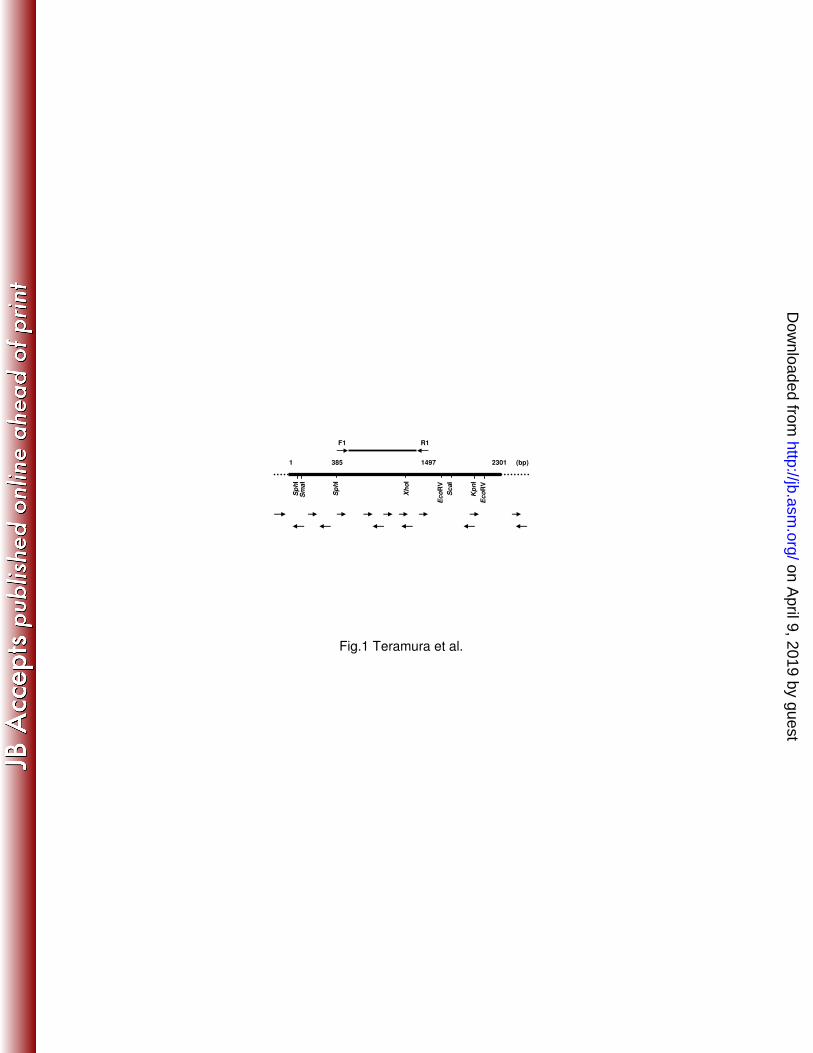
Fig.1 Teramura et al.
(bp)
F1
Sm
aI
1 23011497385
R1
Kp
nI
Ec
oR
V
Sp
hI
Sc
aI
Xh
oI
Sp
hI
Ec
oR
V
on April 9, 2019 by guest
http://jb.asm.org/
Dow
nloaded from

Fig.2 Teramura et al.
7.
4.
3.
6.
2.
5.
1.
-12
89
189
289
389
489
589
689
789
889
989
1089
1189
1289
1389
1489
1589
1689
1789
1889
1989
2089
2189
2289
88
188
288
388
488
588
688
788
888
988
1088
1188
1288
1388
1488
1588
1688
1788
1888
1988
2088
2188
2288
2388
on April 9, 2019 by guest
http://jb.asm.org/
Dow
nloaded from

Fig.3 Teramura et al.
Zn2+NH2 COOH
Pre Pro Catalytic domain PKD PPC
Zn2+NH2
COOH
Pre Pro Catalytic domain PPC
V. parahemolyticus
V. alginolyticus
G. hollisae
A
B
on April 9, 2019 by guest
http://jb.asm.org/
Dow
nloaded from

Fig.4 Teramura et al.
11797
55
37
29
(kDa)
2 31 5 64
19
C
A
21 3
250
150
100
75
50
(kDa)
37
B
21 3
25
20
100
75
50
(kDa)
37
4
on April 9, 2019 by guest
http://jb.asm.org/
Dow
nloaded from


![Spectrofluorometric Assays of Human Collagenase …file.scirp.org/pdf/AER_2015033110440165.pdfV. Ejupi et al. 20 extracellular matrix [1]. Human collagenase is a MMP with the ability](https://static.fdocuments.us/doc/165x107/5aaa03fa7f8b9a77188d970c/spectrofluorometric-assays-of-human-collagenase-filescirporgpdfaer-ejupi.jpg)