Cloning and sequencing of a storage protein receptor ...
71
Cloning .and sequencing of a storage protein receptor fragment from the corn earworm, Helicoverpa zea Zongshu Luo B.Sc., Wuhan University, 1984 M.Sc., Institute of Medicinal Biotechnology, Chinese Academy of Medical Sciences, 1987 \ THESIS SUBMITTED IN PARTIAL FULFILLMENT OF THE REQUIREMENTS FOR THE DEGREE OF MASTER OF SCIENCE I IN THE DEPARTMENT BIOLOGICAL SCIENCES O Zongshu Luo 1997 SIMON FRASER UNIVERSITY All rights reserved. This work may not be reproduced in whole or in part, by photocopy or other means, without permission of the author. 4
Transcript of Cloning and sequencing of a storage protein receptor ...

Cloning .and sequencing of a storage protein receptor fragment from the corn earworm, Helicoverpa zea
Zongshu Luo
B.Sc., Wuhan University, 1984
M.Sc., Institute of Medicinal Biotechnology,
Chinese Academy of Medical Sciences, 1987
\
THESIS SUBMITTED IN PARTIAL FULFILLMENT OF
THE REQUIREMENTS FOR THE DEGREE OF
MASTER OF SCIENCE I
I N THE DEPARTMENT
BIOLOGICAL SCIENCES
O Zongshu Luo 1997
SIMON FRASER UNIVERSITY
All rights reserved. This work may not be
reproduced in whole or in part, by photocopy or other means,
without permission of the author. 4

National Library 1+1 of Canada BibliotMque nationale du Canada
Acquisitions and Aquisitions et . Bibliographic Services servlces bibliographiques
W5 Wellington Street. 395, rue Wellington OttawaON KlAON4 Ottawa ON K l A ON4 Canada CaMda
The author has granted a non- exclusive licence allowing the . National Library of Canada to reproduce, loan, cistribute or sell copies of ths thesis in microform, paper or electronic formats.
The author retains ownershp of the copynght in t h s thesis. Neither the thesis nr# substantial extracts from it may be printed or otherwise reproduced without the author's permission.
Your rib Votrn relerbncm
Our ft& Notre reference
L'auteur a accorde une licence non exclusive pennettant a la Bibliotheque nationale du Canada de reproduire, preter, distribuer ou vendre des copies de cette these sous la forrne de microfiche/film, de reproduction sur papier ou sw format electronique.
L'auteur conserve la propriete du droit d'quteur qui protege cette these. Ni la these ni des extraits substantiels de celle-ci ne doivent itre imprimes ou autrement reproduits sans son autorisation.

APPROVAL
Name: Zongshu &uo '%
C
Degree: MASTER OF SCIENCE
Title of Thesis:
Cloning and sequencing of a storage protein receptor fragment from the corn earworm, Helicoverpa zea.
1 \
Examining Cornrni ttee:
Chair: Dr. B. Crespi, Associate Professor
- Dr. N. H. Haunerland, Associate Professor, Senior supervisor Department of Biological Sciences, S.F.U.
Dr. M.M. Moore, Associate Professor Department of Biological Sciences, S.F.U.
Dr. B. M. Honda, Associate Frofessor Department of Biological Sciences, S.F.U.
u o r Department of Biological Sciences, S.F.U. Public Examiner
Date Approved: .Ai' +,I@?

The very high density lipoprotein (VHDL) receptor from the perivisctrar fat body, of I
the corn earworm, Helicoverpa- tea is the only storage protein receptor found so far in
lepidopteran insects. No cDNAs for this receptor have been isolated to date. In the curfent
research, reverse transcription-polymerase chain reaction (RT-PCR) was used for cloning
partial cDNA sequence for this receptor. The N-terminal sequences from two major CNBr
fragments were used to prepare degenerate primers for RT-PCR. A 1.3 kb PCR product,
obtained with one pair o i these primers, was tloned into a TA plasmid. The PCR product was ' I
sequenced and Northern blot analysis was done with the labeled PCR product. The labeled
PCR product hybridized to mRNA of 2.6-2.8. kb from the perivisceral fat body. This mRNA
first appeared in the 4th day of last larval instar, then reached its highest level in the 7th day.
Sequencing revealed one open reading frame of the 1308 bp, coding for 436 amino
acids. he predicted protein has the mjAecular weight of 50206 dalton and a
8.39. It has one possible transmembrane helix. The composition shows that there are 4%
methionine in this polypeptide. The codon usage was consistent with the preferential codon
usage in related insect families.
Sequence homology search showed that the sequence of 1310 bp has about 25%
identities to several putative RNA-directed RNA polymerases of plant viruses. To exclude the
possibility of virus contamination, further experiments were carried out. PCR with genomic
DNA of fat body cDNA obtained with oligo dT yielded the expected fragment, confirming that
the sequence is a part of the Helicoverpa rea genome and is expected in the fat body.
While the above data are consistent with the storage protein receptor of Helicoverpa,
ultifiate proof will require the cloning and expression of the complete cDNA sequence. e
I

ACKNOWLEDGMENTS . I
s
;b.
I would like to @ve m i thanks.first to my senior supervisor, Dr. Norbert H .
Haunerland, for his guidance, patience and encouragement during my study. My thanks also
go to both of my committee members: Dr. Margo Moore and Dr. Barry Honda for their
valuable comments and suggestions in my research and thesis writing.
I wish to express my thanks to Deryck Persaud in our lab for allowing me to adapt
some of his results in Chapter 8, and to the other lab fellows: Mark, Qiwei, Huarong, Chris
and Rtck for their efforts to keep-@e lab working in a friendly and cooperative environment. I
want thank Dr. S. P. Lee for his critical suggestions and,discussions in my thesis work. +
I also thank Biotechnology Laboratory of UBC for their constant support in the protein I I
sequencing and DNA sequencing through this project.
My special thanks to my family members here in Vancouver: my husband Francis, for
his love and patience; my mother-in-law for her looking after my baby during my last year of
the thesis work; my family members in China: my father and my brother, for their everlasting *
encouragement.
TABLE OF CONTENTS
APPROVAL
ABSTRACT
ACKNOWLEDGMENTS
TABLE OF CONTENTS
LIST OF FIGURES
CHAPTER 1 GENERAL INTRODUCTION
CHAPTER 2 WESTERN BLOTS OF VHDL RECEPTOR PROTEIN Ir.
2 . 1 . Introduction
2 . 2 . Methods
2.2.1. Polyacrylamide gel electrophoresis
2.2.2. Western blots
2 . 3 . Results
2 . 4 . Discpssion
CHAPTER 3 'i
PROTEIN ISOLATION AND N-TERMINAL
SEQUENCING
3 . 1 . Introduction
3 . 2 . Metbods
3.2.1. Insect rearing 13
3.2.2. Preparation and solubilization of fat body membrane
proteins 13
3.2.3. Gel electrophoresis in slab gels and electroelution 13
3.2.4. Separation in the Bio-Rad Model 491 prep Cell 14
3.2.5. N-terminal protein sequence analysis 15

3 .3 Results
3 .4 . Discussion
CHAPTER 4 CHEMICAL CLEAVAGE AND PROTEIN
SEQUENCING
4 .1 . Introduction 'Zr
4 . 2 . Methods
4.2.1 . CNBr digestion
4.2.2. Polyacrylamide gel and membrane blot
4 . 3 . Results
4 . 4 . Discussion
CHAPTER 5 RT-PCR AND CLONING OF THE RECEPTOR
cDNA w
5 . 1 . Introduction
5 . 2 . Methods
5.2.1. Total RNA isolation
5.2.2. Reverse Transcription and polymerase chain reaction
5 . 3 . Results
5.3.1. Quality control for RNA- preparations
5.3.2. Pnmer design and'^^-PCR of actin . 5.3.3. h m e r design and RT-PCR of the receptor
5.3.4. RT-PCR with degenerate primers from internal sequences
of the receptor protein
5 . 4 . Discussion
CHAPTER 6 . CLONING OF PCR PRODUCT AND DNA
SEQUENCING
6 . 1 . ~ntroduction

6 . 2 . Methods
- 6.2.1. Cloning of PCR product
6.2.2. DNA purification and restriction analysis
6.2.3. DNA sequencing and computer analysis
6 . 3 . ~ e s u l t s I
6.3.1. DNA sequencing
6.3.2. Database search
6 . 4 . Discussion
CHAPTER 7 NORTHERN BLOT
7 . 1 . Introduction r
7 . 2 . Methods
7.2.1. Probe preparation and DIG-labeling
7.2.2. Northern blotting
7 . 3 . Results
7 . 4 . Discussion
CHAPTER 8 . GENERAL DISCUSSION
REFERENCES
vii

LIST OF FIGURES AND TABLE
Fig. 2.1
Fig. 3.1
Fig. 3.2
Fig. 4.1
Fig. 4.2
Fig. 5.1
Fig. 5.2
Fig. 5.3
Fig. 5.4
Fig. 5.5
Fig. 5.6
Fig. 6.1
Fig. 6.2
Fig. 6.3
Fig. 6.4
Fig. 7.1
Fig. 7.2
Fig. 8.1
Fig. 8.2
Western blot of VHDL receptor protein in Helicoverpa zea 10
SDS-PAGE of purified VHDL receptor 16
PVDF membrane blot of VHDL receptor protein 17
PVDF meflbrane blot of CNBr fragment 2 1
~ifferent digestion time of CNBr for VHDL receptor 23
Methods for priming cDNA synthesis for RT-PCR 26
Total RNA isolation and agarose gel 30
p-actin primer design from the consensus sequence of Genebank ., 31
RT-PCR with actin primers 33
RT-PCR with degenerate primers 35
The structure of receptor protein and its cDNA 36
Restriction analysis of TA cloning 40
s&tegy of sequencing the cloning of VHDL receptor 4 1
The nucleotide sequence of PCR fragment for VHDL receptor protein
from Helicoverpa lea and their deduced amino acid sequence 42
Hydrophobicity and secondary structure prediction of VHDL receptor B
protein 43
Northern blot of VHDL receptor 49
Comparison of Northern blodin different stage of last larval instar 50'
RT-PCR with specific primers and oligo dT 54
RT-PCR with specific primers and genomic DNA 55
Table 6.1 The percentage of identities among the receptor sequence and'the plant
virus sequences. .. 46

CHAPTER 1 GENERAL INTRODUCTION '
' f ' All insects change in body structure during their development from juvenile to adult. F '
Many in~ects~rnolt directly from their last larval to the adult stage, in a process that is called
incomplete mepmorphosis . In contrast to these hemimetablous insects, 'hot om h bolo us orders, such as flies and moths, undergo complete metamorphosis which involves a discrete
pupal stage between larvae and adults. During the pupal stadium numerous new structures P
(e,g., cuticle, wings) must be formed while others are broken down (Sehnal, 1985; Levenbook
and Bauer, 1984; Scheller et al., 1980). Many new proteins and carbohydrates are synthesized
in pupae, and these activities require large amounts of biosynthetic precursors such as amino
acids, carbohydrates and lipids. Yet pupae are not able to take up any nutrients from their
surroundings. The needed amino acids must therefore come from reserves accumulated in I
feeding larvae (Dean, 1985). 1 .
The insect storage proteins are synthesized in fat body tissue, secreted and released into
hemolymph by the fat body of feeding larvae and reach extraordinary concentrations in the
- hemolyrnph just prior to metamorphosis (Levenbook, 1985). Storage proteins mostly
accumulate in the hernolymph of last instar larvae. These proteins are taken up into the fat body \
during the larva to pupa molt and stored in cytoplasmic protein granules. These frequently
crystalline granules break down later to provide the amino acids needed for adult protein
synthesis. However, they may also be incorporated into cuticle as intact proteins or be diverted
into energy metabolism (Telfer and Kunkel, 1991; Konig et al., 1986; Schenkel and Scheller,
There are several different classes of storage proteins, which were recently reviewed by
Telfer and Kunkel ( 199 1 ) and Haunerland ( 19%). Most storage proteins belong to a family of
hexameric proteins (hexamerins) related to hemocyanin, an oxygen transporting protein found .
in marine arthropods (Van Holdmnd Miller, 1982; Linzen er al., 1985; Beintema er al . , 1994).
These proteins have native molecular weights around 500,000 and are composed of six 70 and

and 85 kDa subunits (see reviews by Telfer and Kunkel, 1991). Before the primary structure
and the evolutionary relationship of the different storage proteins were known, they were - *
classified according to their amino acid composition. All hololllttabolo~s insects possess
arylphorin, a protein that is very rich in the aromatic amino acid &sidues (up to 20 %) that are
needed for the formation of cuticular w e i n s (fot a review, see G l f e r and Kunkel, 1991). It is
noteworthy, however, that lepidopteran and dipteran arylphorin -is not the' same 'prutein.
Dipteran arylphorin has high aromatic and methionine contents (Kinnear and Thomson, 1975;
Munn and Greville, 1969; Munn et al., 1%9), while lepidopteran arylphorin is high in 2
aromatic amino acid and low in methionine content (Haunerland and Bowers, 1986; Karpells et
al., 1990; Kramer et al., 1980; Kunkel et al., 1990; Palli and Locke, 1987; Ryan et al., 1986;
Telfer et al . , 1983; Tojo et al . , 1980). The sequences of lepidopteran arylphorins are quite
different from those of dipteran arylphorin (see a review by Haunerland, 1996). Among other
hexamerins found in lepidopteran insects, methonine-rich proteins (> 4 9% of methionine) are
the most common proteins. This group of proteins has high methionine and low aromatic
amino acid contents but lacks carbohydrates (Bean and Silhacek, 1989; Ryan et al., 1985;
Ryan er al . , 1986; Tojo et al . , 1978; Tojo et al . , 1980). It is not known what specific role I
these proteins play and whether the methionine content is important.
In addition to storage hexamerins, at least one lepidopteran family, the Noctuids, use a
non-hexameric storage protein composed of 4 subunits of 150 kDa and 8 .4 % lipid, hence .
called very high density lipoprotein (VHDL) (Haunerland and Bowers, 1986, Jones et al.,
1988). In the corn eanvorm, Helicoverpa zea , VHDL is colored blue due to bound biliverdin.
The blue color allowed to easily see how VHDL accumulates initially in the hemolymph and
later in fat body tissue. In early larval stages, hemolymph is pale yellow and the fat body,
located peripherally next to the cuticle, is white. During the first half of the last larval instar, w
the hemolymph turns bright blue. Subsequently, the blue color gradually disappears from the
hemolymph, and accumulates in a new perivisceral fat body, located in the body cavity, The

blue tissue becomes dominant in perivisceral fat body during the last 4 days of the last larval
instar. In contrast, the periphera! fat body remains white. Detailed studiks have demonstrated ,
that both known storage proteins of H. zea, VHDL and arylphorin are selectively taken up by
the perivisceral fat body only. The white peripheral fat body, where these and other proteins \
are synthesized earlier, never takes up storage proteins. Instead, it cksintegrates during further
development. VHDL and arylphorin, however,, accumulate in the periviscerai fat body in
dense protein granules that later are partially digested to serve as amino acid reserve for the
synthesis of adult proteins (Wang and Haunerland, 1991; wang and ~aune r l and , 1992).
Since storage proteins are normally present in large concentrations in the insect
hemolymph, non-selective endocytosis alone could assure the import of large amount of m
s t o a p r o t e i n s into the fat body, and initial experiments with horseradish peroxidase ~-
demonstrated this (Locke and Collins, 1968). However, the clearing of proteins from
hemolymph and the accumulation in fat body is not a function of their original concentration,
indicating that the uptake occurs in a selective receptor-mediated process (Pan and Telfer,
1993). Such a process would not exclude the unspecified import of other abundant hemolymph
proteins. When the fat body of H. zea was incubated with equaf amounts of labeled arylphorin
and a foreign protein (IgG) in virro, a small amount of IgG accumulated in the tissue, but a
tenfold excess of arylphorin was taken up (Wang and Haunerland, 1994b). This suggests the
selective uptake must be mediated by specific endocytotic receptors.
Detailed studies of the perivisceral fat body by Wang and Haunerland led to the
identification and isolation of a VHDL receptor protein in H. zea. (Wang and Haunerland,
1993; 1994). Electron micrograph; of irnmunogold-labeled sections show that the receptor is
located in the plasma membrane of perivisceral fat body cells. It was demonstrated in a receptor
binding assay that a large concentration of receptor exists between the 4th and 8th day of last
instar larvae. The'storage protein receptor was identified by ligand blotting and purified to
homogeneity (Wang and Haunerland, 1992). I t is a glycosylated basic protein of 80 kDa with

4
an isoelectric point of pH 8.2. Binding requires Ca2+ and is optimal at pH 5.5. A very
interesting fiqding is that the receptor for VHDL also functions as the receptor for arylphorin, !. , although these storage proteins are completely different in structure.,The binding constants are
similar, 7.8 x lo-* for VHDL and 9.02 x 10' for arylphorin. Binding of both storage proteins
in ligand blots was also competitively reduced by excessive amounts of either unlabeled
protein, but not by bovine serum albumin (Wang and Haunerland, 1994).
To date, storage protein receptors have not been identified in other lepidopteran species.
However, similar reasoning led investigators to propose storage protein receptors in Dipteran
species (Bumester and Scheller, 1992; Ueno et al., 1983; Ueno and Natori, 1984). Dipteran
storage proteins have similar developmental profiles as their lepidopteran counterparts:
synthesis begins in early or mid-larval .stages and terminates in feeding larvae, followed by
sequestration by the fat body (Haunerland, 1996). Unlike the great variety of storage proteins
encountered in Lepidoptera, each dipteran species apparently has only one or two storage
hexamers, arylphorin and another larval serum protein (LSP-1) (Telfer. and Kunkel, 1991;
Haunerland, 1996).
Evidence for receptor mediated uptake of storage proteins by the fat body had earlier
been reported in two dipteran species. A fat body membrane fraction in Sarcophaga peregrina
can bind radiolabeled arylphorin with a Kd of 4 x (Ueno et of.. 1983; Ueno and Natori.
1984; Ueno and Natori, 1987). The binding requires ca2' and is optimal at pH 6.5. This
putative arylphorin receptor has a molecular weight of 120 kDa .and comes from an inactive
precursor of 125 kDa. Recently, a cDNA for this putative receptor protein was cloned and
sequenced (Chung er a l . , 1995). However, these authors failed to detect the protein in the
plasma membrane of fat body cells, and could see it only in protein granules. Hence, they
suggested that the 120 kDa protein may be different from the arylphorin receptor that is needed
for incorporation of arylphorin into fat body; possibly, it binds arylphorin to immobilize it in
the protein granutes of pupal fat body.

5
In addition to the work done with Sarcophaga, Burrnester and Scheller have studied
arylphorin binding proteins in Calliphora vicina (Burmester and Scheller, 1992). Three
proteins with molecular weights of 130 kDa, % kDa and 65 kDa showed binding function with
arylphorin. Later work (Burrnester and Scheller, 1995) suggested that the 96 kDa protein must
be modified before arylphorin uptake can take place, possibly by cleavage to the 65 kDa
protein, which may be the active arylphorin receptor. The cDNA clones of the arylphorin
binding proteins from Sarcophuga and Calliphora are very similar, and the amino acid
sequences of these proteins are very similar too (46% identity) (Haunerland, 1996). Both
proteins are also similar to a protein with unknown function that is encoded by the P1 gene of
Drosophila melanogaster (Maschat et al, 1990).
It is generally assumed that storage protein uptake is essential for adult development.
Therefore the study of the receptor-mediated uptake process will not only lead to the thorough
understanding of this biochemical and physiological process, but also provide a potential way
to control certain lepidopteran species. Based on preliminary results from this laboratory, the
goal of this research was to determine the primary structure of the storage protein receptor
from H. zea, which is apparently quite different from the above described protein found in
Diptera.
In principle, two different strategies could be used to achieve this: construction of a
cDNA expression library and screening with anti-receptor antibodies previously produced in
the laboratory (Wang and Haunerland, 1992), or amplification of receptor cDNA via PCR
primers constructed from amino-terminal sequences of the receptor or some fragments thereof.
At the onset of this study, it was difficult to predict which approach would be more likely to
succeed. Screening of expression libraries is notorious for its low signal to noise ratio, and
excellent antibodies are normally required for success. Although the available antibodies had
been used successfully for immuno-cytochemical applications, no rigorous evaluation of their

specificity and applicability for Western blots had been done. On the other hand, the second
approach was chatlenging since it had previously been shown that the amino-terminus of the
receptor protein is blocked; hence, it was necessary to cleave the protein in controlled ways and % 4
to obtain internal sequences, which in turn could be used f a t h e construction of PCR primers. *
In light of these facts, it was decided to initially evaluate the existing antibodies and proceed
with an expression library if they proved to be strong and specific. Otherwise, the second
approach would be ~ e d .

Chapter 2: Western Bjots of VMDL Receptor Protein
2.1. Introduction *.A *
Initially, it was planned to construct a cDNA library and screen the library to obtain the
cDNA for the storage protein receptor. As Wang and Haunerland (1992) had isolated the
receptor protein and produced antibodies against it, it appeared feasible to use these antibodies.
Ideally, an antibody used for screening of expression libraries should be absolutely \
. specific for conformation-independent epitopes that are displayed on both native and denatured
forms of the protein, and high titers of antibodies should be present in the antiserum.
There were some concerns whether the anti-receptor antibodies produced ,earlier were
appropriate for library screening. Although these antibodies had -been successfully used for
irnrnunocytochemical detection of the storage protein receptor in thin electron microscopy
sections, they had only been used in Western blots of protein fractions rich in storage protein
receptor. Moreover, the production of antibodies had failed several times with aliemative
adjuvants (Ribi imrnunostimulant) and had succeeded only after immunization and several
booster shots with complete Freunds adjuvant, suggesting that the pmtein did not elicit a very
strong immune response in rabbits. These antibodies had been produced 2 years prior to the
beginning of this work and stored at -80 OC; quality losses have frequently been observed for
antibodies that had been stored for extended time periods. To determine whether the antiserum
available was suitable as probes, initial experiments were designed in which serial dilutlon of
antiserum were tested for the specific reactivity with the receptor protein on Western blots.
2.2. Methods
2.2.1 . Polyacrylamide Gel Electrophoresis
Sodium dodecyl sulfate polyacrylamide gel electrophoresis (SDS-PAGE) was canied
out in a mini gel unii (Hoefer Scientific, San Francisco, CA). Acrylamide and N, N'-
methylene bisacrylamide were used to polymerize a 10 % T, 2.6 % C resolvipg gel, pH 8.8 . and a 4 % T, 20 9% C stachng gel, pH 6.8. Samples were diluted with 2 volumes of stock
sample buffer (0.06 M Tris-HCI. pH 6.8, 2 96 SDS, 10 % glycerol. 0.025 '% ' ~romo~heno l , Blue; 50 PI of 2-mercaptoethanol/ml added immediately before use) and were heated in boiling
water for 5 minutes. Electrophoresis was run atfroom temperature under constant current (25
mA) for 2-3 h. The gels were stained with Coqmassie brilliant blue R 250 in methano1:acetic
acid:water (4: 1 5 ) and destained with the same solution.
2.2.2. Western blots
Protein samples were transferred from SDS-PAGE gels onto nitrocellulose on a semi-
dry blotting apparatus (LKB Nova Blot) according to Towbin et al. (1979). The syni-dry
transfer technique of the Nova Blot system uses filter papers soaked in transfer buffer (39 rnM
glycine, 48 mM Tris, 20% vlv methanol, pH 8.9) as the only buffer reservoir; the transfer was
carried out at 0.8 rn~ lcm2 overnight.
The immunodetection was done with a blotting detection kit from Amersham (Arlington
Heights, IL). After transfer, the nitrocellulose blots were incubated for 1 h with blocking
buffer (5 mglml bovine albumin and 0.3% gelatin in Tris-buffered saline-Triton X- 100 (TBS-
T): 20 rnM Tris-HCI, 150 rnM NaCI, 0.1% Triton X-100, pH 7 6 ) . The blots then were
washed three times with TBS-T and incubated for 1 h with diluted rabbit anti-receptor
antibodies in TBS buffer. After three washes with TBS-T buffer, the membranes were
incubated for 20 minutes in diluted biotinylated anti-rabbit IgG antibody solution (1:506 in - TBS). Following another three washes with TBS-T buffer, the blots were incubated for 20
minutes in diluted streptzividin alkaline phosphatase solution (1:3000 in TBS). Finally. the

9
bands were visualized by incubating with a solution of 1 drop (- 50 PI) each bf NBT (Nitro-
blue tetrazolium) and BCIP (5-Bromo-4 chloro-3-indolyl phosphate) in 10 ml diethanolamine
buffer (100 rnM diethanolamine, 5 rnM MgC12, pH 9.5). The reaction was stopped by
washing with distilled pater .
0
2.3. , Results a I
The Western blot results revealed the target protein as well as many unspecified bands.
Many attempts were made to vary the conditions to achieve stronger signal and weaker
background staining. Different dilutions of the anti-receptor antibody (ftom 1 5 0 0 to 1:50,000) r
were tried but failed to display specific antibody-antigen reaction for the receptor protein. A
representative result using dilution 15,000 is shown in Fig. 2.1. The antiserum also showed
cross-reactivity with insect arylphorin, fatty-acid binding protein and some yeast proteins. The
sample was sent to another laboratory and checked with different reagents to exclude
laboratory- or operator-specific problems; however. even those attempts failed to give clear
signals and low background
2.4. Discussion w
+ The results did not show that the anti-receptor antibody has the specific reactivity to the
receptor protein. Even at very low titer ( 1 :50,000), the antibody still gave unspecified binding ~ -
to other membrane proteins. These problems were not only caused by the anti-receptor
antibody since they also existed with other anti bodies. Immunodetection with alkaline
phosphatase, while much more sensitive than horseradish peroxidase. is frequently-re prone
to unspecified interactions with other proteins, possibly because some traces of enzyme bind to
many proteins on the blot. However, in most cases specific antibodies react much stronger I
with their antigen, and it is easy to distinguish signal and background. Hence, it was concluded
that the antibody used here was not very specific, possibly due to low titer or loss of binding

Fig. 2.1: A typical Western blot of VHDL receptor protein from H. zea. Lane 1: Marker protein, stained membrane with Coomassie Blue after
transfer.
Lane 2: Crude membrane fraction from H. zea fat body, expected band size
-80 kDa. 10 pg of protein samples were loaded and separated by SDS-PAGE
(10 % T), transfered onto nitrocellulose and stained with anti-VHDL receptor
antiserum (1: 5,000 dilution).
activity during storage. It is p s i ble that alternative detection methods, e.g. with horseradish
peroxidase, could have given acceptable results in Western blots.
However, high titer and specificity would be an absolute necessity for screening an
expression library, since the receptor protein may be present in positive clones in only small
amounts. Moreover, since the prokaryotic cells of a library will not process the protein in
similar ways as insect cells, the receptor may not be located in the plasma membrane, even if e-?&
the full length cDNA of the receptor, complete with its targeting sequence, is translated.
Therefore, it appeared to be of little benefit for the present study to invest time and money to
evaluate alternative Western detection systems. Itswas considered unlikely that the antibody ., could be successfully used for primary screening of an expression library
Since screening of a cDNA expression library with antibodies was not possible, the
alternative plan was to use PCR to obtain the cDNA sequence of the receptor gene. The
underlying idea was to get partial internal sequences of the protein with chemical cleavage.
These sequences can be used to construct oligo nucleotide primers for PCR. A part of the
cDNA sequence may be amplified in that way, and sequenced or later used as a probe for
library screening. /

Chapter 3: Protein isolation and N-terminal sequencing
3.1. Introduction
For the amplification of cDNA that encodes the VHDL receptor, sequence-specific
primers were required. Ideally, one primer is designed from the amino-terminal sequence of
the protein, but because the N-terminus of the VHDL receptor is blocked (Wang and
Haunerland, l h ) , this sequence was not known. Therefore, it was decided to obtain internal
sequence information, by N-terminal sequencing of fragments of the protein. Such fragments
can be obtained by chemical or enzymatic cleavage of the polypeptide chain. Trypsin or
chymotrypsin are frequently used to cleave the chain at the carboxyl side of a basic or aromatic
amino acid, respectively. Since these residues are normally quite abundant in a protein,
numerous small fragments would be obtained which must be separated by HPLC.
Alternatively, one could attempt to only digest the most accessible residues, thus obtaining a
smaller number of larger fragments. Chemical proteolysis is mostly achieved through treatment
with cyanogen bromide, which cleaves at the carboxyl side of methionine. Since methionine is
a relatively rare amino acid (average of only 2 % of all residues), cyanogen bromide cleavage
offered a better chance of obtaining a few, relatively large fragments that could be separated by
gel electrophoresis.
The latter approach was used in the current study. For the optimization of the cleavage
and to obtain sufficient amounts of fragment for sequencing, milligram amounts of VHDL
receptor were required. The method previously used for receptor isolation (Wang and
Haunerland, 1992) resulted in a very pure protein, but it involved many steps with low overall
yield. I t also included affinity chromatography on an agarose medium which had been
covalently bound with VHDL and this medium was no longer available. Since there was not
enough purified receptor protein left for sequencing studies, efforts were made to purify large
amounts of the protein.
3.2. Methods
3.2.1. Insect rearing
The corn earworm, H. tea was reared in plastic boxes on a 16:8 lightldark cycle .at
260C (Patana and McAda, 1!273). Larvae remain in the fifth larval stage about 7 days then
stop feeding and prepare to pupate. Six or seven day old fifth instar larvae were used for these
experiments.
3.2.2. Preparation and solubilization of fat body membrane proteins
The frozen perivisceral fat body was dissected from last instar larvae and was
homogenized in ice cold extraction buffer (20 mM Tris-HCI, 150 mM NaCI, 1 mM CaC12, pH
8.0 containing 1 mM phenylmethylsulfonyl fluoride (PMSF) and 1 m M ~mercagtoethanol)
with a Potter type glass homogenizer. The homogenate was centrifuged at 800 x g for 10 min.
at 4 OC to remove cell debris. The resulting supernatant was then centrifuged at 30,000 x g for
lh to collect a fraction that contained most of the plasma membranes. The pellet was washed
once with the buffer and solubilized with 2 9% Triton X-100 in the same buffer overnight at 4
OC. Insoluble material was removed by centrifugation at 100, 000 x g for 1 h. The samples
were stored at -80 OC untd the protein gel was run.
3.2.3. Gel electrophoresis in Slab gels and electroelution
Samples containing 1 mg of crude membrane protein were run in the Bio-Rad
PROTEAN I1 xi Cell in a similar method outlined in chapter 2. The gel was stained with
copper stain using the copper stain and destain kit from Bio-Rad. The proteins were therefore
reversibly fixed in the gel, allowing elution after a destain step. The protein bands were
visualized as negatively stained bands on SDS-PAGE gels.

The band of interest (80 kDa) was cut and destained, the gel slice was then put into the
~io- ad Model 422 Electro-Uuter for protein elution. The sample was collected in a 400 p1
volume of elution buffer (same as the eletrode buffer) and lyophilized by freeze drying.
3.2.4. Separation in the Bio-Rad Model 491 Prep cell
Crude membrane protein was run in the Bio-Rad Model 491 Prep Cell, which is
designed to purify proteins or nucleic acids from 'complex mixtures by' a continuous-elution
electrophoresis. Conventional gel electrophoresis buffer systems and media are used with the
Prep Cell.
During a run, samples are electrophoresed through a cylindrical gel. As molecules
migrate through the gel matrix, they separate into ring shaped bands. Individual bands migrate
off the bottom of the gel HIhere they pass directly into an elution chamber for collection.
The sample (2 mg) was mixed with an equal volume of SDS sample buffer (same as in
Chapter 2) and boiled for 5 minutes, then loaded onto a 10 cm long tube gel. The gel was run
for 8- 10 hours at 40 mA constant current at which time the bromophenol blue marker dye was
about 5 rnrn from the bottom of the separating gel. The SDS running buffer (0.025 M Tris,
0.192 M glycine, 0.1 % SDS, pH 8.3) was pumped through the elution chamber at a rate of 1
ml per min.
The elution c h a m M outlet was connected to a fraction collector and 200 x 3 4
fractions were collected. Elution of molecules was monitored with an ultraviolet detector and
chart recorder. Fraction number one was the first fraction containing visible amounts of the
bromophenol blue marker dye (first peak appeared on the chart recorder). In order to locate the
fractions containing the receptor protein, 30 PI of every fourth fraction were analyzed by SDS
gel electrophoresis and silver staining. The best fractions with respect to purity of the putative
receptor protein (80 kDa) were pooled and lyophilized by freeze drying. *

3.2.5. N-terminal protein sequence analysis d
For protein sequencing, the samples were run on SDS-PAGE gels and transferred
unstained to Problot polyvinylidene difluoride (PVDF) membrane (Applied Biosystem) with a
semi-dry blotting apparatus (LKB Nova Blot) according to Towbin et al. (1979). The semi- /
dry transfer technique of the Nova Blot system uses filler papers soaked in CAPS buffer [ I 0
mM 3-(cyclohexylamino)- 1 -propanesulfonic acid in 10 % of methanol, pH 1 1.01 as the only
buffer reservoir. The transfer was carried out overnight at 0.8 r n ~ l c m 2 .
After the transfer, the membrane was removed and rinsed briefly with H20 . The
membrane was stained with Coomassie Brilliant Blue k-25D for 5 min., then destained with
50 % (vlv) methanol for 15 min. The membrane was then washed. with several changes of
H z 0 for 5-10 min. and air dried. Stained bands were excised from the Problot PVDF
membrane and sent to Protein Service Laboratory, University of British Columbia *or micro
sequencing of the proteins (Applied Biosystems, Model 476A).
3.3. Results
With the slab gel and electroelution, purified sample was collected and lyophilized. The
sample was used for trial experiments of cyanogen bromide digestion and protein anaiy,sis.
Figure 3.1 demonstrates the high degree of purity of the receptor protein obtained from
the preparative SDS gel separation. The 80 kDa protein was collected from fractions 163- 170.
Of the 2 mg total protein separated with the Model 491 Prep Cell, 240 pg of nearly .
homogeneous protein was isolated in a single step.
The purified 80 kDa protein was used for N-terminal sequencing of total protein.
Ilowever, sequence was obtained only when a large excess of protein was submitted for ;
sequencing and signal can account only for small percentage of sample.

Fig. 3.1: SDS-PAGE analysis of purified VHDL receptor. Aliquots from fractions 160-170 of the Model 491 preparative electrophoresis Cell
were analysed by SDS-PAGE gels (10 % T) and silver stained.
M: Marker proteins.
153-175: Elution fractions
CM: crude membrane extract from fat body.

Fig. 3.2: PVDF membrane blot of putative VHDL receptor protein purified from fat body tissue of H.zea. Purified receptor (45 yg) was electrophoresed on an SDS-PAGE gel, transferred
onto PVDF membrane and stained with Coomasie blue, as described in 3.2.5.
The putative VHDL receptor bands were cut out and submitted for N-terminal
sequencing.
Lane 1: marker proteins.
Lane 2,3: purified receptor protein.
Lane 4: bovine serum albumin .

t
18
Figure 3.2 displays the blot of the receptor protein used.for sequencing. There was
more than 45 pg (562 pmol) loaded on the gel, however, the sequencing result showed very
low signal, accounting far less than 1 % of the protein loaded.:'
3.4. Discussion
Since the putative receptor band (80 kDa) was the strongest band in an SDS gel, it was
decided to use preparative electrophoresis as the main purification step. Initially, this was
accomplished by electroelution from a preparative gel. However there were concerns about the . efficiency of the elution and the limited amounts that could be processed. Therefore, another
preparativemethod was adopted.
Preparative electrophoresis provided a simple and efficient method to purify relatively
large amount of protein. The proteins purified with this method can be obtained in the
quantities needed for the subsequent studies.
The sequencing results in the current study showed that the N-terminus was indeed
blocked as suggested earlier by Wang and Haunerland (1994). The small signal obtained from
the sequencing of a large excess of the protein is most likely derived from cgtaminating . -
proteins since the apparent purity of the preparation has been observed in Fig. 3.2.
The short sequence obtained is similar to a methionine-rich protein of Trichoplusia ni , a "' f
storage protein present in other species of the same insect family (Noctuidae). Thus, it should
also be present in H. zea.. Although not shown to interact with the receptor, it could also be a
natural ligand, and hence be contained in the membrane protein fraction. Because of its subunit
molecular weight of 80 kDa, it should migrate close to the storage protein receptor during SDS
electrophoresis. - Since the storage protein receptor is N-terminally blocked, chemical cleavage of the
protein was planned to generate internal peptides with unblocked N-termini. Therefore, more
starting material was required than for simple N-terminal sequencing. The method utilized in

19
this study made it possible to supply sufficient amounts (100 pg for each digestion) to do
cyanogen bromide digestion.

Chapter 4: Chemical .f
4.1. Introduction
cleavage and protein sequencing
Since the N-terminus of the receptor protein is apparently blocked, it was necessary to
obtain intemal sequence infobat ion . In this study, the receptor was chemically cleaved to
generate peptides with unblocked N-termini . Cyanogen' bromide (CNBr) cleavage was the
method of choice (Matsudaira, 1990) since the average number of methionine residues in a
protein is relatively low (- 2 %).
Protein ( l 0 0 p g ) was solubilized in 5 0 pl of 7 0 % formic acid and a small crystal of
CNBr was added and dissolved. The tube was flushed with N2 and capped. The sample was
kept in the dark at room temperature for various times, as indicated. Subsequently, the reaction
was quenched by diluting the formic acid to 7 % with H 2 0 . The sample was then dialyzed
against H 2 0 , frozen at -80 O C for 1 h and lyophilized. The freeze-driedprotein was separated
by SDS-PAGE.
4.2.2. Polyacrylamlde gel and membrane blot 9
Gel electrophoresis was carried out as described in Chapter 3 except that 15 % T , 2.6
% C resolving gels were used to separate the fragments.
4.3. Results
The result of the initial 12-hour digestion is shown in Figure 4.1. T w o major bands of
3 1 kDa and 29 kDa fragments appeared on the blot. The bands were cut and then sent to the

Fig. 4.1 PVDF membrane blot of CNBr fragments of the putative VHDL receptor protein. CNBr digestion was done as described in 4.2.1. SDS-PAGE gel (15 % T) was run and samples were transferred onto PVDF membrane and stained with Coomassie blue. Lane 1 & 2: CNBr digestion samples. Two major bands were 29 kDa and 3 1 kDa as indicated.
Biotechnology Laboratory, University of British Columbia for sequencing. Six amino acid b
residues were determined for the 29 kDa peptide, and five residues for the 31 kDa peptide.
t Since these fragments were obtained after cyanogen bromide treatment, which cleaves proteins
at the carboxy-side of a methionine, the preceding residue must have been a methionine.
Hence, the sequences obtained were:
29 kDa : M-Q-D-A-L-D-F.
3 1 kDa : M-T-A-L-P-K.
In order to obtain more sequence information, it was attempted to purify more protein
and repeat the digestion under more controlled conditions with a new batch of CNBr. In
various experiments, cyanogen bromide was weighed and dissolved in formic acid, and known
amounts of the reagent were added to the protein sample. These digestions led to numerous
3 uch smaller fragments which proved difficult to isolate. Only at very dilute concentrations
was it possible to obtain the 29 and 3 1 kDa fragments, but never as prominent as in the initial
digestion. Shorter djgestion times also did not improve the yield of the two fragments. Formic
acid alone did not lead to any degradation (Fig. 4.2). confirming that the 29 kDa and 3 1 kDa
fragments were indeed products of cleavage by cyanogen bromide.
4.4. Discussion
The results of the initial cyanogen bromide digestion were very encouraging, yielding
two N-terminal sequences useful for PCR primer construction. However, attempts to improve
the digestion by using varying digestion times and amounts of reagent failed. Very low
amounts of CNBr did lead to the formation of the two fragments, indicating that these
fragments were the results of partial digestion. Larger amounts of CNBr, or longer digestion
times. led to a more complete digestion and hence much smaller fragments. While the exact
amount of reagent used in the initial digestion is not known, it certainly was much more than

Fig. 4.2. SDS-PAGE of VHDL receptor after CNBr digestion for
different times with new batch of CNBr.
hrified receptor (100 pg) was digested for the indicated time period with CNBr. The
final reaction solution was dialysed against H20 and freeze dried. Aliquotes of the
products were then separated by SDS-PAGE, and the gel was stained with the diarnine
silver staining method (Merril, 1990).
Lane 1, 2: marker proteins.
Lane 3: crude membrane extract.
Lane 4: crude membrane extract after 20 h incubation with formic acid.
Lane 5: crude membrane extract after 3.5 h incubation with CNI3r
Lane 6: crude membrane extract after 7.5 h incubation with CNBr
Lane 7 : crude membrane extract after 20 h incubation with CNI3r

that used later. However, the original CNBr reagent had-been opened and stored at 4 OC for
more than a year. Cyanogen bromide may decompose when exposed to heat, moist air, or r
water, or on prolonged storage. It is therefore likely that this preparation was partly degraded,
and had only weak activity. It was assumed that under those conditions only the most exposed
methionine residues were cleaved. The attempts to reproduce these conditions and to obtain
more sequence consumed a large amount of purified receptor protein. While it should have
been possible to find appropriate conditions that would allow the production of more 29 and 3 1
kDa fragments, such experiments would have required further amount of the protein and
therefore an expansion of the insect colony. Since there was no guarantee that the results would
have been superior, it was decided to go forward with the results from the initial fragments of
CNBr digestion.

i Chapter 5 RT-PCR and cloning of the receptor gene
5.1. Introduction
From the internal sequences of the reckptor protein, primers can be designed to amplify
the cDNA coding for the part of the receptor protein that lies between those sequences (Flic'k
and Anson, 1995; Burden and Whitney, 1995; McPherson, et al. 1991). Reverse transcriptase
(RT) must be used to convert all mRNA contained in a total RNA preparation into sihgle-
stranded complementary DNA (cDNA), which subsequently can then be amplified via
standard PCR techniques. The product is a DNA fragment, visible on an ethidium bromide i
stained gel, of a length determined by the primers used to amplify the cDNA and diagnostic for
the presence of the corresponding mRNA in the starting sample. The overall process is referred
to as RT-PCR. Reverse transcriptase can synthesize DNA complementary to mRNA only in the
presence of a primer specific for the 3' end of the sequence. There are two ways to prime the
synthesis of cDNA from mRNA. Both the oligo'dT and random priming method used in this
study are illustrated in Fig. 5.1. In both methods, the entire population of mRNA molecules is
first converted into cDNA by priming with either oligo (dT) or random sequence hexamers.
Two gene-specific PCR primers are then added for amplification.
Since the successful amplification of mRNA by RT-PCR depends greatly on the quality
of mRNA, primers'and conditions used for the reverse transcription reaction, it was decided to
evaluate the method first using primers for a highly conserved protein, p-actin.' From the
aligned sequences of actin from several insect species it should be possible to identify a
consensus region useful for the construction of actin-specific primers.
Successful amplification of actin mRNA by RT-PCR would indicate that it may also be
possible to obtain DNA encoding a part of the receptor protein with a limited amino acid
sequence. However, there are several possible codons for each amino acid residue
(degeneracy) and the primers designed for PCR amplification must take degeneracy and codon

c DNA 1 TTTTTT -
5' primer a 3' primer
cDNA d
5' primer 1 3' primer
Fig. 5.1. Methods for amplifying cDNA using RT-PCR. I . Oligo(dT) primer method the entire population of mRNA molecules is used as a template for
the synthesis of first strand cDNA. Subsequently, the complementary strand is synthesized and
the double strand cDNA can be used as a template for PCR.
11 . Random primer method: random sequence oligonucleotides are annealed to the mRNA
template and extended with reverse transcriptase. Some, but not all cDNA molecules can serve
as a template for PCR

preferences into account (see 5.3.3.). Thus, a sequence stretch of lowest possible degeneracy
should be chosen. The two fragments of 29 kDa and 31 kDa obtained after CNBr digestion
should belong to the receptor, and their amino-terminal sequences represent internal sequences
of the protein. Therefore, one primer was designed as the upper primer while another one
works as the lower primer. However, since the locations of two fragments in the native protein
were unknown, two pairs of primers had to be constructed. The expected product size depends
on the location of the fragments in the protein; it can be calculated by dividing the protein
fragment size by the average molecular weight of an amino acid (1 15 Da), and then multiplying
the number of amino acid residues with 3 to obtain the number of nucleotides coding for .this
sequence. The expected product size should be between 750 bp (29,000 Da-1115 Da x3) and
1330 bp [(80,000 Da - 29000 Da) I1 15 Da x3 1.
5.2. Methods
5.2.1 . Total RNA isolation
Total RNA was isolated from freshly excised or previously frozen perivisceral fat body tissue
at day 7 by the method of Chomzynski and Sacchi (1987), modified as described below.
1 . The 'tissue was homogenized in RNA extraction buffer 1 (4 M guanidine isothiocyanate,
25 rnM sodium citrate, 0.5 O/c N-laurylsarcosine, sodium salt, 0.1M P;Mercapto-
ethanol) at 50-200 mglml.
2 . The homogenate (5 ml) was added to a 15 ml polypropylene tube.
3 . The following reagents were added in the indicated order:
0.1 vol. 2 M sodium acetate, pH 4.0.
I .O vol. phenol (water saturated), '5
0.2 vol. chloroform (water saturated) P
The sample was mixed between each addition by inversion and shaken thoroughly for
I0 sec.

The sample was left on ice for 15 min.
The sample was centrifuged at 10,000 x g for 20 min. at 4 OC
The aqueous phase (top) was transferred to a fresh tube, avoiding collecting the
interphase.
RNA was precipitated with 1 .O vdl: isopropanol at -20 OC for 1 h or overnight. **
The sample was spun at 10,000 x g for 20 min. at 4 OC.
The pellet was re-suspended by vigorous vortex mixing in 2 ml of 4 M LiCl to
solubilize polysaccharides. The insoluble RNA was pelleted by centrifuging at
3,000 x g for 10 min. I
The resulting pellet was re-dissolved in 2 ml extraction buffer. Chloroform (2 ml) was
added and mixed with the aqueous phase by vortexing. After centrifugation at 3,000 x
g for 10 min, the upper phase was collected and precipitated with 2 ml isopropanol in
the presence of 0.2 M sodium acetate (pH 4.0), overnight.
After centrifugation, the pellet was washed twice with 80 9% ethanol and dried for 5-10
min.
The pellet was dissolved in 400 ml TES (pH 7.0) and transferred to a 1.5 rnl microfuge
tube (may take 10-15 min. at 37 OC).
The sample was precipitated with 2.5 vol. ethanol and 0:l vol. 3.0 M sodium acetate
(pH 5.5) at -20 OC for 1 h. *
The sample was spun for 15 min. in a microfuge at 4 OC. The pellet was washed once
with 80 % ethanol and air dried for 5-10 min., dissolved in sterile, DEPC treated water
and stored at - 80 OC.
5 .2 .2 . Reverse Transcription and polymerase chain reaction
All reactions were performed in one tube in the Perkin-Elmer GeneAmp PCR system
2400. Reverse transcription components included I pg total R N A , 2.5 pM random hexamers,

d 29
1 mM dNTP and 2.5 UIpI MuLV reverse transcriptase. The times and temperatures used were:
42 OC, 15 rnin.; 99 OC, 5 min.; 5 OC, 5 min. one cycle only. The PCR reaction was run by
adding 2.5 UllOO pl Ampl iTq DNA Polymerase and optimum concentration of ~ ~ 2 + and
PCR buffer. The cycling parameters were: 95 OC, 15 sec; 45 OC, 30 sec; 60 O C , 30 sec. 35
cycles. Reaction products were analyzed by electrophoresis through I % agarose.
5.3. Results
5 .3 .1 . Quality control for RNA preparations
Total RNA was analyzed to determine the purity and integrity before running RT-PCR.
The ratio 01)260/280 should be 1.8-2.0 for the final product RNA, and it should exhibit
prominent bands corresponding to 18s and 28s ribosomal RNA ahen run on an agarose gel.
. There should be no evidence of smearing on the gel which would suggest partial degradation of
the RNA. Fig. 5.2 shows the separation of total RNA by agarose gel electrophoresis.
In order to quantify RNA and to assess its purity, UV absorbance was measured. For
each preparation (approx. fat body tissue from 5 larva), an OD260~280 ratio 1.8- 1.9 and a yield
of 80 pg was achieved.
5.3.2. Primer design and RT-PCR of actin
Pnmers for highly conservative g-actin were designed from the consensus sequence of
several related insect species. The primers were designed as shown in Fig. 5.3, with the
OLIGO primer analysis software (Rychlik, 1989; Rychlik, 1990). The expected length of the
amplified product is 3 14 bp.
An RNA template transcribed from the plasmid PAW 109 (included in the lut) was used
as a positive control. Plasmid PAW 109 contains an insert of a synthetic linear array of primer
sequences for multiple target genes constructed such that "upstream" primer sites are followed
by complementary sequences to their "downstream" primer sites in the same order. The

Fig. 5.2. Assessment of the integrity of total RNA samples by agarose gel electrophoresis. Total RNA was loaded on 1 % agarose gel containing formaldehyde.
Lanel-3: different batches of total RNA (3 yg) from H. zea fat body.
Lane 4: 10 yg of total RNA from locust fat body.

27
1
28
5
Ha
nd
uc
a
TC
CK
AC
CC
TG
AA
GT
Bo
mb
yx
T
CC
TC
AC
TC
TC
MC
T
Sp
od
op
tera
T
CC
TC
AC
CC
TC
MC
T
Ap
lys
la
XC
TC
AC
CC
TC
MC
T
Dr o
so
ph
i 1 a
TC
CT
CA
CC
CT
CA
MT
Ae
de
s
TC
CT
CA
CC
CT
CM
GT
up
pe
r p
rim
er
28
6
30
0
30
1
31
5
31
6
33
0 3
31
3
45
3
46
3
60
A
CC
CC
AT
CG
AC
CA
CG
GC
AT
CA
TC
AC
CA
AC
T G
GG
AT
CA
CA
TG
CA
GA
AG
AT
CX
XX
AC
CA
CA
C
CT
TC
TA
CA
AC
GA
GC
AC
CC
CA
TC
CA
GC
AC
C G
TA
TC
AT
CA
CC
MC
T
GW
TG
AC
AT
GC
AG
A
AG
AT
CP
CC
CA
CC
AC
A C
CT
KT
AC
MT
CA
GC
AC
CC
CA
TC
GA
CC
AC
G G
TA
TC
AT
CA
CC
AA
CT
G
GG
AC
CA
CA
TG
CA
GA
A
GA
TC
'ICG
CA
CC
AC
A
CC
TT
CT
AC
MC
GA
GC
AC
CC
CA
TC
GA
CC
AC
G G
CA
TC
GT
CA
CC
AA
CT
GG
GA
TG
AC
AT
GG
AG
A
AG
AT
CK
SC
AT
CA
CA
C
CT
TC
TA
CM
TG
AG
C
AC
CC
CA
TC
GA
GC
AC
C G
TA
TC
AT
CA
CC
MC
T
GG
GA
TC
AT
AT
GG
AG
A A
GA
TC
'ICG
CA
CC
AC
A
CC
TT
CT
AC
MC
GA
GC
AC
CC
CM
TG
AG
CA
CG
G
TA
TC
AT
CA
CC
MC
T
GG
GA
TC
AC
AT
GG
AG
A A
GA
-ATC
AC
A
CC
TT
CT
AC
MT
GM
C
CC
CA
TC
GA
GC
AC
C G
TA
TC
AT
CA
C
----
----
----
- --
----
---
3 6
1
37
5
37
6
39
0
39
1
40
5
40
6
42
0
42
1
43
5
43
6
4 5
0
Ha
nd
dc
a
TG
CQ
TG
TC
CC
CC
CC
C
AG
CA
CC
&C
CC
CG
TC
C
TG
CT
CA
CC
GA
GG
CT
C
CC
CT
CA
AC
CC
CM
GG
CC
MC
AG
CG
MM
CA
T
fXC
CC
AC
AT
CA
TG
T
W~
mb
yx
X
CC
TG
TC
CC
CC
CC
G
AG
CA
CC
AC
CC
CG
TG
C
TG
CT
CA
CC
CA
AG
CC
C C
CC
TC
AA
CC
CC
AA
GG
CC
MC
AC
GC
AC
MG
A
TG
AC
CC
AG
AT
CA
TG
T
Sp
od
op
t e
ra
X
C&
CK
CC
CC
CT
C
A~
AA
CA
CC
CA
GT
CC
TC
CT
GA
CT
GA
GG
CT
C
CC
CIT
MC
CC
TMC
C
CC
MC
AG
OC
AC
MG
A
TG
AC
CC
AG
AT
CA
TG
T
~p
ly
~l
d
~C
C?
Y;T
IY~
CC
CC
AG
AGGA
GCA~
CCCC
GTCC
T
CC
TC
AC
TG
AG
GC
CC
C
CC
PC
MC
CC
CM
CC
C
CM
CA
GA
GA
CM
GA
T
GA
CC
CA
CA
TC
A~
T
Dro
so
yh
i la
T
GC
CT
CT
TG
CC
CC
CG
AG
GA
GC
AC
CC
CC
TC
C
TG
CT
GA
CT
GA
GG
CC
C
CC
C'X
MC
CC
CM
GG
C
TM
CC
GC
OA
CM
GA
T
CA
CC
CA
GA
TC
AT
GT
Ae
drs
T
CC
CT
C;T
IY;C
CC
CC
G A
AG
AG
CA
CC
CA
GT
TC
T
CC
TG
AC
TG
AG
CC
CC
CA
CIY
;AA
CC
CC
MG
G
CC
MC
Co
r(3
AC
MG
A
TG
AC
TC
AG
AT
CA
TG
T
45
1
46
5
46
6
48
0
48
1
- 4
95
4
96
5
10
51
1
52
5
52
6
54
0
Ha
nd
uc
a
TK
AC
AC
CT
R'A
AC
T
CC
CC
CG
CT
AT
CT
AC
C T
CC
CC
AT
CC
AG
GC
CG
TG
CT
CT
CC
CT
GT
AC
C C
CP
Cn;C
TC
GT
AC
CA
C
CG
GT
AT
CC
TG
C'K
G
5&
Bo
mb
yx
TP
CA
GA
CC
TR
'AA
CT
C
CC
CC
CC
CA
TG
TA
CG
TC
CC
CA
TC
CA
GG
CC
G T
GC
TC
TC
GC
rCT
AC
G
CC
rCC
GG
TC
GT
AC
CA
C
CG
GT
AT
CC
IY;C
TC
C
46
3
sp
od
op
r er a
T
M;A
CA
CC
TT
CA
AC
T
CC
CC
CC
CC
AT
GT
AC
G T
CC
CC
AT
CC
AG
GC
TG
TG
CT
CT
CT
CP
CT
AC
G C
CT
CT
OC
TC
GT
AC
CA
CC
GG
TA
TC
GT
CC
TG
G
43
4
Ap
lys
ia
KG
AG
AC
CT
TC
MC
C
CC
CC
CC
CC
AT
GT
AC
C
TT
GC
CA
TC
CA
GG
CC
G T
GC
TC
TC
CC
TG
TA
CG
CC
TC
CC
CA
CG
TA
CC
A
CA
GG
TA
TC
CT
GC
TIY
: 5
19
A
rde
s
'ITG
AG
AC
CT
TC
AA
CT
CC
CC
AG
CC
AT
GT
AC
G T
TC
CC
AT
CC
AA
GC
TG
TT
CT
GT
CC
CT
GT
AC
G C
CP
CC
CC
TC
GT
AC
CA
CT
CC
TA
TC
GT
lYlT
CC
4
63
54 1
Ha
nd
uc
a
Bo
rrb
yx
S
po
do
p t er
a
hp
lys
ia
Dro
so
ph
lla
Ae
de
s
low
er
pr
me
r
55
5
55
6
57
0
57
1.
58
5
58
6 !
60
0 6
01
A
CT
CC
GG
TG
AC
GC
TC
T
CT
CC
CA
CA
CC
CT
GC
CC
AT
CT
AC
GA
AG
GT
T A
CG
CT
CT
GC
CC
CA
CG
AC
TC
CG
CA
CA
TC
GIY
; T
CT
CC
CA
CA
CC
GT
AC
CC
AT
CT
AC
GA
AG
GT
T A
CG
CT
CT
GC
CC
CA
CG
AC
TC
CG
CT
GA
?Y;G
TG
TC
TC
CC
AC
AC
CG
TC
C C
CA
TC
TA
CG
AA
GG
TT
AC
GC
TC
TG
CC
CC
AC
G
AC
TC
TG
GC
CA
TC
GT
C
TC
AC
CC
AC
AC
CG
TC
C
CC
AT
CT
AC
GA
GG
GT
T
AC
GC
TC
TG
CC
CC
AC
C
AC
TC
CG
GT
GA
TC
GT
G T
CT
CC
CA
CA
CC
GT
CC
CC
AT
CT
AT
CA
GG
GT
T A
n;C
rCIY
;CC
CC
AT
G
AT
I'CC
GG
AG
AT
GG
TC
T
CT
CC
CA
CA
CC
GT
CC
CA
AT
CT
AC
GA
AG
GT
T A
TG
CT
CT
WC
AC
AT
G
GA
AG
GTT
A
CG
CT
CK
;CC
CC
AC
G
<--
----
--
----
----
----
-
61
5
61
6
63
0
CC
AT
CC
TC
CG
TC
WX
A
CT
TG
CC
CG
GC
CC
TG
5
99
C
CA
TC
CT
CC
GT
CT
GG
AC
TT
GC
CT
GG
TC
GC
G
55
3
----
----
----
---
----
----
----
---
49
4
CC
AT
CC
TC
CG
TC
TG
G A
CT
M;G
CT
CG
CC
GT
G
60
9
CC
AT
CC
TC
CO
TC
TC
G A
TC
TC
GC
TC
GT
CG
CG
5
83
C
CA
TC
CT
CC
CT
CP
GC
AC
TM
;GC
IY;C
TC
GC
G
5 5
3 C
~i
~.
5.
3. P
CR
pri
mer
s fo
r hi
ghly
con
serv
ed P
-act
in
in H
. ze
a.
The
lin
es w
i~h
arro
ws
repr
esen
t th
e pr
imer
s de
sign
ed fr
om c
onse
nsus
seq
uenc
e of
sev
eral
rel
ated
ins
ect
spec
ies
and
desi
gned
with
OL
EO
pro
gram
.

primers applied in this insert flank an IL- la site and can be used to amplify a 308 bp sequence
within the site.
RT-PCR reactions were run with both control primers and actin primers. As expected,
a 3 14 bp product with actin primers was amplified, as well as a 308 bp band with PAW 109
control primers in F'g. 5.4. Since RT-PCR of total RNA from H. zea was successful with A a~tir?~rirners, RT-PCR reactions with degenerate primers was performed.
5.3 .3 . Primer design and RT-PCR of the receptor
Two pairs of degenerate primers for PCR were derived from the partial amino acid *
sequence of the CNBr fragments.
Degenerate primer design was based on the short amino acid sequences obtained from
the 29 kDa and 31 kDa fragments. Since the relative location of the two fragments in the
protein were unknown, the primers were designed in two directions (as an upper and a lower
primer respectively) for each short sequence. Only one pair of primer combination should work
with the PCR.
From 3 1 kDa, Met-Thr-Ala-Leu-Pro-Lys =
From 29 kDa, Met-Gln-Asp-Ala-Leu-Asp-Phe =
5' ATG CAMG) GAT(Q GCT(CA.G) C(T)TC(GA.'r) GAT(C) n C ( T ) 3'
The degeneracy is 5 12 and 256 respectively. This was reduced by taking into account the
preferential codon usage in a related insect family (B0mbv.r mori) (Wada et a1 , 1990).
The primers were:
'From 3 1 kDa. as Upper Primer. 17-mer
5' ATG ACC(T) G C C ( T ) CTC(G) CCT(C) AA 3' degeneracy 16

Fig. 5.4. RT-PCR with actin primers designed from consensus sequences.
Lane 1 : 100 bp DNA ladder.
Lane 2: 1 kb DNA ladder.
Lane 3: pAW 109 (control), 308 bp.
Lane 4: amplification with 1 pg of total RNA from H. zea fat body . Lane 5: amplification with 2 pg of total RNA .

as Lower Pnmer, 17-mer
5' TTA(G) GGG(C) AGG(A) GCG(A) GTC AT 3' degeneracy 16
- From 2 9 kDa, as Upper Primer, 20-mer
5' ATG CAA(G) GAT(C) GCT(C) CTC(G) GAT(C) TT 3' degeneracy 3 2
as Lower Primer. 20-mer
5 ' AAA(G) TCG(C) AGA(G) GCA(G) TCT(C) TGC AT 3' degeneracy 32 0
5.3.4. RT-PCR with degenerate primers from internal sequences of receptor protein
Both h i g y primer concenb-ation and lower annealing temperature have been tried for
degenerate primers. For PCR reaction, both the combination of F29 upper primerlM1 lower
primer and F3 1 upper primerlF29 lower primer were used. Only one worked with the template.
The result shows a 1.3 kb band on the picture with the primer pair F29 upperIF3 1 lower (Fig.
5.5). There is no product with the other pair of primers. Higher primer concentrntion has a
negative effect on the reaction .
5.4. Discussion
The results of RT-PCR did give a specific product and the band was in the correct
range as expected despite the high degeneracy in primers. Only one pair of primers worked for
PCR (F29 upper primerlF3 1 lower primer) hence the structure of cDNA and relative location of
t w o CNBr fragments in the receptor protein was deduced (Fig. 5.6).
As attempts to sequence the PCR product directly only led to poor results, it was
decided to clone the PCR product and sequence the cloned DNA.
e

Fig. 5.5. RT-PCR with degenerate primers from internal sequences of VHDL receptor protein.
Lane 1 : 100 bp ladder.
Lane 2: 0.5 yM F29 upperIF3 1 lower primer
Lane 3: 1.0 yM F29 upperIF3 1 lower primer
Lane 4: 2.0 yM F29 upperIF3 1 lower primer
Lane 5: 3.0 yM F29 upperIF3 1 lower primer
Lane 6: 0.5 yM F31 upperIF29 lower primer
Lane 7: 1.0 yM F3 1 upperIF29 lower primer
Lane 8: 2.0 yM F31 upperIF29 lower primer
Lane 9: 3.0 yM F3 1 upperIF29 lower primer
Lane 10: 1 kb DNA ladder

Receptor Protein
Receptor cDNA
2.16 kb estimated length of the gene
1 3 kb PCR fragment --I
Fig. 5.6. The structure of the VHDL receptor protein and cDNA.
The location of two CNBr fragments in the entire VHDL receptor protein were determined by
the combination of PCR primers. The size of cDNA was calculated by converting the molecular
weight of amino acids to the length of nucleotides and combining the length of PCR productl

Chapter 6 Cloning of PCR product and DNA sequencing
6 . 1 . Introduction
In the previous chapter the amplification of a 1.3 kb cDNA fragment of the putative
storage protein receptor was described. The PCR product was purified and sent for
sequencing. Since the degenerate PCR primers were used as DNA sequencing primers, direct
sequencing of PCR product produced sequencing results of very poor quality. Therefore it was
decided to clone the PCR fragment into a plasmid and sequence the clone with vector specific
sequencing primers.
Cloning of PCR products can be achieved in various ways, for example after restriction
enzyme digestion or by blunt end cloning. In this study, the TA Cloning Kit with pCRTMII
(Invitrogen) was chosen for this purpose. The advantages of using the TA Cloning Kit to clone
PCR products into a plasmid vector are: 1) it eliminates any enzymatic modifications of the
PCR product and 2) it does not require specially designed PCR primers which contain
restriction sites. TA cloning takes advantage of the fact that Taq polymerase has a template-
independent activity which adds a single deoxy adenosine (A) to the 3' ends of PCR products.
The linearized vector supplied has single 3' deoxy thymidine (T) residues. This allows PCR
inserts to ligate efficiently with the vector.
6 . 2 . Methods
6.2.1 . Cloning of PCR product
Cloning of PCR product has been done as described in the manufacturer's manual. The
fresh PCR reactions containing the 1.3 kb amplification product was ligated directly into the
PCRTxf2.1 vector, a vector containing single 5' dT overhangs, which allows PCR product
with a single 3' dA to ligate efficiently with the vector. I t is essential that the ligation takes place
immediately after the PCR reaction. as the dA overhangs tend to be degraded with time. The

vector also contains the fLgalactosidase gene for bluelwhite color selection. Clones
transformed with recombinant plasmid were identified by growing on LB agar plates
containing 50 pglml. of ampicillin and X-gal. White transformants were selected for plasrnid
DNA purification and further analysis.
6.2.2. DNA purification and restriction analysis
To determine the presence and orientation of insert, white colonies were picked and
grown overnight in 2 rnl LB broth containing 50 pglrnl ampicillin for plasmid isolation and
restriction analysis. Small scale plasrnid DNA isolation was performed by the alkaline lysis
method (Birnboim and Doly, 1979). Purified plasmids (1 pg) were digested with Hind111 and
EcoFU restriction enzymes respectively to verify that the size of the insert was 1.3 kb. White
colonies with the expected insert size were sequenced.
6 .2 .3 . DNA sequencing and Computer analysis
DNA sequencing was conducted by the Biotechnology Laboratory of UBC. Primers
us d were the M13 forward and reverse sequencing primers. From the sequence derived using B the above two sequencing primers. four additional specific primers were synthesized. two on
each strand, and used as sequencing primers. The sequence of the entire 1.3 kb PCR insert
was obtained by aligning all sequencing results with the ClustalW multiple sequence alignment
program. Database search for similar sequences were carried out with the BEAUTY program at
NCBI (Bethesda, USA). Sequence analysis tools also used were Protparam, ProtScale.
Computer p1lMW. ~ e ~ t i d e ~ a s s . Secondary structure prediction and calculation of hydropathy
were done with the method of Kyte and Doolittle ( 1982).
6 . 3 . Results
6.3.1 Cloning and sequencing

The 1.3 kb PCR product was ligated into pCRrM2.1 and transformed into One ShotTM
competent cells (Invitrogen) according to the protocol described in the manufacturer's manual.
Twenty four white colonies were selected for plasmid isolation and restriction analysis (Fig <
6.1). Three (#2, #8 and #15 ) were verified as recombinant plasmids, and these were sent for
sequencing. Fig. 6.2 shows the sequencing strategy. Both ends of the insert were sequenced
by using primers located within the vector (M13 reverse and forward sequencing primer). The
sequences obtained in this way were used to prepare specific primers for sequencing the rest of
the insert (sequences underlined in Fig. 6.3). The sequencing results from three recombinant
plasmids were analyzed and the complete sequence was achieved.
The complete nucleotide sequence of the insert and the putative amino acid sequence of
the protein are shown in Fig. 6.3. There is an open reading frame of 436 residues encoding a
protein with a molecular weight of 50,206 Da, which should represent about two thirds of the
entire protein (80,000 Da). The predicted protein fragment has a theoretical pI 8.39 which was
very close to the value (pl 8.2) reported by Wang and Haunerland for the whole receptor
protein. As seen in the hydropathy profile shown in Fig. 6.4, one hydrophobic motif is
present in and it is a possible transmembrane helix. This is consistent with the characteristics
of a VHDL receptor protein that is located in a membrane.
The sequence, however, did not include a priming site for F29 upper primers at both
ends. The possible reasons for this will be discussed later in this chapter.
6.3.2. Database search
The DNA sequence and translated amino acid sequence were sent to GENBANK and
SWISS-PROT protein sequence database. The sequence of 1308 bp has 24 % identity to a
maize chlorotic mottle virus genomic RNA, and the deduced 436 amino acid sequence has
about 25% identity to several putative RNA-directed KNA polymerases of plant v,iruses.

Fig. 6.1. Restriction analysis for TA clones of RT-PCR product. 24 white colonies were selected for plasmid preparation and digestion with Hind 111. #2, #8 and
#15 were clones with the insert of right size. Other digestions with ECoR I and BamH I also have
been done (pictures not shown ) and these three clones were verified to have the expected size of
insert.

Fig. 6.2. Sequencing strategy.
The sequence coding for VHDL receptor is represented by the box. The shaded area represents
the vector part. Arrows show the extent and direction of each sequence determination.
A: Sequenced regions with primers from the vector.
B. C: Sequenced regions with the primers designed from A .

KA
VB
HH
VT
KQ
HS
PD
NP
CK
CC
YD
TV
AQ
KI
AR
LQ
AB
YP
WC
TG
KN
SC
VT
RT
CB
RY
KV
NC
TR
US
CD
VN
TS
FO
NC
VV
NL
AL
LR
YV
Fig
. 6.
3. N
ucle
otid
e se
quen
ce o
f th
e cD
NA
for
a p
art
of t
he VHDL r
ecep
tor
and
its
dedu
ced
amin
o ac
id s
eque
nce.
T
he d
educ
ed a
min
o ac
id s
eque
nce
of t
he r
ecep
tor
prot
ein
is s
how
n by
one
-lett
er s
ymbo
ls b
elow
the
nuc
leot
idc
sequ
ence
. The
PC
R
a
prim
ers a
nd DNA se
quen
cing
prim
ers a
re u
nder
lined
. N

Fig. 6.4. Hydropathy analysis of the VHDL receptor protein. The distribution of hydrophobic and hydrophilic domains was analysed by the method of Kyte
and Doolittle (1982). Numbers of amino acid residues are shown at the bottom. No. 263-282
(20 amino acids) indicated a strong transmembrane helix. Data presented as hydrophobic and
hydrophilic portions are plotted above and below the vertical line, respectively.

6.4 . Discussion
e In this study, a cDNA sequence of 1310 bp was amplified using RT-PCR. It has been
cloned and sequenced. The 1310 bp cDNA sequence does have an open reading frame of 436
amino acids. Only the F3 1 lower primer could be found in the PCR fragment, however, raising
the question whether this cDNA is really a part of the receptor gene. It should be considered'
that sequencing of a cloned PCR product may contain errors because several factors are
involved in the fidelity o f ' DNA polymerases used in PCR. T q DNA polymerases, for
instance, do not contain 3'-> 5' proofreading exonuclease _activities and therefore are less \
accurate in DNA synthesis in vitro. The error rate of Taq polymerase can be reduced by raising
the reaction temperature, but because degenerate primers were used in this study, the annealing
temperatures were limited to less than 50 OC. Low annealing temperatures increased the
possibility of false priming. It therefore is possible that the F3 1 lower primer did anneal not
only to the correct priming site at the 3' end of the sequence, but also interacted with a false
priming site upstream, i.e. with a complementary sequence at the 5' end of the amplified 4
fragment.
The database search shows that there is about 25 % identity between the sequence of
PCR product and several probable RNA-directed RNA polymerase encoded by plant viruses,
suggesting that these proteins might be related. No other protein was found to have significant
sequence identity to the deduced amino acid sequence. The sequence identities between the
putative receptor and various plant virus polymerase sequences are shown in Table 6.1.
All virus sequences displayed a similar degree of sequence identity with the receptor
sequence ( 4 6 96). a relatively low degree of sequence similarity. In contrast, the viruses are i
much more similar to each other. with identities between 35-53 %. Hence, it is unlikely that the
sequenced clone originated from a novel plant virus. Nevertheless, one cannot exclude the
possibility that the clone is not from H. :ea. To confirm if the clone is indeed from H. :eu and
45
to exclude the possibility o f virus and other resource contamination, additional experiments
were done, as presented and discussed in the following chapters.

RECEPTOR RCNMV CARMV TNVA TCV MCMV TNVD
RCNMV 23.5 100.0
CARMV 23.1 36.5 100.0
TNV A 24.5 37.4 45.5 100.0
TCV 25.4 34.9 52.2 43.9 100.0
MCMV 24.0 34.6 46.7 46.8 52.7
TNVD 24.7 34.9 39.8 44 .O 41 .O
CNV 25.5 3 6.4 40.1 44.9 41.6 .
Table 6.1 The percentage of identities among the putative receptor sequence and RNA-dirated RNA polymerase sequences of several plant viruses -. Sequence identities wem determined by pairwise alignment using ALIGN. All virus sequences
were downloaded fromkenbank files. RCNMV: red clover necrotic mosaic virus (Genbank - I
sequence ID P22956); CARMV: carnation mottle virus (Genbank sequence ID PO45 18);
TNVA: tobacco necrosis virus (strain A) (Genbank sequence ID P22958); TCV: turnip crinkle
virus (Genbank sequence ID P17460); MCMV: maize chlorotic mottle virus (Genbank
sequence ID P11640); TNVD: tobacco necrosis virus (strain D) (Genbank sequence ID
P27209); CNV: cucumber necrosis virus (Genbank sequence ID PI5 187).

Chapter 7 Northern blot
7 . 1 . Introduction:
Although the clone of the putative receptor cDNA was obtained by RT-PCR with total
RNA extracted from the fat body tissue in H. zea, the remote possibility exists that the template
for amplification was not receptor mRNA. For example, the template could be either ribosomal
RNA or genomic DNA. In addition, the sequence homology to viral RNA polymerases,
although weak, made it necessary to consider viral RNA contamination. Moreover, only one
PCR primer was found in the amplified sequence, and hence additional evidence is required to
decide whether receptor cDNA was amplified.
To clarify these points, Northern blotting was therefore performed to determine
whether the transcript amount, size, and temporal expression pattern is consistent with the
exidting data for the storage protein receptor.
7 .2 . Methods
7.2 .1 . Probe preparation and DIG-labeling
The hybridization probe was prepared by recovering the 1.3 kb PCR product from a
low me1 ng agarose gel and doing a random-primed labeling with dikoxigenin-l 1-dUTP 'i, following the manufacturer's instruction (GeniusTM System, Boehringer Mannheim,
Indianapolis. IN). The amount of labeled probe produced was measured by comparing it with
the manufacturer's standard. A 20 h reaction with 0.45 pg of template DNA yielded 500 ng of
DIG-labeled DNA.
7 .2 .2 Northern blotting.
Total RNA was prepared from the fat body tissue as described in Chapter 5 and
separated by electrophoresis on a 1 L7c agarose, 1 . 1 1 % formaldehyde denaturing gel. R N A
*as blotted from the gel to a nylon membrane by capillary transfer overnight. Prehybridization.

hybridization and washing procedures were performed at 50 OC according to the
manufacturer's instructions. The membrane carrying the hybridized probe and bound antibody
conjugate was incubated with the chemiluminescent substrates CSPD (Disodium 3-(4-
methoxyspiro{ I ,Z-dioxetane-3~'-(5~-~hloro)tricyclo[3.3.l.l~~~]decan}4~l lphenylphosphate)
and exposed to X-ray film (30 min) to record the chemiluminescent signal.
7 . 3 . Results
The Northern blot analysis of RNA from late 5th instar larvae shows a single, strong
mRNA band of 2.6-2.8 kb (Fig. 7.1). No signal was detected in RNA from locust fat body,
which was used as a negative control. Expression of this mRNA was analyzed at various days
in the last instar, as the receptor has been reported to be absent at the beginning of the last larval
instar. The receptor signal was compared in Northern blots of RNA from day 1 , day 4 and day
7 of last instar (Fig. 7 . 2 ) . There is no detectable amount of receptor signal at day 1; a
moderately strong signal mRNA appears at day 4 which further intensifies at day 7.
-
7 4 . Discussion
The results of the Northern blots are consistent with the assumption that the cloned
cDNA belongs indeed to the receptor protein. A major mRNA band was shown to hybridize
with the cloned cDNA, indicating that the transcript cannot be a minor coqtaminant. Its size is J
what one would expect for the receptor protein, which has a mokcular weight of 80,000 I3
and hence should be encoded by a mRNA of approximately 2.5 kb (700 amino acid residues.
plus signal sequence and approximately a couple of hundred base pairs of untranslated
sequence). Enally, the. temporal expression pattern also is consistent with previously published
results for the L'HDL receptor (Wang and Haunerland 1993). The protein is absent at the
besinning of the last larval instar, but shows up prominently between day 5 and 8, when
2

Fig. 7.1. Northern blot analysis of total RNA from H.zea fat body. 1.3 kb RT-PCR product was labeled with digoxigenin-1 I-dUTP and the blot was detected by
incubation with the chemiluminescent substrate CSPD. The exposure time was 30 min.
Lane 1: 3 yg of total RNA from locust fat body.
Lane 2: 4 yg of total RNA from fat body of 5th instar larvae.

Fig. 7.2. The mRNA expression profile of the VHDL receptor during the last
larva (5th) instar. Northern blot was done as described in chapter 7.2.2. 1 pg of total RNA was loaded in each
lane. The scale represents the size of RNA ladder.
Lane 1: mRNA from 7 day old last instar larvae.
Lane 2: mRNA from 4 day old last instar larvae
Lane 3: mRNA from 1 day old last instar larvae.

5 1
storage protein uptake takes place. Following endocytosis, the receptor apparently is not
recycled but degraded in the fat M y . Therefore, the protein must be expressed for the entire
time of storage protein uptake. Northern blots showed that mRNA first appears in the middle
of the last larval instar and remains strong for the following days. Hence, the expression level
and developmental profile are as expected for the receptor protein.
Although these results strongly suggest that the clone belongs indeed to an mRNA from
the fat body of H. tea, they cannot unambiguously exclude that ribosomal or viral RNA are
recognized.
Whde ultimate proof is not possible without cloning the full receptor cDNA and
expressing active protein, many of these possibilities can be excluded with appropriate
experiments. For example, if the cloned DNA originated from RNA viral contamination, it v
should not be present in genomic DNA of H. tea . If the clone belongs to ribosomal RNA,
reverse transcription from a poly T primer should not be successful. As mentioned in the
following chapter, other experiments to exclude these possibilities have been done in the
laboratory. First, amplification of PCR must work after reverse transcription with p l y T .
Second. the amplified sequence should be also present in genomic DNA of H . zea, although b
there is the possibility of introns. Third, the putative location and direction of two CNBr
fragments in the receptor protein as shown in Fig. 5.6 could be verified by designing a PCR
reaction with appropriate primers.

52
CHAPTER 8 GENERAL DISCUSSION
The goal of the current study was to clone the receptor responsible for the receptor-
mediated endocytosis of the storage protein YHDL. Although antibodies to this protein were
available, these proved to be not specific enough for a sensitive detection of the receptor protein
on Western blots, and therefore were not suitable for screening an expression library of insect
fat body. Instead, it was attempted to obtain iriternal sequence of the receptor, and use PCR to
amplify a fragment of the receptor.
The PCR product was subsequently cloned and sequenced, yielding a single open
reading frame potentially encoding a fragment of the protein. While the theoretical properties,
such as PI, hydrophobic regions, and amino acid composition are consistent with the
experimentally determined values, some results were rather unexpected, and hence it is not
possible to conclude with certainty that the cloned cDNA is indeed the receptor. The nucleotide
sequence of one end of the cDNA encodes, with the omission of one amino acid, a protein
sequence identical to that obtained from the N-terminus of a 31 kDa CNBr fragment of the
VHDL receptor (Fig. 6.3). This N-terminal sequence was used to construct the lower primer
(F31 lower) for PCR reaction, and it did appear at the 3' end of the amplified cDNA. Since
this primer yielded the amplification product together with the other upper primer (F29 upper)
which was de4gned after the 29 kDa of CNBr fragment, the structure of cDNA and relative
location of two CNBr fragments in the receptor protein were determined, as shown in Fig. 5.6.
However, after the complete sequence of the cloned PCR-product was obtained it
became clear that the F29- upper primer sequence was not part of the clone; instead, it appears
that the lower primer had not only annealed to its priming site at the 3' end of the fragment, but
also acted as upper primer. binding to a false priming site at the 5' end. While this does not
eltclude that the fragment belongs to the receptor cDNA, the fact that only one sequence-
specific primer gave rise to the PCR-product raises the possibility that the cloned fragment
represents something else, either from the insect or from other contaminants. T o shed light on

this issue, further experiments were camed out by the fellows in the laboratory. All of results
obtained support the hypothesis that the cDNA encodes a part of the VHDL receptor gene, as
explained below.
First, the Northern blot analysis showed there was a 2.6-2.8 kb band of mRNA which
is the right size of mRNA encoding a 80 kDa of a protein (Fig. 7.1). The Northern blot
analyses also displays the developmental profile of mRNA which is consistent with that of the
receptor protein: the mRNA is present in small amounts at the beginning of the last instar, but
the band intensity increases dramatically between day 5 and 8, at the same time when high
concentrations of the receptor are found in the fat body.
Secondly, when reverse transcription was primed with oligo dT, which anneals to the
poly A at 3' end of mRNA, the expected 800 bp PCR product was obtained when using non- .II
degenerate, fragment-specific primers F1 and R1 (see Fig. 6.3). Hence, the fragment was
obtained from messenger RNA and not some other intracellular RNA species such as rRNA
(Fig. 8.1).
Thirdly, when genomic DNA from H. zea was used as template for this PCR heaction,
the expected 800 bp band was amplified together with three other bands larger than 800 bp
(Fig. 8.2). While further optimization of the PCR conditions may be necessary to obtain a
single band in the PCR reaction, this experiment nevertheless supports the notion that the
cloned cDNA is encoded by a gene from H. zea.
The database search showed that the deduced amino acid sequence does not have
sim~larity to any sequence in either lepidopteran or dipteran species. Instead, approximately 25
52 sequence identity was detected to 13 plant virus RNA-directed RNA polymerases.
However, the experiments described above have proved that the cloned fragment was
expressed in the insect tissue, and hence cannot be derived from some minor virus
contamination.

Fig. 8.1. RT-PCR of total RNA from H. zea with FlIR1 primers (see Fig. 6.3).
Reverse transcription was done with poly dT which anneals to
the poly A tail at the 3' end of mRNA. Subsequently, PCR
was carried out with primers F1 and R1.
Lane 1: 100 bp DNA marker
Lane 2: The 800 bp PCR product.
(courtesy of D.Persaud)
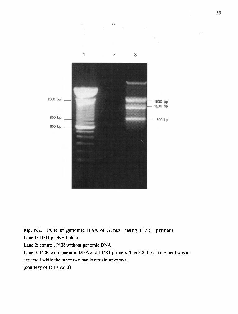
Fig. 8.2. PCR of genomic DNA of H.zea using FUR1 primers Lane 1: 100 bp DNA ladder.
Lane 2: control, PCR without genomic DNA.
Lane.3: PCR with genomic DNA and FUR1 primers. The 800 bp of fragment was as
expected while the other two bands remain unknown.
(courtesy of D.Persaud)

56 + 4
Since the corn earworm was raised on a diet containing wheat germ, plant virus
contamination may appear suggestive. However, the molecular data support the hypothesis that
the clone was derived from H. zta.
T o date, the only known wheat virus which can be transmitted by seed at very low
levels is wheat streak mosaic virus. Its thermal inactivation point is 54 OC. The longevity of the
infectivity of sap in vitro is 4-8 days (Brunt er al., 19%). This excludes the possibility of virus
surviving the dehydration process of manufacturing wheat germ or the process of making hiet
for H. zea, in which the ingredients wedmixed with boiling water. Indeed, when total RNA
was extracted from the wheat germ used for H. tea diet, no intact RNA has been found (data
not shown).
In addition, all of the RNA virus sequences found to be similar to the receptor (Table
6.1) lack p l y A regions in their 3' termini (Guilley et al., 1985; Rochon and Tremaine, 1989;
Lomrnel et al., 1991). Therefore, even had there been virus contamination, RT-PCR still
would not work with virus RNA when using the poly T primer for the reverse transcription i
reaction.
While plant viruses tend to have relatively narrow host ranges, rarely have insects
severed as vector. Within the arder Coleoptera, about 30 out of 55,000 species of plant-eating
beetles are known to transmit plant viruses. and each skc i e s feeds on a limited range% host
plants. Most vector species are found in the sub-families Galerucinase and Halticinae (flea-
beetles). H. zea belongs to Lepidopteran family in which transmission of plant virus has never
been reported.
The fact that plant viruses infect possible host plants for lepidopteran insects, however.
is intriguing, and the evolutionary implications would be interesting if it can be proven that the
cloned fragment indeed codes for the VHDL receptor, or any other protein associated with
receptor-medrated endocytosis of storage proteins.

T o complete this work, it would be necessary to construct a cDNA library from the H.
zea fat body, and use the fragment cloned in this thesis to obtain the full sequence of the
protein. Once expressed in vitro, the properties of the protein can be studied, e.g., by Western
blots or ligand blots, in order to confirm that it is the receptor. However, if the current clone
does not represent the receptor, it would be necessary to re-purify the receptor protein to obtain
additional internal sequence, for example by limited proteolysis with proteolytic enzymes. The
resulting sequences can then be used, in conjunction with the fragment sequences obtained in
this study, to obtain a more specific amplification product. In either case, the current study has
provided valuable information necessary to clone the entire sequence of the VHDL receptor
from H. zea.
58
References
Bean, D. and Silhacek, D. 1989 Changes in the titer of the female-predominant storage protein (8 1 K) during larval and pupal development of the way moth, Galleria mellonella. Arch. Insect Biochem. Physiol. 10:333-348
Beintema, J . J., Stam, W. T., Hazes, B. and Smidt, M. P. 1994 Evolution of arthropod hemocyanins and insect storage proteins (Hexamerins). Mol. Biol. Evol. 1 1:493-503
Birnboim, H.C. and Doly, J . 1979 A rapid alkaline extraction procedure for screening recombinant plasmid DNA. Nucleic Acids Res. 7: 15 13- 1523
Brunt, A.A., Crabtree, K., Dallwitz, M J . , Gibbs, A J . and Watson, L. 19% Viruses of Plants: descriptions and listsfrom the VIDE database. CAB International.
Burden, D. W. and Whitney , D.B. 1995 Biotechnology: proteins to PCR. Birkhauser press \
. b
Burmester, T. and Scheller, K. 1992 Identification of binding proteins involved in the stage- specific uptake of arylphorin by the fat body cells of Calliphora vicina. Insect Biochem. Mol. Biol. 22:211-220
Burmester, T . and ~che l le r , K. 1995a Ecdysteroid-mediated uptake of arylphorin by larval fat bodies of Calliphora vicina. Involvement and developmental regulation of arylphorin binding proteins. Insect Biochem. Mol. Biol. 25:799-806
Burmester, T. and Scheller, K. 1995b Complete cDNA sequence of the receptor responsible for arylphorin uptake by larval fat body of the blowfly Calliphora vicina. Insect Biochem. Mol. Biol. 25: 98 1-989
Chomcynski, P. and Sacchi, N. 1987 Single-step method for RNA isolation by acid guanidinum-thiocyanate-phenol-chloroform extraction. Anal. Biochem. 162: 156
Chung, S.O., Kubo,T. and Natori.S., 1995 Molecular cloning and sequencing of arylphorin- binding protein in protein granules of Sarcophaga fat body. J . Biol. Chem. 270:4624463 1
Dean, R.L; Collins, J.V and Locke, M. 1985 Insect storage protein. Comprehensive Insect Physiology, Biochemistry, and Pharmacology. (Gilbert, L.I., and Kerkut, G .A. eds) Pergamon Press. vol. 10. pp 307-346
Farrell, R.E. Jr., ed 1993 RNA Methodologies: A laboratory guide for isolation and characterization. Academic Press, New York
Flick, P. and Anson, J . 1995 Methods of RNA analysis. Editorial Comments. 22:l-7 Amersham LIFE SCIENCE.
Guilley, H.. Carrington, J .C., Balazs, E., Jonard, G . , Richards. K . and Morris. T.J . 1985 Nucleotide sequence and genome organization of carnation mottle virus RNA. Nuc.1. Acids Res. 1 3: 6663 -6677
Haunerland, N . H . and Bowers. 1V.S. 1986 Arylphorin from the corn earworm. Heliorhis x c l .
Ir~src-t Biochem. 16:6 17-625

Haunerland, N .H. a n d ' ~ o w e r s , W.S. 1986 A larval specific lipoprotein: purification and characterization of a blue chromoprotein from Heliothis zea. Biochem. Biophys. ResCommun. 1 34580-586
Haunerland, N.H. and Shirk, P.D. 1995 Regional and functional differentiation in the insect fat body. Annu. Rev. Entomol. 40: 12 1-45
Haunerland, N.H. 1996 Insect storage proteins: gene families and receptors. Imect Biochem. Mol. Biol. 26:755-765
Karpells, S., Leonard, D. and Kunkel, J . 1990 Cyclic fluctuations in arylphorin. the principal serum storage protein of Lymantria dispar. indicate multiple roles in development. Insect Biochem. 20:73-82
Kinnear, J . and Thomson, J . 1975 Nature, origin itnd fate of major haemolymph proteins in Calliphora . Insect Biochem. 5:53 1 -552
Konig, M., Agrawal, O., Schenkel, H. and Schelier, K. 1986 Incofporation of calliphorin into the cuticle of the developing blowfly, Calliphora vicina. Wilhelm Roux's Arch. Dev. Biol. 195:2%-301
Kramer, S ., Mundall, E. and Law, J . 1980 Purification and properties of manducin, an amino acid storage protein of the haemolymph of larval and pupal Manduca sexta. Insect Biochem. 10:279-288
Kunkel,.J., Grossniklaus-Buergin, C.. Karpells, S. and Lanzrein, B. 1990 Arylphorin of Trichoplusia ni: characterization and parasi te-induced precocious increase in titer. Arch. lmecr Biochem. Physiol. 1 3: 1 17- 125
Kyte, J. and Doolittle, R.F. 1982 A simple method for displaying the hydropathic character of a protein. J. Mol. Biol. 157: 105- 132
Jones, G., Ryan, R.O., Haunerland, N.H. and Law, J .H. 1988 Purification and characterization of a very high density chromolipoprotein from the hemolymph of . ichoplusis ni (Hubner). Arch. Insect Biochem. Physiol. 7: 1- 1 1 p Laemmli, U.K. 1970 Cleavage of structural proteins during the assembly of the head of bacteriophage T4. Nature 227:680-685
Levenbook. L. 1985 Insect storage protein. Comprehensive Insect Physiology. Biochemistry, and Pharmacology. (Gilbert, L. I . and Kerkut, G . A . eds) Pergamon Press
Levenbook, L. and Bauer, A . 1984 The fate of the laval storage protein calliphorin during adult development of Callinphora vicina. Insect Biochem. 14:77-86
Linzen. B. , Soeter. N . , Riggs. A.. Schneider. H.-J. and Schartau. W. et al. 1985 The structure of arthropod hemocyanins. Science 229:5 19-24
Locke. M . and Collins. J . 1968 Protein uptake into multivesicular bodies and storage granules i n the fat body of an insect. J . Cell. Biol. 36:353-483

Lommel, S.A., Kendall, T . L., Xiong, 2. and Nutter, R.C. 1991 Identification of the maize chlorotic mottle virus capsid protein cistron and characterization of its subgenomic messenger RNA. Virology 18 1 :382-385
Maschat, F., Dubertret, M.L., Thrond, P., Claverie, J.M. and Eepesant, J.A. 1990 Structure of the ecdysone inducible P1 gene of Drosophila melanogaster. J. Mol. Biol. 214:359-372
Matsudaira. P. 1990 Limited N-terminal sequence analysis. Methods in Enzymology 182:610- 613 d
Matthew, R. E. F. 1992 Fundamentals of Plant Vrology . ~ c a d e m i c Press, pp 289-307
McPherson. M J., Quirke, P. and Taylor, G. R. 1991 PCR: A practical approach. IRL Press
Meml, C. 1990 Get-staining techniques. Methods in Enzymology 1 8 2 : 4 8 2 4 8
Munn, E. and Greville, G . 1969 The soluble proteins of developing Calliphora erythrocephala, particularly calliphorin, and similar proteins in other insects. J. Insect Physiol. 15: 1935-1950
Munn, E., Price, G . and Greville, G. 1%9 The synthesis in vitro of the protein calliphorin by fat body from the larva of the blowfly, Calliphora erythrocephala. J. Insect Physiol. 15:1601-5
Palli, S . and Locke, M. 1987 Purification and characterization of three major hemolymph proteins of an insect, Calpodes erhlius (Lepid0ptera:Hesperiidae). Arch. Insect Biochem. Physiol. 5:233-244
Pan, M. and Telfer, W.H. 1992 Selectivity in storage hexamerin clearing demonstrated with hemolymph transfusions between Hvalophora cecropia and Act& h a . Arch. Insect Biochem. Physiol. l9:203-2 1
Patana, R . and McAda, W.C. 1973 Tobacco budworm: use of dry diet flakes in rearing.J Econ. Entomol. 66:817-818
Rochon, D.M. and Tremaine, J.H. 1989 Complete nucleotide sequence of the cucumber necrosis virus genome.Virology 169:25 1-259
Ryan, R. , Keim, P. , Wells, M. and Law, J. 1985 Purification and properties of a predominantly female-specific protein from the hemolymph of the larva of the tobacco hornworm, Manduca sexta. J. Biol. Chem. 260:782-787
Ryan, R., Wang, X . , Willott. E. and Law. J . 1986 Major hemolymphproteins from larvae of the black swallowtail butterfly, Papilio pol>l.\-enes. Arch. Insect Biochem. Phy.sio1. 3:539-550
Rychlik. W . and Rhoads, R. E . 1989 A computer program for choosing optimal oligonucleotides for filter hybridization, sequencing and in virro amplification of D N A . ,L'uclt.ic Acids Res. 17:8543-855 1 4
Rychlik. W . , Spencer. W . J . and Rhoads, R. E . 1990 Optimization of the annealing temperature for DNA amplification in virro. N~rcleic Acids Res. 18:6309-6412

Scheller, K., Zimmerman, H. P. and Sekeris, C. 1980 Calliphorin, a protein involved in the . cuticle formation of the blowfly, Calliphora vicina. 2. Naturforsch. 35c:387-389
Schenkel, H. and Scheller, K. 1986 Stage- and tissue-specific expression of genes encoding calliphorin, the major larval serum protein of Calliphora vicina. Wilhelm Roux's Arch. Dev. Biol. 195:290-95
Sehnal 1985. Comprehensive Insect Physiology, Biochemistry, and Pharmacology (Gilbert, L.I., and Kerkut, G. A . eds) Pergamon Press. 3: 1-86
Telfer, W., Keim, P. and Law, J. 1983 Arylphorin, a new protein from Hyalaphora cecropia: Comparisons with calliphorin and manducin. Insect Biochem. 13:601-13
Telfer, W.H. and Kunkel, J.G. 1991 The function and evolution of insect storage hexamers. Annu. Rev. Entomol. 36:205-228
Tojo, S., Betchaku, T., Ziccardi, V. and Wyatt, G . 1978 Fat body protein granules and storage proteins in the sil krnoth, Hyalophora cecropia. J. Cell Biol. 78:823-838
Tojo, S. , Nagata, M. and Kobayashi, M. 1980 Storage proteins in the silkworm, Bombyx mori. Insect Biochem . 10: 289-303
* Towbin, H., Staehelin, T. , and Gordon, J . 1979 Electrophoretic transfer of proteins from polyacry lamide gels to nitrocellulose sheets: procedure and some applications. Proc. Nat . Acad. Sci. USA, 76:4350
Ueno. K.. Oshawa. F.. and Natori. S. 1983 Identification and activation of storage motein receptor of Sarcophaga peregrina' fat body by 20-hydroxyecdysone. J. BioT 'Chem. 258: 12210-14
Ueno, K. and Natori. S . 1984 Identification of storage protein receptor and its precursor in the fat body membrane of Sarcophaga peregrina. J. Biol. Chem. 259: 12107- 1 1 1
Ueno, K.and Natori, S. 1987 Possible involvement of lumichrome in the binding of storage protein to its receptor in Sarcophaga peregrina. J. Biol. Chem. 262: 12780-784
VanHolde, K . and Miller, K. 1982 Hemocyanins. Q. Rev. Biophys. 15:l-12
Wada, K . , Aota, S., Tsuchiya, R. , Ishibashi, F., Gojobori, T. and Ikemura, T. 1990 Codon usage tabulated from the GenBank genetic sequence data. N~ccleic Acids Research, Vol. 18. Supplement 2367-24 10
Wang, Z. and Haunerland, N.H. 1991 Ultrastructural study of storage protein granules in fat body of the corn ear worm Heliothis zea. J. Insect Ph~,siol. 37:353-363
Wang, Z. and Haunerland, N.H. 1992 Fate of differentiated fat body tissue during metamophosis of Helicor!erpa zru. J. lnsrcr Ph~riol . 38: 199-2 13
U'nng. Z. and Hauneriand. N.H. 1993 Storage protein uptake in Hrlicor~erpcl x u . Purification of the very high density lipoprotein receptor from perivisceral fat body. J. Biol. Chrtn . 268: 16673-78

Wang, Z.and Haunerland, N.H. 1994 Storage protein uptake in Helicoverpa zea fat body. A single receptor mediates the uptake of arylphorimn and VHDL. Arch. Inrect Biochem .Physiol. 26: 15-26
Wang, 2. and Haunerland, N.H. 1994b Receptor-mediated endocytosis o f storage proteins by the fat body of Helicoverpa zea. Cell Tissue Res. 278: 107-1 15