Clinicopathological study ofmyoepithelial sialadenitis ... · andchronicsialadenitis...
7
J Clin Pathol 1988;41:403-409 Clinicopathological study of myoepithelial sialadenitis and chronic sialadenitis (sialolithiasis) G M KONDRATOWICZ,* LESLEY A SMALLMAN, D W MORGANt From the *Department of Pathology, The Medical School, The University of Birmingham, and tthe Department of Otorhinolaryngology, The Queen Elizabeth Hospital, Birmingham SUMMARY To determine any overlap in pathological features between myoepithelial sialadenitis and chronic sialadenitis/sialolithiasis histological sections from 69 cases of myoepithelial sialadenitis (MESA) (n = 7) and chronic sialadenitis/sialolithiasis (n = 62) were reviewed over a 10 year period. Three of the cases with MESA contained calculi and four of those originally diagnosed as chronic sialadenitis/sialolithiasis showed epimyoepithelial island formation. The presence of calculi should not rule out a diagnosis of MESA, particularly in the parotid gland where calculi are uncommon; as the incidence of MESA may very well be underestimated and diagnosed as chronic sialadenitis, these patients, who are at increased risk of developing lymphoma, could be lost to follow up. Myoepithelial sialadenitis (MESA) is characterised by the formation of epimyoepithelial islands accompan- ied by chronic lymphocytic infiltration of the salivary parenchyma and often glandular atrophy. It has largely superceded the earlier used term, benign lym- phoepithelial lesion, which was coined by Godwin in 1952.12 Although the histological picture of well established MESA is usually so characteristic that there is little difficulty distinguishing it from other types of sialadenitis, we recently saw a case of MESA in which the excised parotid gland tissue contained several calcified laminated calculi situated within dilated ducts. This feature, which initially prompted some diagnostic confusion, led us to review all the cases of MESA and chronic sialadenitis/sialolithiasis from our files seen over the past 10 years. The aim of this study was to determine the incidence of calculi in cases diagnosed as MESA and the incidence of epimyoepithlial islands in otherwise typical chronic sialadenitis. We were particularly interested in cases which may represent the development of MESA in pre-existing chronic sialadenitis and the possibility of any overlap between these two types of disease. Material and methods Cases diagnosed as chronic sialadenitis, sialolithiasis, MESA, Sj6gren's syndrome, or lymphoma were Accepted for publication 14 October 1987 403 obtained from the files of the department of path- ology, University of Birmingham and the department of histopathology, General Hospital, Birmingham. Sections stained with haematoxylin and eosin were reviewed and the presence or absence of epimyoepith- elial islands, ductal infiltration by lymphoid cells, and calcified material noted. Other features assessed were the prominence of lobular lymphoid infiltration, presence or absence of germinal centres, intra-acinar and ductal acute inflammatory cells, lobular and periductal fibrosis and atrophy, and epithelioid gran- uloma formation. Interval sections were cut in those cases showing epimyoepithelial island formation. The case notes of patients diagnosed as having MESA or chronic sialadenitis/sialolithiasis, in which epimyoepithelial islands were found were reviewed (table). Results Histological sections from a total of 73 salivary glands (56 submandibular, 17 parotid) were reviewed. Two submandibular glands and six parotid glands had been reported as MESA or Sjogren's syndrome. This included one patient with two excisions over a period of several years. Chronic sialadenitis/sialolithiasis had been diagnosed in 54 submandibular glands and eight parotid glands; calculi were noted macroscopically, or microscopically, or a definite clinical history of sialolithiasis had been elicited in 29 (53%) of subman- dibular glands and three (38%) of parotid glands. Of the remaining three salivary glands, one was clearly a on 16 June 2019 by guest. Protected by copyright. http://jcp.bmj.com/ J Clin Pathol: first published as 10.1136/jcp.41.4.403 on 1 April 1988. Downloaded from
Transcript of Clinicopathological study ofmyoepithelial sialadenitis ... · andchronicsialadenitis...

J Clin Pathol 1988;41:403-409
Clinicopathological study of myoepithelial sialadenitisand chronic sialadenitis (sialolithiasis)G M KONDRATOWICZ,* LESLEY A SMALLMAN, D W MORGANt
From the *Department ofPathology, The Medical School, The University of Birmingham, and tthe DepartmentofOtorhinolaryngology, The Queen Elizabeth Hospital, Birmingham
SUMMARY To determine any overlap in pathological features between myoepithelial sialadenitis andchronic sialadenitis/sialolithiasis histological sections from 69 cases of myoepithelial sialadenitis(MESA) (n = 7) and chronic sialadenitis/sialolithiasis (n = 62) were reviewed over a 10 year period.Three of the cases with MESA contained calculi and four of those originally diagnosed as chronicsialadenitis/sialolithiasis showed epimyoepithelial island formation. The presence of calculi shouldnot rule out a diagnosis of MESA, particularly in the parotid gland where calculi are uncommon; as
the incidence ofMESA may very well be underestimated and diagnosed as chronic sialadenitis, thesepatients, who are at increased risk of developing lymphoma, could be lost to follow up.
Myoepithelial sialadenitis (MESA) is characterised bythe formation of epimyoepithelial islands accompan-ied by chronic lymphocytic infiltration of the salivaryparenchyma and often glandular atrophy. It haslargely superceded the earlier used term, benign lym-phoepithelial lesion, which was coined by Godwin in1952.12Although the histological picture ofwell established
MESA is usually so characteristic that there is littledifficulty distinguishing it from other types ofsialadenitis, we recently saw a case ofMESA in whichthe excised parotid gland tissue contained severalcalcified laminated calculi situated within dilatedducts. This feature, which initially prompted somediagnostic confusion, led us to review all the cases ofMESA and chronic sialadenitis/sialolithiasis from ourfiles seen over the past 10 years. The aim of this studywas to determine the incidence of calculi in casesdiagnosed as MESA and the incidence ofepimyoepithlial islands in otherwise typical chronicsialadenitis. We were particularly interested in caseswhich may represent the development of MESA inpre-existing chronic sialadenitis and the possibility ofany overlap between these two types of disease.
Material and methods
Cases diagnosed as chronic sialadenitis, sialolithiasis,MESA, Sj6gren's syndrome, or lymphoma were
Accepted for publication 14 October 1987403
obtained from the files of the department of path-ology, University of Birmingham and the departmentof histopathology, General Hospital, Birmingham.Sections stained with haematoxylin and eosin werereviewed and the presence or absence of epimyoepith-elial islands, ductal infiltration by lymphoid cells, andcalcified material noted. Other features assessed werethe prominence of lobular lymphoid infiltration,presence or absence of germinal centres, intra-acinarand ductal acute inflammatory cells, lobular andperiductal fibrosis and atrophy, and epithelioid gran-uloma formation. Interval sections were cut in thosecases showing epimyoepithelial island formation. Thecase notes of patients diagnosed as having MESAor chronic sialadenitis/sialolithiasis, in whichepimyoepithelial islands were found were reviewed(table).
Results
Histological sections from a total of 73 salivary glands(56 submandibular, 17 parotid) were reviewed. Twosubmandibular glands and six parotid glands had beenreported as MESA or Sjogren's syndrome. Thisincluded one patient with two excisions over a periodof several years. Chronic sialadenitis/sialolithiasis hadbeen diagnosed in 54 submandibular glands and eightparotid glands; calculi were noted macroscopically, ormicroscopically, or a definite clinical history ofsialolithiasis had been elicited in 29 (53%) of subman-dibular glands and three (38%) of parotid glands. Ofthe remaining three salivary glands, one was clearly a
on 16 June 2019 by guest. Protected by copyright.
http://jcp.bmj.com
/J C
lin Pathol: first published as 10.1136/jcp.41.4.403 on 1 A
pril 1988. Dow
nloaded from
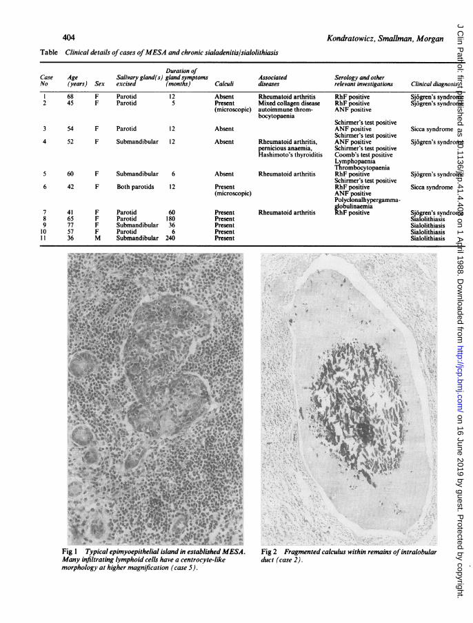
Table Clinical details ofcases ofMESA and chronic sialadenitis/sialolithiasis
Duration ofCase Age Salivary gland(s) gland symptomsNo (years) Sex excised (months)
1 68 F Parotid 122 45 F Parotid 5
3 54 F Parotid 12
4 52 F Submandibular 12
5 60 F Submandibular 6
6 42 F Both parotids 12
7 41 F Parotid 608 65 F Parotid 1809 77 F Submandibular 3610 57 F Parotid 611 36 M Submandibular 240
Associated Serology and otherCalculi diseases relevant investigations Clinical diagnosis
Absent Rheumatoid arthritis RhF positive Sj6gren's syndromePresent Mixed collagen disease RhF positive Sj6gren's syndrome(microscopic) autoimmune throm- ANF positive
bocytopaeniaSchirmer's test positive
Absent ANF positive Sicca syndromeSchirmer's test positive
Absent Rheumatoid arthritis, ANF positive Sj6gren's syndromepernicious anaemia, Schirmer's test positiveHashimoto's thyroiditis Coomb's test positive
LymphopaeniaThrombocytopaenia
Absent Rheumatoid arthritis RhF positive Sj6gren's syndromeSchirmer's test positive
Present RhF positive Sicca syndrome(microscopic) ANF positive
Polyclonalhypergamma-globulinaemia
Present Rheumatoid arthritis RhF positive Sj6gren's syndromePresentPresentPresentPresent
SialolithiasisSialolithiasisSialolithiasisSialolithiasis
14.
'* .
W.-'-*.a
AS - 4Y a a5
Fig I Typical epimyoepithelial island in established MESA. Fig 2 Fragmented calculus within remains ofintralobularMany infiltrating lymphoid cells have a centrocyte-like duct (case 2).morphology at higher magnification (case 5).
404 Kondralowicz, Smallman, Morgan
on 16 June 2019 by guest. Protected by copyright.
http://jcp.bmj.com
/J C
lin Pathol: first published as 10.1136/jcp.41.4.403 on 1 A
pril 1988. Dow
nloaded from

Clinicopathological study ofmyoepithelial sialadenitis and chronic sialadenitis (sialolithiasis)
_. A.
A.%A%_
.-Le, , ,:- 4
Fig 3 Dilated duct denselv infiltrated by lymphoid cells and containing calcified material (case 6).
granulomatous sialadenitis, probably sarcoidosis, andin the two others there was a history oftrauma or localtumour resection.The mean age of patients with MESA was 59 years
(range 43-68 years); all were women (table).Epimyoepithelial islands were found in all eight cases(fig 1). In addition, calculi were present in three (38%)cases, one of which was noted macroscopically on
slicing glandular tissue as a 0-2 cm stone (case 7). In theother two cases calcified material was noted withindilated ducts on histological examination (fig 2). Apartfrom epimyoepithelial islands, an important his-tological feature was the presence of dilated ductsinfiltrated by lymphoid cells. This was seen in six oftheeight cases (75%) and was pronounced in three ofthese. In those cases containing calcified material thiswas characteristically within dilated ducts of this type(fig 3). A dense chronic lymphoid infiltrate was alsopresent around ducts and within lobules, but in mostcases occasional lobules were largely spared or com-
pletely devoid of infiltration. Germinal centre forma-tion varied from abundant to absent, but was presentin most cases (75%). No cases of salivary lymphomahad been seen over the 10 year period.The mean age of patients in the chronic sialadenitis/
sialolithiasis group was 44-6 years (range 13-77) forthe submandibular cases, and 57 8 years (48-65) for
the parotid cases. Male to female ratios were about 1:1and 2:3, respectively. A review of these cases showedthat epimyoepithelial islands were found in two (4%)of the submandibular glands and in two (25%) of theparotid glands. Calculi were identified either macro-scopically or microscopically in all four cases (table).
Epimyoepithelial islands were small and sparse inthese four cases but more typical examples wereusually identified by interval sections through thetissue blocks (figs 4-6). A lymphoid infiltrate in theepithelium of dilated ducts as seen in MESA waspresent in both parotid tissue samples but not insubmandibular tissue; otherwise, lymphoid infiltra-tion of ductal epithelium was conspicuously absent inthe cases of chronic sialadenitis/sialolithiasis.
Occasional intraductal lymphoid cells were alsopresent as part of a granulomatous infiltrate in two offour cases of chronic sialadenitis in which sparseepithelioid granulomas were noted. Acute inflam-matory cells within ducts or acini were rare in thepresence of epimyoepithelial islands, but particularlycommon in those cases of chronic sialadenitis withdefinite calculi (41%). A prominent lobular lymphoidinfiltrate was present in seven of 54 (13%) subman-dibular glands, previously reported as being chronicsialadenitis/sialolithiasis, but this was usually lessprominent than in the cases of MESA.
*
d* .
1--we -v- I.
40)5-Inimqw , .-. ... , I
,-
'i .4i 4 0
i N,I._t
L9,.W
on 16 June 2019 by guest. Protected by copyright.
http://jcp.bmj.com
/J C
lin Pathol: first published as 10.1136/jcp.41.4.403 on 1 A
pril 1988. Dow
nloaded from

Kondratowicz, Smallman, Morgan
Fig 4 Epimyoepithelial island infiltrated by lymphoid cells in patient with clinical diagnosis ofsialolithiasis (case 9).
In the parotid gland specimens this feature wasprominent only in those cases containingepimyoepithelial islands. Germinal centre formationwas present in 42% ofcases ofchronic sialadenitis andwas occasionally of a degree comparable with thatseen in typical MESA. Focal fibrosis, both lobular andperiductal, was common in cases of chronic siala-denitis/sialolithiasis and often present in those withMESA.
Discussion
Although the eye catching feature of epimyoepithelialisland formation makes the histological diagnosis ofMESA quite straightforward, it is a histopathologicalterm without a precise clinical counterpart. The clin-ical setting in which MESA occurs is variable and theterminology confusing.3 Many cases occur in thecontext of Sj6grens's syndrome, a triad of keratocon-junctivitis sicca, xerostomia, and an underlying dis-ease of connective tissue-most often rheumatoidarthritis.4 An incomplete, primary (or limited) form ofSjogren's syndrome without an accompanyingautoimmune disease is also recognised and commonlyreferred to as the sicca syndrome. This can also be partof a "sicca systemic syndrome" in which several otherexocrine gland systems are affected and extraglandular
manifestations can occur. The exact relation betweenthis primary form and the classic secondary form is notfully resolved, but is considered to be a distinct clinicalentity and more common than the latter.SThe clinical manifestations of dry eyes and dry
mouth are secondary to the degree of inflammatorychanges occurring in gandular tissue and may bedifficult to show in mild or early disease. Some workerssuggest that xerostomia should be substituted by themore objective criterion offocal sialadenitis in a minorsalivary gland biopsy specimen.6 In practice, manycases present as salivary gland swelling in a patientwith confirmed connective tissue disease. In our seriesfour of the cases diagnosed as MESA occurred inassociation with rheumatoid arthritis and one withmixed collagen disease; these cases thus fell into theclassic secondary category of Sjogren's syndrome. Theother two could be regarded as examples of theprimary form.Although there was definite evidence of calculi in
apparently half of the cases of chronic sialadenitis/sialolithiasis, this was probably a considerableunderestimate as many stones are passed spontan-eously or are not received by the pathologist. A recentstudy suggests that at least 82% of cases of chronicsialadenitis are related to calculi.7 The four patientswith an original diagnosis of sialadenitis/sialolithiasis,
406
on 16 June 2019 by guest. Protected by copyright.
http://jcp.bmj.com
/J C
lin Pathol: first published as 10.1136/jcp.41.4.403 on 1 A
pril 1988. Dow
nloaded from

Clinicopathological study ofmyoepithelial sialadenitis and chronic sialadenitis (sialolithiasis)
Fig 5 Another epimyoepithelial island in a similar case
(case 10).
in whom epimyoepithelial islands were found on
review, had no pointers towards any underlyingconnective tissue disease (although serology had notbeen carried out) and all four had documented calculi.Although our series is small, it suggests that calculus
formation is not uncommon in otherwise typicalMESA (three of eight cases). All the calculi identifiedwere small and multiple, and in only one case wascalcification noted macroscopically, necessitatingdecalcification of tissue. Thorough sampling of tissuein other cases would probably have shown more
examples of microscopic calcification. Generally, cal-culi of the parotid gland are said to be rare.8 Thereasons for this probably include the downward slopefollowed by Stensen's duct and the relatively lowviscosity of parotid secretions. This paucity of parotidcalculus formation is reflected in our series: after casesdiagnosed as MESA are subtracted only seven parotidglands remain showing features of chronic sialaden-itis/sialolithiasis. Two of these on review contained
Fig 6 Early epimyoepithelial islandformation in salivaryduct (case 8).
scarce epimyoepithelial islands and large dilated ductsinfiltrated by lymphoid cells. In view of this a carefulsearch for features of MESA seems indicated in thepresence of calculi at this site.
Dilated ducts showing variable degrees oflymphoidinfiltration have often been noted in cases of MESAand account for the sialoectasia often seenradiologically5 (fig 7). Almost all our cases of MESAcontained ductal lesions of the type and it is morelikely that the calculi noted in our cases were the resultof calcification of inspissated secretions in such abnor-mal ducts rather than MESA developing on a back-ground of pre-existing sialolithiasis. Ductal lymphoidcell infiltrates, other than the very occasionalintraepithelial cell, were not seen in any of the casesinitially diagnosed as chronic sialadenitis/sialolith-iasis, with the exception of two of the cases in whichepimyoepithelial islands were also found and twocases in which small numbers of lymphoid cells were
present as part of a granulomatous infiltrate. Gran-
407llq'. ,
_S4
on 16 June 2019 by guest. Protected by copyright.
http://jcp.bmj.com
/J C
lin Pathol: first published as 10.1136/jcp.41.4.403 on 1 A
pril 1988. Dow
nloaded from

Kondratowicz, Smallman, Morgan
Fig 7 Sialogram showing sialoectasia in case ofMESA.
ulomas have recently been described as an occasionalfeature of chronic sialolithiasis and are thought to bethe result of duct rupture.9
It is difficult to categorise precisely the four cases inwhich epimyoepethilial islands were found in a clinicalcontext of sialolithiasis. Two of these occurred in theparotid glands of women aged 57 and 65 years. Onehad multiple small stones present in Stensen's duct andintraglandular ducts, the other a larger calculus inStensen's duct. Infiltrated dilated ducts as well assparse epimyoepithelial islands were present. Thesecases most probably represent mild or early examplesof MESA and would best fit into the clinical categoryof primary Sjdgren's syndrome in which the dominantclinical picture of sialolithiasis may have been coin-cidental or the result of calcification of inspissatedsecretions in dilated ducts. These women may developfull blown Sj6gren's syndrome. Epimyoepithelialislands without dilated duct lesions were noted in thesubmandibular glands of two other patients, one, awoman of 77 years, had a radiologically confirmedstone in Stenson's duct. Interestingly, she also hadclinical enlargement ofher other submandibular glandand both parotid glands, again suggesting the pos-sibility of more widespread salivary disease. The onlymale patient in the group (aged 36 years) had undoub-ted well formed epimyoepithelial islands surroundedby a dense lymphoid infiltrate within a gland whichshowed atrophy of occasional lobules but was other-wise unremarkable (fig 8). This case is closest torepresenting an example of the very rare occurrence ofepimyoepithelial islands in otherwise straightforwardsialolithiasis.3
Epimyoepithelial islands occurred in our series withone exception-in middle aged and elderly women, a
* ar -'-, S
Fig 8 Small epimyoepithelial island surrounded by denselymphoid infiltrate in male patient with long history ofsialolithiasis. Much of the surrounding tissue is normal(case 11).
group particularly prone to autoimmune disease. It isinteresting to speculate that even in the absence ofclinically overt MESA those lesions found incidentallyin salivary glands in the clinical context of sialolithiasisare likely to represent an "autoimmune phenomenon"and their relation to full blown Sj6gren's syndromemay parallel that of focal lymphocytic thyroiditis toHashimoto's thyroiditis. In both these entities charac-teristic glandular epithelial changes occur to a lesser orgreater extent and are associated with other organspecific autoimmune diseases."Our study shows that the presence of calculi should
certainly not rule out a diagnosis of MESA, par-ticularly in the parotid gland, where calculi aregenerally uncommon, a careful search should be madefor epimyoepithelial islands and ducts infiltrated bylymphoid cells. Multiple sections may be helpful in thisrespect as these lesions may be very sparse. The overall
408
on 16 June 2019 by guest. Protected by copyright.
http://jcp.bmj.com
/J C
lin Pathol: first published as 10.1136/jcp.41.4.403 on 1 A
pril 1988. Dow
nloaded from

Clinicopathological study ofmyoepithelial sialadenitis and chronic sialadenitis (sialolithiasis) 409
incidence of MESA is probably underestimated assome cases are probably labelled as chronic sialaden-itis. Such patients would be lost to follow up and maybe at a slightly increased risk of developing a lym-phoma.22
We are grateful to Mr M A Atkins for technicalassistance, Mr A A Cooper for help with thephotographs, and Mrs I F Pring for typing themanuscript.
References
I Godwin T. Benign lymphoepithelial lesion of the parotid gland.Cancer 1952;5: 1089-103.
2 Schmid V, Helborn D, Lennert K. Development of malignantlymphoma in myoepithelial sialadenitis (Sjogren's syndrome).Virchows Arch (Pathol Anat) 1982;395:1 1-43.
3 Ferlito A, Cattai N. The so-called "benign lymphoepitheliallesion". (Part 1. Explanation of the term and of its synonymousand related terms). J Laryngol Otol 1980;94:1189-97.
4 Batsakis JG. The pathology of head and neck tumours. Thelymphoepithelial lesion and Sj6gren's syndrome, part 16. Head
Neck Surg 1982;5:150-63.5 Ostberg Y. The clinical picture of benign lymphoepithelial lesion.
Clin Otolaryngol 1983;8:381-90.6 Daniels TE. Labial salivary gland biopsy in Sj6gren's syndrome.
Assessment as a diagnostic criterion in 362 suspected cases.Arthritis Rheum 1984;27: 147-56.
7 Isacsson G, Lundquist P-G. Salivary calculi as an aetiologicalfactor in chronic sialadenitis of the submandibular gland. ClinOtolaryngol 1982;7:231-6.
8 Czernwinski F, Zielinska B. Kamica Mlinianki przyusznej. WiadLek 1983:36:493-6.
9 Van der Walt JD, Leake J. Granulomatous sialadenitis of themajor salivary glands. A clinicopathological study of 57 cases.Histopathology 1987;11:131-44.
10 Thackray AC, Lucas RB. Tumours of the major salivarv glands.Fascicle 10. Washington, DC: Armed Forces Institute of Path-ology, 1974.
11 Doniach I. The thyroid gland. In: W St C Symmers, eds. Systemicpathology. 2nded. Edinburgh: Churchill Livingstone, 1978:197%2037.
12 Isaacson PG, Spencer J. Malignant lymphoma of mucosaassociated lymphoid tissue. Histopathology 1987;1 1:445-62.
Requests for reprints to: Dr G M Kondratowicz, Lecturer inPathology, The Medical School, University of Birmingham,Birmingham B15 2TJ, England.
on 16 June 2019 by guest. Protected by copyright.
http://jcp.bmj.com
/J C
lin Pathol: first published as 10.1136/jcp.41.4.403 on 1 A
pril 1988. Dow
nloaded from