Clinical Virology in NICU, PICU and AICU 20
20
20 Clinical Virology in NICU, PICU and AICU C. Y. W. Tong and S. Schelenz 20.1 Introduction Viruses are significant causes of nosocomial infections, but their importance has been underappreciated in the past. However, outbreak of severe acute respiratory syndrome (SARS), avian and pandemic influenza with high morbidity and mortality rates increased the awareness of intensivists regarding the devastating effects of nosocomial spread of viral infections in intensive care units (ICU). Advances in medicine have led to a large number of immunocompromised patients susceptible to severe viral infections. Many such patients are cared for in the ICU and in turn become an infectious hazard for other vulnerable patients. Health care workers can acquire common viral infections from the community and spread them to susceptible patients in the ICU. Patients in neonatal (NICU) and paediatric (PICU) ICUs are most vulnerable because of the lack of prior immunity against many viruses circulating in the community. Recent improvements in diagnostic methods have enabled the rapid diagnosis and monitoring of many viral infections. Rapid and accurate typing of viral strains using a molecular technique can help identify the source of outbreaks. Also, specific postexposure prophylaxis and treatment are now available for many important nosocomial viral infections. In this chapter, we discuss some of the important viruses that could be associated with nosocomial infections in the ICU, according to their usual route of trans- mission (Table 20.1). Infection control measures recommended for preventing these viral infections are listed in Table 20.2. C. Y. W. Tong (&) Infection, Guy’s and St Thomas’ NHS Foundation Trust and King’s College London School of Medicine, London, UK e-mail: [email protected] H. K. F. van Saene et al. (eds.), Infection Control in the Intensive Care Unit, DOI: 10.1007/978-88-470-1601-9_20, Ó Springer-Verlag Italia 2012 333
Transcript of Clinical Virology in NICU, PICU and AICU 20

20Clinical Virology in NICU, PICUand AICU
C. Y. W. Tong and S. Schelenz
20.1 Introduction
Viruses are significant causes of nosocomial infections, but their importance hasbeen underappreciated in the past. However, outbreak of severe acute respiratorysyndrome (SARS), avian and pandemic influenza with high morbidity andmortality rates increased the awareness of intensivists regarding the devastatingeffects of nosocomial spread of viral infections in intensive care units (ICU).
Advances in medicine have led to a large number of immunocompromisedpatients susceptible to severe viral infections. Many such patients are cared for inthe ICU and in turn become an infectious hazard for other vulnerable patients.Health care workers can acquire common viral infections from the community andspread them to susceptible patients in the ICU. Patients in neonatal (NICU) andpaediatric (PICU) ICUs are most vulnerable because of the lack of prior immunityagainst many viruses circulating in the community. Recent improvements indiagnostic methods have enabled the rapid diagnosis and monitoring of many viralinfections. Rapid and accurate typing of viral strains using a molecular techniquecan help identify the source of outbreaks. Also, specific postexposure prophylaxisand treatment are now available for many important nosocomial viral infections.In this chapter, we discuss some of the important viruses that could be associatedwith nosocomial infections in the ICU, according to their usual route of trans-mission (Table 20.1). Infection control measures recommended for preventingthese viral infections are listed in Table 20.2.
C. Y. W. Tong (&)Infection, Guy’s and St Thomas’ NHS Foundation Trustand King’s College London School of Medicine, London, UKe-mail: [email protected]
H. K. F. van Saene et al. (eds.), Infection Control in the Intensive Care Unit,DOI: 10.1007/978-88-470-1601-9_20, � Springer-Verlag Italia 2012
333
20.2 Viruses Transmitted by Droplets and Airborne Route
20.2.1 Influenza Viruses
Influenza viruses (family Orthomyxoviridae) are classified into types A, B and C.Annual seasonal outbreaks of influenza are caused by minor antigenic changes(antigenic drift) seen in influenza A and B viruses. Major changes in antigenicsubtypes (antigenic shift) are only found in influenza A virus and typically involvethe emergence of novel hemagglutinin (H) and/or neuraminidase (N) proteins onthe viral envelope. Pandemic influenza occurs when a new influenza A strainemerges, to which the majority of the world’s population has little or no immunity.
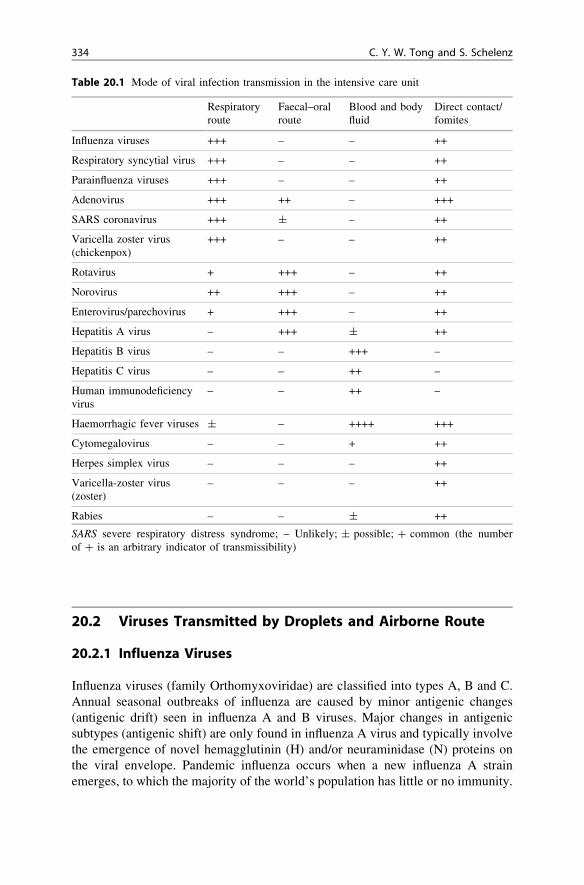
Table 20.1 Mode of viral infection transmission in the intensive care unit
Respiratoryroute
Faecal–oralroute
Blood and bodyfluid
Direct contact/fomites
Influenza viruses +++ – – ++
Respiratory syncytial virus +++ – – ++
Parainfluenza viruses +++ – – ++
Adenovirus +++ ++ – +++
SARS coronavirus +++ ± – ++
Varicella zoster virus(chickenpox)
+++ – – ++
Rotavirus + +++ – ++
Norovirus ++ +++ – ++
Enterovirus/parechovirus + +++ – ++
Hepatitis A virus – +++ ± ++
Hepatitis B virus – – +++ –
Hepatitis C virus – – ++ –
Human immunodeficiencyvirus
– – ++ –
Haemorrhagic fever viruses ± – ++++ +++
Cytomegalovirus – – + ++
Herpes simplex virus – – – ++
Varicella-zoster virus(zoster)
– – – ++
Rabies – – ± ++
SARS severe respiratory distress syndrome; – Unlikely; ± possible; ? common (the numberof ? is an arbitrary indicator of transmissibility)
334 C. Y. W. Tong and S. Schelenz

Ta
ble
20
.2In
fect
ion
cont
rol
mea
sure
sfo
rpr
even
ting
vira
lin
fect
ions
Vir
usIs
olat
ion
orco
hort
ing
Han
dw
ashi
ngA
pron
/go
wn+
Glo
ves
Mas
ks/
gogg
les
Incu
bati
onD
urat
ion
ofin
fect
ivit
y
Infl
uenz
avi
ruse
s4
(neg
ativ
epr
essu
re)
IB4
44
4IB
1–4
days
Pro
drom
alph
ase
and
7da
ysaf
ter
onse
t
Res
pira
tory
sync
ytia
lvi
ruse
s(R
SV
)
4II
4IA
4IB
4IA
2–8
days
48h
befo
resy
mpt
oms
and
7da
ysfr
omon
set;
long
erin
imm
unoc
ompr
omis
ed(u
pto
30da
ys)
Par
ainfl
uenz
avi
ruse
s4
44
42–
4da
ysA
slo
ngas
sym
ptom
sla
st
Ade
novi
rus
44
44
5–10
days
As
long
assy
mpt
oms
last
SA
RS
coro
navi
rus
4(n
egat
ive
pres
sure
)IA
44
44
(FF
P3
orN
95)
IB
4–6
days
(max
.re
port
ed14
days
)
Pea
kat
day
10of
illn
ess,
nore
port
edtr
ansm
issi
on10
days
beyo
ndre
solu
tion
offe
ver
Var
icel
la-z
oste
rvi
rus
(chi
cken
pox)
4(n
egat
ive
pres
sure
)IB
44
410
–21
days
2da
ysbe
fore
firs
tve
sicl
eun
til
all
lesi
ons
are
crus
ted
Rot
avir
us4
44
42–
3da
ys2
days
befo
resy
mpt
oms
and
upto
4–7
days
afte
ron
set
ofil
lnes
s
Nor
ovir
us4
IB4
IA4
IB4
IB±
15–4
8h
Up
to48
haf
ter
beco
min
gsy
mpt
omfr
ee
Ent
erov
irus
/pa
rech
ovir
us4
(you
ngin
fant
sof
ten
requ
ire
care
inS
CB
Uor
NIC
U)
44
42–
25da
ys7–
14da
ysfr
omon
set
ofil
lnes
s;as
ympt
omat
icsh
eddi
ngco
mm
on
Hep
atit
isA
viru
s4
44
42–
6w
eeks
Infe
ctio
us1
wee
kbe
fore
onse
tof
illn
ess,
infe
ctiv
ity
decl
ines
rapi
dly
afte
ron
set
ofil
lnes
s
Hep
atit
isB
viru
s4
42–
3m
onth
sA
slo
ngas
pati
ent
isvi
rem
ic(c
onti
nued
)
20 Clinical Virology in NICU, PICU and AICU 335

Ta
ble
20
.2(c
onti
nued
)
Vir
usIs
olat
ion
orco
hort
ing
Han
dw
ashi
ngA
pron
/go
wn+
Glo
ves
Mas
ks/
gogg
les
Incu
bati
onD
urat
ion
ofin
fect
ivit
y
Hep
atit
isC
viru
s4
42–
3m
onth
sA
slo
ngas
pati
ent
isvi
rem
ic
Hum
anim
mun
odefi
cien
cyvi
rus
(HIV
)
44
3–6
wee
ksIn
defi
nite
ly,
thou
ghvi
rem
iaca
nbe
cont
roll
edby
ther
apy
Vir
alhe
mor
rhag
icfe
ver
viru
ses
(VH
F)
4(h
igh
secu
rity
isol
atio
n)4
44
43–
21da
ysH
igh
infe
ctiv
ity
duri
ngil
lnes
s
Cyt
omeg
alov
irus
(CM
V)
44 (c
onge
nita
lC
MV
)
4 (con
geni
tal
CM
V)
3–6
wee
ksfr
ompr
imar
yin
fect
ion.
Con
geni
tal
infe
ctio
n—fr
ombi
rth.
Asy
mpt
omat
icsh
eddi
ngco
mm
on
Her
pes
sim
plex
viru
s(H
SV
)4
4O
ften
due
tore
acti
vati
onU
ntil
lesi
ons
have
heal
ed
Var
icel
la-z
oste
rvi
rus
(shi
ngle
s)4
44
Due
tore
acti
vati
onU
ntil
vesi
cles
crus
ted
over
Rab
ies
viru
s4
44
44
2–8
wee
ksor
long
erD
urat
ion
ofil
lnes
s
Cat
egor
isat
ion
ofre
com
men
dati
ons:
IAst
rong
lyre
com
men
ded
for
all
hosp
ital
san
dst
rong
lysu
ppor
ted
byw
ell-
desi
gned
expe
rim
enta
lor
epid
emio
logi
cal
stud
ies;
IBst
rong
lyre
com
men
ded
for
all
hosp
ital
san
dvi
ewed
asef
fect
ive
byex
pert
sin
the
fiel
d(t
hese
reco
mm
enda
tion
sar
eba
sed
onst
rong
rati
onal
ean
dsu
gges
tive
evid
ence
,ev
enth
ough
scie
ntifi
cst
udie
sm
ayno
tha
vebe
enpe
rfor
med
).II
sugg
este
dfo
rim
plem
enta
tion
inm
any
hosp
ital
s(t
hese
reco
mm
en-
dati
ons
may
besu
ppor
ted
bysu
gges
tive
clin
ical
orep
idem
iolo
gica
lst
udie
s,a
stro
ngth
eore
tica
lra
tion
ale
orde
fini
tive
stud
ies
appl
icab
leto
som
ebu
tno
tal
lho
spit
als
FF
P3
orN
95hi
gh-fi
ltra
tion
resp
irat
ors,
SCB
Usp
ecia
lcar
eba
byun
it,N
ICU
neon
atal
inte
nsiv
eca
reun
it,4
reco
mm
ende
dan
dsu
gges
ted
for
impl
emen
tati
onin
mos
tse
ttin
gs,
SAR
Sse
vere
acut
ere
spir
ator
ydi
stre
sssy
ndro
me,
HIV
hum
anim
mun
ede
fici
ency
viru
s,V
HF
vira
lha
emor
rhag
icfe
vers
,C
MV
cyto
meg
alov
irus
336 C. Y. W. Tong and S. Schelenz

There were three influenza pandemics in the last century, of which the pandemic in1918 due to the H1N1 virus was the most severe. The first pandemic of thiscentury occurred in 2009 [1] and was due to another H1N1 variant that emergedthrough a quadruple reassortment of viral RNAs derived from human, avian,Eurasian and North American swine influenza sources [2]. The presence of animalinfluenza subtypes, particularly avian influenza viruses such as H5N1, is of con-tinuous concern, as these could be the source of future pandemics. Though withrelatively high case-fatality rate, H5N1 avian influenza virus has so far only causeda limited number of human infections in restricted geographical locations withlittle evidence of human to human spread. However, the 2009 pandemic H1N1virus proved to be a major burden for ICU staff [3].
Clinically, influenza infection is characterised by abrupt onset of fever, sorethroat, myalgia, cough, headache and malaise. Young children may develop croup,pneumonia or middle ear infection. With seasonal influenza, complications areoften seen in the elderly, the immunocompromised and those with pre-existingchronic heart or lung disease or diabetes. During the 2009 H1N1 pandemic,children and young adults were more susceptible [4]. Overall fatality rate was\0.5%, but as many as 9–31% of hospitalised patients needed ICU admission [5].Severe disease and high mortality rates were seen in pregnant women, patientswith underlying medical pulmonary, cardiac, metabolic, neuromuscular illness andsevere obesity, and those in whom the diagnosis and admission was delayed [6–8].Respiratory failures could be caused by viral pneumonia and acute respiratorydistress syndrome (ARDS). In addition, secondary bacterial infection with Strep-tococcus pneumoniae or Staphylococcus aureus (often methicillin resistant) werefound in 20–24% of ICU patients and 26–38% of patients who died [3, 5, 9]. Fatalcases were often complicated by multiorgan failure.
Influenza has a short incubation time of 1–4 days. The virus is transmitted viadroplets, and patients are infectious during the prodromal phase and up to 7 daysafter symptom onset. Rapid antigen detection from respiratory secretions isavailable, but this was found to be insensitive for the 2009 H1N1 pandemic virus[10]. More sensitive and specific real-time polymerase chain reaction (PCR)methods had to be used [11]. Due to the infection-control hazards of takingnasopharyngeal aspirates or bronchoalveolar lavage, the use of throat and nasalswabs were advocated. A complete respiratory diagnostic workup needed to beperformed to exclude other viral, bacterial and noninfectious causes. A singlenegative influenza PCR result on an upper respiratory sample did not definitivelyexclude the diagnosis [12]. In addition, other concurrent or secondary infectionshad to be considered. Protocols needed to be in place to ensure satisfactory triageof patients according to severity [13]. Early administration of specific neuramin-idase inhibitors, such as oral doses of oseltamivir or inhalation zanamivir, seemedto be beneficial [14]. In more refractory cases, the off-license use of intravenouslyadministered zanamivir or peramivir was tried. Extracorporeal membraneoxygenation (ECMO) was found to be useful in very severe cases [12].
The risk of nosocomial transmission to other hospitalised patients and staff iswell documented. Infected patients should ideally be cared for in a single room
20 Clinical Virology in NICU, PICU and AICU 337

or cohorted together. Health care workers should be protected through theproper use of personal protective equipments, including respirators or masks, eyeprotection, gowns/aprons and gloves [15, 16]. High-filtration respirator to FFP3(Europe) or N95/N99 (USA) standard should be used for staff carrying outaerosol-generating procedures after fit testing and training. Surgical masksshould be adequate for nonaerosol contacts [16]. Environmental contamination isan important source of transmission. Good hand hygiene can prevent transmis-sion through this route.
Vaccination is the most specific preventative measure. Annual seasonal influ-enza vaccination to vulnerable individuals and health care workers has beenadvocated. A specific vaccine against the H1N1 pandemic strain was developedwithin months of the onset of the outbreak. However, vaccine uptake ratesamongst health care workers are usually poor, and more needs to be done toeducate both patients and staff.
20.2.2 Respiratory Syncytial Virus
Respiratory syncytial virus (RSV) (family Paramyxoviridae) is a major cause oflower respiratory tract infections in young children and infants. There are twosubtypes, A and B, with varying dominance in different years [17]. The incidenceof RSV is seasonal in temperate climates, and hospital admissions usually peakduring winter months. Prematurity, bronchopulmonary dysplasia and congenitalheart disease are associated with a significant risk for admission to high-depen-dency units or PICU. In Switzerland, it was estimated that approximately 1–2% ofeach annual birth cohort required such admission. RSV can also cause significantdisease in adults, particularly in immunocompromised individuals such as patientsundergoing therapy for haematological malignancies, the elderly and those withchronic pulmonary disease [18].
The most rapid diagnosis of RSV is by direct antigen detection methods such aschromatographic immunoassays. A typical rapid test method is completed within30 min and can be used as a point of care testing method in emergency roomsand ICUs. However, these rapid tests lack sensitivity [19]. More recently, manylaboratories have begun using multiplex real-time nucleic acid amplificationtechniques (NAAT) to diagnose respiratory tract infections, including RSV [20].Although NAAT is highly sensitive, it is not a rapid testing method. Hence, it isdesirable to have a mixed strategy of diagnostic approaches, such as an initial rapiddirect antigen test followed by retesting of negative samples by NAAT.
Nosocomial transmission of RSV in the ICU and haemoncology units hasfrequently been reported. It is important to identify infected patients and to applyprompt and effective infection control measures (Table 20.2). It is recognised thata combination of cohorting patients using dedicated health care staff, contactisolation of patients, strict adherence to hand hygiene; and screening visitors,family members and health care staff for upper respiratory tract infection symp-toms significantly reduce the cross-infection rate of RSV. In haemoncology units,
338 C. Y. W. Tong and S. Schelenz

the practice of enhanced seasonal infection control programs for RSV has beenshown to be effective [21]. The usefulness of wearing masks and goggles is less clear.
There is no safe and effective vaccine to prevent RSV infection. However,immunoprophylaxis in the form of RSV immunoglobulin (RSV-IG) or humanisedmonoclonal antibodies (palivizumab) is available as prophylaxis for some high-risk patients to prevent serious RSV disease or to limit further nosocomial spread.Both palivizumab and RSV-IG have been shown to decrease the incidence of RSVhospitalisation and ICU admission, although there was no significant reduction inthe risk of mechanical ventilation or mortality rate. When given prophylaxis,infants born\35 weeks gestational age and those with chronic lung and congenitalheart disease all had a significant reduction in the risk of RSV hospitalisation [22].
Treating RSV infection is mainly supportive, including oxygen, ventilation andbronchodilatative drugs. Aerosolised ribavirin has often been used in severe cases,with or without gamma globulin i.v. [23]. However, evidence for the clinicalefficacy of ribavirin in RSV infection remains inconclusive [24]. The use ofaerosolised ribavirin needs to be carefully controlled, as there are potentialteratogenic effects on pregnant staff and visitors. Others have tried a combinationof palivizumab i.v. with or without ribavirin [25].
Another paramyxovirus, known as human metapneumovirus (hMPV), shares asimilar spectrum of clinical illness as RSV. It is likely that general infectioncontrol measures against RSV would also be effective against hMPV.
20.2.3 Parainfluenza Viruses
There are four types of human parainfluenza virus (PIV) types: PIV 1–4 (familyParamyxoviridae). Infections with PIV1 and 2 are seasonal, with a peak in autumnaffecting mainly children between 6 months and 6 years of age. Clinically,patients often present with croup or a febrile upper respiratory tract infection.In contrast, PIV3 is endemic throughout the year and infects mostly young infantsin the first 6 month of life and up to 2 years of age. Clinically, there is no specificpresentation in PIV3, but bronchiolitis and pneumonia are not uncommon.In immunocompromised adults, such as stem cell transplant recipients, PIV3 isassociated with a high mortality rate. Such patients often present with severepneumonia and many require admission to the ICU.
The diagnosis of PIV infection can be confirmed by immunofluorescenceantigen detection or NAAT [26]. Nosocomial transmission is often due to PIV3and has been documented in neonatal care and adult haematology units [27].Infection control precautions are the same as for RSV. Despite several uncon-trolled case series of apparent successful use of intravenously, orally or aerosolisedadministration of ribavirin to treat PIV infections, there is no clear evidence thatribavirin with or without immunoglobulin alters mortality rates from PIV3pneumonia or decreases the duration of viral shedding from the nasopharynx [28].Nevertheless, there may be a role for pre-emptive early therapy with ribavirin toprevent progression of upper airway infection to pneumonia.
20 Clinical Virology in NICU, PICU and AICU 339

20.2.4 Adenovirus
Adenovirus (family Adenoviridae) multiplies in the pharynx, conjunctiva or smallintestine. Clinically, the infection is localised and typically presents with phar-yngitis, conjunctivitis or gastroenteritis depending on serotype. However, in younginfants and immunocompromised patients such as organ transplant recipients orAIDS patients, adenovirus can cause severe pneumonia, disseminated infection orhaemorrhagic cystitis. The diagnosis can be confirmed by specific antigen detec-tion tests on respiratory or stool samples. Viremia and viruria can be confirmedand quantified using real-time PCR.
In respiratory infections, the virus spreads via droplets or through contaminatedhands or fomites. Nosocomial adenovirus infections have been reported and can bea particular problem in neonatal units. It is important to adhere to strict infectioncontrol procedures to prevent nosocomial spread (Table 20.2). In vitro, adenovirusis susceptible to antivirals such as cidofovir and ribavirin [29]. Use of cidofovir inselected patients may be successful [30].
20.2.5 Severe Acute Respiratory Syndrome: Coronavirus
A respiratory virus that caused a severe acute respiratory syndrome (SARS)emerged from southern China in 2002. The virus was subsequently identified as anovel virus from the Coronaviridae family and was named SARS coronavirus(SARS CoV) [31]. SARS was associated with a high mortality rate, and of themost concern to the international community was the potential in causing noso-comial infections. From a single index case in a Hong Kong hotel, a seriesof chains of outbreaks occurred in Vietnam, Singapore and Canada [32].Subsequently, infections were reported in major cities in Asia, Europe and USA,transmitted through international travel. In total, 8,422 individuals were infected,with 916 deaths around the world. The emergence of SARS was the first wake-upcall to the medical community regarding the need for comprehensive infectioncontrol policies in hospitals and ICU. This also led to the general provision ofpersonal protective equipment (PPE) with training and fitting programmes forhealth care workers in many countries.
SARS is infectious from the onset of illness and infectiousness correlates withthe degree of viral shedding. Incidences of superspreaders or superspreadingevents may have accounted for most of the large-scale transmissions. Older ageand underlying comorbidity are major risk factors for fatality [33]. Viral loads invarious anatomical sites also correlate with the severity of symptoms andmortality. Shedding of SARS CoV peaks at day 10 after the onset of symptoms.The disease pathology is characterized by uncontrolled viral replication, with amajor proinflammatory response. The optimal therapy for SARS is still not clear,as there were no randomized controlled trials conducted. Treatment with interferon(IFN)-a, steroid, protease inhibitors (such as lopinavir) together with ribavirin,or convalescent plasma containing neutralising antibody, could all be useful.
340 C. Y. W. Tong and S. Schelenz

Prophylaxis with IFN or hyperimmunoglobulin may also be considered as post-exposure prophylaxis [34].
SARS CoV is identified as a zoonosis with a natural reservoir in Chinesehorseshoe bats [35]. Its emergence is associated with local culinary practice insouthern China, leading to captured palm civets acting as the amplifying host andpassing on infection to human. As long as the reservoirs and amplifying hostscoexist, there is a potential for SARS to re-emerge. Intensivists should always beon the lookout for patients with unexplained severe respiratory infections andconsider SARS as a possible differential diagnosis.
20.2.6 Varicella Zoster Virus: Chickenpox
Primary varicella zoster virus (VZV) (family Herpesviridae) infection causeschickenpox. This is a common self-limiting childhood infection characterised by amild fever and a generalised vesicular rash. Risk factors for severe disease includeimmunosuppression, smoking and pregnancy. Complications include bacterialsepsis, pneumonia, encephalitis, ataxia, toxic shock, necrotising fasciitis andhaemorrhagic chickenpox with disseminated coagulopathy and fatality [36].
Chickenpox is highly infectious and can be transmitted via inhalation ofrespiratory secretions or by direct contact. Patients are likely to be infective 48 hbefore the appearance of the rash until the last lesion has crusted over. Outbreaksin the ICU have frequently been reported [37, 38]. Infected patients should bepromptly isolated, preferably in negative-pressure rooms.
A rapid diagnosis of chickenpox can be made by electron microscopy orimmunofluorescence of scrapings from the vesicle base. A person who has hadchickenpox does not develop chickenpox again, but the virus may reactivate aszoster/shingles. Susceptibility to chickenpox can be determined by testing for thepresence of VZV immunoglobulin (Ig)G. Infected patients need to be isolatedimmediately, and exposed patients and staff investigated. Exposed staff who aresusceptible to VZV should be excluded from contact with high-risk patients for8–21 days postexposure. Susceptible individuals at risk of severe disease shouldreceive varicella-zoster immunoglobulin (VZIG) prophylaxis, which could begiven up to 10 days after exposure.
Neonates born to mothers who developed chickenpox 7 days before to 7 daysafter delivery are highly susceptible due to a lack of protective maternal anti-bodies. In such cases, VZIG prophylaxis to the neonate is recommended. The babyshould also be isolated. Intravenously administered acyclovir should be startedpromptly at the first sign of illness. Most childhood chickenpox does not requiretreatment. However, in severe cases (e.g. pneumonitis, disseminated disease withvisceral involvement and patients requiring hospitalisation), intravenouslyadministered acyclovir (10 mg/kg 8 hourly) is the treatment of choice. Treatmentof neonates will require a higher dose (20 mg/kg 8 hourly). A live attenuatedvaccine against VZV is available. Susceptible health care workers should beimmunised.
20 Clinical Virology in NICU, PICU and AICU 341
20.3 Viruses Transmitted by the Faecal–Oral Route
20.3.1 Rotavirus
Rotavirus (family Reoviridae) is highly infectious and a significant cause ofnosocomial gastroenteritis, particularly in children \5 years of age. Patientspresent with sudden onset of fever, vomiting, abdominal pain and watery diar-rhoea. Due to the high viral shedding in the faeces, a diagnosis can be easilyobtained using antigen-detection enzyme-linked immunosorbent assay (ELISA) orelectron microscopy.
In temperate climates the infection is seasonal with peaks in winter, and hos-pital outbreaks often coincide with outbreaks in the community. In Europe, it wasfound that 49–63% of paediatric nosocomial gastroenteritis was positive forrotavirus, with an incidence of 1–2.3 per 1,000 hospital days, leading to prolongedhospitalisation between 1.5 and 4.5 days [39]. Very sick infants with gastroen-teritis may require intensive care and could, in turn, be the source of nosocomialinfection in ICU. Premature and very low birth weight infants (\1,500 g) areparticularly at risk, as severe complications such as necrotising enterocolitis andintestinal perforation are commonly reported. A Dutch study found that amongstall nosocomially acquired viral infections in NICUs, 10% were due to rotavirus,which demonstrates the importance of this infection in the ICU setting [40].
Nosocomial rotavirus infections in adults have also been reported and occa-sionally cause serious complications in the elderly and immunosuppressedpatients. Nosocomial transmission has been previously associated with unglovednasogastric feeding, contaminated toys, shortage of nurses, overcrowding and highpatient turnover. Adherence to effective infection control measures (hand hygiene,enteric precautions; Table 20.3), as well as adequate staffing and patient cohorting/isolation can therefore help prevent or manage an outbreak [41]. The recentlydeveloped rotavirus vaccine could substantially reduce the incidence of nosoco-mial infections [42].
20.3.2 Norovirus
Norovirus (family Caliciviridae) is the most common cause of nosocomial out-breaks of gastroenteritis. Symptoms typically comprise profuse diarrhoea andprojectile vomiting. The diagnosis can be confirmed by ELISA, RT-PCR orelectron microscopy of stool samples. Noroviruses are highly infectious and areusually transmitted by direct contact via the faecal–oral route or via oropharyngealexposure to aerosolised vomit. A number of outbreaks have recently beendescribed in NICUs involving mainly premature neonates, some of whom devel-oped necrotising enterocolitis. Neonates and immunocompromised patients canshed the virus for a prolonged time over months, which emphasises the needfor rigorous adherence to effective infection control measures (Table 20.3).Additional measures such as increased hand hygiene and wiping of floors and
342 C. Y. W. Tong and S. Schelenz

incubators with agents active against caliciviruses have been proven to beparticularly useful in controlling outbreaks in NICU wards [43].
20.3.3 Enteroviruses and Parechoviruses
Both enteroviruses and parechoviruses (family Picornaviridae) have numeroussubtypes. Enteroviruses include polioviruses, coxsackieviruses, echoviruses andother numbered enteroviruses. There are as many as 14 types of human parech-oviruses [44]. Parechovirus type 3, in particular, can cause severe infection inyoung infants [45].
Both viruses are significant causes of nosocomial infections, particularly in theNICU. Enterovirus outbreaks involving up to 23 neonates have been reported [46],and an attack rate of 29% was reported. Enterovirus infections can present asneonatal sepsis, meningoencephalitis, myocarditis, hepatitis or gastroenteritis.Necrotising enterocolitis with pneumatosis intestinalis is a known complication inneonates. Some enteroviruses, such as Enterovirus 71, can cause severe and fatalillness in older children. Parechoviruses can cause meningoencephalitis [47] and asepsis syndrome in young infants [48]. Enteroviruses and parechoviruses aregenetically distinct from each other and require a different RT-PCR for diagnosis.Sequencing of the gene encoding the VP1 region of the virus has been used toidentify outbreak strains.
With the global polio eradication programme, poliomyelitis is no longer acommon nosocomial infection, although health care workers in the ICU who may
Table 20.3 General measures to control outbreak of viral gastroenteritis [41]
• Hand washing (liquid soap) or decontamination (aqueous antiseptic/alcohol based-hand rub)(A). However, alcohol-based products are known to be less effective against nonenvelopedviruses, for which hand washing with soap and water is preferred (B)
• Wear disposable gloves and aprons when contact with stool or vomitus is likely (B)
• Isolate symptomatic individuals (particularly with uncontrolled diarrhoea, incontinence, andchildren) (B)
• Avoid unnecessary movement of patients to unaffected areas (B)
• Staff working in affected areas must not work in unaffected areas within 72 h (B)
• Exclude symptomatic staff members from duty until symptom free for 72 h (B)
• If a large number of patients is involved and no further isolation facilities are available, closethe unit to new admissions or transfers until 72 h after the last new case (B)
• Terminal cleaning of the environment, using freshly prepared hypochlorite (1,000 ppm) on hardsurfaces (B)
• Caution visitors and emphasise hand hygiene (B)
Categorisation of recommendations: A strongly recommended for all hospitals and stronglysupported by well-designed experimental or epidemiological studies; B strongly recommendedfor all hospitals and viewed as effective by experts in the field
20 Clinical Virology in NICU, PICU and AICU 343

be in contact with live vaccine poliovirus shedding infants should ensure that theyare immunised.
Rigorous hand washing (Table 20.3) is the most important measure during anoutbreak. Cohort nursing, source isolation and screening are other measures fre-quently used (Table 20.3). Clearance of the virus by the host is antibody-mediatedand many have advocated the use of normal human immunoglobulin (NHIG).
20.3.4 Hepatitis A Virus
Hepatitis A virus (family Picornaviridae) belongs to the same family as entero-viruses and is usually transmitted via the faecal–oral route. Nosocomial trans-mission of hepatitis A virus is well documented. An outbreak in an adult ICU(AICU) occurred as a result of inadequate precautions taken while handling bile ofa patient not suspected of incubating hepatitis A [49]. Most other outbreaksoccurred in PICUs or NICUs, with attack rates varying between 15 and 25%. Riskfactors for outbreaks have been attributed to handling soiled bed pads, nappies orgowns of an index patient, failure to wash hands, and eating in the ICU. In theNICU, vertical transmission and blood transfusion have been implicated as thecause of infection in the index case. The effect of nosocomial hepatitis A infectionvaries from asymptomatic to classic presentation with acute hepatitis. Diagnosis isby serological detection of hepatitis-A-specific IgM. The use of molecular tech-niques such as RT-PCR can help identify early infection or in difficult cases, suchas those with immunodeficiency. Sequencing of PCR products is useful inestablishing epidemiological linkage during outbreaks. NHIG has been success-fully used for postexposure prophylaxis to control outbreaks. There is nowincreasing evidence that hepatitis A vaccine can be used for prophylaxis if thecontact occurs within 14 days from onset of illness in the index case [50].
20.4 Viruses Transmitted by Blood and Body Fluid
The most commonly encountered nosocomial blood-borne viruses are hepatitis Bvirus (HBV), hepatitis C virus (HCV) and human immunodeficiency virus (HIV).The main risks are transmission from patients to health care workers. However,transmissions between patients and from health care workers to patients have beenreported. The best way to prevent occupational exposure of blood-borne viruses is topractice universal precautions. Blood and body fluids (Table 20.4) from any patient,whether or not there are identifiable risk factors, should be considered as a potentialrisk. This encourages good and safe practice and helps prevent unnecessary acci-dents. Physical isolation of patients with blood-borne virus infection is generally notnecessary unless there is profuse uncontrolled bleeding. Infection-control teams andoccupational health departments should adopt a proactive approach to educate andprevent sharps injury (Table 20.5). There should also be specific instructions on howto deal with blood and body fluid exposure (Table 20.6).
344 C. Y. W. Tong and S. Schelenz
20.4.1 Hepatitis B Virus
HBV is the most infectious of the three common blood-borne viruses. The risk oftransmission depends on the viral load of the source patient. An HBV-infectedindividual with hepatitis B ‘‘e’’ antigen (HBeAg) tends to have a high viral loadand is therefore more infectious than carriers without HBeAg. Estimate of infec-tivity ranges from 2% (HBeAg absent) to 40% (HBeAg present). All health careworkers should be immunised against HBV. Exposed health care workers who aresusceptible (not immunised or vaccine nonresponders) should receive hepatitis Bimmunoglobulin for postexposure prophylaxis. A booster dose of vaccine shouldbe given to those exposed individual who had previously been successfullyimmunised.
20.4.2 Hepatitis C Virus
HCV is probably the commonest blood-borne virus encountered in Westerncountries. In the UK over a 3-year period, 462 incidences of occupational exposureto HCV were reported in comparison with 293 of HIV and 151 of HBV [51].Follow-up studies of health care workers who sustained a percutaneous exposureto blood from a patient known to have HCV infection have reported an averageincidence of seroconversion of 1.8% (range 0–7%). No vaccine or postexposureprophylaxis was available to prevent HCV transmission. Early diagnosis isessential, as early interferon treatment after seroconversion has a high success ratefor eradication [52]. Exposed health care workers should be followed up at 6 and
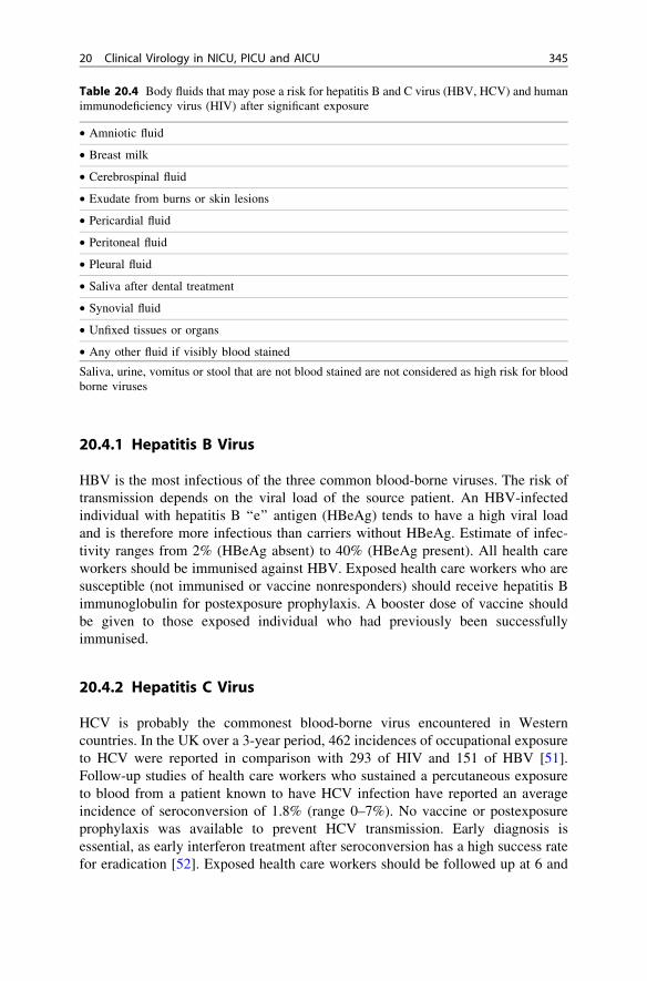
Table 20.4 Body fluids that may pose a risk for hepatitis B and C virus (HBV, HCV) and humanimmunodeficiency virus (HIV) after significant exposure
• Amniotic fluid
• Breast milk
• Cerebrospinal fluid
• Exudate from burns or skin lesions
• Pericardial fluid
• Peritoneal fluid
• Pleural fluid
• Saliva after dental treatment
• Synovial fluid
• Unfixed tissues or organs
• Any other fluid if visibly blood stained
Saliva, urine, vomitus or stool that are not blood stained are not considered as high risk for bloodborne viruses
20 Clinical Virology in NICU, PICU and AICU 345

12 weeks for HCV RNA testing and promptly referred for treatment if foundinfected.
20.4.3 Human Immunodeficiency Virus
The average risk of HIV transmission after percutaneous exposure to HIV-infectedblood is about 0.3%. After mucocutaneous exposure, the risk is estimated to be\0.1%. A case-control study [53] identified four factors with increased risk oftransmission:• deep injury;• visible blood on the device that caused the injury;• injury with a needle that has been placed in a source patient’s artery or vein;• terminal HIV-related illness in the source patient.
This study also showed that the use of zidovudine prophylaxis reduce the risk oftransmission by 80%. Postexposure prophylaxis (PEP) should therefore be offeredto all health care workers who have significant exposure to blood or body fluidfrom a patient known to be at high risk of or to have HIV infection. Various PEPoptions are available depending on national recommendations. This should bestarted as soon as possible after exposure and continued for 4 weeks.
Table 20.6 Example of actions to be taken immediately after a blood/body fluid exposure(adapted from the Infection Control Team of Guys and St. Thomas’ NHS Foundation Trust,London)
• Rinse area thoroughly under running water
• If a wound, wash site with soap, encourage the wound to bleed, and apply a waterproof dressing(if appropriate)
• Report it immediately to:
– Your supervisor (or line manager)
– Occupational health or accident and emergency
• Document incident by completing an incident form with source name and details
Table 20.5 Example of points for sharps safety education (adapted from the Infection ControlTeam of Guys and St. Thomas’ NHS Foundation Trust, London)
• Never resheath needles
• Dispose all sharps in an approved container
• Dispose sharps at the point of use
• If carried, sharps must be placed in a tray
• Do not overfill sharps bins—dispose of sharps bins when three-fourths full
346 C. Y. W. Tong and S. Schelenz

20.4.4 Viral Haemorrhagic Fevers
Viral haemorrhagic fevers (VHFs) are severe and life-threatening diseases causedby a range of viruses. They are either zoonotic or arthropod-borne infections andare often endemic in certain parts of the world. They are often highly infectiousthrough close contact with infected blood and body fluid and therefore pose asignificant risk of hospital-acquired infection. As many patients with VHFpresent with shock and require vigorous supportive treatment, it is a potentialproblem in the ICU. The major viruses of nosocomial concern in this setting areMarburg, Ebola, Rift Valley fever, Lassa and Crimean Congo haemorrhagicfever (Table 20.7). The incubation period for these VHFs ranges from3–21 days. Initial symptoms are often nonspecific but may eventually lead tohaemorrhage and shock. Any febrile patient who has returned from an endemicarea of one of the VHF agents or has a history of contact with cases suspected tohave VHF within 3 weeks should be considered as at risk. However, malariashould always be excluded. A risk assessment needs to be performed, and anypatient known or strongly suspected to be suffering from VHF should beadmitted to a high-security infectious disease unit that is designed to managethese patients. While awaiting transfer to a secure unit, such patients should beplaced in a negative-pressure room with strict source isolation. Specimens forpatient management should be processed in a high-security laboratory designatedfor category 4 pathogens, and the aetiological agent established using PCR,serology and virus culture. All areas and materials in contact with infectedpatients should be autoclaved, incinerated or treated with hypochlorite(10,000 ppm of available chlorine). If the patient dies, the body should be placedin a sealable body bag sprayed or wiped with hypochlorite. Individuals who havebeen in contact with a case of VHF should be put under surveillance for3 weeks. The successful i.v. use of ribavirin has been reported in some cases ofVHFs (Lassa, Crimean Congo haemorrhagic fever and Hantaan). Apart fromyellow fever, no vaccines are available.
Table 20.7 Viruses responsible for viral haemorrhagic fevers with nosocomial concern in theintensive care unit
Virus Geographic distribution Reservoir Vector
Marburg andEbola
Sub-Sahara Africa Bats None
Rift Valley fever Mainland Africa Sheep, cattle Mosquito
Lassa West Africa Rodents None
Crimean Congohaemorrhagicfever
East, West and South Africa, North and CentralAsia, Middle East, India and Pakistan, Balkans,West China
Cows, haresbirds,hedgehogs
Ticks
20 Clinical Virology in NICU, PICU and AICU 347

20.5 Viruses Transmitted by Direct Contact
20.5.1 Varicella Zoster Virus; Shingles
Shingles or zoster is the result of the reactivation of latent VZV (family Herpes-viridae) in the dorsal root or cranial nerve ganglia. The clinical presentation is apainful vesicular eruption covering the affected dermatome. The clinical diagnosiscan be confirmed rapidly by immunofluorescence, electron microscopy or PCR ofthe cellular material obtained from a vesicular scraping. The infection is usuallyself-limiting but can be more severe in immunocompromised patients, in whom itmay present over multiple dermatomes or as a disseminated infection. The lattercases should be managed as if they were chickenpox, and respiratory precautionsfor infection control have to be enforced.
Patients or health care staff members with classic shingles are contagious fromthe day the rash appears until the lesions are crusted over. There is some risk ofnosocomial transmission if the lesions are on exposed areas of the body or inimmunocompromised infected patients. Nonimmune (VZV-IgG negative) patientsor health care staff members with no history of chickenpox are susceptible ifthey have close contact with shingles and should be managed as described forchickenpox contact.
20.5.2 Herpes Simplex Virus
The herpes simplex virus (HSV) (family Herpesviridae) consist of two types:HSV-1 and HSV-2. Clinically, they most commonly manifest with oral (mainlyHSV-1) or genital (mainly HSV-2) ulcerations/vesicles, and reactivation is com-mon, particularly in the ICU. Other presentations include keratitis, encephalitis,meningitis, herpetic whitlow or neonatal infection.
The diagnosis can be confirmed rapidly by immunofluorescence, electronmicroscopy or PCR of vesicle/ulcer scrapings. In the immunocompromisedpatient, HSV can cause life-threatening disseminated infection and, early treatmentwith acyclovir i.v. is recommended. It has also been suggested that occult herpesvirus reactivation may increase the mortality risk of ICU patients [54].
As the infected lesions contain virus, there is an increased risk of nosocomialtransmission until the lesions have crusted over. Standard isolation precautionsshould be in place to reduce transmission (Table 20.2). Patients with active lesionsshould be nursed away from high-risk patients (i.e. immunocompromised, severeeczema, burns, or neonates). As patients can be asymptomatic secretors, healthcare workers should wear gloves when dealing with mucosal secretions (i.e. saliva)to avoid infections such as herpetic whitlow. Infected staff should cover lesions ifpossible and should not attend those at risk.
Neonatal herpes is usually transmitted from mother to the child at the time ofdelivery and may not be noticed until the infant develops the disease. Universalprecautions, in particularly, hand washing, should always be in place to reduce
348 C. Y. W. Tong and S. Schelenz

transmission of infection. To contain or prevent an outbreak, infected cases shouldbe cohorted and nursed by dedicated staff who will not attend noninfected infants.
20.5.3 Rabies Virus
Rabies virus (family Rhabdoviridae) is usually transmitted to humans followingexposure to saliva of a rabid animal (e.g. dog, fox, bat) via a bite or scratch, butonly 40% of exposed people develop disease. The virus spreads from the wound tothe central nervous system causing fatal encephalitis, and the virus may be presentin the patient’s saliva, skin, eye, and brain tissue. The diagnosis can be confirmedby demonstrating the virus directly in brain tissue or saliva by RT-PCR or byimmunofluorescence detection of antigen in skin biopsies from the nape of theneck. Due to the severe and paralysing effect, patients may be admitted to the ICU.To date, no case of nosocomial transmission has been reported apart from twopatients who received corneal transplants from infected donors. Suspected orproven cases should be placed in standard isolation and appropriate precautionstaken when dealing with potential infectious secretions (e.g. wearing of mask ifdealing with oral secretions). Any health care worker with a significant exposure(e.g. splash of secretion onto mucosa or broken skin) should receive rabies vaccineand specific immunoglobulin.
20.6 Summary
Viral infection can cause significant morbidity and mortality and has the potentialto result in cross infection, involving patients as well as health care workers. Goodinfection-control practice is essential to prevent nosocomial infection. Intensivistsshould be on the alert for important viruses causing infections according to agegroup of patients and mode of transmission and should never be complacent. Goodliaison with the laboratory is essential for determining correct diagnostic tests andtimely report of results to help in patient management.
References
1. Fitzgerald DA (2009) Human swine influenza A. Paediatr Respir Rev 10:154–1582. Trifonov V, Khiabanian H, Rabadan R (2009) Geographic dependence, surveillance, and
origins of the 2009 influenza A (H1N1) virus. N Engl J Med 361:115–1193. Webb SA, Pettila V, Seppelt I et al (2009) Critical care services and 2009 H1N1 influenza in
Australia and New Zealand. N Engl J Med 361:1925–19344. Itoh Y, Shinya K, Kiso M et al (2009) In vitro and in vivo characterization of new swine-
origin H1N1 influenza viruses. Nature 460:1021–10255. Writing committee of the WHO consultation on clinical aspects of pandemic (H1N1) 2009
Influenza (2010) Clinical aspects of pandemic 2009 Influenza A (H1N1) virus infection.N Engl J Med 362:1708–1719
6. Siston AM, Rasmussen SA, Honein MA et al (2010) Pandemic 2009 influenza A(H1N1)virus illness among pregnant women in the United States. JAMA 303:1517–1525
20 Clinical Virology in NICU, PICU and AICU 349
7. Campbell A, Rodin R, Kropp R et al (2010) Risk of severe outcomes among patientsadmitted to hospital with pandemic (H1N1) influenza. CMAJ 182:349–355
8. Hanslik T, Boelle PY, Flahault A (2010) Preliminary estimation of risk factors for admissionto intensive care units and for death in patients infected with A(H1N1)2009 influenza virus,France, 2009–2010. PLoS Curr. 2010 March 9; 2:RRN1150
9. Anonymous (2009) Bacterial coinfections in lung tissue specimens from fatal cases of 2009pandemic influenza A (H1N1)–United States, May–August 2009. MMWR Morb MortalWkly Rep 58:1071–1074
10. Anonymous (2009) Evaluation of rapid influenza diagnostic tests for detection of novelinfluenza A (H1N1) virus–United States, 2009. MMWR Morb Mortal Wkly Rep 58:826–829
11. Ellis J, Iturriza M, Allen R et al (2009) Evaluation of four real-time PCR assays for detectionof influenza A (H1N1) viruses. Euro Surveill 14(22): pii: 19230
12. Flagg A, Danziger-Isakov L, Foster C et al (2010) Novel 2009 H1N1 influenza virus infectionrequiring extracorporeal membrane oxygenation in a pediatric heart transplant recipient.J Heart Lung Transpl 29:582–584
13. Christian MD, Joynt GM, Hick JL et al (2010) Critical care triage. Recommendations andstandard operating procedures for intensive care unit and hospital preparations for aninfluenza epidemic or mass disaster. Intensive Care Med 36(Suppl 1):S55–S64
14. Jain S, Kamimoto L, Bramley AM et al (2009) Hospitalized patients with 2009 H1N1influenza in the United States, April–June 2009. N Engl J Med 361:1935–1944
15. Taylor BL, Montgomery HE, Rhodes A, Sprung CL (2010) Protection of patients and staffduring a pandemic. Recommendations and standard operating procedures for intensive careunit and hospital preparations for an influenza epidemic or mass disaster. Intensive Care Med36(Suppl 1):S45–S54
16. Cheng VC, Tai JW, Wong LM et al (2010) Prevention of nosocomial transmission of swine-origin pandemic influenza virus A/H1N1 by infection control bundle. J Hosp Infect 74:271–277
17. Reiche J, Schweiger B (2009) Genetic variability of group A human respiratory syncytialvirus strains circulating in Germany from 1998 to 2007. J Clin Microbiol 47:1800–1810
18. Berger TM, Aebi C, Duppenthaler A, Stocker M (2009) Prospective population-based studyof RSV-related intermediate care and intensive care unit admissions in Switzerland over a4-year period (2001–2005). Infection 37:109–116
19. Caram LB, Chen J, Taggart EW et al (2009) Respiratory syncytial virus outbreak in a long-term care facility detected using reverse transcriptase polymerase chain reaction: an argumentfor real-time detection methods. J Am Geriatr Soc 57:482–485
20. Gadsby NJ, Hardie A, Claas EC, Templeton KE (2010) Comparison of the Luminex RVPFast assay with in-house real-time PCR for respiratory viral diagnosis. J Clin Microbiol48:2213–2216
21. Lavergne V, Ghannoum M, Weiss K et al (2011) Successful prevention of respiratorysyncytial virus nosocomial transmission following an enhanced seasonal infection controlprogram. Bone Marrow Transpl 46(1):137–142
22. Morris SK, Dzolganovski B, Beyene J, Sung L (2009) A meta-analysis of the effect of antibodytherapy for the prevention of severe respiratory syncytial virus infection. BMC Infect Dis 9:106
23. Falsey AR, Walsh EE (2000) Respiratory syncytial virus infection in adults. Clin MicrobiolRev 13:371–384
24. Boeckh M, Englund J, Li Y et al (2007) Randomized controlled multicenter trial ofaerosolized ribavirin for respiratory syncytial virus upper respiratory tract infection inhematopoietic cell transplant recipients. Clin Infect Dis 44:245–249
25. Chavez-Bueno S, Mejias A, Merryman RA et al (2007) Intravenous palivizumab andribavirin combination for respiratory syncytial virus disease in high-risk pediatric patients.Pediatr Infect Dis J 26:1089–1093
26. Terlizzi ME, Massimiliano B, Francesca S et al (2009) Quantitative RT real time PCR andindirect immunofluorescence for the detection of human parainfluenza virus 1, 2, 3. J VirolMethods 160:172–177
350 C. Y. W. Tong and S. Schelenz

27. Maziarz RT, Sridharan P, Slater S et al (2010) Control of an outbreak of human parainfluenzavirus 3 in hematopoietic stem cell transplant recipients. Biol Blood Marrow Transpl16:192–198
28. Nichols WG, Corey L, Gooley T et al (2001) Parainfluenza virus infections afterhematopoietic stem cell transplantation: risk factors, response to antiviral therapy, andeffect on transplant outcome. Blood 98:573–578
29. Lenaerts L, De Clercq E, Naesens L (2008) Clinical features and treatment of adenovirusinfections. Rev Med Virol 18:357–374
30. Williams KM, Agwu AL, Dabb AA et al (2009) A clinical algorithm identifies high riskpediatric oncology and bone marrow transplant patients likely to benefit from treatment ofadenoviral infection. J Pediatr Hematol Oncol 31:825–831
31. Ksiazek TG, Erdman D, Goldsmith CS et al (2003) A novel coronavirus associated withsevere acute respiratory syndrome. N Engl J Med 348:1953–1966
32. Poutanen SM, Low DE, Henry B et al (2003) Identification of severe acute respiratorysyndrome in Canada. N Engl J Med 348:1995–2005
33. Lau EH, Hsiung CA, Cowling BJ et al (2010) A comparative epidemiologic analysis ofSARS in Hong Kong, Beijing and Taiwan. BMC Infect Dis 10:50
34. Wong SS, Yuen KY (2008) The management of coronavirus infections with particularreference to SARS. J Antimicrob Chemother 62:437–441
35. Cheng VC, Lau SK, Woo PC, Yuen KY (2007) Severe acute respiratory syndrome coronavirusas an agent of emerging and reemerging infection. Clin Microbiol Rev 20:660–694
36. Cameron JC, Allan G, Johnston F et al (2007) Severe complications of chickenpox inhospitalised children in the UK and Ireland. Arch Dis Child 92:1062–1066
37. Aly NY, Al Obaid I, Al-Qulooshi N, Zahed Z (2007) Occupationally related outbreak ofchickenpox in an intensive care unit. Med Princ Pract 16:399–401
38. Apisarnthanarak A, Kitphati R, Tawatsupha P et al (2007) Outbreak of varicella-zoster virusinfection among Thai healthcare workers. Infect Control Hosp Epidemiol 28:430–434
39. Fruhwirth M, Heininger U, Ehlken B et al (2001) International variation in disease burden ofrotavirus gastroenteritis in children with community- and nosocomially acquired infection.Pediatr Infect Dis J 20:784–791
40. Verboon-Maciolek MA, Krediet TG, Gerards LJ et al (2005) Clinical and epidemiologiccharacteristics of viral infections in a neonatal intensive care unit during a 12-year period.Pediatr Infect Dis J 24:901–904
41. Boyce JM, Pittet D (2002) Guideline for hand hygiene in health-care settings.Recommendations of the healthcare infection control practices Advisory Committee andthe HIPAC/SHEA/APIC/IDSA Hand Hygiene Task Force. Am J Infect Control 30:S1–S46
42. Cunliffe NA, Both JA, Lowe SJ et al (2010) Healthcare-associated viral gastroenteritis amongchildren in a large pediatric hospital, United Kingdom. Emerg Infect Dis 16:55–62
43. Armbrust S, Kramer A, Olbertz D et al (2009) Norovirus infections in preterm infants: widevariety of clinical courses. BMC Res Notes 2:96
44. Nix WA, Maher K, Pallansch MA, Oberste MS (2010) Parechovirus typing in clinicalspecimens by nested or semi-nested PCR coupled with sequencing. J Clin Virol 48:202–207
45. Harvala H, Robertson I, Chieochansin T et al (2009) Specific association of humanparechovirus type 3 with sepsis and fever in young infants, as identified by direct typing ofcerebrospinal fluid samples. J Infect Dis 199:1753–1760
46. Takami T, Sonodat S, Houjyo H et al (2000) Diagnosis of horizontal enterovirus infections inneonates by nested PCR and direct sequence analysis. J Hosp Infect 45:283–287
47. Gupta S, Fernandez D, Siddiqui A et al (2010) Extensive white matter abnormalities associatedwith neonatal Parechovirus (HPeV) infection. Eur J Paediatr Neurol 14(6):531–534
48. Benschop KS, Schinkel J, Minnaar RP et al (2006) Human parechovirus infections in Dutchchildren and the association between serotype and disease severity. Clin Infect Dis 42:204–210
49. Hanna JN, Loewenthal MR, Negel P, Wenck DJ (1996) An outbreak of hepatitis A in anintensive care unit. Anaesth Intensive Care 24:440–444
20 Clinical Virology in NICU, PICU and AICU 351

50. Victor JC, Monto AS, Surdina TY et al (2007) Hepatitis A vaccine versus immune globulinfor postexposure prophylaxis. N Engl J Med 357:1685–1694
51. Evans B, Duggan W, Baker J et al (2001) Exposure of healthcare workers in England, Wales,and Northern Ireland to bloodborne viruses between July 1997 and June 2000: analysis ofsurveillance data. BMJ 322:397–398
52. Maheshwari A, Thuluvath PJ (2010) Management of acute hepatitis C. Clin Liver Dis14:169–176
53. Cardo DM, Culver DH, Ciesielski CA et al (1997) A case-control study of HIVseroconversion in health care workers after percutaneous exposure. Centers for DiseaseControl and Prevention Needlestick Surveillance Group. N Engl J Med 337:1485–1490
54. Cook CH, Martin LC, Yenchar JK et al (2003) Occult herpes family viral infectionsare endemic in critically ill surgical patients. Crit Care Med 31:1923–1929
352 C. Y. W. Tong and S. Schelenz