Clinical Trials: Cornea Various Landmark Corneal Clinical · PDF filewww. dosonline.org l 49...
12
www. dosonline.org l 47 Clinical Trials: Cornea Various Landmark Corneal Clinical Trials Clinical Trials: Cornea Sandeep Gupta MS,DNB Sandeep Gupta MS,DNB, Parth Patel MBBS, B.V. Rao MS,DNB, Gagandeep Kaur MBBS, V.S. Gurunadh MS, M.A. Khan MS Armed Forces Medical College, Pune, Maharashtra C ornea is the first refractive media and also is the major part of the eye in contact with outer environment. Due to these unique features, it is the site of major sight disabling infections. It also contributes 2/3rd of the total refractive power of the eye and is the site of most of the procedures ending in refractive changes. It is also one of the most successfully transplanted tissue in the world. These characteristics have led to attempts to keep the cornea disease free and clear as far as possible by means of a series of landmark studies which include the control of infections trials and trials related to corneal transplants for maintaining the clear optical media. Following is a brief synopsis of the various studies related to cornea which have showed us the way to manage these ailments. Herpes eye disease study Herpes Eye Disease Study I (HEDS I) 1 Purpose • To study the efficacy of topical steroids eye drops in treating herpes simplex stromal keratitis in conjunction with topical antivirals (trifluridine) 2 . • To study the efficacy of oral acyclovir in treating herpes simplex stromal keratitis in patients receiving concomitant topical corticosteroids and antivirals (trifluridine) 3 . • To evaluate the efficacy of oral acyclovir in treating herpes simplex iridocyclitis in combination with topical steroids and antivirals (trifluridine) 4 . Background Herpes simplex keratitis is a leading cause of corneal infections leading to opacification throughout the world. An estimated 450,000 people in the United States develop recurrent episodes of the disease and about 46,000 episodes of HSV eye infection every year. Herpetic eye disease is the most common infectious cause of corneal blindness in the USA. Despite the availability of antiviral agents that are effective in treating herpes simplex epithelial keratitis, inflammation in the corneal connective tissue and iris that can lead to corneal scarring and visual impairment develops in many patients. Prior to the HEDS-I trials, the role of topical corticosteroids in the management of HSV stromal keratitis was uncertain; some animal and human studies suggested there was a benefit to treatment whereas others suggested harm. The value of adding an oral antiviral agent to treatment with topical corticosteroids and topical antivirals also was unknown. The HEDS-I trials were developed to assess the efficacy of topical corticosteroids and oral acyclovir in treating HSV stromal keratitis and iridocyclitis. Description HEDS-I consisted of three randomized, placebo-controlled trials. The organizational structure consisted of a data coordinating center and eight clinical centers. All patients received the topical antiviral trifluridine as prophylaxis against recurrences of HSV epithelial ulceration. Patients were evaluated weekly for 10 weeks, every other week through week 16, and again at 6 months. The primary outcome was the time to development of preset criteria for treatment failure during the 16-week period of examination. Protocol-specific descriptions of the three trials follow. Definitions followed in the trial Treatment failure: Worsening of stromal keratitis or uveitis at any scheduled visit, no change in stromal inflammation in the first 2 weeks or 3 later consecutive weeks, or occurrence of an adverse event.
Transcript of Clinical Trials: Cornea Various Landmark Corneal Clinical · PDF filewww. dosonline.org l 49...

www. dosonline.org l 47
Clinical Trials: Cornea
Various Landmark Corneal Clinical Trials
Clinical Trials: Cornea
Sandeep GuptaMS,DNB
Sandeep Gupta MS,DNB, Parth Patel MBBS, B.V. Rao MS,DNB, Gagandeep Kaur MBBS, V.S. Gurunadh MS, M.A. Khan MS
Armed Forces Medical College, Pune, Maharashtra
Cornea is the first refractive media and also is the major part of the eye in contact with outer environment.
Due to these unique features, it is the site of major sight disabling infections. It also contributes 2/3rd of the total refractive power of the eye and is the site of most of the procedures ending in refractive changes. It is also one of the most successfully transplanted tissue in the world.
These characteristics have led to attempts to keep the cornea disease free and clear as far as possible by means of a series of landmark studies which include the control of infections trials and trials related to corneal transplants for maintaining the clear optical media. Following is a brief synopsis of the various studies related to cornea which have showed us the way to manage these ailments.
Herpes eye disease study
Herpes Eye Disease Study I (HEDS I)1
Purpose
• To study the efficacy of topical steroids eye drops in treating herpes simplex stromal keratitis in conjunction with topical antivirals (trifluridine)2.
• To study the efficacy of oral acyclovir in treating herpes simplex stromal keratitis in patients receiving concomitant topical corticosteroids and antivirals (trifluridine)3.
• To evaluate the efficacy of oral acyclovir in treating herpes simplex iridocyclitis in combination with topical steroids and antivirals (trifluridine)4.
Background
Herpes simplex keratitis is a leading cause of corneal infections leading to opacification throughout the world. An estimated 450,000 people in the United States develop recurrent episodes of the disease and about 46,000 episodes
of HSV eye infection every year. Herpetic eye disease is the most common infectious cause of corneal blindness in the USA.
Despite the availability of antiviral agents that are effective in treating herpes simplex epithelial keratitis, inflammation in the corneal connective tissue and iris that can lead to corneal scarring and visual impairment develops in many patients. Prior to the HEDS-I trials, the role of topical corticosteroids in the management of HSV stromal keratitis was uncertain; some animal and human studies suggested there was a benefit to treatment whereas others suggested harm. The value of adding an oral antiviral agent to treatment with topical corticosteroids and topical antivirals also was unknown.
The HEDS-I trials were developed to assess the efficacy of topical corticosteroids and oral acyclovir in treating HSV stromal keratitis and iridocyclitis.
Description
HEDS-I consisted of three randomized, placebo-controlled trials. The organizational structure consisted of a data coordinating center and eight clinical centers. All patients received the topical antiviral trifluridine as prophylaxis against recurrences of HSV epithelial ulceration. Patients were evaluated weekly for 10 weeks, every other week through week 16, and again at 6 months. The primary outcome was the time to development of preset criteria for treatment failure during the 16-week period of examination. Protocol-specific descriptions of the three trials follow.
Definitions followed in the trial Treatment failure: Worsening of stromal keratitis or uveitis at any scheduled visit, no change in stromal inflammation in the first 2 weeks or 3 later consecutive weeks, or occurrence of an adverse event.

48 l DOS Times - Vol. 19, No. 10 April, 2014
Clinical Trails: Cornea
Increased severity of stromal keratitis: Any one of the following conditions: development of a new zone of non-necrotizing or necrotizing stromal keratitis of 15 mm2 or greater; increase in total area of stromal keratitis of 7.5 mm 2 or by 75% of the previous area depending on the size ofthe previous lesion; or increase in total area of stromal keratitis of 10 mm 2 or by 100% of the initial area, depending on the size of the initial lesion.
No change: Less than a 10% change in the area of stromal keratitis.
Increased severity of iridocyclitis: A two-step or greater increase of cells in the anterior chamber compared with the best attained measurement or 3+ cells or more in the anterior chamber for 2 weeks.
Significant vision decrease: Deterioration of visual acuity by four lines or more from baseline as measured on the Bailey-Lovie chart.
Treatment success: Completion of the 10-week regimen of study medications, resolution of active inflammation during the 16-week treatment/observation interval, and no reactivation or recurrence for 6 weeks without medication.The three arms of the trial were-
1. Herpes Stromal Keratitis, Not on Steroid Trial (HEDS-SKN): Patients with active HSV stromal keratitis who had not used a topical corticosteroid in the preceding 10 days were randomized to treatment with topical prednisolone phosphate drops or topical placebo drops. A treatment schedule, starting with 8 drops a day of 1% prednisolone phosphate for 7 days, was progressively decreased over 10 weeks in such a way that patients received 1 drop per day of 1/8% prednisolone for the last 3 weeks of treatment. Placebo drops were given by the same schedule.
2. Herpes Stromal Keratitis, on Steroid Treatment (HEDS-SKS): Patients with active HSV stromal keratitis who
already were being treated with a topical corticosteroid were randomized either to oral treatment with acyclovir capsules (400 mg five times daily) for 10 weeks or to the identical dose of placebo capsules. Patients also received topical prednisolone phosphate in the dosage schedule described above for the SKN trial.
3. Herpes Simplex Virus Iridocyclitis, Receiving Topical Steroids (HEDS-IRT): Patients with active HSV iridocyclitis were randomized either to oral treatment with acyclovir capsules (400 mg five times daily) for 10 weeks or to the identical dose of placebo capsules. Patients also received topical prednisolone phosphate in the dosage schedule described above for the SKN trial.
Patient Eligibility: Eligibility criteria common to the three protocols included
• Age 12 years or older
• No active HSV epithelial keratitis
• No prior keratoplasty of the involved eye
• Not pregnant
Patient Recruitment Status: Completed.
Current Status of Study: Completed.
Results
Herpes Stromal Keratitis, Not on Steroid Trial (HEDS-SKN):
Patient recruitment was stopped on the recommendation of the Data and Safety Monitoring Committee after 106 of the originally planned 176 patients were enrolled. Compared with the patients in the placebo group, the patients who received prednisolone phosphate drops had faster resolution of the stromal keratitis and fewer treatment failures. However, delaying the initiation of corticosteroid treatment did not affect the eventual outcome of the
Table 1: HEDS 1
TRIAL Herpes Stromal Keratitis, Not on Steroid Trial (HEDS-SKN)
Herpes Stromal Keratitis, on Steroid Treatment (HEDS-SKS)
Herpes Simplex Virus Iridocyclitis, Receiving Topical Steroids (HEDS-IRT):
PURPOSE Topical corticosteroids in herpes simplex stromal keratitis with topical trifluridine
Oral acyclovir in herpes simplex stromal keratitis with concomitant topical corticosteroids and trifluridine
Oral acyclovir in herpes simplex iridocyclitis with topical corticosteroids and trifluridine
RESULT The patients who received prednisolone phosphate drops had faster resolution of the stromal keratitis and fewer treatment failures. Delaying the initiation of corticosteroid treatment did not affect the eventual outcome of the disease.
There was no apparent benefit in the addition of oral acyclovir to the treatment regimen of a topical corticosteroid and a topical antiviral.
Apparent benefit in adding oral acyclovir in the treatment of HSV iridocyclitis to topical corticosteroids and trifluridine.
www. dosonline.org l 49
Clinical Trials: Cornea
disease, in that visual acuity was similar in the two groups at 26 weeks.
Herpes Stromal Keratitis, on Steroid Treatment (HEDS-SKS):
One hundred and four patients were enrolled in this trial. Over the 16-week followup period, there was no difference in the rate of treatment failure between the two groups. Thus, there was no apparent benefit in the addition of oral acyclovir to the treatment regimen of a topical corticosteroid and a topical antiviral.
Herpes Simplex Virus Iridocyclitis, Receiving Topical Steroids (HEDS-IRT):
The trial was stopped because of slow recruitment after only 50 of the originally planned 104 patients were enrolled during a 4-year recruitment period. Treatment failures occurred at a higher rate in the placebo group than in the acyclovir group. Although the number of patients enrolled in this trial was too small to achieve statistically conclusive results, the trend in the results suggests a benefit in adding oral acyclovir to the treatment of HSV iridocyclitis in patients receiving topical corticosteroids and trifluridine prophylaxis.
Herpes Eye Disease Study II(HEDES II)5
Purpose
• To find if early treatment (with oral acyclovir) of herpes simplex virus (HSV) ulcerations of the corneal epithelium prevents progression to the blinding complications of stromal keratitis and iridocyclitis6.
• To evaluate the efficacy of low-dose oral acyclovir in preventing recurrent HSV eye infection in patients with previous episodes of herpetic eye disease7.
• To evaluate the role of external factors (such as ultraviolet light or corneal trauma) and behavioural factors (such as life stress) on the induction of ocular recurrences of HSV eye infections and disease.
Background
Infection of the eye by herpes simplex virus (HSV) is a leading cause of corneal morbidity. The infection can lead to corneal scarring and neovascularization, permanent endothelial dysfunction and corneal edema, secondary glaucoma, and cataract. Despite the availability of topical antiviral agents that are highly active against HSV keratitis, there was no known effective method for reducing the frequency of recurrence or severity of stromal keratitis and iridocyclitis. In addition, the prognosis was poor for recovery of good vision following penetrating keratoplasty for actively inflamed or highly vascularized herpetic corneas. On the basis of both animal and human studies, the antiviral agent acyclovir might treat and prevent recurrence of HSV disease. However, no consensus existed on the use
of acyclovir in the management and prevention of herpetic eye disease prior to this study.
Description
HEDS-II consists of two randomized, placebo-controlled trials that assessed the role of oral acyclovir in the management of herpetic eye disease and one epidemiologic study that investigated the risk factors, including stress, for the development of ocular recurrences of the disease.
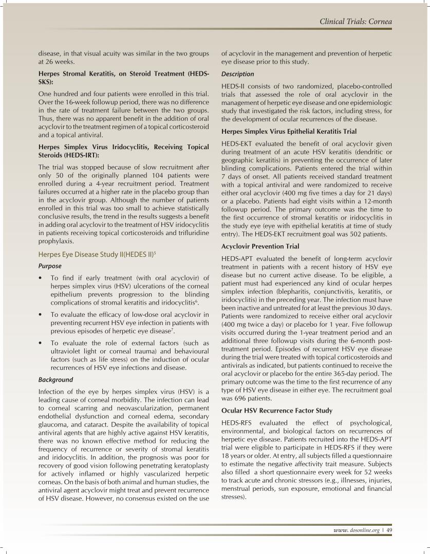
Herpes Simplex Virus Epithelial Keratitis Trial
HEDS-EKT evaluated the benefit of oral acyclovir given during treatment of an acute HSV keratitis (dendritic or geographic keratitis) in preventing the occurrence of later blinding complications. Patients entered the trial within 7 days of onset. All patients received standard treatment with a topical antiviral and were randomized to receive either oral acyclovir (400 mg five times a day for 21 days) or a placebo. Patients had eight visits within a 12-month followup period. The primary outcome was the time to the first occurrence of stromal keratitis or iridocyclitis in the study eye (eye with epithelial keratitis at time of study entry). The HEDS-EKT recruitment goal was 502 patients.
Acyclovir Prevention Trial
HEDS-APT evaluated the benefit of long-term acyclovir treatment in patients with a recent history of HSV eye disease but no current active disease. To be eligible, a patient must had experienced any kind of ocular herpes simplex infection (blepharitis, conjunctivitis, keratitis, or iridocyclitis) in the preceding year. The infection must have been inactive and untreated for at least the previous 30 days. Patients were randomized to receive either oral acyclovir (400 mg twice a day) or placebo for 1 year. Five followup visits occurred during the 1-year treatment period and an additional three followup visits during the 6-month post-treatment period. Episodes of recurrent HSV eye disease during the trial were treated with topical corticosteroids and antivirals as indicated, but patients continued to receive the oral acyclovir or placebo for the entire 365-day period. The primary outcome was the time to the first recurrence of any type of HSV eye disease in either eye. The recruitment goal was 696 patients.
Ocular HSV Recurrence Factor Study
HEDS-RFS evaluated the effect of psychological, environmental, and biological factors on recurrences of herpetic eye disease. Patients recruited into the HEDS-APT trial were eligible to participate in HEDS-RFS if they were 18 years or older. At entry, all subjects filled a questionnaire to estimate the negative affectivity trait measure. Subjects also filled a short questionnaire every week for 52 weeks to track acute and chronic stressors (e.g., illnesses, injuries, menstrual periods, sun exposure, emotional and financial stresses).
50 l DOS Times - Vol. 19, No. 10 April, 2014
Clinical Trails: Cornea
Patient Recruitment Status: Completed.
Current Status of Study: Completed
Results
Herpes Simplex Virus Epithelial Keratitis Trial (HEDS-EKT)
Patient recruitment in the HEDS-EKT trial was stopped on the recommendation of the Data and Safety Monitoring Committee after 287 of the originally planned 502 patients were enrolled because of a lack of any suggestion of efficacy of the treatment protocol in the accrued results. The study found that in the treatment of acute HSV epithelial keratitis, there was no benefit from the addition of oral acyclovir to treatment with topical trifluridine in preventing the development of stromal keratitis or iritis. Importantly, the study found that the risk of stromal keratitis or iridocyclitis was quite low in the year following an episode of epithelial keratitis treated with topical trifluridine alone. This risk is much lower than the published risk in the literature.
Acyclovir Prevention Trial (HEDS-APT)
Of the 703 patients, 357 were randomly assigned to the acyclovir group, and 346 to the placebo group. All patients were followed for all forms of ocular and nonocular recurrences during a 12-month treatment period, followed by a six-month observation period. The acyclovir group received oral acyclovir 400 mg twice daily for 12 months. Only 4% of patients in the acyclovir group and 5% in the placebo group stopped treatment because of side effects.Scientists found that the drug acyclovir, taken by mouth, reduced by 41 % the probability that any form of herpes of the eye would return in patients who had the infection in the previous year. Importantly, researchers noted a 50% reduction in the rate of return of the more severe form of the disease -- stromal keratitis -- among patients who had
this infection during the past year.In addition to the main findings, doctors also noted that during the 12 months of treatment:
• Oral acyclovir reduced the incidence of epithelial keratitis from 11% to 9%, and the incidence of stromal keratitis from 13% to 8%.
• 4% of patients in the acyclovir group and 9% in the placebo group had more than one recurrence.
The Mycotic Ulcer Treatment Trial -A Randomized Trial Comparing Natamycin vs Voriconazole
Mycotic keratitis is a major group contributing to ocular morbidity esp in developing world and also in patients with ocular trauma, long term ocular steroid therapy and in immunocompromised patients. Most of these infections are diagnosed late and treated inappropriately leading to considerable morbidity.Natamycin was the only topical antifungal approved by the US Food and Drug Administration for topical ophthalmic use. However,most isolates from fungal keratitis have shown good in-vitro susceptibility to newer azoles, including voriconazole.
Fungal corneal ulcers tend to have very poor outcomes with commonly used treatments. The triazole voriconazole had recently become the treatment of choice for systemic fungal infections such as pulmonary aspergillosis. The use of topical ophthalmic preparations of voriconazole had been described in numerous case reports, however there had been no systematic attempt to determine whether it is more or less clinically effective than natamycin. Additionally, there had been many case reports of the use of oral voriconazole in the treatment of fungal corneal ulcers, however there had been no systematic attempt to determine if it improves outcomes in severe ulcers.
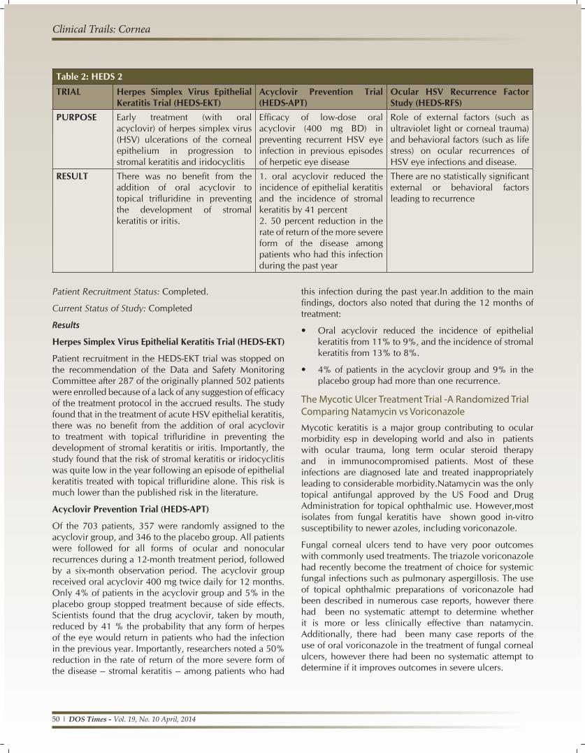
Table 2: HEDS 2
TRIAL Herpes Simplex Virus Epithelial Keratitis Trial (HEDS-EKT)
Acyclovir Prevention Trial (HEDS-APT)
Ocular HSV Recurrence Factor Study (HEDS-RFS)
PURPOSE Early treatment (with oral acyclovir) of herpes simplex virus (HSV) ulcerations of the corneal epithelium in progression to stromal keratitis and iridocyclitis
Efficacy of low-dose oral acyclovir (400 mg BD) in preventing recurrent HSV eye infection in previous episodes of herpetic eye disease
Role of external factors (such as ultraviolet light or corneal trauma) and behavioral factors (such as life stress) on ocular recurrences of HSV eye infections and disease.
RESULT There was no benefit from the addition of oral acyclovir to topical trifluridine in preventing the development of stromal keratitis or iritis.
1. oral acyclovir reduced the incidence of epithelial keratitis and the incidence of stromal keratitis by 41 percent2. 50 percent reduction in the rate of return of the more severe form of the disease among patients who had this infection during the past year
There are no statistically significant external or behavioral factors leading to recurrence

www. dosonline.org l 51
Clinical Trials: Cornea
This study was a randomized, double-masked, placebo-controlled trial to determine if the use natamycin or voriconazole results in better outcomes for fungal corneal ulcers. Fungal corneal ulcers with baseline visual acuity between 6/12 (20/40, logMAR 0.3) and 6/120 (20/400, logMAR 1.3) presenting to the Aravind Eye Hospitals and the UCSF Proctor Foundation were randomized to receive either topical natamycin or topical voriconazole8.
Methods
• Phase 3, double-masked, multicenter trial .
• Comparing outcomes in patients with fungal corneal ulcers receiving topical natamycin 5% and topical voriconazole 1%
• Primary outcome -- best spectacle-corrected visual acuity (BSCVA) 3 months from enrollment.
• Secondary out comes -- BSCVA at 3 weeks, infiltrate or scar size at 3 weeks and 3 months, time to reepithelialization, microbiological cure at 6 days (+/-1 day), and corneal perforation and/or therapeutic penetrating keratoplasty (TPK).
• BSCVA protocol was adapted from the Age-Related Eye Disease Study using Early Treatment Diabetic Retinopathy Study tumbling “E” charts (charts 2305 and 2305A; PrecisionVision) at 4 m, using a protocol identical to that used in the Steroids for Corneal Ulcers Trial, with low- vision testing at 0.5 m.
• Dosing schedules were identical in both the arms and consisted of 1 drop applied to the affected eye every 1 hour while awake for 1 week, then every 2 hours while awake until 3 weeks from enrollment.
Measurement of outcomes
• Re-epithelialization was defined as the absence of an epithelial defect with the administration of fluorescein.
• Depth was assessed in 3 categories: more than 0% to 33%; more than 33% to 67%; and more than 67% to 100%.
• Corneal scrapings were obtained from the leading edge and base of the corneal ulcer.
Results
• 323 patients were enrolled.
• 34 perforations and/or TPKs had occurred among patients randomized to voriconazole and 18 had occurred among those randomized to natamycin. Recruitment was stopped immediately in these cases.
• Natamycin was added to the treatment regimens for all patients currently enrolled. Thus, only patients
enrolled on or before this, were included in the final primary analysis (323 patients).
• The most commonly isolated organisms were Fusarium species (128 patients [40%]), followed by Aspergillus species (54 patients [17%]) .
• The median duration of treatment was 31 days in the natamycin treated arm and 39 days in the voriconazole treated arm.
• At 3 weeks, the mean BSCVA among patients randomized to voriconazole was 1.1 lines poorer compared with those randomized to natamycin.
• At 3 months, patients randomized to receive voriconazole did 1.8 lines worse than those randomized to receive natamycin.
• BSCVA for Fusarium-infected patients randomized to natamycin was 4.1 lines better than for such patients randomized to voriconazole.
• No evidence of a difference in adjusted BSCVA between the 2 treatments in non-Fusarium cases.
• A higher fraction of individuals randomized to voriconazole tested culture positive at 6 days than individuals randomized to natamycin: 23 of 155 patients (15%) for natamycin vs 69 of 144 patients (48%) for voriconazole9.
• No compelling evidence of a difference in time to reepithelialization.
• At 3 months, there was evidence of a difference in scar size between patients randomized to the 2 treatments. Fusarium cases had significantly smaller scars at 3 months when treated with natamycin, no evidence of larger scars at 3 months for non-Fusarium cases.
• 12 patients randomized to voriconazole had an increase of at least 2 mm in hypopyon size, while only 5 patients randomized to natamycin showed such an increase.
Important take home points• Significantly better visual acuity at 3 months in patients
randomized to receive topical natamycin compared with those randomized to receive topical voriconazole.
• Voriconazole-treated cases were more likely to have a perforation.
• Reepithelialization time and 3-month infiltrate or scar size were not significantly different between the 2 treatments.
• The difference in efficacy noted in this trial was primarily attributable to cases caused by Fusarium species.

52 l DOS Times - Vol. 19, No. 10 April, 2014
Clinical Trails: Cornea
• Natamycin was significantly more successful at clearing culture positivity after 6 days than was voriconazole.
• Findings suggest that, in vivo, topical voriconazole is substantially less effective at clearing Fusarium species and should not be considered appropriate monotherapy for Fusarium keratitis. (voriconazole was still more effective against Fusarium species in vitro than natamycin and other antifungals)
Mycotic ulcer treatment trial II (MUTT II)This study is a randomized, double-masked, placebo-controlled trial to determine if the use of oral voriconazole in severe ulcers reduces the rate of perforations. 240 fungal corneal ulcers with baseline visual acuity worse than 6/120 presenting to the Aravind Eye Hospitals and the UCSF Proctor Foundation would be randomized to receive oral voriconazole plus topical voriconazole and topical natamycin, or oral placebo plus topical voriconazole and topical natamycin. The primary outcome is the rate of perforation over the three month follow-up period.
Status-Ongoing trial
Primary objective-Comparison of rate of perforation between the treatment groups (topical voriconazole with oral voriconazole vs. topical voriconazole with oral placebo) in mycotic keratitis.
Secondary objectives-
• Best spectacle-corrected logMAR visual acuity [Time Frame: 3 weeks after enrolment]
• Hard contact-lens corrected visual acuity measured in logMAR [Time Frame: 3 months after enrolment]
• Size of infiltrate/scar [Time Frame: 3 weeks and 3 months after enrolment]
• Time to resolution of epithelial defect [Time Frame: At the time of resolution of epithelial defect]
• Number of adverse events [Time Frame: At the time of adverse event]
• Minimum inhibitory concentration of isolates [Time Frame: 3 months after enrolment]
• Microbiological cure at 7 days [Time Frame: 7 days after enrolment]
Steroids in corneal ulcer trial(SCUT)
Purpose
To determine if topical corticosteroids in addition to antibacterials as adjunctive therapy for bacterial keratitis improves long-term clinical outcomes10.
Design
Randomized, placebo-controlled, double-masked clinical trial.
Methods
This multicentre trial compared 1.0% prednisolone sodium phosphate to placebo in the treatment of bacterial keratitis among 500 patients with culture-positive ulcers receiving 48 hours of eye drop moxifloxacin before randomization. The primary endpoint was 3 months from enrolment, and 399 patients were evaluated at 12 months. The outcomes examined were best spectacle-corrected visual acuity (BSCVA) and scar size at 12 months.
Results
• No significant differences in clinical outcomes by treatment group were seen with the prespecified regression models (BSCVA: −0.04 logMAR, 95% CI, −0.12 to 0.05, P = .39; scar size: 0.03 mm, 95% CI, −0.12 to 0.18, P = .69).
• A regression model including a Nocardia-treatment arm interaction found corticosteroid use associated with a mean 1-line improvement in BSCVA at 12 months among patients with non-Nocardia ulcers (−0.10 logMAR, 95% CI, −0.19 to −0.02, P = .02).
• No significant difference was observed in 12-month BSCVA for Nocardia ulcers (0.18 logMAR, 95% CI, −0.04 to 0.41, P = .16).
• Corticosteroids were associated with larger mean scar size at 12 months among Nocardiaulcers (0.47 mm, 95% CI, 0.06-0.88, P = .02) and no significant difference was identified by treatment for scar size for non-Nocardia ulcers (−0.06 mm, 95% CI, −0.21 to 0.10, P = .46).
Table 3: Mycotic Ulcer Treatment TrialSTUDY Mycotic Ulcer Treatment Trial (MUTT)PURPOSE Compared natamycin 5% and voriconazole
1% in patients with a smear-positive filamentous fungal ulcer and visual acuity of 20/40 to 20/400
RESULT Natamycin-treated cases had significantly better 3-month BSCVA than voriconazole-treated cases (P=.006).Natamycin-treated cases were less likely to have perforation or require therapeutic penetrating keratoplasty (P=.009).Fusarium cases fared better with natamycin than with voriconazole for 3-month BSCVA (P<.001) and perforation (P=.001).Non-Fusarium cases fared similarly for 3-month BSCVA (P=.81) and perforation (P=.86).

www. dosonline.org l 53
Clinical Trials: Cornea
Conclusions drawn by the studyAdjunctive topical corticosteroid therapy may be associated with improved long-term clinical outcomes in bacterial corneal ulcers not caused by Nocardia species. However, it is left to the discretion of the treating Ophthalmologist to add steroids as per the case.
Collaborative Corneal Transplantation Studies (CCTS)
Purpose
To find out whether histocompatibility matching of corneal transplant donors and recipients can reduce the incidence of graft rejection in high-risk patients.
Background
Approximately 20% of corneal transplant patients, about 6,000 per year, face donor tissue rejection at rates of up to 60% because of corneal vascularization or prior graft rejection. Histocompatibility antigen matching and/or crossmatching may offer these patients an improved chance for successful outcome in form of less rejection episodes.
Description
The Collaborative Corneal Transplantation Studies Group conducted two controlled, double-masked studies addressing donor-recipient histocompatibility matching.
• The Crossmatch Study was a randomized study assessing the effectiveness of crossmatching in preventing graft rejection among high-risk patients with lymphocytotoxic antibodies11.
• The Antigen Matching Study was a prospective, double-masked, observational study of the effectiveness of HLA-A, -B, and -DR donor-recipient matching in high-risk patients who had no lymphocytotoxic antibodies12.
Six clinical centres recruited high-risk patients and collaborated with their local eye banking and organ procurement agencies in procuring donor corneal tissue. For each of the two studies, a total of 400 patients were sought. Blood samples from each enrolled patient were sent to the local CCTS tissue typing laboratory for HLA typing, and serum samples were sent to the Central Laboratory to be screened for preformed lymphocytotoxic antibodies. Depending on the results of the testing, patients were entered into the Crossmatch Study or the Antigen Matching Study.
As corneal donors became available, donor blood samples were HLA typed at the local laboratories and cross matched against all CCTS patients who awaited transplantation. Results of the testing were entered in a national, 24-hour computerized allocation system operated by the United Network for Organ Sharing (UNOS). Patients in the Crossmatch Study received a cornea from either a positively crossmatched donor or a negatively crossmatched donor.
Patients in the Antigen Matching Study received a cornea with 0 to 6 matched antigens.
Transplant patients were followed intensively during the first months after surgery. The number of clinic visits was tapered to 2 during the third and final year of followup, resulting in a total of 17 postoperative visits. Irreversible failure of the corneal allograft due to all causes was the primary outcome variable in both studies. Allograft reaction episodes, irreversible failure due to rejection, and visual acuity were secondary outcome variables.
Patient Eligibility• Males and females age 10 years or older with two to
four quadrants of corneal stroma vascularization or a history of allograft rejection in the eye considered for surgery were eligible for both studies in the CCTS.
• Patients must have been willing to participate in 3 years of followup. No one was eligible for the CCTS who had a condition that would greatly increase the risk of nonrejection graft failure, such as xerophthalmia or severe exposure keratopathy. Also excluded were patients with systemic diseases or with medication usage that might alter their immune response.
Patient Recruitment StatusRecruitment began in May 1986 and was completed September 30, 1989.
Results
Researchers report that donor-recipient tissue typing had no significant long-term effect on the success of corneal transplantation in more than 400 patients at high risk for rejection.
Many apparent risk factors did not maintain their association with graft outcomes after adjustment for other risk factors. Young recipient age, the number of previous grafts, history of previous anterior segment surgery, preoperative glaucoma, quadrants of anterior synechiae, quadrants of stromal vessels, a primary diagnosis of chemical burn, and blood group ABO incompatibility were among the strongest risk factors identified for graft failure. Donor and corneal preservation characteristics had little influence on graft outcome.
The Collaborative Corneal Transplantation Studies (CCTS) suggested instead that matching patient and donor blood types (ABO compatibility), a test that is not currently standard practice in corneal transplantation, might be effective in improving patient outcome. Treating patients with high-dose topical steroid therapy for several months after surgery may have improved transplant survival in this study.
After 3 years of patient followup, CCTS researchers found that people who received corneal transplants with well-

54 l DOS Times - Vol. 19, No. 10 April, 2014
Clinical Trails: Cornea
matched antigens did not fare significantly better than those with a poor match. Each patient group had similar rates of initial immune reactions, graft rejection, and graft failure due to rejection or other causes. The researchers also noted that CCTS patients who were compatible with the donor’s blood type had a better outcome than unmatched patients.
The CCTS also reported a 65% rate of graft survival at its 3-year mark--higher than the usual average of about 50% in high-risk patients. The study’s high success rate might be due to rigorous high-dose steroid therapy given to control the patients’ immune reaction to the donor cornea several months after surgery.
The most important conclusion to be drawn from the CCTS is that high-dose, postoperative topical steroids, good patient compliance with a self-administered steroid regimen, and close patient followup are the keys to successful corneal transplantation in high-risk individuals.
Data from the CCTS indicate that matching patient and donor blood types combined with treating patients with high-dose topical steroids after surgery may be potentially more effective in improving high-risk corneal transplantation. These two inexpensive strategies are considerably more economical than the more expensive donor-recipient tissue typing. These findings provide clinicians with solid information on managing patients at high-risk for graft failure, while keeping health care costs to a minimum.
Australian corneal graft registry
The Australian Corneal Graft Registry (ACGR) was opened in May198514 and thus has now been in operation for over 26 years. It constitutes probably the largest and the most organised data on corneal grafts in the world.The dates for the latest report were 01/06/2010 for penetrating grafts and 12/10/2011 for lamellar grafts. Over the years,
it has collected data on more than 23,000 corneal grafts. The majority of grafts registered have been penetrating, but increasing numbers of lamellar grafts have also been registered over recent years (Table 5).
Following a standard practice in the ACGR, at registration, information is elicited on the recipient, the donor, the eye bank practices and the operative procedure. Follow-up then occurs at approximately yearly intervals for an indefinite period, and ceases upon loss of the graft, or the death or loss-to-follow-up of the patient. At each round of follow-up, information is requested on the graft and visual outcome, and upon relevant post-operative events and treatments.The data is entered into an Access database and checked for consistency. Descriptive, univariate and multivariate analyses are subsequently performed using SPSS and Stata software, and the periodic reports are eventually collated15.
Definitions used in ACGR
Vessel ingrowth into the cornea at the time of graft–
• It is scored on a scale of 0-4
• 0 representing no growth in any quadrant extending to the graft-host junction
• 1 representing such growth in 1 quadrant
• 2 representing growth in 2 quadrants
• 3 being vessel ingrowth in 3 quadrants
• 4 being vessel ingrowth in 4 quadrants
Table 4: Collaborative Corneal Transplantation StudiesSTUDY Collaborative Corneal Transplantation
Studies PURPOSE To determine whether histocompatibility
matching of corneal transplant donors and recipients can reduce the incidence of graft rejection in high-risk patients.
RESULT High-dose, postoperative topical steroids, good patient compliance and close patient followup are the keys to successful corneal transplantation in high-risk individuals13.Matching patient and donor blood types combined with treating patients with high-dose topical steroids after surgery are more effective in improving high-risk corneal transplantation.HLA matching does not change the prognosis
Table 5: Synopsis of the current database (census date for penetrating and limbal grafts 01/06/2010 and 12/10/2011 for lamellar grafts) in ACGR

www. dosonline.org l 55
Clinical Trials: Cornea
No distinction is made between superficial or deep vessels, patent or ghost vessels, or single or multiple vessel leashes. After corneal transplantation, the presence of even one vessel leash extending into the graft is considered enough to classify that graft as vascularized.
The intraocular pressure (IOP)-
It is generally considered to be raised if a reading of 25 mm Hg or greater is made by applanation tonometry, but the decision is at the discretion of the ophthalmologist.
Primary graft non-functions
It is defined as grafts that never thin and clear in the post- operative period. The trial time for such grafts is arbitrarily adjusted to one day. Any existing graft that is replaced by another in the same eye, irrespective of graft clarity and for whatever reason, is classified as a failed graft. An example in this category would be a clear graft with an unacceptably high degree of irregular astigmatism, not improved by refractive surgery, which is then replaced. In all other cases, graft failure is defined as oedema and irremediable loss of clarity in a previously thin, clear graft. The day of failure is the first day the patient is seen with an oedematous, opaque graft that subsequently fails to thin and clear.
Rejection-
It is defined as the development of a rejection line (epithelial or endothelial) or a unilateral anterior chamber reaction with corneal infiltrates and spreading corneal oedema in a previously thin, clear graft.
Any development with the potential to compromise graft outcome is considered to be a complication.
Post-operative complications are collected in two ways.
• A number of specified complications (stitch abscess, microbial keratitis, neovascularization of the graft, synechiae, uveitis, rise in IOP, cataract, rejection episode, herpetic recurrence, early changes of bullous keratopathy),
• Refractive and related errors (anisometropia, >5 dioptres astigmatism) and factors potentially affecting visual outcome but unrelated to the graft (cataract, amblyopia, retinal detachment, age related macular degeneration and diabetic retinopathy).
Results of data collected in ACGR
• Penetrating corneal graft survival is 73% at 5 years.
• Lamellar corneal graft survival is 67% at 5 years.
• Limbal allograft survival is 43% at 5 years.
• Surgeons who performed 25 or more penetrating grafts per year on average achieved better results in terms of graft survival than did those who performed fewer
grafts, although the difference in terms of graft survival was small.
Donor and eye-banking information by ACGR data• The most common causes of donor death were related
to diseases of the cardiac/circulatory system, followed by malignancy and strokes and other hemorrhages. No influence of cause of donor death on penetrating graft survival was observed, other than that corneas collected from victims of traumatic or accidental death appeared to show improved survival. However, such corneas were preferentially used for recipients with keratoconus.
• Approximately 69% of corneas were retrieved from donors aged 51-80 years at the time of death. Donor age exerted no influence on survival of grafts performed for keratoconus, but for all other indications for penetrating keratoplasty, better survival was achieved with corneas from younger donors, at least in univariate analysis. However, the effect disappears in multivariate analysis.
• Median death-to-enucleation time for penetrating grafts was 7 hours. Median death-to-graft time was 79 hours. Neither death-to-enucleation nor death-to-graft time affected corneal graft survival significantly.
• Most corneas are currently stored in Optisol in Eye Banks. Corneal storage time in Optisol did not affect graft survival.
• Corneas from multi-organ donors exhibited better survival after transplantation than did those from cardiac death donors, however, 44% of these cornea were used for recipients with keratoconus. Fewer than 1% of corneas used in penetrating grafts failed to function in the immediate post-operative period. Donor corneal endothelial cell count has no significant influence on penetrating graft survival.
Recipient age and sex at graft• The distribution of recipient age at graft was bimodal,
with peaks at around 20-40 years and around 60-80 years.
• Although recipient age appeared to influence graft survival, it is more likely that the indication for graft associated with different age groups affected survival. Keratoconus was the main indication for graft in 58% of grafts performed in recipients <60 years of age and 73% of recipients <20 years of age.
• Recipient sex had no influence on graft survival.
Recipient-related factors in graft survival• The sequelae of corneal vascularization, inflammation
and raised IOP prior to, or at the time of graft, all exerted significant negative influences on graft survival.

56 l DOS Times - Vol. 19, No. 10 April, 2014
Clinical Trails: Cornea
• The main indications for penetrating keratoplasty were keratoconus (30%), bullous keratopathy (26%), failed previous graft (20%), corneal dystrophy (8%), and herpetic eye disease (4%). These broad indications accounted for 88% of penetrating grafts (Table 6).
• The number of previous ipsilateral grafts influenced the survival of subsequent grafts significantly. However, repeat grafts that had previously failed due to primary non-function or astigmatism showed better graft survival than those that had failed due to rejection or infection.
• Although corneal grafts for keratoconus showed better survival than grafts for all other indications, these grafts were also influenced by rejection episodes, number of previous grafts, age at graft, other pre-graft morbidities and complications.
• Graft survival for uncomplicated keratoconus and for the corneal dystrophies was very good. Graft survival for bullous keratopathy, previous failed graft, burns, trauma, corneal ulcers, active HSV, infection, corneal perforations, endophthalmitis and mycotic ulcers was relatively poor.
Effect of procedures at time of graft and influence of intraocular lenses• Graft size was correlated with graft survival. Grafts
within the size range of about 7.5 to 8.5 mm in
diameter fared best. By comparison, larger and smaller grafts showed reduced survival. Rejection was a major cause of graft failure across all groups, but infection also caused the loss of very large and very small grafts.
• In 34% of corneal grafts, some other specified procedure was carried out at the same time. The most frequently-performed of these procedures were a manipulation involving the crystalline lens or an intraocular lens (10%) and vitrectomy (9%).
• Graft survival was better in phakic than in aphakic or pseudophakic recipients.
• Graft survival did not differ greatly between the cohorts who had undergone a triple procedure or a staged lens implant but the difference was significant (p=0.01),favoring the staged procedure.
Post-graft events• The major reasons for failure of penetrating grafts are
irreversible rejection (31%), endothelial cell failure (15%), infection including recurrent HSV keratitis (8%), and glaucoma (8%).
• Removal of graft sutures within 6 months of the time of transplantation is a risk factor for graft failure.
• A variety of post-operative events are associated with graft failure. These include development of microbial keratitis or a stitch abscess (affecting 3% of grafts), uveitis (1%), synechiae (2%), graft neovascularization (6%), rejection episodes (18%) and a rise in intraocular pressure (16%) in the grafted eye (Table 7).
• Over 33% of grafted eyes have undergone some form of operative procedure since the time of corneal transplantation.
• In 12% of cases, a refractive procedure has been performed on the grafted eye.
Visual outcome after corneal transplantation• In 74% of cases, the major reason for transplantation
was improved vision and in a further 15%, the reasons were improved vision plus relief of pain.
• A best corrected visual acuity (BCVA) of 6/12 or better at the time of most recent follow-up was achieved in 44% of these eyes, and of 6/18 or better in 54%. A best corrected acuity of less than 6/60 was achieved by 27% of grafted eyes with follow-up BCVA available.
• Snellen acuity improved after corneal transplantation in 73% of cases, remained the same in 13% of cases and worsened in 14% of cases. Visual outcome after corneal transplantation depended, to some extent, on indication for graft, lens status and on co-morbidities.
• The most important factors with a negative influence on the visual outcome of the grafted eye were: graft
Table 6: Indications for Corneal graft
Table 7: Variables predicting Penetrating graft failure

www. dosonline.org l 57
Clinical Trials: Cornea
failure, major astigmatism (≥5D), and maculopathy. A refractive surgical procedure was performed on 12% of grafts. By 10 years post-graft, 14% of grafted eyes either required no form of correction or had been prescribed none, while a further 9% of eyes had an IOL in situ but were otherwise uncorrected.
Results specifically related to lamellar grafts
Visual acuity when all lamellar grafts were considered:
• A best corrected Snellen acuity of 6/12 or better was achieved in 48% of cases at most recent follow-up.
• Fifty eight percent of cases achieved a follow-up visual acuity of 6/18 or better and 28% recorded a visual acuity of less than 6/60 at last follow-up.
• Follow-up visual acuities were not advised in 20% of cases.
Visual acuity when broken down into type of lamellar graft:
• A best corrected Snellen acuity of 6/12 or better at most recent follow-up was achieved in 34% of cases for traditional lamellar grafts; 37% of deep anterior lamellar grafts (DALK), and 18% of endothelial lamellar grafts.
• Forty seven percent of traditional grafts achieved 6/18 or better, while 62% of DALK and 45% of endothelial lamellar grafts achieved this level of acuity.
• A visual acuity of less than 6/60 at last follow-up was recorded in 21% of traditional lamellar grafts, 16% of DALKs, and 33% of endothelial lamellar grafts.
• Follow-up visual acuities were not advised in 26% of cases for traditional lamellar grafts, 5% of DALK and 6% of endothelial lamellar grafts.
• Endothelial cell count had no significant effect on corneal graft survival of DALK or endothelial lamellar grafts. There were insufficient traditional lamellar grafts with endothelial cell count recorded to analyze in this type of lamellar graft.
Traditional lamellar:
• The main indications for traditional lamellar grafts were pterygium (16%), failed previous graft (16%), corneal ulcers (15%), trauma (12%), and keratoconus (9%).
• Grafts sized between 6.0 mm and 7.9 mm fared better than grafts that were larger or smaller.
• The main reasons for failure were corneal degeneration (14%), corneal ulcers (12%) and infection (11%).
• Pre-graft inflammation, a history of raised IOP and post graft microbial keratitis or stitch abscess were associated with poor corneal graft survival.
DALK:
• The main indication for (DALK) was keratoconus (76%).
• When compared to penetrating grafts for keratoconus, DALKs had poorer graft survival.
• Fifty six percent of DALKs exhibited a post graft best corrected Snellen visual acuity of 6/12 or better compared to 67% of penetrating grafts performed since 1996.
• The main reasons for failure were primary graft failure (17%), infections, endothelial cell failure and scars & opacities (9% each).
Endothelial keratoplasty:
• The main indications for endothelial lamellar grafts were reported to be corneal dystrophy (47%), bullous keratopathy (33%) and failed previous graft (10%).
• Endothelial lamellar grafts performed for Fuchs’ dystrophy and bullous keratopathy had poorer corneal graft survival than penetrating grafts performed for the same indications.
• Thirty two percent of endothelial lamellar grafts exhibited a post graft best corrected Snellen visual acuity of 6/12 or better compared to 34% of penetrating grafts performed since 2004.
• A significant difference in graft survival was apparent amongst surgeons who had performed a larger number of endothelial lamellar grafts per year (over 15 grafts) and surgeons who had performed fewer grafts per year, favoring the higher volume surgeons.
Table 8: Variables predicting Lamellar graft failure

58 l DOS Times - Vol. 19, No. 10 April, 2014
Clinical Trails: Cornea
• Different types of procedures for endothelial lamellar grafts had no significant influence on corneal graft survival.
• Endothelial lamellar grafts with a larger graft size had better corneal graft survival than smaller grafts.
• The main reason for failure of these grafts was primary graft failure (49%).
• Overall, endothelial lamellar grafts exhibited poorer graft survival and displayed a larger proportion of primary non-function grafts than any other type of lamellar graft (Table 8).
ConclusionThese landmark trials have changed the way we treat corneal infections and approach corneal transplantations. There is an ongoing endeavor to refine the salient points brought up in these studies. However, the results brought out in these trials are just guidelines on which most of us will formulate our treatment protocols and not rigid rules.
References1. Dawson CR: The Herpetic Eye Disease Study. Arch Ophthalmol
108; 1990: 191-192.
2. Wilhelmus KR, Gee L, Hauck WW, Herpetic Eye Disease Study: A controlled trial of topical corticosteroids for herpes simplex stromal keratitis. Ophthalmology 101; 1994: 1883-96.
3. Barron BA, Gee L, Hauck WW, Herpetic Eye Disease Study: A controlled trial of oral acyclovir for herpes simplex stromal keratitis. Ophthalmology 101; 1994: 1871-82.
4. Herpetic Eye Disease Study Group: A controlled trial of oral acyclovir for iridocyclitis caused by herpes simplex virus. Arch Ophthalmol 114; 1996: 1065-72.
5. Dawson CR, Beck R, Wilhelmus KR, Cohen EJ: Herpetic Eye Disease Study. You can help. Arch Ophthalmol 114; 1996: 89-90.
6. Herpetic Eye Disease Study Group: A controlled trial of oral acyclovir for the prevention of stromal keratitis or iritis in patients with herpes simplex virus epithelial keratitis. Arch Ophthalmol 115; 1997: 703-12.
7. The Herpetic Eye Disease Study Group: Acyclovir for the prevention of recurrent herpes simplex virus eye disease. N Engl J Med 339; 1998: 300-06.
8. Prajna NV, Krishnan T, Mascarenhas J, et al; Mycotic Ulcer Treatment Trial Group. The mycotic ulcer treatment trial: a randomized trial comparing natamycinvsvoriconazole. JAMA Ophthalmol. 131; 2013: 422-9.
9. Sun CQ, Prajna NV, Krishnan T, et al. Expert prior elicitation and Bayesian analysis of the Mycotic Ulcer Treatment Trial I. Invest Ophthalmol Vis Sci. 2013;14;54: 4167-73.
10. MuthiahSrinivasan, MD; JeenaMascarenhas, MD; RevathiRajaraman, MD;et al; for the Steroids for Corneal Ulcers Trial Group.The Steroids for Corneal Ulcers Trial.Arch Ophthalmol. 2012;130(2):151-57.
11. Klein PK, Stark WJ, Maguire MG, Stulting RD. Collaborative Corneal Transplantation Research Group: Donor-recipient crossmatching and typing to avoid corneal allograft rejection, in Cavanaugh HD (ed). The Cornea: The World Congress on the Cornea III, New York, Raven Press, 1988: 395-98.
12. Fink N, Stark WJ, Maguire MG, et al: Effectiveness of histocompatibility matching in high-risk corneal transplantation: A summary of results from the Collaborative Corneal Transplantation Studies. Cesk Oftalmol 50; 1994: 3-12.
13. Kamp MT, Fink NE, Enger C, et al. Patient-reported symptoms associated with graft reactions in high-risk patients in the Collaborative Corneal Transplantation Studies. Collaborative Corneal Transplantation Studies Research Group. Cornea 14; 1995: 43-48.
14. Coster DJ, Williams KA.The Australian Corneal Graft Registry (ACGR).KlinMonblAugenheilkd. 1994; 205:271-4.
15. The Australian Corneal Graft Registry. 1990 to 1992 report.Aust N Z J Ophthalmol. 1993; 21:1-48.