Clinical Study Dosimetric Coverage of the External Anal...
7
Clinical Study Dosimetric Coverage of the External Anal Sphincter by 3-Dimensional Conformal Fields in Rectal Cancer Patients Receiving Neoadjuvant Chemoradiation: Implications for the Concept of Sphincter-Preserving Radiation Therapy Yi-Jen Chen, 1 Michelle B. Chen, 1 Alan J. Liu, 1 Julian Sanchez, 2 Peter Tsai, 1 and An Liu 1 1 Radiation Oncology, City of Hope Medical Center, 1500 East Duarte Road, Duarte, CA 91010, USA 2 Colorectal Surgery, City of Hope Medical Center, 1500 East Duarte Road, Duarte, CA 91010, USA Correspondence should be addressed to Yi-Jen Chen; [email protected] Received 26 April 2014; Accepted 7 June 2014; Published 25 June 2014 Academic Editor: Tsair-Fwu Lee Copyright © 2014 Yi-Jen Chen et al. is is an open access article distributed under the Creative Commons Attribution License, which permits unrestricted use, distribution, and reproduction in any medium, provided the original work is properly cited. Background. We evaluated the anatomic location of the external anal sphincter (EAS) to pelvic bony landmarks related to 3- dimensional conformal radiotherapy (3DRT) and studied the dosimetric coverage of the EAS in patients undergoing neoadjuvant chemoradiation for rectal cancer. Methods. Sixty-four consecutive rectal cancer patients treated with neoadjuvant chemoradiation were included. All patients were treated in a prone position on a bellyboard by 3DRT. e inferior border of the RT fields was at least 3–5cm inferior to the gross tumorous volume (GTV) or at the inferior border of the obturator foramen (IBOF), whichever was more inferior. e EAS was contoured and dose distributions were determined using dose-volume histograms. Results. In 53 out of 64 cases (82.8%), the EAS was completely inferior to the IBOF. In the remaining 11 cases, the EAS was either overlapping the IBOF (10 cases; 15.6%) or completely superior to the IBOF (1 case; 1.7%). e average mean dose delivered to the EAS was 2795 cGy. Lower mean doses were delivered to the EAS when the center of the EAS was located more distant from the GTV. Conclusions. Meticulous planning to define the inferior border of the RT field is recommended to avoid irradiating the EAS. 1. Introduction Neoadjuvant chemoradiation (CRT) followed by surgical intervention is recommended for patients with stage II or III rectal cancer. Compared to postoperative CRT, neoadjuvant CRT is associated with a significantly reduced local recur- rence, reduced treatment-related acute and chronic toxicity, and an increased rate of sphincter preservation [1]. Although treatment-related toxicity was reduced, neoadjuvant CRT still caused 40% acute and 24% chronic grade 3 or 4 toxicity. Recently, Bruheim et al. compared patients without a stoma who were treated by pre- or postoperative CRT or radiother- apy (RT) to patients who had surgery alone. e study con- firmed that patients who had CRT or RT experienced signif- icantly poorer long-term effects on anorectal function, espe- cially in terms of bowel frequency, urgency, and fecal incon- tinence, which negatively impacted their quality of life [2]. e design and delivery of pelvic RT for patients with rectal cancer are based on the anatomic location of the cancer, the pathways of lymphatic spreading, and patterns of cancer recurrence. Techniques including multiple-field RT and placing the patient in a prone position are generally used to reduce RT toxicity to the small intestine [3]. To define RT fields, margins from primary cancer are included to make sure the targets are well covered. General guidelines indicate that the inferior border of the RT field should be at least 3 to 5 cm inferior to the primary tumor or at the inferior border of the obturator foramen (IBOF), whichever is more inferior [4]. Depending on the location of the primary tumor and its anatomic relation to the external anal sphincter (EAS), the EAS could be located within, at the border of, or outside the RT fields. Rectal sensation, rectal storage capacity, and sphincter pressure determine normal anorectal continence. e EAS Hindawi Publishing Corporation BioMed Research International Volume 2014, Article ID 578243, 6 pages http://dx.doi.org/10.1155/2014/578243
Transcript of Clinical Study Dosimetric Coverage of the External Anal...

Clinical StudyDosimetric Coverage of the External Anal Sphincter by3-Dimensional Conformal Fields in Rectal Cancer PatientsReceiving Neoadjuvant Chemoradiation: Implications forthe Concept of Sphincter-Preserving Radiation Therapy
Yi-Jen Chen,1 Michelle B. Chen,1 Alan J. Liu,1 Julian Sanchez,2 Peter Tsai,1 and An Liu1
1 Radiation Oncology, City of Hope Medical Center, 1500 East Duarte Road, Duarte, CA 91010, USA2Colorectal Surgery, City of Hope Medical Center, 1500 East Duarte Road, Duarte, CA 91010, USA
Correspondence should be addressed to Yi-Jen Chen; [email protected]
Received 26 April 2014; Accepted 7 June 2014; Published 25 June 2014
Academic Editor: Tsair-Fwu Lee
Copyright © 2014 Yi-Jen Chen et al. This is an open access article distributed under the Creative Commons Attribution License,which permits unrestricted use, distribution, and reproduction in any medium, provided the original work is properly cited.
Background. We evaluated the anatomic location of the external anal sphincter (EAS) to pelvic bony landmarks related to 3-dimensional conformal radiotherapy (3DRT) and studied the dosimetric coverage of the EAS in patients undergoing neoadjuvantchemoradiation for rectal cancer.Methods. Sixty-four consecutive rectal cancer patients treated with neoadjuvant chemoradiationwere included. All patients were treated in a prone position on a bellyboard by 3DRT. The inferior border of the RT fields was atleast 3–5 cm inferior to the gross tumorous volume (GTV) or at the inferior border of the obturator foramen (IBOF), whicheverwas more inferior. The EAS was contoured and dose distributions were determined using dose-volume histograms. Results. In 53out of 64 cases (82.8%), the EAS was completely inferior to the IBOF. In the remaining 11 cases, the EAS was either overlapping theIBOF (10 cases; 15.6%) or completely superior to the IBOF (1 case; 1.7%).The average mean dose delivered to the EAS was 2795 cGy.Lower mean doses were delivered to the EAS when the center of the EAS was located more distant from the GTV. Conclusions.Meticulous planning to define the inferior border of the RT field is recommended to avoid irradiating the EAS.
1. Introduction
Neoadjuvant chemoradiation (CRT) followed by surgicalintervention is recommended for patients with stage II or IIIrectal cancer. Compared to postoperative CRT, neoadjuvantCRT is associated with a significantly reduced local recur-rence, reduced treatment-related acute and chronic toxicity,and an increased rate of sphincter preservation [1]. Althoughtreatment-related toxicitywas reduced, neoadjuvant CRT stillcaused 40% acute and 24% chronic grade 3 or 4 toxicity.Recently, Bruheim et al. compared patients without a stomawho were treated by pre- or postoperative CRT or radiother-apy (RT) to patients who had surgery alone. The study con-firmed that patients who had CRT or RT experienced signif-icantly poorer long-term effects on anorectal function, espe-cially in terms of bowel frequency, urgency, and fecal incon-tinence, which negatively impacted their quality of life [2].
The design and delivery of pelvic RT for patients withrectal cancer are based on the anatomic location of thecancer, the pathways of lymphatic spreading, and patternsof cancer recurrence. Techniques including multiple-field RTand placing the patient in a prone position are generally usedto reduce RT toxicity to the small intestine [3]. To define RTfields, margins from primary cancer are included to makesure the targets are well covered. General guidelines indicatethat the inferior border of the RT field should be at least 3 to5 cm inferior to the primary tumor or at the inferior borderof the obturator foramen (IBOF), whichever is more inferior[4]. Depending on the location of the primary tumor and itsanatomic relation to the external anal sphincter (EAS), theEAS could be located within, at the border of, or outside theRT fields.
Rectal sensation, rectal storage capacity, and sphincterpressure determine normal anorectal continence. The EAS
Hindawi Publishing CorporationBioMed Research InternationalVolume 2014, Article ID 578243, 6 pageshttp://dx.doi.org/10.1155/2014/578243
2 BioMed Research International
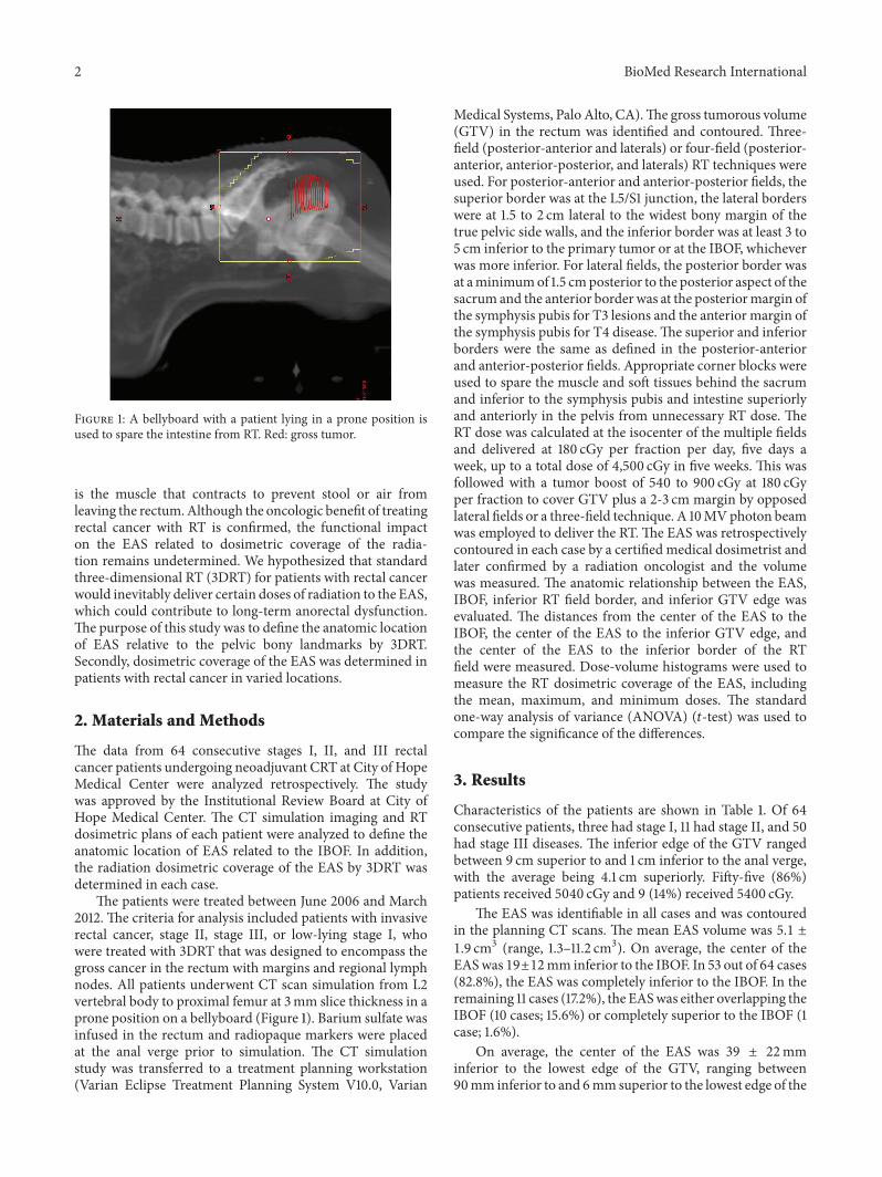
Figure 1: A bellyboard with a patient lying in a prone position isused to spare the intestine from RT. Red: gross tumor.
is the muscle that contracts to prevent stool or air fromleaving the rectum.Although the oncologic benefit of treatingrectal cancer with RT is confirmed, the functional impacton the EAS related to dosimetric coverage of the radia-tion remains undetermined. We hypothesized that standardthree-dimensional RT (3DRT) for patients with rectal cancerwould inevitably deliver certain doses of radiation to the EAS,which could contribute to long-term anorectal dysfunction.The purpose of this study was to define the anatomic locationof EAS relative to the pelvic bony landmarks by 3DRT.Secondly, dosimetric coverage of the EAS was determined inpatients with rectal cancer in varied locations.
2. Materials and Methods
The data from 64 consecutive stages I, II, and III rectalcancer patients undergoing neoadjuvant CRT at City of HopeMedical Center were analyzed retrospectively. The studywas approved by the Institutional Review Board at City ofHope Medical Center. The CT simulation imaging and RTdosimetric plans of each patient were analyzed to define theanatomic location of EAS related to the IBOF. In addition,the radiation dosimetric coverage of the EAS by 3DRT wasdetermined in each case.
The patients were treated between June 2006 and March2012. The criteria for analysis included patients with invasiverectal cancer, stage II, stage III, or low-lying stage I, whowere treated with 3DRT that was designed to encompass thegross cancer in the rectum with margins and regional lymphnodes. All patients underwent CT scan simulation from L2vertebral body to proximal femur at 3mm slice thickness in aprone position on a bellyboard (Figure 1). Barium sulfate wasinfused in the rectum and radiopaque markers were placedat the anal verge prior to simulation. The CT simulationstudy was transferred to a treatment planning workstation(Varian Eclipse Treatment Planning System V10.0, Varian
Medical Systems, Palo Alto, CA).The gross tumorous volume(GTV) in the rectum was identified and contoured. Three-field (posterior-anterior and laterals) or four-field (posterior-anterior, anterior-posterior, and laterals) RT techniques wereused. For posterior-anterior and anterior-posterior fields, thesuperior border was at the L5/S1 junction, the lateral borderswere at 1.5 to 2 cm lateral to the widest bony margin of thetrue pelvic side walls, and the inferior border was at least 3 to5 cm inferior to the primary tumor or at the IBOF, whicheverwas more inferior. For lateral fields, the posterior border wasat aminimumof 1.5 cmposterior to the posterior aspect of thesacrum and the anterior border was at the posteriormargin ofthe symphysis pubis for T3 lesions and the anterior margin ofthe symphysis pubis for T4 disease.The superior and inferiorborders were the same as defined in the posterior-anteriorand anterior-posterior fields. Appropriate corner blocks wereused to spare the muscle and soft tissues behind the sacrumand inferior to the symphysis pubis and intestine superiorlyand anteriorly in the pelvis from unnecessary RT dose. TheRT dose was calculated at the isocenter of the multiple fieldsand delivered at 180 cGy per fraction per day, five days aweek, up to a total dose of 4,500 cGy in five weeks. This wasfollowed with a tumor boost of 540 to 900 cGy at 180 cGyper fraction to cover GTV plus a 2-3 cm margin by opposedlateral fields or a three-field technique. A 10MVphoton beamwas employed to deliver the RT. The EAS was retrospectivelycontoured in each case by a certified medical dosimetrist andlater confirmed by a radiation oncologist and the volumewas measured. The anatomic relationship between the EAS,IBOF, inferior RT field border, and inferior GTV edge wasevaluated. The distances from the center of the EAS to theIBOF, the center of the EAS to the inferior GTV edge, andthe center of the EAS to the inferior border of the RTfield were measured. Dose-volume histograms were used tomeasure the RT dosimetric coverage of the EAS, includingthe mean, maximum, and minimum doses. The standardone-way analysis of variance (ANOVA) (𝑡-test) was used tocompare the significance of the differences.
3. Results
Characteristics of the patients are shown in Table 1. Of 64consecutive patients, three had stage I, 11 had stage II, and 50had stage III diseases. The inferior edge of the GTV rangedbetween 9 cm superior to and 1 cm inferior to the anal verge,with the average being 4.1 cm superiorly. Fifty-five (86%)patients received 5040 cGy and 9 (14%) received 5400 cGy.
The EAS was identifiable in all cases and was contouredin the planning CT scans. The mean EAS volume was 5.1 ±1.9 cm3 (range, 1.3–11.2 cm3). On average, the center of theEASwas 19±12mm inferior to the IBOF. In 53 out of 64 cases(82.8%), the EAS was completely inferior to the IBOF. In theremaining 11 cases (17.2%), the EASwas either overlapping theIBOF (10 cases; 15.6%) or completely superior to the IBOF (1case; 1.6%).
On average, the center of the EAS was 39 ± 22mminferior to the lowest edge of the GTV, ranging between90mm inferior to and 6mmsuperior to the lowest edge of the

BioMed Research International 3
Table 1: Characteristics of the patients.
Characteristics NumberNumber of patients 64Staging
T2N0 3T2N1 2T3N0 9T3N1 45T3N2 2T4N0 2T4N1 1
GTV to anal vergeMean 4.1 cmRange −1 to 9 cm
RT dose5040 cGy 555400 cGy 9
GTV.On average, the center of the EASwas 2±7mmsuperiorto the inferior border of the RT field, ranging between 42mminferior to and 27mm superior to the inferior border of theRT field. In 19 cases (30%), the EAS was completely outsideof the inferior border of the RT field. The average mean dosedelivered to the EAS was 2795 cGy (range, 245–5441 cGy).Lower mean doses delivered to the EAS were noted for casesthat had larger distances from the center of the EAS to theGTV inferior border (Figure 2). The average mean dosedelivered to the EAS for cases with a distance more than 4 cmwas 1264 ± 993 cGy, and for a distance less than 4 cm it was4045 ± 1087 cGy (𝑃 < 0.00001). For cases where the centerof the EAS was located more than 5mm inferior to the field’sinferior border, the mean EAS dose was 607 cGy. In contrast,for cases where the center of the EAS was located more than5mm superior to the field’s inferior border, the mean EASdose was more than 4000 cGy (𝑃 < 0.00001, Figure 3). Atypical case showing the anatomic relationship between theEAS, GTV, and pelvic bony landmarks for RT is illustrated inFigure 4. Please note that in this case the EAS was completelydistal to IBOF.
4. Discussion
To our knowledge, this is the first study to define the anatomiclocation of the EAS relative to pelvic bony landmarks by3DRT in a prone position setup. This is also the first study tosummarize the dosimetric coverage of EAS for patients withrectal cancer treated by 3DRT. Our data indicate that in allcases the EAS was identifiable in CT scans. In 82.8% of cases,the EAS was completely located inferior to the IBOF, whichmeans, inmajority of cases, that the EAS could be spared fromunnecessary radiation exposure if the lower edges of the fieldwere set at IBOF.
While the oncologic benefits of neoadjuvant pelvic RT forpatients with rectal cancer have been confirmed by severalrandomized studies [1, 5, 6], there are only a few studies
0
1000
2000
3000
4000
5000
6000
0 20 40 60 80 100
Mea
n do
se o
f the
sphi
ncte
r (cG
y)
Distance between center of the sphincter and the GTV inferior border (mm)
−20
Figure 2: Mean dose of EAS versus distance between center of theEAS and the GTV inferior border.
0
1000
2000
3000
4000
5000
6000
0 10 20 30 40 50
Mea
n do
se o
f the
sphi
ncte
r (cG
y)
Distance between center of the sphincterand the inferior border of RT field (mm)
−20 −10−30−40
Figure 3: Mean dose of EAS versus distance between center of theEAS and the inferior border of the RT field.
IBOFCenter of sphincter
Lowest edge of GTV
AB
A: the center of anal sphincter was 19mm
B: the center of anal sphincter was 39mm inferior to the lowest edge of GTV.
inferior to the IBOF.
Figure 4: A typical case showing the anatomic relationship betweenthe EAS, GTV, and pelvic bony landmarks for RT; solid red: GTV;magenta: EAS.

4 BioMed Research International
addressing the impact of RT on anorectal function. Certainly,cancer per se and surgery alone, such as total mesorectalresection, can compromise normal anorectal function andcause different levels of fecal frequency, urgency, inconti-nence, or emptying difficulties [7, 8]. It is generally believedthat neoadjuvant RT could contribute a negative impact onanorectal function as well.Through a questionnaire survey bythe Swedish Rectal Cancer Trial, compared to surgery-alonegroup, patients with preoperative short-course irradiation(5Gy × 5) had a significantly worsened long-term bowelfunction including increasing bowel frequency, incontinencefor loose stools, urgency, and emptying difficulties [9]. Inaddition, irradiated patients more commonly required astoma later because of poor anorectal function after surgery.More importantly, 30% of irradiated patients reported animpaired social life that was related to bowel dysfunction,compared to 10% of the surgery-alone group.
In another questionnaire survey by Peeters et al. from theDutch Colorectal Cancer Group Study [10], patients reportedconsiderable long-term adverse effects of preoperative RT by5Gy × 5 on anorectal functional outcome. Compared withnonirradiated patients, irradiated patients had increased ratesof fecal incontinence (62% versus 38%), had to wear a paddue to incontinence (56% versus 33%), and experienced analblood loss (11% versus 3%) and mucus loss (27% versus 15%).In addition, satisfactionwith bowel functionwas significantlylower in the irradiated group. While, from a radiobiologicalpoint of view, one can argue larger fraction size, 5 Gy pertreatment, could be the reason for poorer chronic side effects,more recently, Bruheim et al. from Norway reported consid-erable long-termdetrimental effects on anorectal function forpatients receiving conventional long-course RT (2Gy × 25 or1.8 Gy × 28) [2]. In patients without a stoma, with a mediantime of 4.8 years since surgery, compared to patients whohad surgery alone, a higher proportion of irradiated patientswere incontinent for liquid stools (49% versus 15%), neededa sanitary pad (52% versus 13%), and lacked the ability todefer defecation (44% versus 16%). Poorer global quality oflife and social function were also noted for these irradiatedpatients.
Indeed, irradiating normal structures could cause endo-vascular injury and overproduction of fibrogenic cytokines,such as transforming growth factors, which could lead toradiation-induced fibrosis as an end result [11]. Specifically,there has been quantitative evidence confirming sphincterdysfunction frompelvic RT for cervical, prostate, and anorec-tal cancers [12–16]. It is generally believed that anal canalpressure, especially basal resting pressure, could be reducedsignificantly by RT compared to baseline preirradiated state.In a prospective study for patients with mid and low rectalcancer, Ammann et al. compared anorectal manometricvalues before and after surgery [16]. Anorectal manome-try was performed preoperatively and at a median of 383days postoperatively. The mean resting pressure for patientswith neoadjuvant CRT decreased significantly from 89 ±35mmHg preoperatively down to 53 ± 17mmHg postop-eratively. In contrast, no statistically significant manometricdifferences occurred before and after surgery for patientswho underwent surgery alone. Therefore, shielding of the
anal sphincter was recommended whenever a sphincter-preserving procedure was considered.
Radiation portals for patients with rectal cancer aregenerally defined by anatomic locations of cancer, lymphaticpathways of drainage, and patterns of locoregional cancerrecurrence. Reviewing patterns of failure in 75 rectal cancerpatients using second or symptomatic look operations follow-ing curative surgery, Gunderson and Sosin found that localfailure and/or regional lymph node metastases occurred asthe only failure in nearly 50% of the failure group [17]. Thebenefits of radiation treatment were suggested, and, based onfailure patterns, appropriate radiation portals were defined.Specifically, it was suggested that the inferior border of thefield be set at 3 to 5 cm inferior to the primary tumor or atthe IBOF, whichever was more inferior [4, 17]. The currentstandard is to use a multiple beam setup and the borders ofthe fields are determined based on the anatomic locations ofcancer and pelvic bony landmarks.
With the increasing need to implement target-directedRT for better results, recently, consensus on structures thatshould be included in the target volume for patients withrectal cancer has been defined [18, 19]. Based on the recur-rence data from a systematic review of 18 studies, Roelset al. concluded that the primary tumor, the mesorectum,and the presacral and internal iliac nodal areas shouldbe covered in all cases [18]. With respect to the inferiorextent of the target volume it is agreed that a minimumof 2 cm inferior to the gross disease is needed and theentire mesorectum to the pelvic floor should be includedeven for cases with upper rectal cancer [19]. Mesorectum isthe mesentery with lymphovascular and neural structuressupporting and connecting the midupper portion of therectum with the sacrum. It is cylindrical in shape startingat the level of sacral promontory and ending at the levelwhere the levator ani muscle meets with the rectal wall.For patients with positive pelvic lymph nodes, by systematicreview, the mesorectal lymph nodes were involved in up to87% of the cases [18]. After evaluating surgical specimens in311 consecutive patients with colorectal cancer, Morikawa etal. reported that no lymphatic metastasis was noted morethan 4 cm distal to the tumor in the mesorectum [20]. Todefine the inferior border of RT fields, in addition to theanatomic location of the primary tumor and the extent ofmesorectum, the internal iliac lymph nodes, specifically theareas with obturator lymph nodes, need to be taken intoconsideration. Inferiorly, it is recommended to include theobturator lymph nodes till the level where the obturatorartery enters the obturator canal [18]. In any case, becausethe EAS is located inferior to the levator ani and in thecurrent study in 98.4% of cases the EASwas either completelyinferior to (82.8%) or overlapping with (15.6%) the IBOF,theoretically, fields of 3DRT should be designed to spare theEAS for mid/upper rectal cancer where there is no evidenceof EAS cancerous involvement. Of note, for cases in this studythat had the tumor locatedmore than 4 cmabove the EAS, theEAS was spared from unnecessary radiation (average meandose 1264 cGy) in the majority of cases, even though theEAS was not identified in the original RT planning. If theEAS was identified and RT plans were designed to avoid it,

BioMed Research International 5
(a) (b)
(c) (d)
Figure 5: A patient with a rectal cancer located 2.7 cm above the EAS. (a) The original RT field covered 4 cm inferior to the gross tumor. (b)The EAS can be spared from radiation treatment easily by reducing the inferior border superiorly by 2 cm. (c) Axial CT image through theEAS shows that no draining lymph nodes need to be covered. (d) Coronal scout view shows the level of axial image (red line). Solid red: grosstumor; green: EAS.
it is likely that the dose of RT to the EAS would be evenlower in such a patient population. For cases where the tumoris located within 4 cm of the EAS, given that margins areneeded to add to clinical target volume to account for setupvariation, it will not be easy to spare EAS by conventional3DRT. However, with daily image guidance to reduce theneed of adding planning target margins and by designatingthe EAS as an avoidance structure, sparing the EAS fromRT would be possible in certain cases that have the cancerlocated 2 to 4 cm superior to EAS (Figure 5). Apparently, ifthe cancer is within 2 cm of the EAS, or an abdominoperinealresection is planned after neoadjuvant CRT, the EAS shouldbe considered as part of RT targets and covered.
One of the limitations of the current study is that wewere not able to define the internal anal sphincter by the CTscans. It is essential to point out that while sparing the EASfrom unnecessary RT is recommended when it is feasible,the cephalad extent of the internal anal sphincter is at thelevel of the caudad extent of the mesorectum and, therefore,at least the upper portions of the internal anal sphincter willinevitably be included in the RT fields. Another limitation is
that the correlation between RT dose distribution to the EASand anal-rectal dysfunctionwas not studied, whichwill be thefocus of our future studies.
5. Conclusions
In summary, we have evaluated the anatomic location of theEAS to pelvic bony landmarks related to 3DRT and studiedthe dosimetric coverage of the EAS in patients undergoingneoadjuvant CRT for rectal cancer. 3D planning under CTguidance allows accurate delineation of the EAS. In 82.8% ofpatients, the EAS is located completely inferior to the IBOF.Because RT can cause anorectal dysfunction, meticulousplanning to define the inferior border of the RT field isrecommended to spare the EAS.
Abbreviations
EAS: External anal sphincter3DRT: 3-Dimensional conformal radiotherapy

6 BioMed Research International
GTV: Gross tumorous volumeIBOF: Inferior border of the obturator foramenCRT: ChemoradiationRT: Radiotherapy.
Conflict of Interests
All the authors declare that there is no conflict of interestsrelated to the current study.
Authors’ Contribution
Yi-Jen Chen and An Liu designed and coordinated the study.Yi-Jen Chen drafted the paper. An Liu and Peter Tsai carriedout the statistical analysis of the data. Julian Sanchez reviewedthe design and revised the paper critically. Michelle B. Chenand Peter Tsai delineated contours of critical organs andcollected data. Alan J. Liu participated in data collection andinterpretation of the results. Michelle B. Chen and Alan J.Liu edited the paper. All authors have read, reviewed, andapproved the final paper.
References
[1] R. Sauer, H. Becker,W. Hohenberger et al., “Preoperative versuspostoperative chemoradiotherapy for rectal cancer,” The NewEngland Journal of Medicine, vol. 351, no. 17, pp. 1731–1810, 2004.
[2] K. Bruheim, M. G. Guren, E. Skovlund et al., “Late sideeffects and quality of life after radiotherapy for rectal cancer,”International Journal of Radiation Oncology Biology Physics, vol.76, no. 4, pp. 1005–1011, 2010.
[3] M. J. Gallagher, H. D. Brereton, R. A. Rostock et al., “Aprospective study of treatment techniques to minimize thevolume of pelvic small bowel with reduction of acute and lateeffects associated with pelvic irradiation,” International Journalof Radiation Oncology, Biology, Physics, vol. 12, no. 9, pp. 1565–1573, 1986.
[4] B. D. Minsky, M. L. Welton, and A. P. Venook, “Cancer of therectum,” in Radiation Oncology, R. T. Hoppe, T. L. Phillips, andM. Roach III, Eds., pp. 851–869, Elsevier Saunders, Philadelphia,Pa, USA, 3rd edition, 2010.
[5] L. Pahlman, “Improved survival with preoperative radiother-apy in resectable rectal cancer,” The New England Journal ofMedicine, vol. 336, no. 14, pp. 980–987, 1997.
[6] E. Kapiteijn, C. A. M. Marijnen, I. D. Nagtegaal et al., “Preoper-ative radiotherapy combined with total mesorectal excision forresectable rectal cancer,”The New England Journal of Medicine,vol. 345, no. 9, pp. 638–646, 2001.
[7] P. J. McDonald and R. J. Heald, “A survey of postoperativefunction after rectal anastomosis with circular stapling devices,”British Journal of Surgery, vol. 70, no. 12, pp. 727–729, 1983.
[8] G. Batignani, I. Monaci, F. Ficari, and F. Tonelli, “What affectscontinence after anterior resection of the rectum?” Diseases ofthe Colon and Rectum, vol. 34, no. 4, pp. 329–335, 1991.
[9] M. Dahlberg, B. Glimelius, W. Graf, and L. Pahlman, “Preoper-ative irradiation affects functional results after surgery for rectalcancer: results from a randomized study,” Diseases of the Colonand Rectum, vol. 41, no. 5, pp. 543–551, 1998.
[10] K. C.M. J. Peeters, C. J. H. van deVelde, J.W.H. Leer et al., “Lateside effects of short-course preoperative radiotherapy combined
with totalmesorectal excision for rectal cancer: Increased boweldysfunction in irradiated patients—a Dutch Colorectal CancerGroup Study,” Journal of Clinical Oncology, vol. 23, no. 25, pp.6199–6206, 2005.
[11] G. C. Blobe, W. P. Schiemann, and H. F. Lodish, “Role oftransforming growth factor beta in human disease,” The NewEngland Journal of Medicine, vol. 342, no. 18, pp. 1350–1358,2000.
[12] T. Iwamoto, S. Nakahara, R. Mibu, M. Hotokezaka, H. Nakano,andM. Tanaka, “Effect of radiotherapy on anorectal function inpatients with cervical cancer,”Diseases of the Colon and Rectum,vol. 40, no. 6, pp. 693–697, 1997.
[13] E. K. Yeoh, A. Russo, R. Botten et al., “Acute effects oftherapeutic irradiation for prostatic carcinoma on anorectalfunction,” Gut, vol. 43, no. 1, pp. 123–127, 1998.
[14] P. Broens, E. van Limbergen, F. Penninckx, and R. Kerremans,“Clinical and manometric effects of combined external beamirradiation and brachytherapy for anal cancer,” InternationalJournal of Colorectal Disease, vol. 13, no. 2, pp. 68–72, 1998.
[15] P. Gervaz, N. Rotholtz, M. Pisano et al., “Quantitative short-term study of anal sphincter function after chemoradiation forrectal cancer,” Archives of Surgery, vol. 136, no. 2, pp. 192–196,2001.
[16] K. Ammann, W. Kirchmayr, A. Klaus et al., “Impact of neoad-juvant chemoradiation on anal sphincter function in patientswith carcinoma of the midrectum and low rectum,” Archives ofSurgery, vol. 138, no. 3, pp. 257–261, 2003.
[17] L. L. Gunderson and H. Sosin, “Areas of failure found atreoperation (second or symptomatic look) following “curativesurgery” for adenocarcinoma of the rectum: clinicopathologiccorrelation and implications for adjuvant therapy,” Cancer, vol.34, no. 4, pp. 1278–1292, 1974.
[18] S. Roels, W. Duthoy, K. Haustermans et al., “Definition anddelineation of the clinical target volume for rectal cancer,”International Journal of Radiation Oncology Biology Physics, vol.65, no. 4, pp. 1129–1142, 2006.
[19] R. J. Myerson, M. C. Garofalo, I. El Naqa et al., “Electiveclinical target volumes for conformal therapy in anorectalcancer: a radiation therapy oncology group consensus panelcontouring atlas,” International Journal of Radiation OncologyBiology Physics, vol. 74, no. 3, pp. 824–830, 2009.
[20] E. Morikawa, M. Yasutomi, K. Shindou et al., “Distribution ofmetastatic lymph nodes in colorectal cancer by the modifiedclearing method,” Diseases of the Colon and Rectum, vol. 37, no.3, pp. 219–223, 1994.

Submit your manuscripts athttp://www.hindawi.com
Stem CellsInternational
Hindawi Publishing Corporationhttp://www.hindawi.com Volume 2014
Hindawi Publishing Corporationhttp://www.hindawi.com Volume 2014
MEDIATORSINFLAMMATION
of
Hindawi Publishing Corporationhttp://www.hindawi.com Volume 2014
Behavioural Neurology
EndocrinologyInternational Journal of
Hindawi Publishing Corporationhttp://www.hindawi.com Volume 2014
Hindawi Publishing Corporationhttp://www.hindawi.com Volume 2014
Disease Markers
Hindawi Publishing Corporationhttp://www.hindawi.com Volume 2014
BioMed Research International
OncologyJournal of
Hindawi Publishing Corporationhttp://www.hindawi.com Volume 2014
Hindawi Publishing Corporationhttp://www.hindawi.com Volume 2014
Oxidative Medicine and Cellular Longevity
Hindawi Publishing Corporationhttp://www.hindawi.com Volume 2014
PPAR Research
The Scientific World JournalHindawi Publishing Corporation http://www.hindawi.com Volume 2014
Immunology ResearchHindawi Publishing Corporationhttp://www.hindawi.com Volume 2014
Journal of
ObesityJournal of
Hindawi Publishing Corporationhttp://www.hindawi.com Volume 2014
Hindawi Publishing Corporationhttp://www.hindawi.com Volume 2014
Computational and Mathematical Methods in Medicine
OphthalmologyJournal of
Hindawi Publishing Corporationhttp://www.hindawi.com Volume 2014
Diabetes ResearchJournal of
Hindawi Publishing Corporationhttp://www.hindawi.com Volume 2014
Hindawi Publishing Corporationhttp://www.hindawi.com Volume 2014
Research and TreatmentAIDS
Hindawi Publishing Corporationhttp://www.hindawi.com Volume 2014
Gastroenterology Research and Practice
Hindawi Publishing Corporationhttp://www.hindawi.com Volume 2014
Parkinson’s Disease
Evidence-Based Complementary and Alternative Medicine
Volume 2014Hindawi Publishing Corporationhttp://www.hindawi.com


![[MC-DRT]: Distributed Routing Table (DRT) Version 1€¦ · Distributed Routing Table (DRT) Version 1.0 Intellectual Property Rights Notice for Open Specifications Documentation](https://static.fdocuments.us/doc/165x107/603ec42a19edb942e54403aa/mc-drt-distributed-routing-table-drt-version-1-distributed-routing-table-drt.jpg)