Clinical Study A Randomised Controlled Trial of...
10
Clinical Study A Randomised Controlled Trial of Efficacy of Cognitive Rehabilitation in Multiple Sclerosis: A Cognitive, Behavioural, and MRI Study J. Campbell, 1 D. Langdon, 2 M. Cercignani, 1 and W. Rashid 3 1 Clinical Imaging Sciences Centre, Brighton and Sussex Medical School, Falmer, UK 2 Department of Psychology, Royal Holloway, University of London, London, UK 3 Department of Neurology, Brighton and Sussex University Hospitals NHS Trust, Brighton, UK Correspondence should be addressed to J. Campbell; [email protected] Received 21 July 2016; Revised 29 October 2016; Accepted 17 November 2016 Academic Editor: Malgorzata Kossut Copyright © 2016 J. Campbell et al. is is an open access article distributed under the Creative Commons Attribution License, which permits unrestricted use, distribution, and reproduction in any medium, provided the original work is properly cited. Aim. To explore the efficacy of home-based, computerised, cognitive rehabilitation in patients with multiple sclerosis using neuropsychological assessment and advanced structural and functional magnetic resonance imaging (fMRI). Methods. 38 patients with MS and cognitive impairment on the Brief International Cognitive Assessment for MS (BICAMS) were enrolled. Patients were randomised to undergo 45 minutes of computerised cognitive rehabilitation using RehaCom soſtware ( = 19) three times weekly for six weeks or to a control condition ( = 19). Neuropsychological and MRI data were obtained at baseline (time 1), following the 6-week intervention (time 2), and aſter a further twelve weeks (time 3). Cortical activations were explored using fMRI and microstructural changes were explored using quantitative magnetisation transfer (QMT) imaging. Results. e treatment group showed a greater improvement in SDMT gain scores between baseline and time 2 compared to the control group ( = 0.005). e treatment group exhibited increased activation in the bilateral prefrontal cortex and right temporoparietal regions relative to control group at time 3 ( < 0.05 FWE corrected ). No significant changes were observed on QMT. Conclusion. is study supports the hypothesis that home-based, computerised, cognitive rehabilitation may be effective in improving cognitive performance in patients with MS. Clinical trials registration is ISRCTN54901925. 1. Introduction Cognitive impairment is present in 40–65% of individuals with MS [1]. Studies have shown cognitive deficits (in partic- ular deficits in information processing speed, concentration, and working memory) to be present in the early stages of MS [2–4]. Cognitive impairment has a negative impact on quality of life (QOL) independent of physical symptoms [5, 6]. ere exists mounting evidence for neuroplasticity as a mechanism to compensate for accumulating pathology in MS and some tentative evidence that cognitive rehabilitation may be effective in preserving or improving cognitive function in patients with MS [7–9]. Computer-assisted cognitive rehabilitation has the potential to provide a structured and standardised approach to rehabilitation. RehaCom is one par- ticular type of soſtware designed and utilised for treatment of cognitive impairment in a number of disease states such as stroke, brain injury, and psychiatric disorders [10, 11]. It has been used in a growing number of trials of cognitive rehabilitation in MS as a more standardised intervention [8, 9, 12, 13]. e difficulty level of the computerised tasks adapts to an individual’s performance, only increasing in difficulty in response to improving performance. Few studies have examined the structural basis of cog- nitive rehabilitation and longitudinal studies are relatively lacking [8, 14]. Animal data suggest that myelination is, at least in part, regulated by neuronal activity [15]. It is therefore conceivable that techniques, such as magnetisation transfer Hindawi Publishing Corporation Neural Plasticity Volume 2016, Article ID 4292585, 9 pages http://dx.doi.org/10.1155/2016/4292585
Transcript of Clinical Study A Randomised Controlled Trial of...

Clinical StudyA Randomised Controlled Trial of Efficacy ofCognitive Rehabilitation in Multiple Sclerosis: A Cognitive,Behavioural, and MRI Study
J. Campbell,1 D. Langdon,2 M. Cercignani,1 and W. Rashid3
1Clinical Imaging Sciences Centre, Brighton and Sussex Medical School, Falmer, UK2Department of Psychology, Royal Holloway, University of London, London, UK3Department of Neurology, Brighton and Sussex University Hospitals NHS Trust, Brighton, UK
Correspondence should be addressed to J. Campbell; [email protected]
Received 21 July 2016; Revised 29 October 2016; Accepted 17 November 2016
Academic Editor: Malgorzata Kossut
Copyright © 2016 J. Campbell et al. This is an open access article distributed under the Creative Commons Attribution License,which permits unrestricted use, distribution, and reproduction in any medium, provided the original work is properly cited.
Aim. To explore the efficacy of home-based, computerised, cognitive rehabilitation in patients with multiple sclerosis usingneuropsychological assessment and advanced structural and functional magnetic resonance imaging (fMRI).Methods. 38 patientswithMS and cognitive impairment on the Brief International Cognitive Assessment forMS (BICAMS) were enrolled. Patients wererandomised to undergo 45 minutes of computerised cognitive rehabilitation using RehaCom software (𝑛 = 19) three times weeklyfor six weeks or to a control condition (𝑛 = 19). Neuropsychological and MRI data were obtained at baseline (time 1), followingthe 6-week intervention (time 2), and after a further twelve weeks (time 3). Cortical activations were explored using fMRI andmicrostructural changes were explored using quantitative magnetisation transfer (QMT) imaging. Results. The treatment groupshowed a greater improvement in SDMT gain scores between baseline and time 2 compared to the control group (𝑝 = 0.005).The treatment group exhibited increased activation in the bilateral prefrontal cortex and right temporoparietal regions relative tocontrol group at time 3 (𝑝 < 0.05FWE corrected). No significant changes were observed on QMT. Conclusion. This study supportsthe hypothesis that home-based, computerised, cognitive rehabilitation may be effective in improving cognitive performance inpatients with MS. Clinical trials registration is ISRCTN54901925.
1. Introduction
Cognitive impairment is present in 40–65% of individualswith MS [1]. Studies have shown cognitive deficits (in partic-ular deficits in information processing speed, concentration,and working memory) to be present in the early stages of MS[2–4]. Cognitive impairment has a negative impact on qualityof life (QOL) independent of physical symptoms [5, 6].
There exists mounting evidence for neuroplasticity as amechanism to compensate for accumulating pathology inMSand some tentative evidence that cognitive rehabilitationmaybe effective in preserving or improving cognitive functionin patients with MS [7–9]. Computer-assisted cognitiverehabilitation has the potential to provide a structured and
standardised approach to rehabilitation. RehaCom is one par-ticular type of software designed and utilised for treatmentof cognitive impairment in a number of disease states suchas stroke, brain injury, and psychiatric disorders [10, 11]. Ithas been used in a growing number of trials of cognitiverehabilitation in MS as a more standardised intervention [8,9, 12, 13].The difficulty level of the computerised tasks adaptsto an individual’s performance, only increasing in difficultyin response to improving performance.
Few studies have examined the structural basis of cog-nitive rehabilitation and longitudinal studies are relativelylacking [8, 14]. Animal data suggest that myelination is, atleast in part, regulated by neuronal activity [15]. It is thereforeconceivable that techniques, such as magnetisation transfer
Hindawi Publishing CorporationNeural PlasticityVolume 2016, Article ID 4292585, 9 pageshttp://dx.doi.org/10.1155/2016/4292585
2 Neural Plasticity
(MT), which is sensitive tomyelin content, might be sensitiveto structural plasticity in MS [16].
In this study we combined neuropsychological assess-ment, functional MRI (fMRI), and quantitative magneti-sation transfer (QMT) imaging to explore whether home-based, computerised cognitive rehabilitation is an effectivemeans of promoting cognitive rehabilitation and whetherthe structural basis for rehabilitation can be better defined[17]. The primary outcome of the study was measured asany improvement in cognition after the training, while thesecondary outcomes included changes in fMRI, fatigue, andquality of life assessments.
2. Subjects and Method
Participants. Thirty-eight patients with objective evidence ofcognitive impairment were invited to participate in this studybetween February 2014 and February 2015. All participantssigned informed written consent before undergoing testing.Inclusion criteria were as follows: (a) age between 18 and 65,(b) clinically definiteMS, according to theMcDonald criteria[18], (c) Expanded Disability Status Scale (EDSS) ≤ 6.5, and(d) cognitive impairment defined as scores below the 5thpercentile for normative data adjusted for age, sex, and yearsof formal education [19] on one ormore of the BICAMS tests.
Patients were excluded if they had a history of significantpsychiatric disorders, alcohol or substance abuse, visual acu-ity less than 6/18 corrected, oscillopsia, or diplopia that wouldinterfere with testing. Patients were also excluded if they hada MS relapse, received corticosteroids, or changes made topsychoactive medications within the previous month.
The studywas approved by theNorthern IrelandResearchEthics Committee.
Study Design. An open-design, randomised, controlled trialwas conducted. Neuropsychological and MRI data wereobtained at baseline (time 1), immediately following a 6-weekintervention period (time 2) and after an additional 12-weekfollow-up period (time 3), during which no additional inter-vention was administered (supplementary Figure 1 in Supple-mentaryMaterial available online at http://dx.doi.org/10.1155/2016/4292585).
It was not possible for the cognitive assessments to becompleted by a blinded assessor. The MRI analysis was con-ducted by a researcher blind to the patients’ group allocation.
Cognitive and Behavioural Assessments. At entry all partici-pants underwent a detailed clinical neurological assessmentincluding EDSS conducted by an experienced neurologist.Patients were screened for cognitive impairment using theBrief International Cognitive Assessment for MS (BICAMS).BICAMS is a brief (15-minute) screening tool to identifycognitive impairment in patients with MS and comprises thefirst five learning trials of the California Verbal Learning TestII (CVLT-II), the first three recall trials of Brief VisuospatialMemory Test Revised (BVMT-R), and the Symbol DigitsModalities Test (SDMT) [20].
The BICAMS assessment was conducted by a neurologyclinical fellow with almost ten years of clinical experience
(J.C). The assessing neurologist was trained in BICAMSassessing methods by an experienced neuropsychologist(D.L).
At baseline participants also completed a number ofbehavioural and QOL assessments including EuroQOL five-dimension questionnaire (EQ-5D), a generic health-relatedquality of life scale [21], Functional Assessment of MS(FAMS) (a MS specific quality of life scale) [22], Patient Acti-vationMeasure (PAM-13) (a 13-item generic scale for chronicillness management), a measure of patient “empowerment”in MS [23], Unidimensional Self-Efficacy scale for MS (USE-MS) [24], theHospital Anxiety andDepression Scale (HADS)[25], Multiple Sclerosis Neuropsychological Questionnaire(MSNQ) self-report (a patient self-reported measure of cog-nitive function) [26], and the Fatigue Severity Scale [27].
At each subsequent time point participants underwentrepeat cognitive assessment using BICAMS (same test forms)as well as repeat behavioural and QOL assessments.
Randomisation. Following baseline MRI, patients were ran-domised to either the treatment or control groups. Randomi-sation was performed using a random number generator andallocations were placed inside sealed folders. Folders wereopened following baseline MRI.
Intervention. The treatment group underwent six weeksof home-based, computer-assisted cognitive rehabilitationusing RehaCom software (https://www.fixxl.co.uk/). Thisconsisted of 45-minute sessions, three times weekly. Thecontrol group were asked to watch a series of natural historyDVDs of corresponding duration and frequency to therehabilitation sessions performed by the treatment group forsix weeks. The need to evaluate MRI parameters in studieswith active control conditions has been highlighted [14].
Treatment sessions consisted of training in three specificmodules involving working memory, visuospatial memory,and divided attention. In all tasks the level of difficultyis tailored to the individuals performance and increasesautomatically but only in line with satisfactory progress.Real-time data pertaining to performance, progress, andcompliance is transmitted to the investigator over the Internetduring the intervention period.
“Divided Attention” Module. In the divided attention taskthe patient is asked to drive a simulated car using keyboardinputs. Multiple distractions must be navigated and thespeed and direction of the vehicle altered according to roadconditions. As the complexity of the task increases, moredistractors are introduced with increased multitasking skillsrequired.
“Working Memory” Module. The working memory task con-sists of remembering a series of playing cards presentedbriefly on screen.The participant is then asked to select whichcardswere presented froma longer series of options includingdistractor cards. As the complexity of the task increases,participants are asked to remember only cards of a particularvalue or suit and the number of items to remember increases.Higher levels involve having to remember the cards in reverseorder.

Neural Plasticity 3
“Topological Memory” Module. Visuospatial memory is asimilar task involving various objects presented briefly onscreen with the patient asked to remember the object as wellas its position in the sequence. As the complexity of the taskincreases, the number of items on screen increases and moreabstract shapes are introduced.
MRI Imaging Protocol. The following sequences wereacquired in an order designed to minimise the potentialfor fatigue on the fMRI task: (1) dual-echo turbo spin-echofor lesion identification; (2) high-resolution T1-weightedmagnetisation-prepared rapid-acquisition gradient echo(MPRAGE); (3) functional MRI with echo-planar imaging(EPI) acquired during a 𝑛-visually presented back task; (4)quantitative magnetisation transfer (QMT) with balancedsteady-state free precession (bSSFP) [28].
T2 lesion volume was measured at baseline for each par-ticipant using the software package JIM (Version 3.0, XinapseSystems Ltd., Northamptonshire, UK, http://www.xinapse.com/).
𝑁-Back Task. A visual 𝑛-back test was presented duringfunctional imaging acquisition.This was adapted from Sweetet al. [29] and involved three conditions: 0-back, 1-back, and2-back tasks.The 0-back condition was designed to act as thebaseline condition and would provide the baseline activationfor comparison in fMRI analysis. The 1-back and 2-backconditions provided increasing working memory demands.
The 𝑛-back task did not constitute part of the cognitiverehabilitation. All participants were allowed to briefly prac-tice the 𝑛-back task under supervision for five minutes priorto the MRI scan to ensure comprehension of the task andallow familiarity with it.
The visual 𝑛-back task was presented using Cogent Vand MATLAB 2013a. Stimuli were projected onto a mirroredscreen inside the MRI scanner 45 cm from a participant’snose. An MRI compatible button box was placed in theparticipant’s right hand.
White letters were projected onto a black backgroundin bold size 200 Arial font. This involved of a series ofpseudo-randomised consonants in both upper and lowercases. The stimulus duration was 1000ms with a betweenstimulus interval of 2000ms. Instructions were presented for3000ms before each new 𝑛-back task.
fMRI data were acquired during three 9-minute runs.0-back, 1-back, and 2-back tasks were presented in a ran-domisedmanner resulting in six blocks per nine-minute run.Each block consisted of 126 stimuli, one-third of which weretargets. Twice as many 0-back tasks were presented as 1-backor 2-back. There was a rest period of 90 seconds betweenblocks.
Statistical Analysis. The primary outcome was cognitiveperformance as measured by improvement in SDMT, BVMT,and CVLT between groups compared to baseline. Secondaryoutcomes were QOL, fMRI, and QMT measures as detailedbelow.
(i) Behavioural Data. Descriptive statistics for normallydistributed continuous variables are expressed as mean
and standard deviation. Skewed continuous variables weresummarised using median and interquartile range (IQR).Categorical variables are summarised by frequencies andpercentages.
Normality of continuous variables was assessed using theKolmogorov-Smirnov test.
Baseline cognitive and behavioural measures were com-pared between the treatment and control groups. Categoricalvariables were compared by the Pearson 𝜒2 test.Themeans ofcontinuous variables were compared using the independentsamples 𝑡-test or the Mann–Whitney 𝑈 test for skewed data.
All tests were two-tailed; 𝑝-values less than 0.05 wereconsidered significant.
Outcomes were compared between the two groups usingindependent samples 𝑡-test to compare gain scores for cog-nitive data between groups. To compare differences betweengroups for other behavioural and QOL data, a 2 × 3 repeatmeasures analysis of variance (ANOVA)was usedwith “time”as the within-subject factor and “treatment” as the betweenfactor (active rehabilitation versus control).
Analyses were performed using SPSS version 21(Armonk, NY: IBM Corp).
(ii) Functional MRI Analysis. fMRI data were analysed usingSPM8 (Wellcome Department of Cognitive Neurology, UCL,London, http://www.fil.ion.ucl.ac.uk/spm/).
For each time series, the first five EPIs were discardedto ensure steady-state magnetisation. Individual EPIs werethen realigned to the first remaining image of the series byrigid-body transformation to correct for involuntary headmovements during acquisition before normalisation into astandard anatomical space (Montreal Neurological Institute[MNI]) using linear and nonlinear transformations. Finally,images were smoothed with an 8mm3 full-width-at-half-maximum (FWHM) 3D Gaussian kernel.
First-Level Analysis. For each participant, the difference inblood oxygen level-dependent (BOLD) response between the0-back, 1-back, and 2-back conditions was estimated at everyvoxel across the whole brain using the general linear model(GLM).This produced a series of contrasts representingmeanactivation during each 𝑛-back condition minus the 0-backcondition.
Second-Level Analysis. Each contrast obtained at the first-level was entered into a second-level GLM to generatesummary statistical parametric maps (SPMs). For between-group analysis of difference between the time points, we useda 3× 2 ANOVAflexible factorial design with group (between-subject) and time (within-subject) as separate factors toexamine themain effects on group (treatment versus control),time and the interaction between them to evaluate areasof relative change in activity after cognitive training versuscontrol.
The threshold for significance was set at alpha of 0.05corrected formultiple comparisons (family-wise error (FWE)corrected). Results are reported at cluster level throughout.Within each region of statistical significance, the location oflocal maxima of signal intensity increase is expressed as 𝑥, 𝑦,and 𝑧 coordinates in MNI space.

4 Neural Plasticity
(iii) Quantitative MT Analysis. The MT data were analysedusing SPM8.MT and T1mapping data from all three sessionswere first realigned to subject specific MPRAGE structuralimages using the SPM8 rigid-body registration function.The MPRAGE were then segmented into white matter, greymatter, and CSF to yield a parenchymal mask.
A T1 map was calculated for all datasets by fitting thetheoretical spoiled gradient echo as a function of the flip angleto the signal measured by the 3D FLASH sequences [30].MT parameters were obtained by performing a voxelwisenonlinear least squares fitting (Levenberg-Marquardt) to abinary spin bath model for bSSFP.
The statistical analysis was performed voxelwise in SPM8on the resulting warped and smoothed MT maps. The sameGLMs used for the second-level fMRI analysis and describedin the previous section were used for estimating the maineffects of time and group and the interaction between thesetwo factors.
3. Results
Baseline Characteristics. 38 patients were included in thestudy. The majority of the participants were female (71.1%).At entry 27 patients (70.3%) had RRMS, and 11 patients(29.7%) had SPMS. Patients were aged between 32 and 62(mean 47.37, SD 8.23). The duration of MS from diagnosisto enrolment ranged from 12 months to 40 years (mean11.61 years, SD ± 8.2 yrs). Median EDSS was 5.0 (3.5–6.0).20 patients (52.6%) were on disease modifying therapy atenrolment (natalizumab 𝑛 = 6, beta-interferon 𝑛 = 7,fingolimod 𝑛 = 6, and teriflunomide 𝑛 = 1).
After randomisation to either computer-assisted cogni-tive training (treatment group, 𝑛 = 19) or the active controlcondition (𝑛 = 19), there were no significant differences interms of baseline demographics (Table 1) or quality of lifemeasures (supplementary Table 3) between the two groups.
The treatment group had higher baseline cognitive scoreson the BICAMS battery; however, these did not differ signif-icantly from the control group.
The most frequently failed component of the BICAMStest battery was the SDMT with 33 (86.8%) of participantsscoring below the 5th centile, 18 (47.4%) failing the CVLT-II, and 13 (34.2%) failing the BVMT-R. Overall 21 (55.2%)failed one test, 10 (26.3%) failed two tests, and 7 (18.4%)failed all three tests of the BICAMS test battery. This level ofimpairment is consistent with other publishedMS samples onBICAMS [31–33].
Overall, 88.9% of patients (16/18) in the interventiongroup completed at least 75% of the prescribed sessions with66.7% (12/18) completing all the prescribed sessions.
Behavioural Outcomes. The main behavioural outcomes areshown in Table 2.
Time 2 versus Time 1. Compared to time 1, the treatment groupshowed a significantly greater improvement in gain scoresbetween baseline and early follow-up (time 2) compared tothe control group on the SDMT (treatment 3.94 (SD 5.08),
Proportion of patients showing greater than 10% improvementin SDMT immediately after intervention by group.
Percentage improvement in SDMT
SDMT% gain
Treatment Control
−30
−20
−10
0
10
20
30
40
Proportion of patients showing greater than 10% improvemenin SDMT immediately after intervention by group.
SDMT% gain
Figure 1: Improvement in SDMT slope immediately after interven-tion.
controls −0.63 (SD 3.30), 1.47 to 7.66, (95% CI 1.47 to 7.66), p= 0.005) illustrated in Figure 1.
Similar gain scores in the CVLT and BVMT-R werenot significantly different between the groups although theBVMT-R gain scores did approach significance (p = 0.098).
Cognitive Outcomes: Time 3 versus Time 1. Overall, there wasan improvement in BICAMSperformance across participantsat follow-up. The gain scores between the groups at time 3compared to baseline were, however, not statistically signifi-cantly different.
QOLOutcomes. At time 2 and time 3 there were no significantdifferences in QOL outcome measures, measures of self-efficacy, or subjective cognitive performance between the twogroups (supplementary Table 4).
N-Back Outcomes. The baseline error rate between the treat-ment and control groups was low (8.64% versus 9.48%, p =0.814). No significant differences were observed in the errorrate during the 𝑛-back task between the groups at baseline orat follow-up (supplementary Table 5).
Functional MRI
Baseline: Main Effect of Task. The 𝑛-back task was associatedwith robust activations of several cortical areas. The 1-backtask was associated with activations involving the dorsolat-eral prefrontal cortex bilaterally as well as bilateral inferiorparietal lobule and insular and cerebellar regions relative tothe 0-back contrast. The same regions were activated in the2-back condition but the spatial extent and magnitude ofthe responses were greater, particularly over the frontopari-etal regions (supplementary Figure 2 and supplementaryTable 2).

Neural Plasticity 5
Table 1: Baseline demographic characteristics and cognitive performance.
Treatment group (𝑛 = 19) Control group (𝑛 = 19) Mean difference (95% CI) pMean (SD) Mean (SD)
Age (years) 46.21 (6.59) 48.53 (9.63) −2.31 (−7.75 to 3.12) 0.588Disease duration (years) 10.53 (6.13) 12.68 (9.87) −2.16 (−7.56 to 3.25) 0.424EDSS 4.42 (1.75) 4.45 (1.77) −0.26 (−1.18 to 1.13) 0.964Education (years) 14.05 (2.76) 13.63 (2.89) 0.42 (−1.43 to 2.28) 0.649SDMT 43.39 (7.39) 38.21 (11.39) 5.18 (−1.27 to 11.63) 0.112CLVT 45.32 (9.56) 43.89 (9.73) 1.42 (−4.93 to 7.77) 0.653BVMT 20.63 (5.77) 18.05 (7.37) 2.58 (−1.77 to 6.93) 0.237
N/19 (%) N/19 (%) Odds ratio (95% CI) pGender (female) 13 (68.4) 14 (73.6) 0.74 (0.19 to 3.15) 0.721Unemployed 13 (68.4) 11 (57.9) 1.58 (0.42 to 5.95) 0.501Disease subtype
Relapsing-remitting 14 (73.6) 13 (68.4) 1.29 (0.32 to 5.28) 0.721Secondary-progressive 5 (26.3) 6 (31.6)
On treatment at enrolment 12 (63.2) 8 (42.1) 2.38 (0.64 to 8.68) 0.194Interferon∗ 5 2Fingolimod 5 1Natalizumab 2 4Teriflunomide 0 1∗ Includes Interferon (IF)-1b SC, IF-1A IM and IF-1A SC.
Table 2: BICAMS outcomes in treatment versus control groups.
Treatment (𝑛 = 17) Control (𝑛 = 18) Mean difference 95% CI pMean (SD) Mean (SD)
BICAMS improvement at follow-up (time 2 versus time 1)SDMT gain 3.94 (5.08) −0.63 (3.30) 4.56 1.47 to 7.66 0.005CVLT gain 6.67 (7.56) 4.06 (10.10) 2.71 −3.45 to 8.87 0.377BVMT gain 4.65 (5.18) 1.94 (4.17) 2.70 −0.52 to 5.93 0.098
Treatment (𝑛 = 17) Control (𝑛 = 14) Mean difference 95% CI pMean (SD) Mean (SD)
BICAMS improvement at follow-up (time 3 versus time 1)SDMT gain 3.35 (4.17) 4.57 (7.21) −1.28 −5.45 to 3.01 0.582CVLT gain 6.94 (7.01) 7.50 (8.83) −0.56 −6.38 to 5.26 0.849BVMT gain 7.29 (5.07) 4.14 (5.32) 3.15 −0.68 to 6.98 0.105SDMT: Symbol Digits Modalities Test; CVLT: California Verbal Learning Test; BVMT: Brief Visuospatial Memory Test.
Time 2 versus Time 1. At time 2, increased activation was seenin the right temporoparietal regions (right supramarginal andangular gyri (𝑝 < 0.005FWE corrected at cluster level (𝑘 =228))) in the 1-back in the treatment group relative to controls(group-by-time interaction). No significant change was seenin the 2-back task.
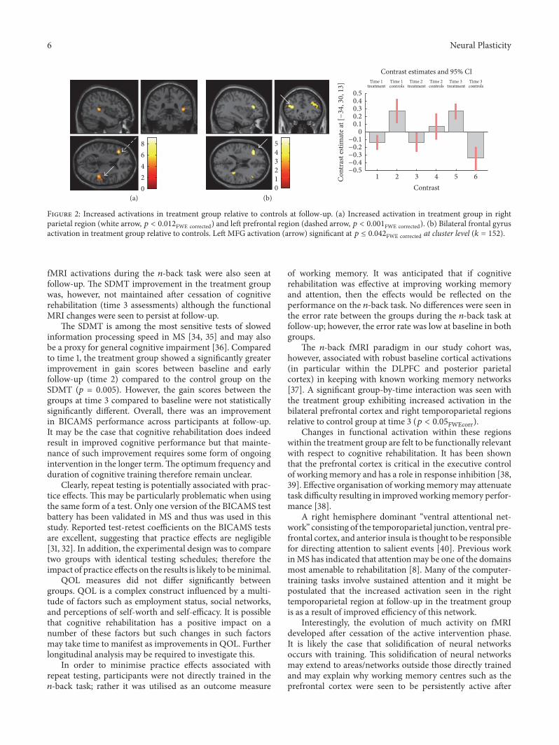
Time 3. At time 3 significant increases in activation were seenin both the 1-back and 2-back conditions in the treatmentgroup relative to controls. In the 1-back task, increasedactivation was seen in the left frontal (𝑝 < 0.001FWE correctedat cluster level (𝑘 = 294)) and right temporoparietal regions(𝑝 < 0.012FWE corrected at cluster level (𝑘 = 187)). In the2-back task, increases in activation were seen in bilateralprefrontal (𝑝 < 0.013FWE corrected at cluster level (𝑘 = 206))
and right temporoparietal regions (𝑝 < 0.024FWE corrected atcluster level (𝑘 = 178)) (Figure 2).
Quantitative Magnetisation Transfer. No significant between-group changes were seen in the QMT at time 2 or time 3, withrespect to time 1. Overall QMT measures showed stabilityacross all participants over the course of the study inmeasuresof all indices.
4. Discussion
In line with previouswork [8], themain outcome of this studywas that 6 weeks of computerised cognitive rehabilitationwas associated with improvement in cognitive performanceas measured on the SDMT. Significant alterations in brain
6 Neural Plasticity
Time 1treatment
Time 1controls
Contrast estimates and 95% CITime 2
treatmentTime 2controls
Time 3treatment
Time 3controls
Con
tras
t esti
mat
e at [
−34
,30
,13
]
Contrast1 2 3 4 5 6
−0.5
−0.4
−0.3
−0.2
−0.1
0
0.1
0.2
0.3
0.4
0.5
8
6
4
2
0
5
4
3
2
0
(a) (b)
1
Figure 2: Increased activations in treatment group relative to controls at follow-up. (a) Increased activation in treatment group in rightparietal region (white arrow, 𝑝 < 0.012FWE corrected) and left prefrontal region (dashed arrow, 𝑝 < 0.001FWE corrected). (b) Bilateral frontal gyrusactivation in treatment group relative to controls. Left MFG activation (arrow) significant at 𝑝 ≤ 0.042FWE corrected at cluster level (𝑘 = 152).
fMRI activations during the 𝑛-back task were also seen atfollow-up. The SDMT improvement in the treatment groupwas, however, not maintained after cessation of cognitiverehabilitation (time 3 assessments) although the functionalMRI changes were seen to persist at follow-up.
The SDMT is among the most sensitive tests of slowedinformation processing speed in MS [34, 35] and may alsobe a proxy for general cognitive impairment [36]. Comparedto time 1, the treatment group showed a significantly greaterimprovement in gain scores between baseline and earlyfollow-up (time 2) compared to the control group on theSDMT (p = 0.005). However, the gain scores between thegroups at time 3 compared to baseline were not statisticallysignificantly different. Overall, there was an improvementin BICAMS performance across participants at follow-up.It may be the case that cognitive rehabilitation does indeedresult in improved cognitive performance but that mainte-nance of such improvement requires some form of ongoingintervention in the longer term.The optimum frequency andduration of cognitive training therefore remain unclear.
Clearly, repeat testing is potentially associated with prac-tice effects. This may be particularly problematic when usingthe same form of a test. Only one version of the BICAMS testbattery has been validated in MS and thus was used in thisstudy. Reported test-retest coefficients on the BICAMS testsare excellent, suggesting that practice effects are negligible[31, 32]. In addition, the experimental design was to comparetwo groups with identical testing schedules; therefore theimpact of practice effects on the results is likely to beminimal.
QOL measures did not differ significantly betweengroups. QOL is a complex construct influenced by a multi-tude of factors such as employment status, social networks,and perceptions of self-worth and self-efficacy. It is possiblethat cognitive rehabilitation has a positive impact on anumber of these factors but such changes in such factorsmay take time to manifest as improvements in QOL. Furtherlongitudinal analysis may be required to investigate this.
In order to minimise practice effects associated withrepeat testing, participants were not directly trained in the𝑛-back task; rather it was utilised as an outcome measure
of working memory. It was anticipated that if cognitiverehabilitation was effective at improving working memoryand attention, then the effects would be reflected on theperformance on the 𝑛-back task. No differences were seen inthe error rate between the groups during the 𝑛-back task atfollow-up; however, the error rate was low at baseline in bothgroups.
The 𝑛-back fMRI paradigm in our study cohort was,however, associated with robust baseline cortical activations(in particular within the DLPFC and posterior parietalcortex) in keeping with known working memory networks[37]. A significant group-by-time interaction was seen withthe treatment group exhibiting increased activation in thebilateral prefrontal cortex and right temporoparietal regionsrelative to control group at time 3 (𝑝 < 0.05FWEcorr).
Changes in functional activation within these regionswithin the treatment group are felt to be functionally relevantwith respect to cognitive rehabilitation. It has been shownthat the prefrontal cortex is critical in the executive controlof workingmemory and has a role in response inhibition [38,39]. Effective organisation of workingmemorymay attenuatetask difficulty resulting in improvedworkingmemory perfor-mance [38].
A right hemisphere dominant “ventral attentional net-work” consisting of the temporoparietal junction, ventral pre-frontal cortex, and anterior insula is thought to be responsiblefor directing attention to salient events [40]. Previous workinMS has indicated that attentionmay be one of the domainsmost amenable to rehabilitation [8]. Many of the computer-training tasks involve sustained attention and it might bepostulated that the increased activation seen in the righttemporoparietal region at follow-up in the treatment groupis as a result of improved efficiency of this network.
Interestingly, the evolution of much activity on fMRIdeveloped after cessation of the active intervention phase.It is likely the case that solidification of neural networksoccurs with training. This solidification of neural networksmay extend to areas/networks outside those directly trainedand may explain why working memory centres such as theprefrontal cortex were seen to be persistently active after

Neural Plasticity 7
cessation of formal training [41]. Debate remains however asto the possible interplay between adaptive and maladaptiveresponses during functional brain reorganisation [14].
The discrepancy between the apparent lack of clinicaldifference between the groups at time 3 and the sustainedfMRI effect at time 3 may reflect the fact that BICAMS doesnot adequately measure working memory which is primarilydomain utilised during the 𝑛-back fMRI paradigm.
Some studies have identified structural changes on diffu-sion tensor imaging as a result of rehabilitation in the contextof physiotherapy [42, 43]. Our study attempted to explorethe role of myelin in rehabilitation and repair. We did notdetect any structural change on QMT after training. Due tothe short duration of follow-up, this is not entirely surprising.Functional alterations in cortical activity may subsequentlymodulate brain structure at the microstructural level butsuch changes in structural brain architecture might only bedetectable over the longer term.
In contrast to many previous studies, which often relyon one-to-one or outpatient administered cognitive reha-bilitation, this study sought to explore whether a home-based approach to cognitive rehabilitation was feasible. Thecompliance rate of those undertaking the rehabilitation wasexcellent. A home-based approach to cognitive rehabilitationis significantly less resource intensive and may pave the wayto greater access for a greater number of patients to suchinterventions in the future.
Limitations. This work has some limitations. Firstly, thegroups were relatively small and there was a dropout ofpatients mainly in the control group between time 2 andtime 3. There was heterogeneity with regard to the cognitivedomains that showed deficits among participants in the study.It is likely therefore that theymay not have benefited from therehabilitation in the same way. Unfortunately the sample sizeof the study is too small to perform subgroup analysis.
As the study was largely exploratory in nature, it utilisedan open design and is therefore subject to a number oflimitations inherent to this type of design. For pragmaticreasons blinding of the investigating neurologist was notestablished due to the potential need for interaction betweenpatient and investigator. This does present the potential forobserver bias, particularly where repeat testing is required.
SPM analysis of MRI data offers objective, largely auto-mated measures, which are independent of measurementbias. Investigator blinding was maintained for any methodssuch as assessment of white matter lesion volumes thatinvolved manual interpretation.
In many respects, the SDMT may provide a proxy foroverall cognitive functioning [44] but a more detailed cog-nitive assessment of the domains directly trained may haveprovided additional insight into effectiveness of cognitivetraining. BICAMS is primarily designed as a screening toolfor cognitive impairment in MS assessing a limited numberof domains. However, strong ecological validity has beendemonstrated in relation to everyday task performance andemployment, suggesting that the three domains are stronglypredictive of comprehensive real-world performance [45, 46].BICAMS may not necessarily be sensitive to change over the
short-term, although the reported test-retest coefficients areexcellent which would suggest sensitivity over this period[31, 32, 47].
It is postulated that alterations in fMRI activity resultfrom microstructural changes. The lack of significantchange in QMT measurements, however, suggests thatthe microstructural changes thought to underpin adaptiveresponses may, at present, be beyond the resolution of eventhe most advanced MRI techniques or not manifest withinthe timescale of this study. Additional follow-up of thiscohort is planned to determine what, if any, changes areobserved in terms of both cortical activation as measuredby fMRI and structural changes measurable with QMT.Longer-term studies may also provide insight into thetrue functional impact of cognitive rehabilitation such asmaintenance of employment.
Competing Interests
Dr. J. Campbell and Professor M. Cercignani have nothing todisclose. Dr W. Rashid has accepted educational grants andtravel bursaries from Genzyme, Biogen-Idec, Novartis, andTeva and has also participated in advisory boards with Bayer,Novartis, Biogen-Idec, and Genzyme. Professor D. Langdon(i) has received research grants from Bayer, Novartis, andBiogen and has also participated in advisory board withBayer, Novartis, and Teva is also in the speaker Bureau forTeva, Roche, Bayer, Novartis, and Biogen.
References
[1] R. H. B. Benedict and R. Zivadinov, “Risk factors for and man-agement of cognitive dysfunction in multiple sclerosis,” NatureReviews Neurology, vol. 7, no. 6, pp. 332–342, 2011.
[2] C. Potagas, E. Giogkaraki, G. Koutsis et al., “Cognitive impair-ment in different MS subtypes and clinically isolated syn-dromes,” Journal of the Neurological Sciences, vol. 267, no. 1-2,pp. 100–106, 2008.
[3] M. P. Amato, G. Ponziani, G. Pracucci, L. Bracco, G. Siracusa,and L. Amaducci, “Cognitive impairment in early-onset multi-ple sclerosis: pattern, predictors, and impact on everyday life ina 4-year follow-up,”Archives of Neurology, vol. 52, no. 2, pp. 168–172, 1995.
[4] M. P. Amato, B. Hakiki, B. Goretti et al., “Association of MRImetrics and cognitive impairment in radiologically isolatedsyndromes,” Neurology, vol. 78, no. 5, pp. 309–314, 2012.
[5] D. W. Langdon, “Cognition in multiple sclerosis,” CurrentOpinion in Neurology, vol. 24, no. 3, pp. 244–249, 2011.
[6] S. M. Rao, G. J. Leo, L. Ellington, T. Nauertz, L. Bernardin, andF. Unverzagt, “Cognitive dysfunction in multiple sclerosis. II.Impact on employment and social functioning,”Neurology, vol.41, no. 5, pp. 692–696, 1991.
[7] W. Staffen, A. Mair, H. Zauner et al., “Cognitive functionand fMRI in patients with multiple sclerosis: evidence forcompensatory cortical activation during an attention task,”Brain, vol. 125, no. 6, pp. 1275–1282, 2002.
[8] M. Filippi, G. Riccitelli, F. Mattioli et al., “Multiple sclerosis:effects of cognitive rehabilitation on structural and functionalMR imaging measures—an explorative study,” Radiology, vol.262, no. 3, pp. 932–940, 2012.

8 Neural Plasticity
[9] A. Cerasa, M. C. Gioia, P. Valentino et al., “Computer-assistedcognitive rehabilitation of attention deficits for multiple sclero-sis: a randomized trial with fMRI correlates,” Neurorehabilita-tion and Neural Repair, vol. 27, no. 4, pp. 284–295, 2013.
[10] T. D’Amato, R. Bation, A. Cochet et al., “A randomized,controlled trial of computer-assisted cognitive remediation forschizophrenia,” Schizophrenia Research, vol. 125, no. 2-3, pp.284–290, 2011.
[11] C.Modden,M. Behrens, I. Damke, N. Eilers, A. Kastrup, andH.Hildebrandt, “A randomized controlled trial comparing 2 inter-ventions for visual field loss with standard occupational therapyduring inpatient stroke rehabilitation,” Neurorehabilitation andNeural Repair, vol. 26, no. 5, pp. 463–469, 2012.
[12] A. Solari, A. Motta, L. Mendozzi et al., “Computer-aidedretraining of memory and attention in people with multiplesclerosis: a randomized, double-blind controlled trial,” Journalof the Neurological Sciences, vol. 222, no. 1-2, pp. 99–104, 2004.
[13] F. Mattioli, C. Stampatori, D. Zanotti, G. Parrinello, and R.Capra, “Efficacy and specificity of intensive cognitive rehabilita-tion of attention and executive functions in multiple sclerosis,”Journal of the Neurological Sciences, vol. 288, no. 1-2, pp. 101–105,2010.
[14] C. Enzinger, D. Pinter, M. A. Rocca et al., “Longitudinal fMRIstudies: exploring brain plasticity and repair in MS,” MultipleSclerosis Journal, vol. 22, no. 3, pp. 269–278, 2016.
[15] X. Qu, S.-W. Sun, H.-F. Liang, S.-K. Song, and D. F. Gochberg,“The MT pool size ratio and the DTI radial diffusivity mayreflect the myelination in shiverer and control mice,” NMR inBiomedicine, vol. 22, no. 5, pp. 480–487, 2009.
[16] M. S. A. Deloire, E. Salort, M. Bonnet et al., “Cognitiveimpairment as marker of diffuse brain abnormalities in earlyrelapsing remitting multiple sclerosis,” Journal of Neurology,Neurosurgery and Psychiatry, vol. 76, no. 4, pp. 519–526, 2005.
[17] J. Campbell,A Randomised Controlled Trial of Efficacy of Cogni-tive Rehabilitation inMultiple Sclerosis: A Cognitive, Behaviouraland MRI Study, Brighton and Sussex Medical School, 2016.
[18] C. H. Polman, S. C. Reingold, B. Banwell et al., “Diagnosticcriteria for multiple sclerosis: 2010 revisions to the McDonaldcriteria,” Annals of Neurology, vol. 69, no. 2, pp. 292–302, 2011.
[19] B. A. Parmenter, S. M. Testa, D. J. Schretlen, B. Weinstock-Guttman, andR.H. B. Benedict, “Theutility of regression-basednorms in interpreting the minimal assessment of cognitivefunction in multiple sclerosis (MACFIMS),” Journal of theInternational Neuropsychological Society, vol. 16, no. 1, pp. 6–16,2010.
[20] D. W. Langdon, M. P. Amato, J. Boringa et al., “Recommenda-tions for a brief international cognitive assessment for multiplesclerosis (BICAMS),”Multiple Sclerosis Journal, vol. 18, no. 6, pp.891–898, 2012.
[21] R. Rabin and F. de Charro, “EQ-5D: a measure of health statusfrom the EuroQol Group,” Annals of Medicine, vol. 33, no. 5, pp.337–343, 2001.
[22] D. F. Cella, K. Dineen, B. Arnason et al., “Validation ofthe functional assessment of multiple sclerosis quality of lifeinstrument,” Neurology, vol. 47, no. 1, pp. 129–139, 1996.
[23] L. Stepleman, M.-C. Rutter, J. Hibbard, L. Johns, D. Wright,and M. Hughes, “Validation of the patient activation measurein a multiple sclerosis clinic sample and implications for care,”Disability and Rehabilitation, vol. 32, no. 19, pp. 1558–1567, 2010.
[24] C. A. Young, R. J. Mills, J. Woolmore, C. P. Hawkins, and A.Tennant, “The unidimensional self-efficacy scale for MS (USE-MS): developing a patient based and patient reported outcome,”Multiple Sclerosis Journal, vol. 18, no. 9, pp. 1326–1333, 2012.
[25] A. S. Zigmond and R. P. Snaith, “The hospital anxiety anddepression scale,” Acta Psychiatrica Scandinavica, vol. 67, no. 6,pp. 361–370, 1983.
[26] R. H. B. Benedict, D. Cox, L. L. Thompson, F. Foley, B.Weinstock-Guttman, and F. Munschauer, “Reliable screeningfor neuropsychological impairment in multiple sclerosis,”Mul-tiple Sclerosis, vol. 10, no. 6, pp. 675–678, 2004.
[27] D. Amtmann, A. M. Bamer, V. Noonan, N. Lang, J. Kim, and K.F. Cook, “Comparison of the psychometric properties of twofatigue scales in multiple sclerosis,” Rehabilitation Psychology,vol. 57, no. 2, pp. 159–166, 2012.
[28] M. Gloor, K. Scheffler, and O. Bieri, “Quantitative magnetiza-tion transfer imaging using balanced SSFP,”Magnetic Resonancein Medicine, vol. 60, no. 3, pp. 691–700, 2008.
[29] L. H. Sweet, S. M. Rao, M. Primeau, A. R. Mayer, and R. A.Cohen, “Functional magnetic resonance imaging of workingmemory among multiple sclerosis patients,” Journal of Neu-roimaging, vol. 14, no. 2, pp. 150–157, 2004.
[30] R. Venkatesan, W. Lin, and E. M. Haacke, “Accurate determina-tion of spin-density and T1 in the presence of RF-field inhomo-geneities and flip-angle miscalibration,” Magnetic Resonance inMedicine, vol. 40, no. 4, pp. 592–602, 1998.
[31] J. B. Dusankova, T. Kalincik, E. Havrdova, and R. H. B. Bene-dict, “Cross cultural validation of the minimal assessment ofcognitive function in multiple sclerosis (MACFIMS) and thebrief international cognitive assessment for multiple sclerosis(BICAMS),” The Clinical Neuropsychologist, vol. 26, no. 7, pp.1186–1200, 2012.
[32] K. O’Connell, D. Langdon, N. Tubridy, M. Hutchinson, and C.McGuigan, “A preliminary validation of the brief internationalcognitive assessment formultiple sclerosis (BICAMS) tool in anIrish populationwithmultiple sclerosis (MS),”Multiple Sclerosisand Related Disorders, vol. 4, no. 6, pp. 521–525, 2015.
[33] D. Sandi, T. Rudisch, J. Fuvesi et al., “The Hungarian validationof the brief international cognitive assessment for multiplesclerosis (BICAMS) battery and the correlation of cognitiveimpairment with fatigue and quality of life,” Multiple Sclerosisand Related Disorders, vol. 4, no. 6, pp. 499–504, 2015.
[34] L. Strober, J. Englert, F. Munschauer, B. Weinstock-Guttman, S.Rao, andR.H. B. Benedict, “Sensitivity of conventionalmemorytests in multiple sclerosis: comparing the rao brief repeatableneuropsychological battery and the minimal assessment ofcognitive function in MS,” Multiple Sclerosis, vol. 15, no. 9, pp.1077–1084, 2009.
[35] B. A. Parmenter, B. Weinstock-Guttman, N. Garg, F. Mun-schauer, and R. H. B. Benedict, “Screening for cognitive impair-ment in multiple sclerosis using the Symbol Digit ModalitiesTest,”Multiple Sclerosis, vol. 13, no. 1, pp. 52–57, 2007.
[36] N. D. Chiaravalloti and J. DeLuca, “The influence of cognitivedysfunction on benefit from learning and memory rehabilita-tion in MS: a sub-analysis of the MEMREHAB trial,” MultipleSclerosis, vol. 21, no. 12, pp. 1575–1582, 2015.
[37] A.M. Owen, K.M.McMillan, A. R. Laird, and E. Bullmore, “N-back working memory paradigm: a meta-analysis of normativefunctional neuroimaging studies,” Human Brain Mapping, vol.25, no. 1, pp. 46–59, 2005.
Neural Plasticity 9
[38] D. Bor,N.Cumming,C. E. L. Scott, andA.M.Owen, “Prefrontalcortical involvement in verbal encoding strategies,” EuropeanJournal of Neuroscience, vol. 19, no. 12, pp. 3365–3370, 2004.
[39] D. Swick, V. Ashley, and A. U. Turken, “Left inferior frontalgyrus is critical for response inhibition,” BMCNeuroscience, vol.9, article 102, 2008.
[40] M. I. Posner and S. E. Petersen, “The attention system of thehuman brain,” Annual Review of Neuroscience, vol. 13, pp. 25–42, 1990.
[41] E. Dahlin, A. S. Neely, A. Larsson, L. Backman, and L. Nyberg,“Transfer of learning after updating training mediated by thestriatum,” Science, vol. 320, no. 5882, pp. 1510–1512, 2008.
[42] L. Bonzano, A. Tacchino, G. Brichetto et al., “Upper limbmotorrehabilitation impacts white matter microstructure in multiplesclerosis,” NeuroImage, vol. 90, pp. 107–116, 2014.
[43] I. Ibrahim, J. Tintera, A. Skoch et al., “Fractional anisotropyand mean diffusivity in the corpus callosum of patients withmultiple sclerosis: the effect of physiotherapy,” Neuroradiology,vol. 53, no. 11, pp. 917–926, 2011.
[44] J. Van Schependom, M. B. D’Hooghe, M. De Schepper et al.,“Relative contribution of cognitive and physical disability com-ponents to quality of life in MS,” Journal of the NeurologicalSciences, vol. 336, no. 1-2, pp. 116–121, 2014.
[45] Y. Goverover, N. Chiaravalloti, and J. DeLuca, “Brief interna-tional cognitive assessment for multiple sclerosis (BICAMS)and performance of everyday life tasks: actual reality,”MultipleSclerosis Journal, vol. 22, no. 4, pp. 544–550, 2016.
[46] R. H. Benedict, A. S. Drake, L. N. Irwin et al., “Benchmarks ofmeaningful impairment on the MSFC and BICAMS,” MultipleSclerosis Journal, 2016.
[47] E. Polychroniadou, C. Bakirtzis, D. Langdon et al., “Validationof the Brief International Cognitive Assessment for MultipleSclerosis (BICAMS) in Greek population with multiple sclero-sis,” Multiple Sclerosis and Related Disorders, vol. 9, pp. 68–72,2016.

Submit your manuscripts athttp://www.hindawi.com
Neurology Research International
Hindawi Publishing Corporationhttp://www.hindawi.com Volume 2014
Alzheimer’s DiseaseHindawi Publishing Corporationhttp://www.hindawi.com Volume 2014
International Journal of
ScientificaHindawi Publishing Corporationhttp://www.hindawi.com Volume 2014
Hindawi Publishing Corporationhttp://www.hindawi.com Volume 2014
BioMed Research International
Hindawi Publishing Corporationhttp://www.hindawi.com Volume 2014
Research and TreatmentSchizophrenia
The Scientific World JournalHindawi Publishing Corporation http://www.hindawi.com Volume 2014
Hindawi Publishing Corporationhttp://www.hindawi.com Volume 2014
Neural Plasticity
Hindawi Publishing Corporationhttp://www.hindawi.com Volume 2014
Parkinson’s Disease
Hindawi Publishing Corporationhttp://www.hindawi.com Volume 2014
Research and TreatmentAutism
Sleep DisordersHindawi Publishing Corporationhttp://www.hindawi.com Volume 2014
Hindawi Publishing Corporationhttp://www.hindawi.com Volume 2014
Neuroscience Journal
Epilepsy Research and TreatmentHindawi Publishing Corporationhttp://www.hindawi.com Volume 2014
Hindawi Publishing Corporationhttp://www.hindawi.com Volume 2014
Psychiatry Journal
Hindawi Publishing Corporationhttp://www.hindawi.com Volume 2014
Computational and Mathematical Methods in Medicine
Depression Research and TreatmentHindawi Publishing Corporationhttp://www.hindawi.com Volume 2014
Hindawi Publishing Corporationhttp://www.hindawi.com Volume 2014
Brain ScienceInternational Journal of
StrokeResearch and TreatmentHindawi Publishing Corporationhttp://www.hindawi.com Volume 2014
Neurodegenerative Diseases
Hindawi Publishing Corporationhttp://www.hindawi.com Volume 2014
Journal of
Cardiovascular Psychiatry and NeurologyHindawi Publishing Corporationhttp://www.hindawi.com Volume 2014