CLINICAL REVIEW - Food and Drug Administration · Therapeutic Class Radioactive diagnostic imaging...
42
CLINICAL REVIEW Application Type Efficacy Supplemental New Drug Application Application Number(s) NDA 018511 s28 Priority or Standard Standard Submit Date(s) February 26, 2017 Received Date(s) February 27, 2017 PDUFA Goal Date December 27, 2017 Division / Office DMIP/ODE IV Reviewer Name(s) August Hofling, MD, PhD Review Completion Date November 17, 2017 Established Name Kit for the preparation of technetium Tc 99m pentetate injection Trade Name DraxImage DTPA Therapeutic Class Radioactive diagnostic imaging agent Applicant Jubilant DraxImage, Inc. Formulation(s) 10 mL multiple dose vials containing 20 mg of pentetic acid and up to 9250 MBq/mL (250 mCi/mL) at time of preparation (reconstitution) Dosing Regimen Adult 25-50 mCi in nebulizer, pediatric 25 mCi in nebulizer, administered by inhalation Indication(s) Lung ventilation imaging and evaluation of pulmonary embolism when paired with perfusion imaging Intended Population(s) Adult and pediatric Template Version: March 6, 2009 Reference ID: 4183449
Transcript of CLINICAL REVIEW - Food and Drug Administration · Therapeutic Class Radioactive diagnostic imaging...

CLINICAL REVIEW
Application Type Efficacy Supplemental New Drug Application
Application Number(s) NDA 018511 s28Priority or Standard Standard
Submit Date(s) February 26, 2017Received Date(s) February 27, 2017
PDUFA Goal Date December 27, 2017Division / Office DMIP/ODE IV
Reviewer Name(s) August Hofling, MD, PhDReview Completion Date November 17, 2017
Established Name Kit for the preparation of technetium Tc 99m pentetate injection
Trade Name DraxImage DTPATherapeutic Class Radioactive diagnostic imaging agent
Applicant Jubilant DraxImage, Inc.
Formulation(s) 10 mL multiple dose vials containing 20 mg of pentetic acid and up to 9250 MBq/mL (250 mCi/mL) at time of preparation (reconstitution)
Dosing Regimen Adult 25-50 mCi in nebulizer, pediatric 25 mCi in nebulizer, administered by inhalation
Indication(s) Lung ventilation imaging and evaluation of pulmonary embolism when paired with perfusion imaging
Intended Population(s) Adult and pediatric
Template Version: March 6, 2009
Reference ID: 4183449

Clinical ReviewAugust Hofling, MD, PhDNDA 018511 s28 DraxImage DTPA (kit for the preparation of technetium Tc-99m pentetate injection)
2
Table of Contents
1 RECOMMENDATIONS/RISK BENEFIT ASSESSMENT..........................................61.1 Recommendation on Regulatory Action ..............................................................61.2 Risk Benefit Assessment .....................................................................................61.3 Recommendations for Postmarket Risk Evaluation and Mitigation Strategies ....61.4 Recommendations for Postmarket Requirements and Commitments.................6
2 INTRODUCTION AND REGULATORY BACKGROUND .........................................62.1 Product Information .............................................................................................62.2 Tables of Currently Available Treatments for Proposed Indications....................72.3 Availability of Proposed Active Ingredient in the United States ...........................92.4 Important Safety Issues with Consideration to Related Drugs ............................92.5 Summary of Presubmission Regulatory Activity Related to Submission ...........102.6 Other Relevant Background Information ...........................................................11
3 ETHICS AND GOOD CLINICAL PRACTICES........................................................163.1 Submission Quality and Integrity .......................................................................163.2 Compliance with Good Clinical Practices ..........................................................163.3 Financial Disclosures.........................................................................................16
4 SIGNIFICANT EFFICACY/SAFETY ISSUES RELATED TO OTHER REVIEW DISCIPLINES...........................................................................................................16
4.1 Chemistry Manufacturing and Controls .............................................................164.3 Preclinical Pharmacology/Toxicology ................................................................164.4 Clinical Pharmacology .......................................................................................17
4.4.1 Mechanism of Action ...................................................................................174.4.2 Pharmacodynamics.....................................................................................174.4.3 Pharmacokinetics ........................................................................................17
5 SOURCES OF CLINICAL DATA.............................................................................175.1 Tables of Studies/Clinical Trials.........................................................................175.2 Review Strategy.................................................................................................20
6 REVIEW OF EFFICACY ..........................................................................................21Efficacy Summary .......................................................................................................216.1 Indication ...........................................................................................................22
6.1.1 Review of Selected Individual Studies ........................................................236.1.10 Additional Efficacy Issues/Analyses ............................................................32
7 REVIEW OF SAFETY..............................................................................................32Safety Summary..........................................................................................................327.1 Methods .............................................................................................................33
7.1.1 Studies/Clinical Trials Used to Evaluate Safety ..........................................33
Reference ID: 4183449

Clinical ReviewAugust Hofling, MD, PhDNDA 018511 s28 DraxImage DTPA (kit for the preparation of technetium Tc-99m pentetate injection)
3
7.2 Adequacy of Safety Assessments .....................................................................337.2.1 Overall Exposure at Appropriate Doses/Durations and Demographics of
Target Populations ......................................................................................337.2.4 Routine Clinical Testing...............................................................................34
7.3 Major Safety Results..........................................................................................347.3.1 Deaths .........................................................................................................347.3.2 Nonfatal Serious Adverse Events................................................................347.3.3 Dropouts and/or Discontinuations ...............................................................347.3.4 Significant Adverse Events..........................................................................34
7.4 Supportive Safety Results .................................................................................357.4.1 Common Adverse Events............................................................................357.4.2 Laboratory Findings.....................................................................................357.4.3 Vital Signs ...................................................................................................357.4.4 Electrocardiograms (ECGs) ........................................................................35
8 POSTMARKET EXPERIENCE................................................................................35
9 APPENDICES..........................................................................................................399.1 Literature Review/References ...........................................................................399.2 Labeling Recommendations ..............................................................................409.3 Advisory Committee Meeting.............................................................................41
Reference ID: 4183449
Clinical ReviewAugust Hofling, MD, PhDNDA 018511 s28 DraxImage DTPA (kit for the preparation of technetium Tc-99m pentetate injection)
4
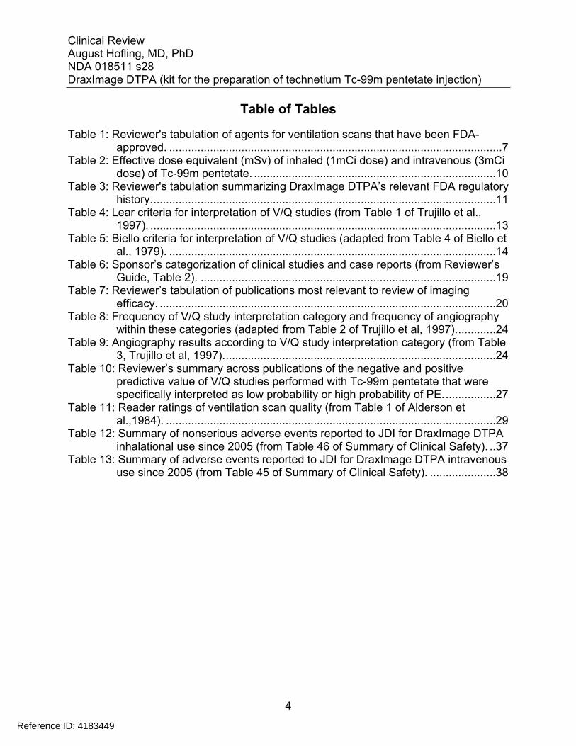
Table of Tables
Table 1: Reviewer's tabulation of agents for ventilation scans that have been FDA-approved. ..........................................................................................................7
Table 2: Effective dose equivalent (mSv) of inhaled (1mCi dose) and intravenous (3mCi dose) of Tc-99m pentetate. .............................................................................10
Table 3: Reviewer's tabulation summarizing DraxImage DTPA’s relevant FDA regulatory history..............................................................................................................11
Table 4: Lear criteria for interpretation of V/Q studies (from Table 1 of Trujillo et al., 1997). ..............................................................................................................13
Table 5: Biello criteria for interpretation of V/Q studies (adapted from Table 4 of Biello et al., 1979). ........................................................................................................14
Table 6: Sponsor’s categorization of clinical studies and case reports (from Reviewer’s Guide, Table 2). ..............................................................................................19
Table 7: Reviewer’s tabulation of publications most relevant to review of imaging efficacy. ...........................................................................................................20
Table 8: Frequency of V/Q study interpretation category and frequency of angiography within these categories (adapted from Table 2 of Trujillo et al, 1997).............24
Table 9: Angiography results according to V/Q study interpretation category (from Table 3, Trujillo et al, 1997).......................................................................................24
Table 10: Reviewer’s summary across publications of the negative and positive predictive value of V/Q studies performed with Tc-99m pentetate that were specifically interpreted as low probability or high probability of PE.................27
Table 11: Reader ratings of ventilation scan quality (from Table 1 of Alderson et al.,1984). .........................................................................................................29
Table 12: Summary of nonserious adverse events reported to JDI for DraxImage DTPA inhalational use since 2005 (from Table 46 of Summary of Clinical Safety). ..37
Table 13: Summary of adverse events reported to JDI for DraxImage DTPA intravenous use since 2005 (from Table 45 of Summary of Clinical Safety). .....................38
Reference ID: 4183449

Clinical ReviewAugust Hofling, MD, PhDNDA 018511 s28 DraxImage DTPA (kit for the preparation of technetium Tc-99m pentetate injection)
5
Table of Figures
Figure 1: Schematic of sponsor’s literature search results (from Reviewer’s Guide, Figure 1). .........................................................................................................18
Figure 2: Sponsor’s estimate over time of the percentage of Tc-99m pentetate ventilation studies in the U.S. that specifically used DraxImage DTPA (derived from distribution data, adapted from Figure 1 of information amendment dated July 13, 2017)..................................................................................................36
Reference ID: 4183449

Clinical ReviewAugust Hofling, MD, PhDNDA 018511 s28 DraxImage DTPA (kit for the preparation of technetium Tc-99m pentetate injection)
6
1 Recommendations/Risk Benefit Assessment
1.1 Recommendation on Regulatory Action
We recommend approval of this 28th supplementary application to NDA 018511; specifically, that DraxImage DTPA, after radiolabeling with technetium-99m, be indicated for lung ventilation imaging and evaluation of pulmonary embolism when paired with perfusion imaging in adults and pediatric patients when administered by nebulizer for inhalation.
1.2 Risk Benefit Assessment
Our recommendation is based primarily on selected publications from the sponsor’s submitted review of literature that evaluate the clinical efficacy, safety, and pharmacokinetics of inhaled technetium-99m pentetate as well as postmarketing safety data for DraxImage DTPA. The body of evidence supports a favorable risk-benefit balance for performing ventilation scans with inhaled technetium-99m pentetate produced by the DraxImage DTPA kit, particularly when considering its already long history of widespread off-label clinical use for this purpose.
1.3 Recommendations for Postmarket Risk Evaluation and Mitigation Strategies
None.
1.4 Recommendations for Postmarket Requirements and Commitments
None.
2 Introduction and Regulatory Background
2.1 Product Information
The active ingredient in DraxImage DTPA is DTPA itself, known formally as diethylenetriaminepentaacetic acid and by other names including pentetate and pentetic acid. This small molecule is a chelator of metal ions and is used in several FDA-approved drugs including the gadolinium-based contrast agent, gadopentetic acid, and drugs that treat internal contamination with certain radiometals. DraxImage DTPA is a kit through which manufacture-supplied, non-radioactive pentetate is combined with
Reference ID: 4183449

Clinical ReviewAugust Hofling, MD, PhDNDA 018511 s28 DraxImage DTPA (kit for the preparation of technetium Tc-99m pentetate injection)
7
user-supplied radioactive technetium-99m, referred to hereafter as Tc-99m, to form the final administered drug product, Tc-99m pentetate.
DraxImage DTPA was originally approved as a kit for preparation of Tc-99m pentetate for intravenous injection “to perform kidney imaging, brain imaging, to assess renal perfusion, and to estimate glomerular filtration rate”. These original indications remain unchanged by the current supplement and apply only to adults and not to pediatric patients. The sponsor would now like to additionally indicate Tc-99m pentetate produced by the DraxImage DTPA kit for the purpose of ventilation imaging of the lungs in both adults and children when administered by nebulizer for inhalation. The same Tc-99m pentetate product as would be injected intravenously is to be placed in a nebulizer, leading to its aerosolization. Thus, this efficacy supplement aims to add a new indication and new route of administration for Tc-99 pentetate produced by the DraxImage DTPA kit in both adult and pediatric patients.
2.2 Tables of Currently Available Treatments for Proposed Indications
Several radioactive gases have been FDA-approved for ventilation imaging as appear in Table 1 below.
Table 1: Reviewer's tabulation of agents for ventilation scans that have been FDA-approved.Non-proprietary Name Commercial AvailabilityXenon-133 (Xe-133) Currently availableXenon-127 (Xe-127) Withdrawn in 2008Krypton-81m (Kr-81m) Withdrawn in 2001
Withdrawal of Kr-81m from the market was influenced by its expense and the fact that its own half-life as well as that of its generator-based parent, rubidium-81, are impractically short. Withdrawal of Xe-127 from the market was influenced by its expense as well as its long-half-life and relatively high energy which generate radiation safety hazards and make storage of waste during decay burdensome. The still commercially available Xe-133 gas is produced as a fission product in nuclear reactors but is fairly easily distributed due to its 5.3 day radioactive half-life.
A Nuclear Regulatory Commission (NRC) Final Rule published in the Federal Register on February 4, 1983 (Vol 48, No.25, 5217), excluded certain regulatory requirements such that inhaled Tc-99m pentetate could be used by properly credentialed practitioners for ventilation imaging despite lack of FDA approval. In contrast to gases including those listed in Table 1, aerosols like Tc-99m pentetate physically consist of particulates suspended in air. Also unlike a gas, inhaled Tc-99m pentetate aerosol is deposited on epithelial surfaces of the lung rather than being exhaled to a significant degree. No other radioactive agents are FDA-approved for aerosolization and inhaled delivery,
Reference ID: 4183449
Clinical ReviewAugust Hofling, MD, PhDNDA 018511 s28 DraxImage DTPA (kit for the preparation of technetium Tc-99m pentetate injection)
8
although a Tc-99m labeled aerosol of fine carbon particles marketed as Technegas is clinically approved in certain foreign countries.
From a technical standpoint, use of aerosolized Tc-99m pentetate for ventilation imaging has both several advantages and disadvantages compared to use of the only approved and commercially available alternative, Xe-133 gas. Perhaps the major advantage of an aerosol likeTc-99m pentetate is the lack of necessary containment equipment other than a closed nebulizer system. Gases like Xe-133, on the other hand, require a negative pressure room with an additional trap or atmospheric vent. This convenient feature of Tc-99m pentetate also allows its use in portable imaging settings. While inhalation of gases like Xe-133 mandates careful adherence to breathing instructions, Tc-99m pentetate aerosol has an additional advantage of requiring relatively little patient cooperation, allowing ventilation studies to be performed even in infants and mechanically ventilated patients. A further advantage of ventilation scans performed with Tc-99m pentetate is the ability to collect images in a variety of projections while Xe-133 images are typically obtained only in posterior or posterior oblique projections due to the low energy of Xe-133 photons. This feature allows Tc-99m pentetate ventilation imaging to match the multiple projections of perfusion imaging, increasing ease of diagnostic interpretation in the common application of ventilation-perfusion studies as further discussed below in Section 2.6. Ventilation scans performed with Tc-99m pentetate can also be performed before or after perfusion imaging in such combined studies while Xe-133 ventilation scans must always be performed first due to down scatter from perfusion imaging agent.
Of the few disadvantages of Tc-99m pentetate aerosol relative to gases like Xe-133, perhaps the most notable is the propensity for aerosol to deposit in central airways that experience turbulent airflow. Such deposition is typically only significant in certain settings of underlying airway disease, such as COPD, where it can negatively impact diagnostic quality. Gases, on the other hand, are not associated with significant central airway deposition, even in the setting of airway disease. Xe-133 gas also allows dynamic wash-in and wash-out imaging to be performed for further sensitive evaluation of airway disease. Tc-99m pentetate aerosol lacks this capability and only provides images comparable to the steady-state phase of Xe-133 administration. However, for the common application of detecting areas of ventilation-perfusion mismatch, the steady-state imaging information provided by either Tc-99m pentetate or Xe-133 is typically sufficient.
As evidenced by current practice patterns, the advantages of Tc-99m pentetate aerosol make it a popular choice for ventilation imaging outside of large, predominantly academic medical centers that meet containment requirements to safely use gas agents. According to the sponsors cited market research, approximately of ventilation studies in the US are currently performed with Tc-99m pentetate, with the remainder performed with Xe-133.
Reference ID: 4183449
(b) (4)
Clinical ReviewAugust Hofling, MD, PhDNDA 018511 s28 DraxImage DTPA (kit for the preparation of technetium Tc-99m pentetate injection)
9
2.3 Availability of Proposed Active Ingredient in the United States
Several kits for the preparation of Tc-99m pentetate have been FDA-approved with varying salt formulations and manufacturers since the 1970s. The current sponsor, Jubilant DraxImage, Inc., referred to hereafter as JDI, holds an active NDA not only for DraxImage DTPA, which is formulated with pentetic acid, but also a similar kit, An-DTPA (NDA 017714), which is formulated with pentetate calcium trisodium. An-DTPA has not been produced since its acquisition by JDI in 2013 and all Tc-99m pentetate kits from other manufacturers have been discontinued. In an information amendment dated July 13, 2017, the sponsor presented marketing data indicating Tc-99m pentetate ventilation scans performed in the US from 2010 to the present have used DraxImage DTPA.
The publications reviewed in this submission, most of which date prior to 2010, typically do not indicate which manufacturer’s kit was used to generate Tc-99m pentetate. We feel that this lack of manufacturer identification has no significant impact on clinical evaluation of the literature, as the possible kits vary only in salt formulation and excipients. In discussion of published data, use of the final radioactive product’s name, Tc-99m pentetate, will be used without reference to a specific kit manufacturer.
Outside of the US, DraxImage DTPA is approved in Canada, Hong Kong, Israel, and Mexico for both intravenous and inhalational routes in adults.
2.4 Important Safety Issues with Consideration to Related Drugs
Radioactive imaging agents in general, including Tc99m-pentetate and other agents used to perform ventilation scans, all carry the risk of radiation exposure. Aligning with recommendations from the American College of Radiology (ACR), the Society for Pediatric Radiology (SPR), and the Society of Thoracic Radiology (STR) published in 2014 (ACR, 2014; 1-9), the sponsor estimates that 0.5 mCi to 1 mCi of Tc-99m pentetate radioactivity should be delivered to the lungs in a typical ventilation scan performed either by itself or prior to a perfusion scan. Below in Table 2, the effective dose equivalent, a measure of whole-body radiation exposure, is compared for 1 mCi of Tc-99m pentetate delivered to the lungs by inhalation and 3 mCi of Tc-99m pentetate injected intravenously, the lowest approved intravenous dose for imaging (ICRP, 1988 Ann ICRP 18:217-219).
Reference ID: 4183449
(b) (4)

Clinical ReviewAugust Hofling, MD, PhDNDA 018511 s28 DraxImage DTPA (kit for the preparation of technetium Tc-99m pentetate injection)
10
Table 2: Effective dose equivalent (mSv) of inhaled (1mCi dose) and intravenous (3mCi dose) of Tc-99m pentetate.
AGE GROUP INHALED (1 mCi) INTRAVENOUS (3 mCi)Adult 0.26 mSv 0.70 mSv15 years 0.34 mSv 0.87 mSv10 years 0.48 mSv 1.2 mSv5 years 0.74 mSv 1.9 mSv1 year 1.3 mSv 3.3 mSv
The critical organ of inhaled Tc-99m pentetate has been reported as the trachea with an estimated 3.0 mGy of absorbed radiation from a 1 mCi inhaled dose in an adult (Atkins et al., 1992 J Nucl Med; 33:1717-1719). A separate study determined that the oral cavity receives a similar absorbed radiation dose as that of the trachea (Bondesson et al., 2007 Br J Clin Pharmacol; 63:722-731). Organs with the next greatest absorbed dose are the urinary bladder wall (approximately 1.7 mGy from a 1 mCi inhaled dose in an adult) and the lungs (approximately 0.6 mGy from a 1 mCi inhaled dose in an adult).
For reference, the average American is estimated to receive an effective dose of 3.1 mSv annually from completely natural sources of radiation. For further reference, a standard 18F-FDG PET scan in an adult with a 10 mCi intravenous dose would yield an estimated whole-body effective dose of 7 mSv, with the urinary bladder wall receiving an absorbed radiation dose of roughly 27 mGy as the critical organ (Mettler et al., 2008 Radiology; 248:254-263; Hays et al., 2002 J Nucl Med; 43:210-214). Compared to these figures, whole-body and critical organ radiation exposures for a typical ventilation study with Tc-99m pentetate are quite small. As such, associated radiation-related cancer risk from inhalation of Tc-99m pentetate is on the low end of the current range for nuclear medicine diagnostic agents.
2.5 Summary of Presubmission Regulatory Activity Related to Submission
Table 3 provides a timeline of the pertinent FDA regulatory history related to DraxImage DTPA.
Reference ID: 4183449

Clinical ReviewAugust Hofling, MD, PhDNDA 018511 s28 DraxImage DTPA (kit for the preparation of technetium Tc-99m pentetate injection)
11
Table 3: Reviewer's tabulation summarizing DraxImage DTPA’s relevant FDA regulatory history.Date Application Description12/29/1989 NDA 018511 Merck Frosst Canada, Inc., receives initial marketing
approval for Tc-99m pentetate under the tradename Frosstimage for intravenous indications of kidney imaging, brain imaging, to assess renal perfusion, and to estimate glomerular filtration rate
6/3/1999 NDA 018511 Transfer of NDA sponsor to DraxImage, Inc.9/23/2011 NDA 018511 Change of sponsor name to Jubilant DraxImage, Inc.
4/20/2015 pIND 125711 Pre-IND meeting held with FDA to discuss the intent to submit a literature-based 505(b)(2) efficacy supplement for inhalation of Tc-99m pentetate produced by the DraxImage DTPA kit for ventilation imaging
12/18/2015 pIND 125711 Agreement reached for the initial pediatric study plan to consist of 505(b)(2) pathway literature review
5/17/2016 pIND 125711 Pre-NDA meeting held with FDA to further discuss the intent to submit a literature-based 505(b)(2) efficacy supplement for inhalation of Tc-99m pentetate produced by the DraxImage DTPA kit for ventilation imaging
2/27/2107 NDA 018511 Current 505(b)(2) literature-based efficacy supplement application s28 received
2.6 Other Relevant Background Information
Ventilation scans image the distribution of an inhaled radioactive agent within the bronchopulmonary air spaces of the lungs. As reflected in the only clinical indication to be categorized as “most common” in the latest Society of Nuclear Medicine Practice Guideline for Lung Scintigraphy (Parker et al., 2012 J Nucl Med Technol 40:57-65), the current use of ventilation scans is dominated by its contribution to combined ventilation-perfusion (V/Q) studies for evaluation of pulmonary embolism (PE). Below, a clinical and diagnostic overview of PE is presented, followed by a brief discussion of other less common diagnostic roles of ventilation scans.
Pulmonary EmbolismPE is obstruction of a pulmonary artery or its branches by materials including thrombus, tumor, air, or fat. Most cases of PE are caused by thrombus and can be acute, subacute, or chronic. Typically, thrombotic emboli arise from veins in the proximal lower extremities although other veins and the right heart are also possible sources. Estimates of PE incidence vary, in part seemingly due to improvement in diagnostic testing over time, with an estimated 112 cases per 100,000 over the period of 1998 to
Reference ID: 4183449

Clinical ReviewAugust Hofling, MD, PhDNDA 018511 s28 DraxImage DTPA (kit for the preparation of technetium Tc-99m pentetate injection)
12
2006 (Wiener et al., 2011 Arch Intern Med 171:831-837). Mortality of PE is significant, particularly when diagnosis or treatment is delayed, with one study calculating total PE-related deaths per year in the United States ranging from roughly 35,000 in 1979 to 25,000 in 1998. (Horlander et al., 2003 Arch Intern Med 163:1711-1717).
Diagnosis of acute or subacute PE involves sources of information aside from imaging. Initially, history and physical examination are used to generate a clinical pretest probability that will determine the need for further testing as well as influence the clinical impact of such testing. Laboratory measurement of D-dimer elevation is a routine test that has a high sensitivity but low specificity for PE. As such, a normal D-dimer level effectively excludes PE in subjects with low or intermediate pretest probability. Conversely, the low specificity of a D-dimer elevation would necessitate subsequent diagnostic imaging in these same pretest groups. Imaging is also often recommended for patients with high pretest probability for PE regardless of D-dimer results.
A variety of imaging modalities including V/Q studies offer useful diagnostic information for detection of PE. A V/Q study is a nuclear medicine imaging technique in which a separately acquired ventilation scan and perfusion scan are interpreted jointly. Perfusion scans image the distribution of intravenously injected macroaggregated albumin (MAA) labeled with Tc-99m, which localizes in the lung through temporary blockade of a tiny fraction of pulmonary capillaries. While a perfusion scan alone provides valuable information regarding pulmonary blood flow, pairing it with a ventilation scan in a V/Q study greatly increases specificity for pathology like PE by identifying areas of mismatch in the patterns of perfusion and ventilation. PE decreases blood flow to a lung region, but the airways and alveoli in the affected region typically remain patent and continue to be ventilated. Thus, a lung region that demonstrates relatively preserved ventilation but decreased or absent perfusion constitutes a V/Q mismatch that is suspicious for PE although other pathology is possible. Conversely, a lung region with matched decrease in both perfusion and ventilation is unlikely to represent PE with other pathology like pneumonia, bullous change of COPD, asthma, and mucous plugging being more likely.
Number, size, and shape of lung regions demonstrating mismatched perfusion and ventilation contribute to V/Q study interpretation with multiple, larger, and segmental abnormalities increasing suspicion for PE. To further increase specificity, V/Q scans are typically read in conjunction with recent chest radiographs to correlate any defects in perfusion and/or ventilation with radiographic findings such as infiltrate, mass, or effusion. Traditionally, V/Q images are collected through planar scintigraphy although more recently, single photon emission computed tomography (SPECT) has been studied as a method for reconstruction of three-dimensional imaging data.
Several established reading methods exist for interpreting V/Q studies that classify results as one of several categories of PE probability such as low, intermediate, or high probability. Such a probabilistic reporting strategy remains dominant in clinical practice
Reference ID: 4183449

Clinical ReviewAugust Hofling, MD, PhDNDA 018511 s28 DraxImage DTPA (kit for the preparation of technetium Tc-99m pentetate injection)
13
today although some groups are moving towards a dichotomized positive or negative reporting system. Two established reading methods for V/Q study interpretation which are relevant to publications discussed later in this review appear below in Tables 4 and 5; namely, Lear criteria (Trujillo et al., 1997 J Nucl Med. 38:1781-1783) in Table 4 and Biello criteria (Biello et al. 1979 Am J Roentgenol. 133:1033-1037) in Table 5.
Table 4: Lear criteria for interpretation of V/Q studies (from Table 1 of Trujillo et al., 1997).
Reference ID: 4183449

Clinical ReviewAugust Hofling, MD, PhDNDA 018511 s28 DraxImage DTPA (kit for the preparation of technetium Tc-99m pentetate injection)
14
Table 5: Biello criteria for interpretation of V/Q studies (adapted from Table 4 of Biello et al., 1979).
Other diagnostic imaging modalities for PE include the historical gold standard technique of catheter-based pulmonary angiography. With the advent of V/Q studies, however, this invasive and operator-dependent test was essentially relegated only to cases with indeterminate or conflicting clinical and imaging data. Subsequently, PE protocol computed tomography (CT) was established as the imaging technique of choice for diagnosis of PE due to its speed, constant availability, simple interpretative criteria, and added ability to detect a gamut of chest pathology other than PE. Catheter-based angiography is now rarely performed for PE diagnosis and is typically reserved for settings of planned intervention such as embolectomy. V/Q studies are now also generally performed only in certain specialized but important clinical scenarios as discussed below.
Reference ID: 4183449
Clinical ReviewAugust Hofling, MD, PhDNDA 018511 s28 DraxImage DTPA (kit for the preparation of technetium Tc-99m pentetate injection)
15
Most often, V/Q scanning is performed when PE protocol CT is contraindicated, including patients with significant renal impairment or known severe allergy to iodinated contrast. Severe claustrophobia and patient weight exceeding CT table limits may also make V/Q scanning a favorable imaging option. Additionally, the much lower radiation exposure of a V/Q study (roughly 2 mSv effective dose) compared to PE protocol CT (roughly 8-10 mSv effective dose) also may make its use preferable in certain patients including pregnant women and children. Recommended clinical use of V/Q studies is further detailed in the recent “Appropriate Use Criteria for Ventilation-Perfusion Imaging in Pulmonary Embolism”, published by the Society of Nuclear Medicine and Molecular Imaging (Waxman et al., 2017 J Nucl Med 58:13N-15N).
A few other imaging tests are less well established or second-line for diagnosis of PE. Magnetic resonance (MR) pulmonary angiography can detect PE without ionizing radiation exposure but currently remains limited in use due to factors including relatively longer scanning times and lesser availability of required equipment and technical expertise. Lower extremity Doppler and echocardiography are occasionally used when CT or V/Q scans cannot be performed or are inconclusive. These imaging techniques look for peripheral thrombus or other findings associated with PE rather than directly visualizing PE itself.
Other Uses of Ventilation ImagingWhile ventilation scans have been employed for a wide variety of diagnostic purposes outside of evaluation for PE, these other applications represent only a small proportion of current clinical practice. Many of these less common clinical uses still occur in the context of a V/Q study, such as evaluation of subjects undergoing lung resection or lung transplantation or the evaluation of congenital heart disease. Often, quantitative analysis of ventilation and perfusion data is performed in these applications. Certain other clinical scenarios may sometimes rely on ventilation imaging alone without perfusion imaging, such as the evaluation of airway disease like COPD or cystic fibrosis or the detection of bronchopleural fistulas. However, alternative imaging modalities like CT often provide more detailed structural information in these situations.
The natural mechanism by which Tc-99m pentetate is absorbed through the lung epithelium into the blood has also led to exploration into its potential diagnostic use for detecting epithelial injury. In this offshoot of ventilation imaging with Tc-99m pentetate, clearance of radioactivity from the lungs following inhaled administration is measured as an indicator of alveolar permeability. The half-time of Tc99m-pentetate in the lungs of healthy nonsmokers in roughly 80 ± 20 minutes (Ziessman et al., 2013 Chapter 10: Pulmonary System, in Nuclear Medicine, The Requisites). A wide range of pulmonary epithelial pathology typically increases the lung clearance rate and thereby decreases the lung half-time of Tc-99m pentetate, including endogenous causes like pulmonary infection and inflammation as well as exogenous causes like smoking and other inhalational toxins. Some lung pathology may decrease Tc-99m pentetate clearance rate, such as certain phases of pulmonary fibrosis. While representing an interesting
Reference ID: 4183449

Clinical ReviewAugust Hofling, MD, PhDNDA 018511 s28 DraxImage DTPA (kit for the preparation of technetium Tc-99m pentetate injection)
16
feature that is unique to inhalation of Tc-99m pentetate as opposed to approved gases, evaluation of alveolar permeability appears to remain predominantly research-based without widespread clinical adoption.
3 Ethics and Good Clinical Practices
3.1 Submission Quality and Integrity
Based on the filing review meeting held on April 6, 2017, the sponsor’s application was found to be sufficiently complete to allow substantive review. The sponsor was notified on May 8, 2017.
3.2 Compliance with Good Clinical Practices
Compliance with Good Clinical Practices is not applicable to this 505(b)(2) pathway submission as it is based on published clinical studies that were not conducted or contributed to by the sponsor.
3.3 Financial Disclosures
Traditional financial disclosures are not applicable to this 505(b)(2) pathway submission that relies on published literature.
4 Significant Efficacy/Safety Issues Related to Other Review Disciplines
4.1 Chemistry Manufacturing and Controls
DTPA (pentetic acid) contained in DraxImage DTPA chelates added Tc-99m to form the complete radioactive product used for imaging, Tc-99m pentetate. Tc-99m radioactively decays by isomeric transition with a half-life of 6.0 hours. The principal photon that is useful for imaging studies has a mean energy of 140.5 keV.
4.3 Preclinical Pharmacology/Toxicology
The sponsor identified a number of published toxicology studies for DTPA. Certain of these studies specifically evaluated the inhalation route in rats for both single and repeat dose regimens (Smith et al., 1980 Toxicol Lett 7:9-16; Bene and Burnett, 2008
Reference ID: 4183449

Clinical ReviewAugust Hofling, MD, PhDNDA 018511 s28 DraxImage DTPA (kit for the preparation of technetium Tc-99m pentetate injection)
17
Int J Toxicol 27 Suppl 2:71-92). These inhalation studies demonstrated wide safety margins for DTPA relative to intended human dosing as fully discussed in the separate pharmacology/toxicology review document.
4.4 Clinical Pharmacology
4.4.1 Mechanism of Action
Beyond its role as a chelating agent, DTPA has no inherent pharmacological properties. For inhalation, the same Tc-99m pentetate solution used for intravenous injection is administered using a nebulizer with a target aerosolized particle size of µm. Larger particles display undesirable deposition in central airways. During several minutes of normal to deep breathing, the aerosolized product deposits on pulmonary epithelium, predominantly at the level of alveoli. Of note, less than 10% of the Tc-99m pentetate that is loaded into a nebulizer is intended to be delivered to the patient’s lungs. Unlike gases, there is no significant exhalation of the Tc-99m pentetate aerosol.
4.4.2 Pharmacodynamics
Acquired images reflect the distribution of aerated lung volume.
4.4.3 Pharmacokinetics
Tc-99m pentetate aerosol deposited in the lungs is absorbed across the pulmonary epithelium into the blood with a half-time of approximately 80 ± 20 minutes in healthy, nonsmokers (Ziessman et al., 2013). Once in the blood, Tc-99m pentetate is removed by the kidneys through glomerular filtration without significant metabolism. As stated in Section 3.2.5 of the sponsor’s Clinical Overview, plasma half-life in subjects with normal renal function is approximately 2.1 hours.
5 Sources of Clinical Data
5.1 Tables of Studies/Clinical Trials
The sponsor conducted a PubMed search on April 12, 2016, using synonyms for Tc-99m pentetate, a series of terms related to lungs or ventilation, and a filter for human species. This search returned 3564 citations that ultimately contained 655 unique publications once repeated results were accounted for. References in these 655 publications were then reviewed by the sponsor and brought the number of unique
Reference ID: 4183449
(b) (4)

Clinical ReviewAugust Hofling, MD, PhDNDA 018511 s28 DraxImage DTPA (kit for the preparation of technetium Tc-99m pentetate injection)
18
publications to 1,090. A total of 528 of these articles were considered relevant by the sponsor through meeting one of the following criteria:
Tc-99m pentetate single agent in inhalation diagnostic image acquisition (ventilation or V/Q scan); and/or
Tc-99m pentetate single agent administered via inhalation to image the lung to identify whether or not ventilation defects are present.
Several appropriate criteria for irrelevance of publications were also used by the sponsor including conjugation of Tc-99m pentetate to another drug product, route of administration other than inhalation, and presentation of only in vitro or nonclinical data. Of the 528 relevant publications, 519 were available including 389 clinical studies, 46 case reports, 17 editorials, 56 review articles, and 11 guideline publications. A schematic of the sponsor’s literature search results appears below in Figure 1.
Figure 1: Schematic of sponsor’s literature search results (from Reviewer’s Guide, Figure 1).
Reference ID: 4183449

Clinical ReviewAugust Hofling, MD, PhDNDA 018511 s28 DraxImage DTPA (kit for the preparation of technetium Tc-99m pentetate injection)
19
To correspond with specific purposes of ventilation imaging listed in the sponsor’s proposed indications, the sponsor categorized clinical studies and case reports as being relevant to pulmonary embolism, lung structure, alveolar permeability, or ventilation distribution. Many publications were placed into more than one category. For each category except structure, the sponsor designated two key studies. The sponsor’s general categorization scheme is reproduced in Table 6 below.
Table 6: Sponsor’s categorization of clinical studies and case reports (from Reviewer’s Guide, Table 2).
In this clinical review, all publications identified by the sponsor were included in the review of safety. Publications that provided the strongest evidence for the review of diagnostic efficacy were of two types, both of which were conducted in the context of V/Q studies for evaluation of suspected PE. One of these study types assessed the diagnostic performance of V/Q studies conducted with Tc-99m pentetate for detection of PE through use of an angiographic or autopsy truth standard. The other of these study types determined the level of diagnostic agreement between V/Q studies performed with Tc-99m pentetate and V/Q studies performed with the approved gases, Xe-133 or Kr-81m, again in the setting of suspected PE. Publications of these types that featured the largest numbers of subjects and the strongest experimental designs formed the basis of detailed efficacy review and are listed in Table 7 below.
Reference ID: 4183449

Clinical ReviewAugust Hofling, MD, PhDNDA 018511 s28 DraxImage DTPA (kit for the preparation of technetium Tc-99m pentetate injection)
20
Table 7: Reviewer’s tabulation of publications most relevant to review of imaging efficacy.
Publication Number of subjects
Angiographic/autopsy truth standardMain studyTrujillo et al., 1997 455Supportive studiesLear et al., 1996 33Freitas et al., 1995 133Selby et al., 1990 72
Approved comparatorMain studyAlderson et al., 1984 107Supportive studiesRamanna et al., 1986 54Finn et al., 1986 40
5.2 Review Strategy
The two publications listed in Table 7 as main studies were designated as such because of their larger sample size and more clearly described experimental design. Publications listed as supportive used smaller study populations and often had weaker or less well specified study design. Each individual publication listed in Table 7 is reviewed sequentially in Section 6 below. Of note, the sponsor’s designated key publications for PE imaging are included in these reviewed publications.
Reference ID: 4183449
(b) (4)

Clinical ReviewAugust Hofling, MD, PhDNDA 018511 s28 DraxImage DTPA (kit for the preparation of technetium Tc-99m pentetate injection)
21
the following review of efficacy will focus only on the publications listed in Table 7, all of which involve V/Q imaging for suspected PE. While this strategy correspondingly narrows the potential labeling of specific indications for DraxImage DTPA, it is important to note that the vast majority of the current and anticipated future clinical use of inhaled Tc-99m pentetate is indeed for the exact purpose of evaluating for PE through V/Q imaging.
6 Review of Efficacy
Efficacy SummarySelected publications support the approval of inhaled Tc-99m pentetate for use in ventilation imaging, particularly in the common application of detecting PE as a component of V/Q studies. In this clinical setting, publications using a truth standard of conventional angiography or autopsy indicate adequate diagnostic performance clearly exceeding that of chance. In this same clinical setting, other publications demonstrate comparable utility of ventilation imaging performed with Tc-99m relative to approved gas comparators.
However, confidence in the ability to quantitate discrete performance metrics is reduced by certain methodological weaknesses of relied upon studies as well as limitations inherent to their older dates of publication, such as unavailability of primary data for verification of results and potentially incomplete generalizability to more modern patient populations and technical equipment.
Reference ID: 4183449
(b) (4)
(b) (4)
(b) (4)

Clinical ReviewAugust Hofling, MD, PhDNDA 018511 s28 DraxImage DTPA (kit for the preparation of technetium Tc-99m pentetate injection)
22
Despite these limitations, support for the efficacy of ventilation imaging performed with Tc-99m pentetate as provided by published literature is concordant with the long history of extensive off-label clinical use of this product in such a fashion. Although published pediatric studies are less robust than those of adults, extrapolation of efficacy from adults to children appears reasonable.
6.1 Indication
The label for DraxImage DTPA currently indicates several uses for intravenous injection as appear in the following indented text:
Technetium Tc 99m Pentetate Injection may be used to perform kidney imaging, brain imaging, to assess renal perfusion, and to estimate glomerular filtration rate.
The sponsor proposes to add additional indications to the DraxImage DTPA label to describe uses for inhalation in both adults and children as appear in the following indented text:
Based on efficacy data available in the published literature as organized by the sponsor’s supplement submission, rather than use the sponsor’s above proposed language, we recommend adding alternative indications for DraxImage DTPA as appear in the following indented text:
DraxImage DTPA, after radiolabeling with Tc-99m, is indicated for lung ventilation imaging and evaluation of pulmonary embolism when paired with perfusion imaging in adults and pediatric patients when administered by nebulizer for inhalation.
Reference ID: 4183449
(b) (4)

Clinical ReviewAugust Hofling, MD, PhDNDA 018511 s28 DraxImage DTPA (kit for the preparation of technetium Tc-99m pentetate injection)
23
6.1.1 Review of Selected Individual Studies
Main study using an angiographic truth standard:
Trujillo et al., 1997 J Nucl Med. 38:1781-1783.
Study Design:
In this prospective trial, pulmonary angiography was performed in 455 of the 5,017 patients who underwent a V/Q study to evaluate suspected PE using inhaled Tc-99m pentetate for the ventilation component and intravenous Tc-99m MAA for the perfusion component at the University of Colorado between 1988 and 1997. Of the total population receiving V/Q studies, 2145 were male and 2872 were female with a mean age of 52 ± 18 years for men and 48 ± 16 years for women. These patients included both inpatients (48%) and outpatients (52%).
Each V/Q study along with companion chest radiographs was interpreted by one of three nuclear medicine physicians using Lear criteria (Table 4) that categorize studies as normal, low probability for PE, indeterminate probability for PE, medium probability for PE, or high probability for PE. Perfusion scans were always performed before ventilation scans, and normal classification was assigned to patients with normal perfusion scans. Ventilation scans were not performed in these patients classified as normal by perfusion scans.
Patients were referred for the truth standard of pulmonary angiography based on the ordering physician’s suspicion for PE as determined not only by clinical information but also by the results of the V/Q study. Angiograms were performed within two days of V/Q studies and were classified by the angiographer as positive or negative for PE.
Study Results:
Table 8 below displays the percentage of the 5017 V/Q studies that were interpreted as each of the possible five categories and the percentage of the 455 subjects in each of these categories that had angiography performed.
Reference ID: 4183449

Clinical ReviewAugust Hofling, MD, PhDNDA 018511 s28 DraxImage DTPA (kit for the preparation of technetium Tc-99m pentetate injection)
24
Table 8: Frequency of V/Q study interpretation category and frequency of angiography within these categories (adapted from Table 2 of Trujillo et al, 1997).
V/Q study interpretation Frequency of V/Q study interpretation n= 5017
Frequency of angiographyn=455
Normal 16% <1%Low 54% 4%Indeterminate 11% 21%Medium 10% 30%High 9% 17%
Of the 455 angiography cases, 172 (38%) were positive for PE while 283 (62%) were negative for PE. Angiography results are listed by V/Q study interpretation category in Table 9 below.
Table 9: Angiography results according to V/Q study interpretation category (from Table 3, Trujillo et al, 1997).
Reviewer Comments:Evaluation of the diagnostic contribution of Tc-99m pentetate ventilation studies in this publication should not consider patients categorized as normal, as this determination was made solely with perfusion imaging. Given the four remaining interpretative categories in the reading system, several methods can be employed for calculating sensitivity and specificity of V/Q scans performed with Tc-99m pentetate for detection of PE. The two most clinically relevant methods are discussed below.
In the first method, V/Q studies read as indeterminate or medium probability for PE are considered nondiagnostic and are not included in calculations of sensitivity and specificity. This strategy is generally reflective of clinical practice where readings of indeterminate or medium probability would typically call for additional testing or follow up whereas readings of low or high probability typically would be considered conclusive.
Reference ID: 4183449

Clinical ReviewAugust Hofling, MD, PhDNDA 018511 s28 DraxImage DTPA (kit for the preparation of technetium Tc-99m pentetate injection)
25
Of note, only 21% of the 5017 V/Q studies performed in this publication would be classified as nondiagnostic with this method. Using only the categories of low and high probability, sensitivity for PE detection of 90% (95% CI 81%-96%) and specificity for PE detection of 95% (95% CI 88%-98%) were calculated using MedCalc online statistical software (https://www.medcalc.org/calc/diagnostic test.php).
In another method that considers all V/Q studies as diagnostic, indeterminate probability scans are combined with low probability scans in a negative interpretation category and medium probability scans are combined with high probability scans in a positive interpretation category. When examining the Lear reading criteria, this categorization of interpretations is the most clinically intuitive. Indeterminate probability scans are characterized by segmental but matched defects corresponding to a radiographic abnormality, findings that are typically lower in suspicion for PE. Conversely, medium probability scans are characterized by segmental, mismatched defects, findings that are typically higher in suspicion for PE. Using this categorization strategy, sensitivity for PE detection of 78% (95% CI 72%-84%) and specificity for PE detection of 67% (95% CI 61%-73%) were calculated using MedCalc online statistical software.
While both of the above methods confidently demonstrate the good diagnostic performance of V/Q studies conducted with Tc-99m pentetate scans for evaluation of PE, certain protocol weaknesses limit the ability to completely adopt the resulting sensitivity and specificity calculations. Most importantly, the 455 out of 5017 patients who underwent angiography were selected for this test by referring physicians who considered not only clinical information but also V/Q study results. It would seem that in the clinical setting that the trial was conducted, angiography would be ordered more often in cases with either equivocal or discordant clinical or imaging data, thereby likely underestimating expected general diagnostic performance. Indeed, angiography was most often performed in subjects with indeterminate or medium probability reads. However, selection bias cannot be excluded regarding referral of patients for angiography, particularly bias related to V/Q study results. Additionally, the use of just one reader for each V/Q study and one reader for each angiography truth standard also weakens the collected data.
Supportive studies using an angiographic or autopsy truth standard:
Lear et al., 1996 J Nucl Med 1996; 37:295P.
This pilot study by the same group that published Trujillo et al., 1997, was conducted in the two years preceding that publication at the University of Colorado. Results were described in this abstract by Lear et al., 1996, and were briefly overviewed in Trujillo et al, 1997. Detailed protocol design was not provided but was presumably similar to that of the subsequent study published in Trujillo et al., 1997. Of 33 patients who underwent both a V/Q study using inhaled Tc-99m pentetate for ventilation as well a pulmonary
Reference ID: 4183449
Clinical ReviewAugust Hofling, MD, PhDNDA 018511 s28 DraxImage DTPA (kit for the preparation of technetium Tc-99m pentetate injection)
26
angiography study, PE was found at angiography in 0 out of 7 patients with low probability V/Q studies, 7 out of 8 patients with high probability V/Q studies, 4 out of 7 patients with indeterminate probability V/Q studies, 4 out of 10 patients with medium probability V/Q studies, and 0 out 1 patient with a normal V/Q study.
Freitas et al., 1995 J Nucl Med 1995; 9:1573-1578.
This study prospectively performed pulmonary angiography in 133 out of 1000 adult patients who underwent a V/Q study for suspected PE using inhaled Tc-99m pentetate for ventilation imaging and intravenous Tc-99m MAA for perfusion imaging between September 1, 1992, and February 2, 1994 at several US hospitals. Studies were interpreted using modified PIOPED criteria as either normal, low probability for PE, intermediate probability for PE, or high probability for PE. Normal interpretations were again based only on perfusion imaging without ventilation imaging. The method of selecting subjects for angiography was not described but presumably involved evaluation of clinical and V/Q study data as in Trujillo et al., 1997. The number of readers for each V/Q and angiographic study was not specified but presumably was one for each study type. PE was present on angiography in 2 out of 36 patients with low probability V/Q studies, 5 out of 6 patients with high probability V/Q studies, and 29 out of 91 patients with intermediate probability V/Q studies. Only 17.4% of the 1000 total V/Q studies were interpreted as intermediate probability.
Selby et al., 1990 Clin Nucl Med 15:143-149
This study prospectively performed pulmonary angiography or autopsy in 72 out of 422 adult patients who underwent a V/Q study for suspected PE using inhaled Tc-99m pentetate for ventilation imaging and intravenous Tc-99m MAA for perfusion imaging between January 1983 and May 1998 at the Veterans Administration Medical Center of the University of South Carolina. Over 90% of patients were male with an age range of 26-90 years. For each V/Q study, at least two nuclear medicine physicians came to a consensus interpretation using normal, low probability, intermediate, or high probability categories through pre-specified criteria. Normal interpretations were based only on perfusion imaging without ventilation imaging. The method of selecting subjects for angiography was not described but presumably involved evaluation of clinical and V/Q study data like the other publications discussed above. The number of readers for each angiographic study was not specified but presumably was one. Autopsies were conducted within 2 weeks of a patient’s V/Q study with otherwise unspecified selection criteria. PE was confirmed by angiography or autopsy in 2 out of 31 patients with low probability V/Q studies, 24 out of 25 patients with high probability V/Q studies, and 5 out of 16 patients with intermediate V/Q studies. Of these 72 patients, 35 underwent angiography only, 34 underwent autopsy only, and 3 underwent both autopsy and angiography. Only 14% of the 422 total V/Q studies were interpreted as intermediate.
Reference ID: 4183449

Clinical ReviewAugust Hofling, MD, PhDNDA 018511 s28 DraxImage DTPA (kit for the preparation of technetium Tc-99m pentetate injection)
27
Reviewer Comments:Results of these supportive publications are comparable to the main publication of Trujillo et al., 1997, with high negative predictive value for V/Q studies interpreted as low probability for PE and high positive predictive value for V/Q studies interpreted as high probability for PE. Although diagnostic performance is degraded upon inclusion of categories with probabilities between that of low and high, as in Trujillo et al., 1997, these supportive publications also indicate that the majority of patients who completed V/Q studies with Tc-99m pentetate ventilation imaging were assigned to low and high probability categories.
Confident pooling of data among the above supportive publications with the main publication of Trujillo et al., 1997, is precluded by varied or unspecified components of the protocols used in each. Table 10 below compares diagnostic performance of the main and supportive publications through specific analysis of patients who had truth standard assessment as well as V/Q studies categorized as low or high probability for PE. The number of such patients in each publication is listed with resultant negative and positive predictive values for PE detection. Calculations were performed with MedCalc online statistical software. The percentage of patients with low or high probability V/Q results out of all those who completed V/Q scans with ventilation imaging is also listed, with exclusion of normal studies based on perfusion imaging alone.
Table 10: Reviewer’s summary across publications of the negative and positive predictive value of V/Q studies performed with Tc-99m pentetate that were specifically interpreted as low probability or high probability of PE.
Publication Number of patients with low or high probability V/Q reads and truth standard assessment
Percentage of patients with low or high probability reads out of those undergoing complete V/Q studies
Negative Predictive Value (95% CI)
Positive Predictive Value (95% CI)
Trujillo et al., 1997 188 75% 93%(87%-96%)
92%(85%-96%)
Lear et al., 1996 15 Not available 100% 88% (53%-98%)
Freitas et al., 1995 42 73% 94% (84%-98%)
83% (41%-97%)
Selby et al; 1990 56 82% 96% (78%-99%)
94% (79%-98%)
Reference ID: 4183449

Clinical ReviewAugust Hofling, MD, PhDNDA 018511 s28 DraxImage DTPA (kit for the preparation of technetium Tc-99m pentetate injection)
28
As an example of intended interpretation of the data presented in Table 10, the publication of Trujillo et al., 1997, found that 75% of patients who underwent a Tc-99m pentetate ventilation scan as part of a complete V/Q study ultimately fell into low or high probability categories for PE with a negative predictive value of 93% and positive predictive value of 92%. The other 25% of patients who underwent Tc-99m pentetate ventilation as part of a V/Q study fell into less diagnostically meaningful categories between low and high probability and likely would require further diagnostic imaging or clinical follow up.
Of note, all of the above supportive publications share at least some of the methodological weaknesses of the main publication of Trujillo et al.,1997.
Main study using an approved comparator:
Alderson et al., 1984 Radiology 153:515-521.
Study design:
A total of 107 adult patients referred for V/Q studies to evaluate for possible PE were prospectively recruited from four US academic medical centers. Slightly more than half of these patients were women. Of the total study population, 31% were current smokers, 31% were past smokers, 15% had a history of COPD, and 11% had a history of asthma.
All 107 patients underwent a ventilation scan with Tc-99m pentetate aerosol. All 107 patients also underwent a ventilation scan with an approved gas, 81 patients with Xe-133 and 26 patients with Kr-81m. One set of perfusion imaging using Tc99m-MAA was also collected in each patient. Chest radiographs were additionally performed in each patient for use in V/Q study interpretations.
Initially, one reader at each of the four study sites interpreted both the gas-perfusion V/Q and aerosol-perfusion V/Q study for each patient that was imaged locally at their site. At least six weeks later, a single complete V/Q study for all 107 patients using either Tc-99m pentetate ventilation images or gas comparator ventilation images was sent to each of the 4 independent readers. After at least another six weeks later, complete V/Q studies for each of the 107 patients using the ventilation technique that was not previously distributed were sent to each of the four readers. In all instances, V/Q studies were interpreted as low, intermediate or high probability for PE according to established Biello criteria. Readers were not informed of their previous interpretations. The quality of both aerosol and gas ventilation imaging was also rated by the four readers as excellent, acceptable, or uninterpretable.
Reference ID: 4183449

Clinical ReviewAugust Hofling, MD, PhDNDA 018511 s28 DraxImage DTPA (kit for the preparation of technetium Tc-99m pentetate injection)
29
Study Results:
Reader ratings of quality among the different types of ventilation scans are displayed in Table 11.
Table 11: Reader ratings of ventilation scan quality (from Table 1 of Alderson et al.,1984).
Agreement in the interpretation for probability of PE was compared between V/Q studies performed with aerosol and gas ventilation scans. For the 81 patients who had V/Q studies performed with both Tc-99m pentetate and Xe-133, a total of 299 reads were collected from the four interpreters, indicating a relatively small amount of data loss compared to the possible 324 reads. In 86% of these reads, the assigned probability of PE was the same between V/Q studies performed with Tc-99m pentetate and V/Q studies performed with Xe-133. A Kappa score (κ) of 0.74 for this comparison indicated a good level of agreement that was significantly greater than that expected by chance alone (p<0.001).
Of note, this 86% agreement from 299 reads is described in the abstract, text, and footnote of Table 3 of the publication. However, the raw data presented in Table 3 of the publication consist of 245 reads which yield a manually calculated agreement rate of 82% between V/Q studies performed with Tc-99m pentetate and Xe-133. While it seems likely that the tabulated data contain a numerical error, there is ultimately very little difference between the agreement rate calculated from these data and the slightly higher agreement rate reported multiple times elsewhere in the publication.
For the 26 patients who had V/Q studies performed with both Tc-99m pentetate and Kr-81m, a total of 99 reads were collected from the four interpreters, indicating a relatively small amount of data loss compared to the possible 104 reads. In 80% of these reads, the assigned probability of PE was the same between V/Q studies performed with Tc-99m pentetate and V/Q studies performed with Kr-81m. A Kappa score of 0.65 for this comparison indicated a good level of agreement that was significantly greater than that expected by chance alone (p<0.001).
Reference ID: 4183449
Clinical ReviewAugust Hofling, MD, PhDNDA 018511 s28 DraxImage DTPA (kit for the preparation of technetium Tc-99m pentetate injection)
30
Of the total 398 V/Q study reads comparing Tc-99m pentetate to Xe-133 or Kr-81m, only 7 reads differed by two probability categories (low versus high probability).
Inter-observer agreement for V/Q studies performed with either Xe-133 or Kr-81m was calculated as 72% (κ =0.51, p<0.001). Inter-observer agreement for V/Q studies performed with Tc-99m pentetate was calculated as 70% (κ=0.45, p<0.001). Using the initial site reads of V/Q studies, intra-observer agreement was calculated as 87% for V/Q studies performed with Tc-99m pentetate (κ =0.76, p<0.001) and 84% for V/Q studies performed with Xe-133 or Kr-81m (κ =0.71, p<0.001). The authors report that no significant differences in agreement were found between V/Q studies performed with Xe-133 relative to Kr-81m, allowing pooling of data from these gases.
Reviewer Comments:The levels of interpretative agreement of V/Q studies performed with Tc-99m pentetate relative to either Xe-133 or Kr-89 are comparable to the levels of intra-observer reproducibility for V/Q studies performed with any of these agents individually. While this study did not directly compare the diagnostic accuracy of V/Q studies performed with Tc-99m pentetate, Xe-133, and Kr-89 through use of a truth standard for PE, results suggest comparable performance of ventilation imaging performed with Tc-99m pentetate and the approved gas agents for determining the probability of PE in the context of a V/Q study.
In regard to image quality ratings displayed in Table 11, there was a small but statistically significant increase in the number of ventilation scans rated as uninterpretable when performed with Tc-99m pentetate compared to those performed with Xe-133 or Kr-81m. There was also a related small but statistically significant decrease in ventilation scans rated as excellent quality when performed with Tc-99m pentetate compared to those performed with Xe-133 or Kr-81m. However, 94% of ventilation scans performed with Tc-99m pentetate were still considered excellent or acceptable in quality. Also, advances in nebulizer technology since the time of this 1984 publication may further reduce the reported small difference in image quality between Tc-99m pentetate aerosol and gas agents.
Supportive studies using an approved comparator:
Ramanna et al., 1986 J Nucl Med 27:1391-1396.
This publication elaborated on the work of Alderson et al., 1984, by applying a zonal reading model to compare ventilation imaging collected with gas and aerosol agents in a subset of patients from two of the four US medical centers in the original publication. The study population consisted of 54 subjects, 30 of whom were imaged with Xe-133 in addition to Tc-99m pentetate and 24 of whom were imaged with Kr-81m in addition to
Reference ID: 4183449

Clinical ReviewAugust Hofling, MD, PhDNDA 018511 s28 DraxImage DTPA (kit for the preparation of technetium Tc-99m pentetate injection)
31
Tc-99m pentetate. Each lung was divided into three zones and separately scored on a three-point scale for each ventilation agent using ratings of either normal activity, mild deficit or inhomogeneity of activity, or marked deficit or inhomogeneity of activity. The number of readers and method of blinding were not specified. Of the total 180 lung zones that compared Xe-133 to Tc-99m pentetate, 64% were interpreted identically. The majority of discordant zones (47 of 65) were interpreted as normal on Tc-99m pentetate imaging but abnormal on Xe-133 scans due to findings on only washout images. This finding can be explained by the extreme sensitivity of Xe-133 washout imaging for airway disease and the lack of this imaging phase in Tc-99m pentetate ventilation imaging. Of the total 144 lung zones that compared Kr-81m to Tc-99m pentetate, 85% were interpreted identically. Most of the discordant cases were suggested to have resulted from the higher resolution of Tc-99m pentetate imaging relative to Kr-81m, with Tc-99m pentetate typically indicating a greater frequency or severity of ventilation abnormality than Kr-81m. Of important note, for every case of discordance between Tc-99m pentetate and either Xe-133 or Kr-81m, the authors concluded that the overall interpretation of the V/Q study for probability of PE would not have changed.
Finn et al., 1986 Br J Radiol 59:19-24.
This study compared V/Q studies performed with Tc-99m pentetate and Kr-81m in 40 adult patients with suspected PE in a British hospital. Of the 37 patients who provided their smoking history, 15 were smokers and 22 were nonsmokers. All 40 patients first underwent Tc-99m pentetate ventilation imaging followed by Kr-81m ventilation imaging and subsequent perfusion imaging with Tc-99m MAA. V/Q studies performed with each ventilation agent were categorized as those that displayed a segmental or greater ventilation-perfusion mismatch and those that did not. The number of readers and method of blinding were not specified. No truth standard for PE was employed. While all 40 Kr-81m scans were felt to be interpretable, 6 of 40 Tc-99m pentetate scans were felt to be uninterpretable due to excess deposition in large airways with 4 of these uninterpretable cases being known smokers. Treating these uninterpretable Tc-99m pentetate cases as discordant with Kr-81m imaging, V/Q studies performed with each agent agreed on the presence or absence of at least a segmental mismatch in 32 of 40 patients (80%). Subgroup analysis calculated 91% agreement in the 22 known nonsmokers and 67% agreement in the 15 known smokers.
Reviewer Comments:These studies support conclusions drawn from Alderson et al., 1984, regarding the comparable utility for assigning the probability of PE among V/Q studies that use Tc-99m pentetate or approved gases, Xe-133 and Kr-81m. These supportive studies highlight certain differences in these agents as well. For example, Ramanna et al, 1986, demonstrated the greater ability of Xe-133 to characterize airway disease, although this property appeared to have no impact on assigning the probability of PE. Finn et al., 1986, further emphasized the technical issues that can be encountered when using Tc-
Reference ID: 4183449

Clinical ReviewAugust Hofling, MD, PhDNDA 018511 s28 DraxImage DTPA (kit for the preparation of technetium Tc-99m pentetate injection)
32
99m pentetate in the setting of airway disease, particularly in populations such as smokers. Both supportive studies are limited by unspecified details of their study designs, such as the number of readers and conditions of blinding.
6.1.10 Additional Efficacy Issues/Analyses
While the above reviewed publications provide the best available support of diagnostic efficacy of Tc-99m pentetate ventilation imaging, they involve only adult patients. A small subset of the other publications identified in the sponsor’s literature search study pediatric patients, including 19 clinical studies and 6 case reports that exclusively describe pediatric experience. An additional 26 published clinical studies involve mixed populations of adult and pediatric subjects, although relative distribution of subjects within these groups is often not specified. Available pediatric publications typically involve relatively small study populations, lack an appropriate truth standard or comparator, and are distributed across a fairly broad range of diseases including PE, pneumonia, asthma, and HIV. As such, no specific publications stand out as being readily generalizable to pediatric efficacy analysis.
However, extrapolation of pediatric efficacy from the higher quality adult studies reviewed above appears appropriate for the following reasons. First, the collective findings of publications involving pediatric patients aged from birth to 17 years are generally concordant with adult results. Second, the very simple mechanism of action of an inhaled aerosol like Tc-99m pentetate enhances applicability of adult data to pediatric patients. Lastly, the long history of successful off-label clinical use of Tc-99m pentetate for ventilation imaging in both adult and pediatric patients suggests similar diagnostic utility in these populations.
Certain features of the pediatric population also make the use of Tc-99m pentetate for ventilation scans potentially advantageous in this group. As a population with heightened radiosensitivity, pediatric patients could most benefit from the relatively low radiation exposure of Tc-99m pentetate ventilation scanning compared to other lung imaging techniques, such as CT. Also, the lower level of patient cooperation required from ventilation scans performed with Tc-99m compared to Xe-133 makes Tc-99m use favorable in younger children.
7 Review of Safety
Safety SummaryAvailable safety data from sources including the published literature, safety reports submitted to the sponsor, and a FAERS search are limited but do not identify a new safety concern for inhaled administration of Tc-99m pentetate compared to approved
Reference ID: 4183449
Clinical ReviewAugust Hofling, MD, PhDNDA 018511 s28 DraxImage DTPA (kit for the preparation of technetium Tc-99m pentetate injection)
33
intravenous administration. No suspected adverse reactions to inhaled Tc-99m pentetate were identified in the published literature. The few suspected adverse reactions to inhaled Tc-99m pentetate revealed through postmarketing sources as presented in Section 8 appear to be mainly related to allergy. The relatively limited availability of safety data for inhaled Tc-99m pentetate is offset by its ongoing and extensive off-label use in clinical practice as well as prior approval of intravenous use.
7.1 Methods
7.1.1 Studies/Clinical Trials Used to Evaluate Safety
Safety assessment for this submission is based on the 389 published clinical studies and 46 case reports identified in the sponsor’s literature search, as well as the postmarketing data discussed in Section 8. No new safety studies were conducted by the sponsor.
7.2 Adequacy of Safety Assessments
Data presented in this section are derived from Section 2 of the sponsor’s Summary of Clinical Safety as well as a clinical information amendment dated September 28, 2017.
7.2.1 Overall Exposure at Appropriate Doses/Durations and Demographics of Target Populations
A total of 21,684 subjects were reported to have had Tc-99m pentetate administered by inhalation in 389 published clinical studies and 46 published case reports. A total of 17,924 subjects were identified as adults while 905 were identified as pediatric subjects. Of note, the designation of pediatric in these 905 subjects reflects the authors’ own definition within each publication. The designation of adult or pediatric was unspecified for the remaining 2,855 subjects.
From clinical studies that reported mean age data, the average adult mean age was 46.1 years and the average pediatric mean age was 4.6 years. Of published pediatric clinical studies, 18 included subjects aged from birth to one month.
Of known adult subjects in clinical studies, 63.9% did not have gender reported, 20.8% were reported as male, and 15.3% were reported as female. Of known pediatric subjects in clinical studies, 67.2% did not have gender reported, 19.0% were reported as male, and 13.8% were reported as female.
Reference ID: 4183449
Clinical ReviewAugust Hofling, MD, PhDNDA 018511 s28 DraxImage DTPA (kit for the preparation of technetium Tc-99m pentetate injection)
34
Of known adult subjects in clinical studies, race was not reported in 98.6%, with race otherwise reported as white in 0.8%, African American in 0.2%, and Asian in 0.4%. Race was not reported for any known pediatric subjects in clinical studies.
Of the total 21,684 subjects who underwent Tc-99m pentetate ventilation imaging, 9,194 subjects did so for evaluation of suspected pulmonary embolism, the most common disease indication in the 389 clinical studies.
Total radioactivity placed in the nebulizer and estimated dose delivered to the lungs was inconsistently reported in the publications. However, available dose information from the publications typically approximates the sponsor’s proposed dosing for clinical use. Furthermore, all 389 published clinical studies and 46 published case reports involved collection of interpretable ventilation images, implying that clinically relevant doses of Tc-99m pentetate were administered. A total of 4,726 subjects received more than one inhaled dose of Tc-99m pentetate in 139 published clinical studies.
7.2.4 Routine Clinical Testing
Aside from one case report describing an adverse event as detailed below in Section 7.4.1, only 26 of all remaining available publications included a statement that specifically indicated a lack of safety concerns. These 26 clinical studies involved 1,287 subjects and did not provide details of safety monitoring measures. For the remaining large majority of publications that did not include a safety statement, it is assumed that no safety concerns were raised.
7.3 Major Safety Results
7.3.1 Deaths
No deaths were reported in the published literature.
7.3.2 Nonfatal Serious Adverse Events
No serious adverse events were reported in the published literature.
7.3.3 Dropouts and/or Discontinuations
Dropout rates were not reported in the published literature.
7.3.4 Significant Adverse Events
No significant adverse events were reported in the published literature.
Reference ID: 4183449

Clinical ReviewAugust Hofling, MD, PhDNDA 018511 s28 DraxImage DTPA (kit for the preparation of technetium Tc-99m pentetate injection)
35
7.4 Supportive Safety Results
7.4.1 Common Adverse Events
Only one adverse event was described through published literature results in a case report by Wasserstrom and Tulchinsky (1994 Clin Nucl Med 19:240-242). In this report, a 49 year-old male with cerebral palsy and baseline swallowing dysfunction underwent ventilation-perfusion imaging with inhaled Tc-99m Pentetate aerosol. The study was performed to rule out pulmonary embolism in the setting of acute shortness of breath approximately one month following cervical spine surgery. During the ventilation scan, Tc-99m pentetate activity was noted to descend from the oral cavity to the posterior basal segment of the lower lobe of the right lung. These findings were interpreted as tracheobronchial aspiration. In conjunction with perfusion imaging, the scan was read as low probability for PE. Chest radiographs the day after ventilation-perfusion imaging demonstrated a new right basilar infiltrate consistent with aspiration pneumonia and sufficient to explain the patient’s initial shortness of breath.
While this case of aspiration pneumonia was reported as an adverse event, the sponsor describes it as unrelated to Tc-99m pentetate administration. The reviewer concurs as Tc-99m pentetate administration appears to have simply aided in the diagnosis of a baseline problem.
7.4.2 Laboratory Findings
No laboratory abnormalities were reported in published studies. Laboratory safety data, if collected, was otherwise not included in publications.
7.4.3 Vital Signs
No vital sign abnormalities were reported in published studies. Vital sign safety data, if collected, was not otherwise not included in publications.
7.4.4 Electrocardiograms (ECGs)
No ECG abnormalities were reported in published studies. ECG safety data, if collected, was not otherwise not included in publications.
8 Postmarket Experience
Postmarketing data are provided by the sponsor for the period of 2005 to 2014 in Section 7 of the Summary of Clinical Safety. The sponsor cites marketing research estimating that V/Q studies were performed in the U.S. in 2005 with roughly
Reference ID: 4183449
(b) (4)


Clinical ReviewAugust Hofling, MD, PhDNDA 018511 s28 DraxImage DTPA (kit for the preparation of technetium Tc-99m pentetate injection)
37
Following successful CPR, he remained unresponsive until his death in the hospital approximately one month later. The reporter assessed causality as related to DraxImage DTPA. Indeed, this case would appear to be consistent with a severe, idiosyncratic allergic reaction to inhalation of components of DraxImage DTPA, a labeled risk of intravenous use as well.
The remaining four adverse events reported to the sponsor since 2005 were categorized as nonserious and are listed in Table 12 below.
Table 12: Summary of nonserious adverse events reported to JDI for DraxImage DTPA inhalational use since 2005 (from Table 46 of Summary of Clinical Safety).
Three of these events consisted of abnormal distribution. The remaining event of swelling in the lips and face is suggestive of an allergic reaction, again a labeled risk of intravenous use.
Consultation with the Division of Pharmacovigilance in the Office of Surveillance and Epidemiology at the FDA revealed three FAERS cases involving inhalation of Tc-99m pentetate, not specifically identified as prepared with the DraxImage DTPA kit, that were not included in the sponsor’s submission. One case occurred in 1997 in a 26 year-old female with asthma who experienced choking sensation, shortness of breath, and nausea one minute following aerosol inhalation. Administration was stopped prematurely and the patient recovered in two to three minutes. Upon resuming inhalation of Tc-99m pentetate, the same symptoms recurred and again resolved upon cessation of inhalation, strongly supporting a causal association. While this event may be related to allergy, it seems more consistent with bronchoreactivity that can be experienced with inhaled medications in general, particularly in asthmatics.
The remaining two FAERS cases were submitted by the same reporter on the same day and were related to abnormal distribution upon imaging resulting in non-diagnostic studies. One of these reports hypothesized that only unconjugated Tc-99m was administered, suggesting a radiopharmacy error.
Reference ID: 4183449

Clinical ReviewAugust Hofling, MD, PhDNDA 018511 s28 DraxImage DTPA (kit for the preparation of technetium Tc-99m pentetate injection)
38
Of note, the sponsor indicated that nine adverse events were reported to them for intravenous use of DraxImage DTPA since 2005 as listed in Table 13 below.
Table 13: Summary of adverse events reported to JDI for DraxImage DTPA intravenous use since 2005 (from Table 45 of Summary of Clinical Safety).
The single serious adverse event case consisting of neurological problems including ventriculitis was reported in a patient undergoing brain imaging, likely indicating pre-existing underlying neurological pathology. Two other nonserious adverse events appear related to allergic reaction while the other events involve unexpected imaging findings or off-label use without associated adverse events.
Reference ID: 4183449

Clinical ReviewAugust Hofling, MD, PhDNDA 018511 s28 DraxImage DTPA (kit for the preparation of technetium Tc-99m pentetate injection)
39
9 Appendices
9.1 Literature Review/References
ACR (2014) ACR-SPR-STR Practice parameter for the performance of pulmonary scintigraphy. 1-9.
Alderson P, et al. (1984) Tc-99m-DTPA aerosol and radioactive gases compared as adjuncts to perfusion scintigraphy in patients with suspected pulmonary embolism. Radiology 153:515-521.
Atkins H et al. (1992) MIRD dose estimate report no. 16: radiation absorbed dose from technetium-99m-diethylenetriaminepentaacetic acid aerosol. J Nucl Med 33:1717-1719.
Benes D and Burnett C (2008) Final report on the safety assessment of pentasodium pentetate and pentetic acid as used in cosmetics. Int J Toxicol 27 Suppl 2:71-92.
Biello D et al. (1979) Ventilation-perfusion studies in suspected pulmonary embolism. Am J Roentgenol 133:1033-1037.
Bondesson E et al. (2007) Site of deposition and absorption of an inhaled hydrophilic solute. Br J Clin Pharmacol 63:722-731.
Finn J et al. (1986) Clinical comparison of a new technetium 99m DTPA aerosol delivery system with krypton 81m. Br J Radiol 59:19-24.
Freitas J et al. (1995) Modified PIOPED criteria used in clinical practice. J Nucl Med 9:1573-1578.
Hays M et al. (2002) MIRD dose estimate report no. 19: radiation absorbed dose estimates from 18F-FDG. J Nucl Med 43:210-214.
Horlander K et al. (2003) Pulmonary embolism mortality in the United States, 1979-1998: an analysis using multiple-cause mortality data. Arch Intern Med 163:1711-1717.
ICRP (1988) Radiation dose to patients from radiopharmaceuticals. ICRP Publication 53. Ann ICRP 18:217-219.
Lear J et al. (1996) Validation criteria for diagnosis of pulmonary embolism using DTPA aerosol. J Nucl Med 37:295P.
Reference ID: 4183449

Clinical ReviewAugust Hofling, MD, PhDNDA 018511 s28 DraxImage DTPA (kit for the preparation of technetium Tc-99m pentetate injection)
40
Mettler F et al. (2008) Effective doses in radiology and diagnostic nuclear medicine: a catalog. Radiology 248:254-263.
Parker J et al. (2012) SNM practice guideline for lung scintigraphy 4.0. J Nucl Med Technol 40:57-65.
Ramanna L et al. (1986) Regional comparison of technetium-99m DTPA aerosol and radioactive gas ventilation (xenon and krypton) studies in patients with suspected pulmonary embolism. J Nucl Med 27:1391-1396.
Selby J et al. (1990) Utility of Tc-99m DTPA aerosol inhalation scans following perfusion lung scans in the diagnosis of pulmonary embolism. Clin Nucl Med 15:143-149.
Smith V et al. (1980) Early effects of inhaled Ca-DTPA on the rat lung. Toxicol Lett 7:9-16.
Trujillo N et al. (1997) DTPA aerosol in ventilation/perfusion scintigraphy for diagnosing pulmonary embolism. J Nucl Med. 38:1781-1783.
Wasserstrom R and Tulchinsky M (1994) Tracheobronchial aspiration observed during ventilation perfusion lung scanning. Clin Nucl Med 19:240-242.
Waxman A et al. (2017) Appropriate use criteria for ventilation-perfusion imaging in pulmonary embolism: summary and excerpts. J Nucl Med 58:13N-15N. (full version available through SNMMI website at http://snmmi.files.cms-plus.com/Quality/jnm191437 v7.pdf)
Wiener R et al. (2011) Time trends in pulmonary embolism in the United States: evidence of overdiagnosis. Arch Intern Med 171:831-837.
Ziessman H et al., (2013) Chapter 10: Pulmonary System, in Nuclear Medicine, The Requisites, Fahey F (ed), Elsevier Health Sciences (Amsterdam, Netherlands)
9.2 Labeling Recommendations
Labeling changes for this supplement will occur in the context of a PLR conversion. Clinical labeling recommendations relevant to use of DraxImage DTPA for ventilation imaging are as follows:
Section 1 Indications and Usageo Revise the sponsor’s proposed added indication to reflect clinical efficacy
review as follows:
Reference ID: 4183449


---------------------------------------------------------------------------------------------------------This is a representation of an electronic record that was signedelectronically and this page is the manifestation of the electronicsignature.---------------------------------------------------------------------------------------------------------/s/----------------------------------------------------
AUGUST A HOFLING11/17/2017
ANTHONY F FOTENOS11/20/2017
Reference ID: 4183449