clinical review - BioHorizons
40
biologics clinical review
Transcript of clinical review - BioHorizons
biologics
clinical review
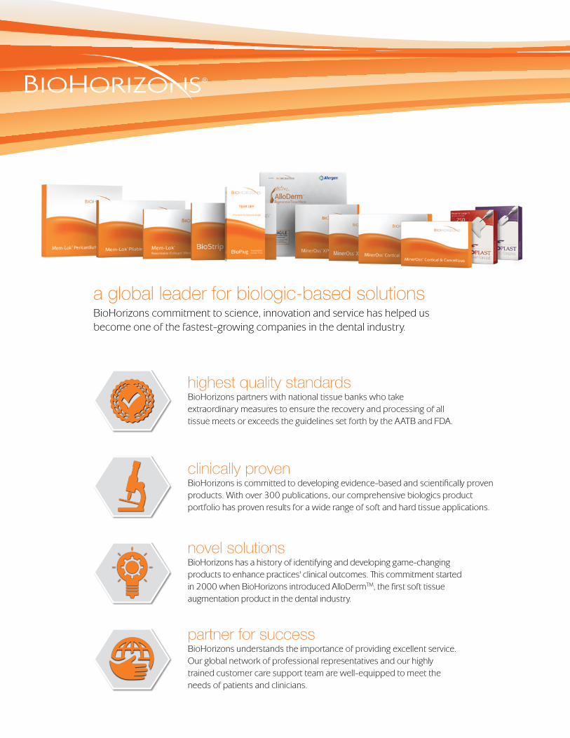
BioHorizons commitment to science, innovation and service has helped us become one of the fastest-growing companies in the dental industry.
clinically provenBioHorizons is committed to developing evidence-based and scientifically proven products. With over 300 publications, our comprehensive biologics product portfolio has proven results for a wide range of soft and hard tissue applications.
a global leader for biologic-based solutions
novel solutionsBioHorizons has a history of identifying and developing game-changing products to enhance practices' clinical outcomes. This commitment started in 2000 when BioHorizons introduced AlloDermTM, the first soft tissue augmentation product in the dental industry.
partner for successBioHorizons understands the importance of providing excellent service. Our global network of professional representatives and our highly trained customer care support team are well-equipped to meet the needs of patients and clinicians.
highest quality standardsBioHorizons partners with national tissue banks who take extraordinary measures to ensure the recovery and processing of all tissue meets or exceeds the guidelines set forth by the AATB and FDA.

The following review summarizes many of the studies and presentations related to the Biologics product portfolio.
Study Review
Foundational Research
Healing and integration of acellular scaffolds 2
A new alternative in the management of periodontal soft tissues 3
Adhesion, proliferation, and differentiation of MSCs into osteoblasts 4
Autograft Equivalency
A substitute for autogenous grafts in mucogingival surgeries 5
Long-term effectiveness and predictability of ADMA and CTG 6
Histological results of CT grafts, ADM grafts, coronally advanced flaps 7
Clinical efficacy of ADM in the augmentation of PMT 8
Outcomes of SCTG and ADM for the application of dental implants 9
Comparison to Other Membranes
Extraction socket healing and alveolar ridge alteration 10
Extraction sockets covered with ADMA or an ePTFE membrane 11
Comparison of AlloDerm & Allopatch 12
Technique
Continuous sling suturing method with the tunneling technique 13
Orientation of an acellular dermal matrix (ADM) allograft 14
Double papilla lateral sliding flap with ADM for root coverage 15
Double layer technique using an ADM 16
Latest Research
Stability of ADM for root coverage in smokers 17
Crestal bone level after soft tissue thickening 18
Clinical benefits using ADM with tenting screw technique 19
Use of ADM with or without mineralized bone allograft 20
GBR technique using allograft and AlloDerm 21
New bone formation with DBM 22
Treatment of periodontal intrabony defects 23
DBM used alone or in combination with platelet-rich fibrin 24
Sinus augmentation using a novel allogenic bone substitute 25
Mixture of mineralized allograft bone chips in maxillary sinus augmentation 26
Platelet-rich fibrin and freeze-dried bone allograft in bone augmentation 27
Anorganic bovine bone mineral (ABBM) as a grafting material 28
In vitro and in vivo studies conducted for PCA 29
Xenografts grafted in adjacent extraction sockets 30
Flapless tooth extraction procedures with or without a xenograft 31
Aspects of bone healing in extraction sites 32
Evaluation of porcine collagen membrane compared to Bio-Gide 33
Collagen membrane offers advantages 34
Soft Tissue Grafting
Hard Tissue Grafting
Dental Membranes
L-PRF Block for bone augmentation 35
Antimicrobial properties of L-PRF membranes 36
L-PRF Block for sinus augmentation 37
L-PRF Applications

222
Introduction: Human Acellular Dermal Matrix (HADM) is produced from skin recovered from tissue donors and gently processed to remove cellular elements while retaining the architecture and key biochemical components of the dermis.
Purpose: The purpose of this study is to present the first primate (Old World monkey) model for ventral hernia repair as a functional and immunologically appropriate surrogate for clinical implantation of HADM.
Procedure: A longitudinal midabdominal incision was made to expose an area of the abdominal muscle wall, and a bilateral longitudinal full thickness defect (3x7 cm) was created by removal of fascia, rectus muscle, and peritoneum. Defects were repaired with hydrated test articles (HADM, n=53; PADM, n=8; HCDM, n=12) in a manner that imparted a uniform tension across the graft to close the defect.
Host Response to Human Acellular Dermal Matrix Transplantation in a Primate Model of Abdominal Wall RepairXu H, Wan H, Sandor M, Qi S, Ervin F, Harper J, Silverman R, and McQuillan D.Tissue Engineering: Part A. Volume 14, Number 12, 2008.
FOUNDATIONAL RESEARCH
Results: HADM appeared off-white at the earliest time point (10 days), similar to preimplant material, but had changed to a reddish color similar to adjacent host tissue at 20 and 35 days. The surface of the graft facing the peritoneal space was smooth, and vascular structures were apparent at 35 days on the surface of HADM implants, similar to a normal peritoneal surface. The strength of the graft-tissue interface was measured. At 10 days the average maximal breaking strength of the repaired abdominal wall was low (5.2 ± 0.2 N, n = 3), but rapidly increased with time to reach a plateau between 35 and 90 days. The breaking strength at 90 days (39.7±8.SN, n = 23) was greater than for primarily healed fascia (21.4 ± 4.8 N, n = 7), indicating that sufficient healing between implant and host tissue had been achieved. (D; a higher magnification view of HADM (F; Cytokeratin-19 expression on HADM at day 35)
Discussion: A number of key questions have been addressed in the current study to advance our understanding of healing and integration of acellular scaffolds in tissue regeneration. The data indicate that cellular repopulation begins early with fibroblasts clearly infiltrating the edge of HADM at 10 days and reaching a plateau between 1 and 3 months. Functional blood vessels appear to form in parallel with host cell re-population, with clearly delineated channels lined with endothelial cells at 1 month. With time, the scaffold assumed characteristics of the surrounding host tissue. For example, the surface of the graft placed in apposition to bowel contents exhibited cellular components consistent with peritoneum, and collagen fiber architecture was distinct from the original reticular orientation and similar to the alignment of native fascia. The gross observations and positive clinical assessment of the healing response to HADM are consistent with an active regenerative process being undertaken during the first 3 months. The HADM provides a scaffold for rapid tissue integration and host cell repopulation without observable herniation, laxity, or attenuation of the graft. HADM and PADM did not induce any chronic immune response, but were repopulated with fibroblasts, and exhibited an extensive vascular network.
Healing and integration of acellular scaffolds
333
Introduction: Soft tissue ridge defects often hamper ideally shaped artificial crowns and are basically treated using autogenous soft tissue grafts or alloplastic materials. Different methods for reconstruction of ridge defects can be divided into three basic categories: 1) reconstruction using autogenous soft tissue grafts 2) reconstruction with non-resorbable alloplastic materials and 3) reconstruction using guided bone regeneration with membrane alone or associated with osseous grafts. Connective tissue grafts meet demands for uniformity of color with the receptor site and cause less postoperative discomfort; however, shrinkage of the grafts is frequently observed. Free gingival onlay grafts also have the disadvantage of tissue blanching with esthetic drawbacks. These techniques require an additional surgical procedure for harvesting the grafts, which may increase postoperative pain, hemorrhage, or infection, accounting for the reluctance of a significant number of patients.
Purpose: This investigation evaluated the use of acellular dermal matrix (ADM) in the treatment of soft tissue ridge defects.
Methods: Eight patients, non-smokers with noncontributory medical history, provided 18 sites corresponding to missing teeth in the anterior maxillary arch. After raising partial-thickness flaps, the ADM material was rehydrated and folded to fill the defect and reproduce the desired gain. Flaps were sutured with no tension, and part of the material was intentionally left exposed to avoid pressure on the incision line and prevent height loss. Patients used local and systemic antimicrobials, and the sutures were removed at 7 days.
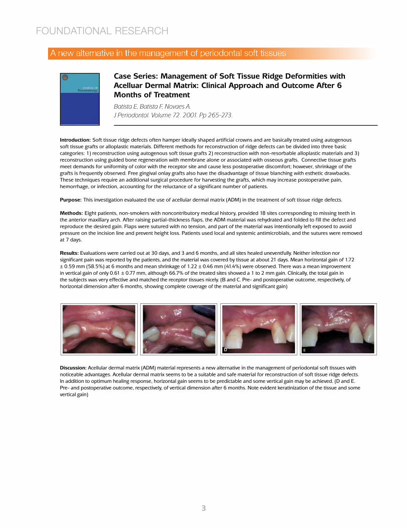
Results: Evaluations were carried out at 30 days, and 3 and 6 months, and all sites healed uneventfully. Neither infection nor significant pain was reported by the patients, and the material was covered by tissue at about 21 days. Mean horizontal gain of 1.72 ± 0.59 mm (58.5%) at 6 months and mean shrinkage of 1.22 ± 0.46 mm (41.4%) were observed. There was a mean improvement in vertical gain of only 0.61 ± 0.77 mm, although 66.7% of the treated sites showed a 1 to 2 mm gain. Clinically, the total gain in the subjects was very effective and matched the receptor tissues nicely. (B and C. Pre- and postoperative outcome, respectively, of horizontal dimension after 6 months, showing complete coverage of the material and significant gain)
Case Series: Management of Soft Tissue Ridge Deformities with Acelluar Dermal Matrix: Clinical Approach and Outcome After 6 Months of Treatment Batista E, Batista F, Novaes A.J Periodontol. Volume 72. 2001. Pp 265-273.
FOUNDATIONAL RESEARCH
Discussion: Acellular dermal matrix (ADM) material represents a new alternative in the management of periodontal soft tissues with noticeable advantages. Acellular dermal matrix seems to be a suitable and safe material for reconstruction of soft tissue ridge defects. In addition to optimum healing response, horizontal gain seems to be predictable and some vertical gain may be achieved. (D and E. Pre- and postoperative outcome, respectively, of vertical dimension after 6 months. Note evident keratinization of the tissue and some vertical gain)
A new alternative in the management of periodontal soft tissues

444
Introduction: The need for bone regeneration in cranial, oral and maxillo-facial and orthopedic surgery is one of the central clinical issues in regenerative and rehabilitation medicine. Four stages have been described to successfully regenerate bone and other tissues: (1) Primary closure of the wound to promote undisturbed and uninterrupted healing. (2) Angiogenesis to provide necessary blood supply and undifferentiated mesenchymal cells. (3) Space creation and maintenance to facilitate space for bone in-growth. (4) Stability of the wound to induce blood clot formation and allow uneventful healing. Since 1982, when the guided bone regeneration (GBR) technique was first introduced, the expanded polytetrafluoroethylene (e-PTFE) membrane has been considered the gold standard for barrier function materials. e-PTFE membranes have certain limits, such as the need for a second surgical operation to remove them and the possibility of bacterial infection. Resorbable membranes were developed to avoid some of these limitations. A membrane made of resorbable material is intended primarily as temporary support during the regeneration phase. Recently an ADM, AlloDerm® GBR, has been proposed as a resorbable membrane for the GBR technique.
Purpose: The aim of the present in vitro study was to examine adhesion, proliferation, and differentiation of Meshenchymal Stem Cells (MSCs) into osteoblasts on an acellular dermal matrix (ADM) used for guided bone regeneration.
Methods: MSCs were cultured in MEM medium+10% fetal bovine serum, Fungizone and ascorbic acid, incubated in humidified atmosphere 95%/5% air/CO2 at 37 placed in sterile siliconized tubes with ADM, and transferred into the Nunc plates wells in incubator at 37° C until the end of experimental times. Test samples were subjected to intermittent treatment in an osteogenic medium, and control test samples to intermittent treatment in a regular medium to determine whether the introduction of osteogenic factors in the culture mean expands the answer given by osteoblastic differentiation of MSCs on an ADM. Cultures were analyzed after 14 days and 35 days using a Scanning Electron Microscope (SEM) to observe the ultrastructural morphology, and X-ray microanalysis to assess the deposition of calcium.
Proliferation Assessment of Meshenchymal Stem Cells on an Acellular Dermal Matrix (AlloDerm GBR) Used for Guided Bone RegenerationPappalardo S, Guarnieri R.Journal of Biomaterials and Tissue Engineering. Volume 3. 2013. PP 1-8.
Results: After 14 days of culture, the sample treated in a regular medium showed a large number of MSCs with well-spread morphology on the ADM. After 35 days, the ADM was completely covered by several layers of MSCs that seemed to have strong adhesion to the membrane surface. In samples treated in an osteogenic medium, the ADM increased the osteoblastic type morphology with the appearance of numerous bundles of collagen, many of which presented grainy formations. Surfaces of the plasma membranes showed micro and macro exocytosis vesicles with X-ray microanalysis that were characterized by the presence of calcium and phosphorus.
Discussion: During the bone formation phase, osteoblasts are recruited from MSC present in bone marrow. The procedure of GBR was developed to ensure that osteoprogenitor cells repopulate the bone wound area by using a membrane to exclude the unwanted cells. In addition to maintaining a space that should be invaded by osteogenetic cells from the surrounding bone, GBR principles also underlined the importance of membrane biocompatibility and surface topography. ADM (AlloDerm GBR®) may influence the adhesion, differentiation and proliferation of mesenchymal stem cells into osteoblasts.
FOUNDATIONAL RESEARCH
SEM images of MSC growth on an ADM in an osteogenic medium after 14 days.
SEM images of MSC growth on an ADM in an osteogenic medium after 35 days.
Adhesion, proliferation, and differentiation of MSCs into osteoblasts
555
Study or Subcategory N
CTG Mean (SD) N
ADM-RCMean (SD)
WMD (random)95% Cl
Weight %
WMD (random)95% Cl
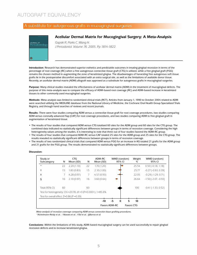
* 22 2.20 (1.10) 22 1.70 (1.20) 25.54 0.50 [-0.18, 1.18]
† 15 1.83 (0.83) 15 2.10 (1.00) 25.77 -0.27 [-0.93, 0.39]
‡ 7 4.28 (0.97) 7 4.57 (0.93) 22.05 -0.29 [-1.29, 0.71]
§ 16 2.10 (0.97) 16 3.60 (0.64) 26.64 -1.50 [-2.07, -0.93]
Total (95% Cl) 60 60 100 -0.41 [-1.33, 0.52]
Test for heterogeneity: Chi =20.78, df =3 (P=0.0001), 1=85.6%
Test for overall effect: Z=0.86 (P =0.39)
Introduction: Research has demonstrated superior esthetics and predictable outcomes in treating gingival recession in terms of the percentage of root coverage (RC) when a free autogenous connective tissue graft (CTG) is utilized, while a free gingival graft (FGG) remains the chosen method in augmenting the zone of keratinized gingiva. The disadvantages of harvesting free autogenous soft tissue grafts lie in the postoperative discomfort associated with an extra surgical site, as well as the limitations of available donor tissue. Recently, an acellular dermal matrix (ADM) allograft was approved as a substitute for autogenous grafts in mucogingival surgeries.
Purpose: Many clinical studies revealed the effectiveness of acellular dermal matrix (ADM) in the treatment of mucogingival defects. The purpose of this meta-analysis was to compare the efficacy of ADM-based root coverage (RC) and ADM-based increase in keratinized tissues to other commonly used mucogingival surgeries.
Methods: Meta-analysis was limited to randomized clinical trials (RCT). Articles from January 1, 1990 to October 2004 related to ADM were searched utilizing the MEDLINE database from the National Library of Medicine, the Cochrane Oral Health Group Specialized Trials Registry, and through hand searches of reviews and recent journals.
Results: There were four studies comparing ADM versus a connective tissue graft for root coverage procedures, two studies comparing ADM versus coronally advanced flap (CAF) for root coverage procedures, and two studies comparing ADM to free gingival graft in augmentation of keratinized tissue.
Acellular Dermal Matrix for Mucogingival Surgery: A Meta-Analysis
Gapski R, Parks C, Wang H.J Periodontol. Volume 76. 2005. Pp 1814-1822.
The results of four studies that compared ADM versus CTG totalized 60 sites for the ADM group and 60 sites for the CTG group. The combined data indicated no statistically significant differences between groups in terms of recession coverage. Considering the high heterogeneity values among the studies, it is interesting to note that three out of four studies favored the ADM-RC group. The results of four studies that compared ADM-RC versus CAF totaled 25 sites for the ADM group and 25 sites for the CTG group. The results revealed no statistically significant differences between groups in terms of recession coverage. The results of two randomized clinical trials that compared ADM versus FGG for an increase in KG totaled 21 grafts for the ADM group and 21 grafts for the FGG group. The results demonstrated no statistically significant differences between groups.
•
•
•
Discussion:
AUTOGRAFT EQUIVALENCY
Meta-analysis of recession coverage comparing ADM versus connective tissue grafting procedures. *Aichelmann-Reidy et al. ; †Novaes et al. ; ‡Tal et al. ; §Barros et al.
A substitute for autogenous grafts in mucogingival surgeries
-10 -5 0 5 10
Favors ADM-RC Favors CTG
Conclusions: Within the limitations of this study, ADM-based mucogingival surgery can be used successfully to repair gingival recession defects and to increase keratinized gingiva.

666
Introduction: Although gingival recession seldom results in tooth loss, marginal tissue recession is associated with thermal and tactile sensitivity, esthetic complaints, and a tendency toward root caries. Gingival recession may be due to several etiologic factors including periodontal disease, mechanical forces such as faulty toothbrushing, tooth malposition, and frenum pull, to mention just a few. Therefore, coverage of exposed root surfaces is performed not to increase keratinized epithelium but to ameliorate the patient’s esthetic troubles, dentinal hypersensitivity, or root caries. A variety of highly predictable and esthetically acceptable mucogingival grafting procedures exists for treating exposed roots, whether intact, carious, or restored. One of the problems with root coverage grafting is the limited supply of donor connective tissue. Acellular Dermal Matrix Allograft (ADMA) has been compared to connective tissue grafts (CTG) in root coverage procedures. Short-term results are esthetically similar and acceptable as well as achieving a similar extent of root coverage. At 12 months postoperatively, ADMA results in a similar extent of coverage as CTG, but CTG results in a significantly greater gain of keratinized mucosa. A recent report indicates that after 48 to 49 months, only 32% of the cases treated with ADMA improved or remained stable with time.
Purpose: The purpose of the present study was to compare the long-term (2 years) effectiveness and predictability of ADMA and CTG in the treatment of relatively severe recessions.
A 2-Year Follow-Up of Root Coverage Using Subpedicle Acellular Dermal Matrix Allografts and Subepithelial Connective Tissue AutograftsHirsch A, Goldstein M, Goultschin J, Boyan B.D., Schwartz Z.J Periodontol. Volume 76. 2005. Pp 1323-1328.
Methods: One hundred one (101) patients were treated with dermal matrix allografts (mean age, 28.4 ± 0.7 years; mean recession, 4.2 mm) and 65 patients treated with connective tissue graft (mean age, 30.1 ± 1.4 years; mean recession, 4.9 mm). All patients underwent full periodontal evaluation and presurgical preparation, including oral hygiene instruction and scaling and root planing. The exposed roots were thoroughly planed and covered by a graft without any further root treatment or conditioning. There were no differences in the average age, time of follow-up, or gender between the two groups. Patients were evaluated periodically between 1 and 2 years. Residual recession and defect coverage were assessed.
Results: Mean residual root recession after root coverage with acellular dermal matrix allograft was 0.2 ± 0.04 mm, with defect coverage of 95.9% ± 0.9%. Frequency of defect coverage was 82.2%. Root coverage was 98.8% ± 0.2%, resulting in a frequency of root coverage of 100%. Gain in keratinized gingiva was 2.2 ± 0.04 mm and attachment gain was 4.5 ± 0.1 mm per patient. No significant differences in final recession and root coverage between the two treatment methods were found. The clinical results were stable for the 2-year follow-up period.
Conclusions: These results indicate that coverage of root by subpedicle acellular dermal matrix allografts or subepithelial connective tissue autografts is a very predictable procedure which is stable for 2 years postoperatively. However, subepithelial connective tissue autografts resulted in significant increases in defect coverage, keratinized gingival gain, attachment gain, and residual probing depth.
AUTOGRAFT EQUIVALENCY
Allograft: A) 38-year-old woman presented with gingival recessions of 4 and 5 mm in teeth #11 and #12, respectively. B) A full-thickness flap with mesial and distal vertical releasing incisions was elevated. C) An acellular dermal matrix allograft was placed to cover the exposed roots and was stabilized by sutures. D) Twenty-four month postoperative photograph shows complete defect coverage.
Long-term effectiveness and predictability of ADMA and CTG
777
Introduction: Successful coverage of exposed roots for esthetic as well as functional reasons has been the objective of various mucogingival procedures. This has been performed through lateral or coronal repositioning of the adjacent attached gingiva via a pedicle flap, coronal advancement of previously placed gingival grafts, gingival grafts placed directly over the root surface, and gingival grafting performed in conjunction with flap advancement for submersion. Traditionally, this augmentation of the gingival complex at the time of root coverage has been performed with autogenous connective tissue (CT) harvested from the palate or edentulous ridge. ADM has been used by dentists as a substitute for palatal connective tissue in gingival augmentation. Although the clinical aspects of various techniques of autogenous CT grafts and ADM grafts for root coverage have been well documented in the literature, only a few studies in humans have evaluated the cellular events at a histologic level.
Purpose: The purpose of this study is to document the histological results of CT grafts, ADM grafts, and coronally advanced flaps to cover denuded roots in humans.
Methods: This study included four patients previously treatment planned for extractions of three or more anterior teeth. Three teeth in each patient were selected and randomly designated to receive either a CT or ADM graft beneath a coronally advanced flap (tests) or coronally advanced flap alone (control). Six months postoperatively, block section extractions were performed and the teeth processed for histologic evaluation with hematoxylin-eosin and Verhoeff’s stains.
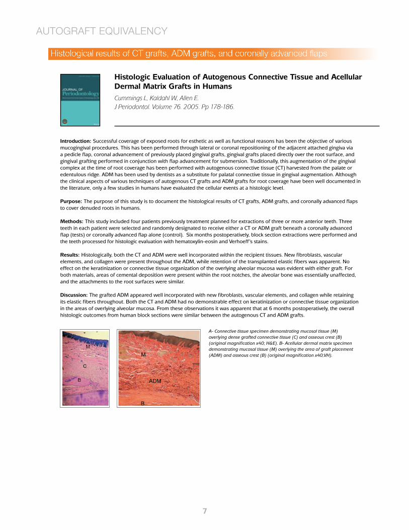
Results: Histologically, both the CT and ADM were well incorporated within the recipient tissues. New fibroblasts, vascular elements, and collagen were present throughout the ADM, while retention of the transplanted elastic fibers was apparent. No effect on the keratinization or connective tissue organization of the overlying alveolar mucosa was evident with either graft. For both materials, areas of cemental deposition were present within the root notches, the alveolar bone was essentially unaffected, and the attachments to the root surfaces were similar.
Discussion: The grafted ADM appeared well incorporated with new fibroblasts, vascular elements, and collagen while retaining its elastic fibers throughout. Both the CT and ADM had no demonstrable effect on keratinization or connective tissue organization in the areas of overlying alveolar mucosa. From these observations it was apparent that at 6 months postoperatively, the overall histologic outcomes from human block sections were similar between the autogenous CT and ADM grafts.
Histologic Evaluation of Autogenous Connective Tissue and Acellular Dermal Matrix Grafts in Humans
Cummings L, Kaldahl W, Allen E.J Periodontol. Volume 76. 2005. Pp 178-186.
A- Connective tissue specimen demonstrating mucosal tissue (M) overlying dense grafted connective tissue (C) and osseous crest (B) (original magnification x40; H&E). B- Acellular dermal matrix specimen demonstrating mucosal tissue (M) overlying the area of graft placement (ADM) and osseous crest (B) (original magnification x40;VH).
AUTOGRAFT EQUIVALENCY
Histological results of CT grafts, ADM grafts, and coronally advanced flaps

888
Introduction: Tooth replacement therapy using endosseous implants has become a routine component of contemporary dental practice. While a plethora of factors may determine success, the bucco-lingual and apico-coronal dimensions of the peri-implant mucosa seem to play a critical role in both the maintenance of peri-implant health and esthetics. The autologous subepithelial connective tissue graft (sCTG) is generally regarded as the gold standard for soft tissue augmentation around natural teeth and dental implants. However, harvesting an autologous soft tissue graft necessarily entails additional pre-operative preparation, a second surgical site, longer operative duration and increased morbidity, regardless of the surgical technique employed and the expertise of the operator. Several studies comparing sCTG with acellular dermal matrix (ADM) as an alternative to autologous soft tissue grafts for the treatment of mucogingival defects in the natural dentition, have shown similar clinical outcomes.
Purpose: The primary aim of this randomized clinical trial was to determine the clinical efficacy of ADM in the augmentation of PMT as compared to an autologous sCTG.
Methods: Patients in need of peri-implant mucosa augmentation at the time of implant placement were recruited. Subjects were randomized to the control (simultaneous sCTG) or test (simultaneous ADM) group. The primary outcome in this study was changes in PMT between baseline and 16 weeks later. Keratinized mucosa width (KMW) changes, modified wound healing index (MWHI) variations and patient reported outcome measures (PROMs) were recorded as well.
Results: A total of 20 subjects were recruited per a priori power analysis. There were no statistically significant differences between groups at baseline for any of the parameters analyzed. No statistically significant differences in terms of PMT, KMW and MWHI changes were observed between groups. The perceived discomfort was higher at 2 and 4 weeks for patients in the sCTG group.
Discussion: The presence of initially thicker crestal mucosa or augmenting thin tissue at the time of placement may attenuate marginal bone changes after implant placement. Implants placed in sites with thin crestal tissue (< 2 mm) that were simultaneously thickened with a soft tissue allograft behaved similarly to sites with initially thick tissue. Sites with initially thin tissue that were not grafted lost a significantly greater amount of bone (1.2 ± 0.08mm) as compared to the thin-grafted and thick groups (0.22 ± 0.06mm). This study offers clinical evidence that in adult patients in need of peri-implant mucosal augmentation at the time of implant placement in tooth bound sites, ADM produces similar outcomes to sCTG in terms of PMT, KMW and PROMs (i.e. perception of discomfort and overall satisfaction).
Comparison of Two Different Surgical Approaches to Increase Peri-Implant Mucosal Thickness: A Randomized Controlled Clinical TrialHutton C, Johnson G, Barwacz C, Allareddy V, Avila-Ortiz G.J Periodontol. 2018. https://doi.org/10.1002/JPER.17-0597.
Photographic sequence of a representative clinical case for both the control and test group, including a control radiograph upon provisionalization after implant uncovering at 16 weeks postplacement. PMT using the periodontal probe and the endodontic spreader at 1, 3 and 5 mm from the crestal mucosal.
AUTOGRAFT EQUIVALENCY
Clinical efficacy of ADM in the augmentation of PMT

999
Introduction: Although there is no “gold standard” graft technique, research showed that treatment with subepithelial connective tissue grafts (SCTG) was the most predictable approach for root coverage outcomes. Because this autogenous technique requires an additional donor site and has associated morbidity, allogenic graft materials have been introduced. Specifically, acellular dermal matrix (ADM) was identified as an adequate alternative to autogenous techniques for gingival augmentation. Although there is an abundance of research concerning gingival augmentation around natural teeth, there are few studies investigating the outcomes of similar procedures around dental implants and none of these studies addressed grafting implants restored with definitive crowns.
Purpose: The objective of this study was to compare the outcomes of SCTG and ADM, 2 methods of soft tissue grafting established on natural teeth for the novel application on dental implants.
Methods: Thirteen patients presenting with implants displaying recession, thin biotype, concavity defects, or a combination thereof associated with single crowned dental implants randomly received subepithelial connective tissue grafts (SCTG) in the control group (N. 7) or acellular dermal matrix (ADM) allografts in the test group (N. 6), both under coronally positioned flaps. Data regarding soft tissue, hard tissue, esthetics, and quality of life (QoL) parameters were collected over 6 months.
Implant Associated Soft Tissue Defects in the Anterior Maxilla: A Randomized Control Trial Comparing Subepithelial Connective Tissue Graft and Acellular Dermal Matrix Allograft Anderson L, Inglehart M, El-Kholy K, Eber R, Wang H.Implant Dentistry. Volume 0. Number 0. 2014.
Results: Both groups gained tissue thickness (SCTG: 63% and ADM: 105%), reduced concavity measures (SCTG: 82% and ADM: 96%), and improved recessions (SCTG: 40% and ADM: 28%) from baseline to 6 months.
Discussion: Although the number of subjects in this pilot study was small, the findings go beyond the understanding in the current literature. Our study showed that a thin soft tissue thickness, or biotype, was not necessarily associated with a thin buccal plate dimension measured by CBCT evaluation. Moreover, we found that a thicker biotype did not necessarily imply better recession or concavity correction outcomes, but that hard tissue morphology dictated the outcome to a greater degree. Major findings of our analysis include: (1) SCTG and acellular dermal matrix allograft (ADM) are suitable graft materials for the treatment of peri-implant soft tissue discrepancies as demonstrated by their ability to increase tissue thickness, decrease concavity dimensions, and provide partial recession correction; (2) there were no statistical differences between SCTG and acellular dermal matrix allograft (ADM) regarding clinical treatment and esthetic outcomes; (3) underlying hard tissue morphology dictated soft tissue treatment outcomes; (4) there were no statistically significant differences in pain, medication, and overall QoL between the 2 groups; and (5) wound healing was less eventful in the control group.
AUTOGRAFT EQUIVALENCY
Outcomes of SCTG and ADM for the application of dental implants

101010
Introduction: Extraction socket augmentation has been proposed as a means of controlling alveolar ridge degradation, preserving crestal buccal plate integrity, improving vital bone fill, and reducing the need for future ridge augmentation. Research has evaluated the use of membrane, bone grafts, and a combination of the two for controlling buccal plate loss. The placement of wound dressing over the grafted extraction socket is critical in preventing bone graft loss. Numerous bioabsorbable and non-resorbable materials, along with various grafting techniques, have been used; they showed varying degrees of success with regard to graft retention.
Purpose: The purpose of this study was to compare extraction socket healing and alveolar ridge alteration after socket augmentation using bone allograft covered with an acellular dermal matrix (ADM) or polytetrafluoroethylene (PTFE) membrane.
Comparison of dermal matrix and polytetrafluoroethylene membrane for socket bone augmentation: A clinical and histologic studyFotek P, Neiva R, Wang H.J Periodontol. Volume 80. 2009. Pp 776-785.
Methods: Twenty non-smoking healthy subjects were selected. Each subject required maxillary premolar, canine, or central incisor tooth extraction. The extraction sites were debrided and grafted with a mineralized bone allograft that was covered with an ADM or PTFE membrane. Postoperative appointments were scheduled at 2, 4, and 8 weeks. After 16 weeks of healing, final measurements were performed and trephine core biopsies were obtained for histomorphometric analysis. Implants were placed immediately after biopsy harvesting.
Results: Eighteen subjects completed the study. All sites healed without adverse events and allowed for implant placement. PTFE membranes exfoliated prematurely, with an average retention time of 16.6 days, whereas the ADM membranes appeared to be incorporated into the tissues. Buccal plate thickness loss was 0.44 and 0.3 mm, with a vertical loss of 1.1 and 0.25 mm, for ADM and PTFE, respectively. Histomorphometric analysis revealed 41.81% versus 47.36% bone, 58.19% versus 52.64% marrow/fibrous tissue, and 13.93% versus 14.73% particulate graft remaining for ADM and PTFE, respectively.
Discussion: For an implant to remain successful over time, an intact buccal bone plate is necessary to maintain a bony wall and soft tissue drape. This bone plate thickness was determined by Spray et al. to be ‡ 1.8 mm thick to preserve the buccal plate height and soft tissue margin and prevent future tissue loss. Therefore, it is critical to maintain the buccal bone integrity at all stages, from tooth extraction to final implant restoration. This study examined the alveolar dimensional changes after tooth extraction. All sites evaluated showed minimal ridge alterations, with no statistical difference between the two treatment modalities with respect to bone composition and horizontal and vertical bone loss, indicating that both membranes are suitable for alveolar ridge augmentation.
COMPARISON TO OTHER MEMBRANES
Histology of bone cores. A) Bone core specimen from ADM group showing allograft particles (p) with new bone (nb) formation in its surface. B) Magnified view of rectangle in A. C) Bone core specimen from PTFE group showing allograft particles (p) with new bone (nb) formation in its surface. D) Magnified view of rectangle in C. Calcified bone stained bright red with variations in intensity depending on the maturity of the bone. Non-calcified bone and osteoid (os) stained bright green: osteoblasts stained blue. (Stevenel’s blue and Van Gieson’s picro fuchsin; original magnification: A and C, x 40; B and D, x200.)
Extraction socket healing and alveolar ridge alteration

111111
Introduction: As a result of the bone resorption and soft tissue shrinkage that occurs after routine atraumatic tooth extraction, ideal implant placement and implant esthetics are often compromised. Controlled clinical studies have documented an average of 4.4 mm of horizontal and 1.2 mm of vertical bone resorption 6 months after tooth extraction. Various materials have been used to prevent or minimize ridge collapse after tooth extraction in an attempt to improve implant placement and the subsequent esthetics of the final implant prosthesis. It is therefore of interest to see if Acellular Dermal Matrix Allograft (ADMA) and/or expanded polytetrafluoroethylene (ePTFE) barriers are able to produce an improved healing result in fresh extraction sockets when primary coverage is purposely not attempted.
Purpose: The purpose of this pilot study was to compare, and histologically evaluate, the healing of extraction sockets implanted with either an absorbable or nonabsorbable hydroxyapatite and covered by an ADMA or an ePTFE membrane.
Methods: Following tooth extraction, a total of 76 sockets in 15 patients with deficient buccal plates of 5 mm were randomly divided into 4 treatment groups: 1) absorbable hydroxyapatite (AH) covered with ADMA, 2) AH covered with an ePTFE membrane, 3) anorganic bovine bone mineral (ABB) covered with ADMA, and 4) ABB covered with an ePTFE membrane. Primary coverage was not attempted or obtained in any of the 16 treated sockets. Six to 8 months post extraction at the time of implant placement, histologic cores of the treatment sites were obtained. These cores were processed, stained with Stevenel’s blue/van Gieson’s picro fuchsin, and histo-morphometrically analyzed. Vital bone, connective tissue and marrow, and residual graft particles were reported at a percentage of the total core.
Extraction Sockets and Implantation of Hydroxyapatites with Membrane Barriers: A Histologic StudyFroum S, Cho S, Elian N, Rosenberg E, Rohrer M, Tarnow D.Implant Dentistry. Volume 13. 2004. Pp 153-164.
Results: The average percentage of vital bone in the 8 sockets covered with ADMA was 38% compared with an average percentage vital bone of 22% in the 8 sockets covered with ePTFE membrane barriers. ADMA covered sites resulted in more vital bone present 6 to 8 months post socket treatment than obtained in the ePTFE covered sites regardless of bone replacement materials used.
Discussion: Extraction socket treatment with ABMA barriers produced more vital bone 6 to 8 months post-extraction than did ePTFE membranes, whether placed over AH or nonabsorbable ABB mineral. The combination of ABMA covering ABB produced the greatest amount of vital bone at 6 to 8 months (41.7%) followed by ABMA covering AH (34.5%), ePTFE covering AH (27.67%), and ePTFE covering ABB 17.8%. Without primary flap coverage over the extraction socket, 1 of 8 ABMA barriers and 6 of 8 ePTFE barriers had to be removed prematurely because of infection before the 6-8 month time period when implants were placed.
COMPARISON TO OTHER MEMBRANES
Barriers Average Percent Vital
Bone
Grafts Vital Bone
Percent (range)
ADMA 38% AH
ABB
34.5 (19-57)
41.7 (19.5-62.4)
ePTFE 27.7% AH
ABB
27.6 (14-40.1)
17.8 (10-25)
Average Percent of Vital Bone in Sockets Covered With Acellular Dermal Matrix Allograft (ADMA) or Expanded Polytetrafluoroethylene (ePTFE) Membranes and Filled With Absorbable Hydroxyapatite (AH) or Anorganic Bovine Bone (ABB)
This table summarizes the average percent vital bone in sockets treated with ADMA compared with sockets treated with ePTFE barriers. Average vital bone obtained from sockets filled with AH or ABB and covered with either ADMA or ePTFE barriers is also recorded.
ADMA, Acellular Dermal Matrix Allograft; ePTFE, expanded Polytetrafluoroethylene; AH, Absorbable Hydroxyapatite; ABB, Anorganic Bovine Bone mineral.
Extraction sockets covered with ADMA or an ePTFE membrane

121212
Introduction: Soft tissue, such as oral mucosa and skin, may be necessary for reconstruction after surgeries for tumor removal, congenital defects (cleft lip), or by trauma. The availability of the healthy autografts to repair these defects is limited. Scaffolds are important to support cellular growth in the manufacture of 3D tissue-engineered products. Scaffolds can be synthetic, such as biodegradable polymer; or non-synthetic, such as collagen, fibrin, or gelatin-based scaffolds; or naturally derived scaffolds, such as acellular human cadaver skin with a preserved basement membrane and the extracellular matrix of the dermis. Acellular human cadaver skin scaffolds are immunologically inert with a long history of clinical applications. Two examples of commercially available human cadaver skin derived scaffolds are AlloDerm® and Allopatch.
Purpose: Acellular human cadaver skin scaffolds are immunologically inert with a long history of clinical applications. Two examples of commercially available human cadaver skin derived scaffolds are AlloDerm® and Allopatch.
Methods: In this report, we evaluated the cellular growth on AlloDerm® and Allopatch, two acellular scaffolds derived from human cadaver skin, using a fabricated 3D organotypic culture with primary human oral keratinocytes to produce an Ex Vivo Produced Oral Mucosa Equivalent (EVPOME). A well stratified epithelium could be constructed on both scaffolds. AlloDerm® and Allopatch EVPOMEs were also implanted into SCID (Severe Combined Immunodeficiency) mice to compare the ingrowth of blood vessels into the dermal component of the two EVPOMEs. Blood vessel counts were 3.3 times higher (p=0.01) within Allopatch EVPOMEs than within AlloDerm® EVPOMEs. An oral and skin keratinocyte co-culture, separated by a physical barrier to create a cell-free zone, was used to evaluate cell migration on AlloDerm® and Allopatch.
Results: Hemotoxylin and Eosin (H&E) histology demonstrated that the AlloDerm® EVPOME has thicker cellular layers and keratin structure overall than the Allopatch EVPOME, which lacked homogeneous cellular layers and keratin.
Comparison of Two Decellularized Dermal Equivalents.Kuo S, Kim H, Wang Z, Bingham E, Miyazawa A, Marcelo C, Feinberg S.Journal of Tissue Engineering and Regenerative Medicine. Volume 12. 2018. Pp 983-990.
Discussion: It is critical to choose the appropriate scaffold with ideal physical properties, such as biocompatibility and porosity, to ensure the success of tissue engineering products in hosts. Natural derived scaffolds can offer molecular complexity and architecture of the native tissue matrices to support cellular growth that synthetic scaffolds cannot for tissue engineering products. An adequate formation of blood vessels on an implanted graft is required to allow the above phenomena to happen. Allopatch could be an advantageous choice compared with AlloDerm®, since there are more blood vessels formed within Allopatch than AlloDerm®. On the other hand, when cell migration is expected to happen in a timely manner on the scaffold surface, then Allopatch may lose its competitive edge to AlloDerm®.
Figure: Evaluation of in vivo development of implanted AlloDerm® and Allopatch EVPOMEs. Before (in vitro) and corresponding one week after (in vivo) EVPOMEs implantation are shown in H&E and pan-keratin immunohistochemistry (IHC) (only in vivo shown for pan-keratin IHC). Red arrows indicate the areas of continuous development of implanted EVPOMEs examined by anti-pan keratin antibody. D represents AlloDerm®, and P Allopatch.
COMPARISON TO OTHER MEMBRANES
Comparison of AlloDerm & Allopatch
131313
Introduction: Site preparation for root coverage grafting has evolved from the original surgical dissection of an open vascular bed, used for placement of an exposed graft overlying the recipient bed, to the current coronally advanced flap and tunnel methods used for submerged grafts. Along with the advancement of soft tissue grafting methods, a variety of suturing techniques have been described.
Purpose: This paper describes a new suturing method, the subpapillary continuous sling suture, for use with soft tissue grafts in tunnel procedures to treat gingival recession. With the introduction of acellular dermal matrix (AlloDerm, LifeCell) for root coverage grafting, site design and suturing techniques changed to accommodate the different requirements for successful outcomes with allografts.
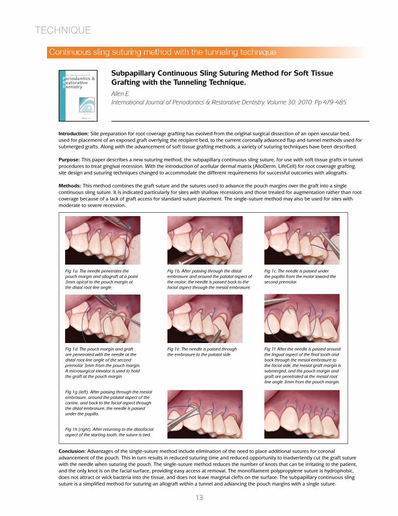
Methods: This method combines the graft suture and the sutures used to advance the pouch margins over the graft into a single continuous sling suture. It is indicated particularly for sites with shallow recessions and those treated for augmentation rather than root coverage because of a lack of graft access for standard suture placement. The single-suture method may also be used for sites with moderate to severe recession.
Subpapillary Continuous Sling Suturing Method for Soft Tissue Grafting with the Tunneling Technique.Allen E.International Journal of Periodontics & Restorative Dentistry. Volume 30. 2010. Pp 479-485.
TECHNIQUE
Continuous sling suturing method with the tunneling technique
Conclusion: Advantages of the single-suture method include elimination of the need to place additional sutures for coronal advancement of the pouch. This in turn results in reduced suturing time and reduced opportunity to inadvertently cut the graft suture with the needle when suturing the pouch. The single-suture method reduces the number of knots that can be irritating to the patient, and the only knot is on the facial surface, providing easy access at removal. The monofilament polypropylene suture is hydrophobic, does not attract or wick bacteria into the tissue, and does not leave marginal clefts on the surface. The subpapillary continuous sling suture is a simplified method for suturing an allograft within a tunnel and advancing the pouch margins with a single suture.
Fig 1a. The needle penetrates the pouch margin and allograft at a point 3mm apical to the pouch margin at the distal root line angle.
Fig 1b. After passing through the distal embrasure and around the palatal aspect of the molar, the needle is passed back to the facial aspect through the mesial embrasure.
Fig 1c. The needle is passed under the papilla from the molar toward the second premolar.
Fig 1d. The pouch margin and graft are penetrated with the needle at the distal root line angle of the second premolar 3mm from the pouch margin. A microsurgical elevator is used to hold the graft at the pouch margin.
Fig 1e. The needle is passed through the embrasure to the palatal side.
Fig 1f. After the needle is passed around the lingual aspect of the final tooth and back through the mesial embrasure to the facial side, the mesial graft margin is submerged, and the pouch margin and graft are penetrated at the mesial root line angle 3mm from the pouch margin.
Fig 1g (left). After passing through the mesial embrasure, around the palatal aspect of the canine, and back to the facial aspect through the distal embrasure, the needle is passed under the papilla.
Fig 1h (right). After returning to the distofacial aspect of the starting tooth, the suture is tied.
141414
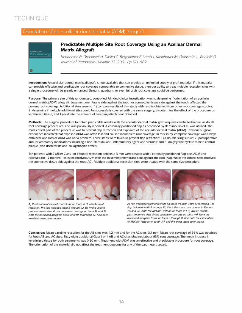
Introduction: An acellular dermal matrix allograft is now available that can provide an unlimited supply of graft material. If this material can provide effective and predictable root coverage comparable to connective tissue, then our ability to treat multiple recession sites with a single procedure will be greatly enhanced. Sextant, quadrant, or even full arch root coverage could be performed.
Purpose: The primary aim of this randomized, controlled, blinded clinical investigation was to determine if orientation of an acellular dermal matrix (ADM) allograft, basement membrane side against the tooth or connective tissue side against the tooth, affected the percent root coverage. Additional aims were to: 1) compare results of this study with results obtained from other root coverage studies; 2) determine if multiple additional sites could be successfully covered with the same surgery; 3) determine the effect of the procedure on keratinized tissue; and 4) evaluate the amount of creeping attachment obtained.
Methods: The surgical procedure to obtain predictable results with the acellular dermal matrix graft requires careful technique, as do all root coverage procedures, and was previously reported. A coronally positioned flap as described by Bernimoulin et al. was utilized. The most critical part of the procedure was to prevent flap retraction and exposure of the acellular dermal matrix (ADM). Previous surgical experience indicated that exposed ADM was often lost and caused incomplete root coverage. In this study, complete coverage was always obtained, and loss of ADM was not a problem. Three steps were taken to prevent flap retraction: 1) a double sling suture; 2) postoperative anti-inflammatory medications including a non-steroidal anti-inflammatory agent and steroids; and 3) doxycycline hyclate to help control plaque (also used for its anti-collagenolytic effect).
Ten patients with 2 Miller Class I or II buccal recession defects ≥ 3 mm were treated with a coronally positioned flap plus ADM and followed for 12 months. Test sites received ADM with the basement membrane side against the root (AB), while the control sites received the connective tissue side against the root (AC). Multiple additional recession sites were treated with the same flap procedure.
Predictable Multiple Site Root Coverage Using an Acelluar Dermal Matrix Allograft.Henderson R, Greenwell H, Dirsko C, Regennitter F, Lamb J, Mehlbauer M, Goldsmith L, Rebitski G.Journal of Periodontol. Volume 72. 2001. Pp 571-582.
Conclusion: Mean baseline recession for the AB sites was 4.2 mm and for the AC sites, 3.7 mm. Mean root coverage of 95% was obtained for both AB and AC sites. Sixty-eight additional Class I or II AB and AC sites obtained about 93% root coverage. The mean increase in keratinized tissue for both treatments was 0.80 mm. Treatment with ADM was an effective and predictable procedure for root coverage. The orientation of the material did not affect the treatment outcome for any of the parameters tested.
TECHNIQUE
A) Pre-treatment view of control site on tooth #11 with 4mm of recession. The flap included teeth 5 through 12. B) Twelve-month past-treatment view shows complete coverage on teeth 11 and 12. Note the thickened marginal tissue of teeth 9 through 12. Also note excellent tissue color match.
A) Pre-treatment view of test site on tooth #6 with 3mm of recession. The flap included teeth 5 through 12; this is the same case as seen in Figures 2A and 2B. Note the McCalls’ festoon on tooth #7. B) Twelve-month post-treatment view shows complete coverage on tooth #6. Note the thickened marginal tissue on teeth 5 through 8. Also note the elimination of McCalls’ festoon on tooth #7 and the exact tissue color match.
Orientation of an acellular dermal matrix (ADM) allograft
151515
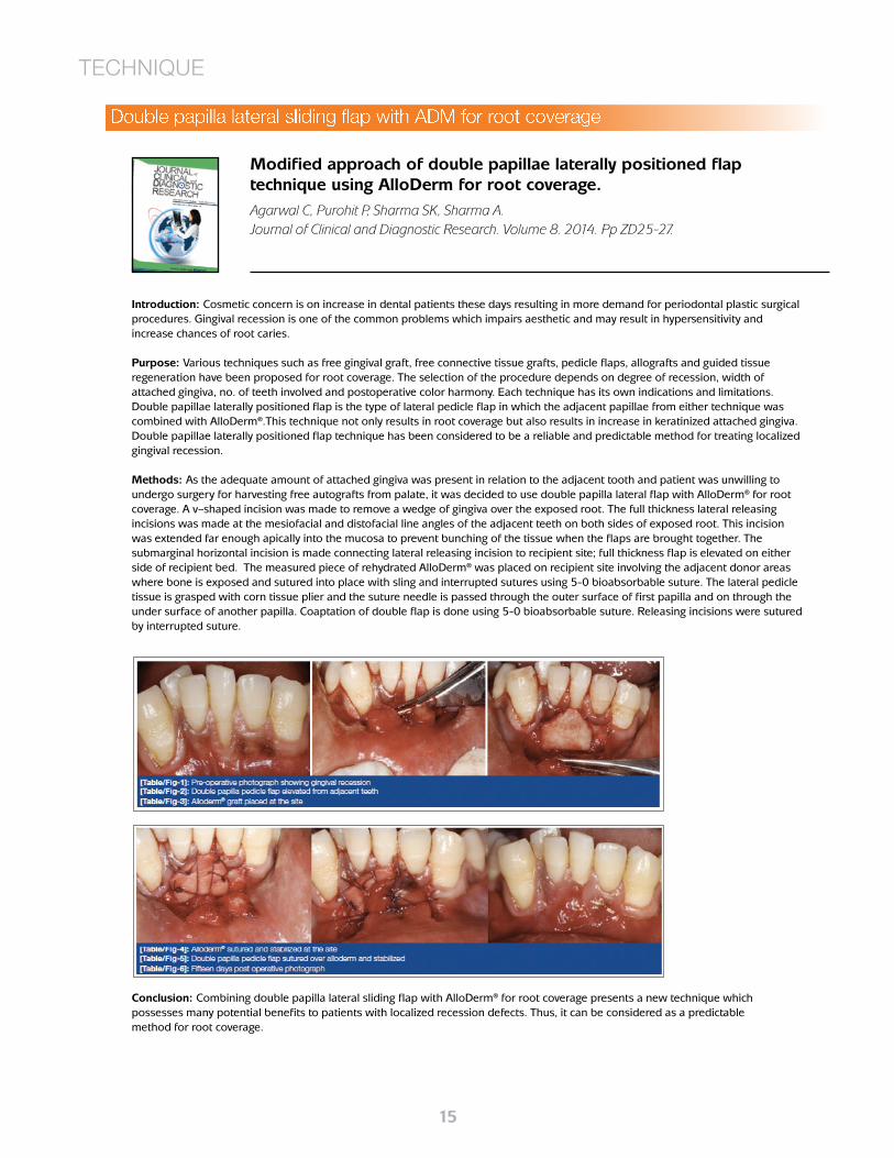
Introduction: Cosmetic concern is on increase in dental patients these days resulting in more demand for periodontal plastic surgical procedures. Gingival recession is one of the common problems which impairs aesthetic and may result in hypersensitivity and increase chances of root caries.
Purpose: Various techniques such as free gingival graft, free connective tissue grafts, pedicle flaps, allografts and guided tissue regeneration have been proposed for root coverage. The selection of the procedure depends on degree of recession, width of attached gingiva, no. of teeth involved and postoperative color harmony. Each technique has its own indications and limitations. Double papillae laterally positioned flap is the type of lateral pedicle flap in which the adjacent papillae from either technique was combined with AlloDerm®.This technique not only results in root coverage but also results in increase in keratinized attached gingiva. Double papillae laterally positioned flap technique has been considered to be a reliable and predictable method for treating localized gingival recession.
Methods: As the adequate amount of attached gingiva was present in relation to the adjacent tooth and patient was unwilling to undergo surgery for harvesting free autografts from palate, it was decided to use double papilla lateral flap with AlloDerm® for root coverage. A v–shaped incision was made to remove a wedge of gingiva over the exposed root. The full thickness lateral releasing incisions was made at the mesiofacial and distofacial line angles of the adjacent teeth on both sides of exposed root. This incision was extended far enough apically into the mucosa to prevent bunching of the tissue when the flaps are brought together. The submarginal horizontal incision is made connecting lateral releasing incision to recipient site; full thickness flap is elevated on either side of recipient bed. The measured piece of rehydrated AlloDerm® was placed on recipient site involving the adjacent donor areas where bone is exposed and sutured into place with sling and interrupted sutures using 5-0 bioabsorbable suture. The lateral pedicle tissue is grasped with corn tissue plier and the suture needle is passed through the outer surface of first papilla and on through the under surface of another papilla. Coaptation of double flap is done using 5-0 bioabsorbable suture. Releasing incisions were sutured by interrupted suture.
Modified approach of double papillae laterally positioned flap technique using AlloDerm for root coverage.Agarwal C, Purohit P, Sharma SK, Sharma A.Journal of Clinical and Diagnostic Research. Volume 8. 2014. Pp ZD25-27.
Conclusion: Combining double papilla lateral sliding flap with AlloDerm® for root coverage presents a new technique which possesses many potential benefits to patients with localized recession defects. Thus, it can be considered as a predictable method for root coverage.
TECHNIQUE
Double papilla lateral sliding flap with ADM for root coverage
161616
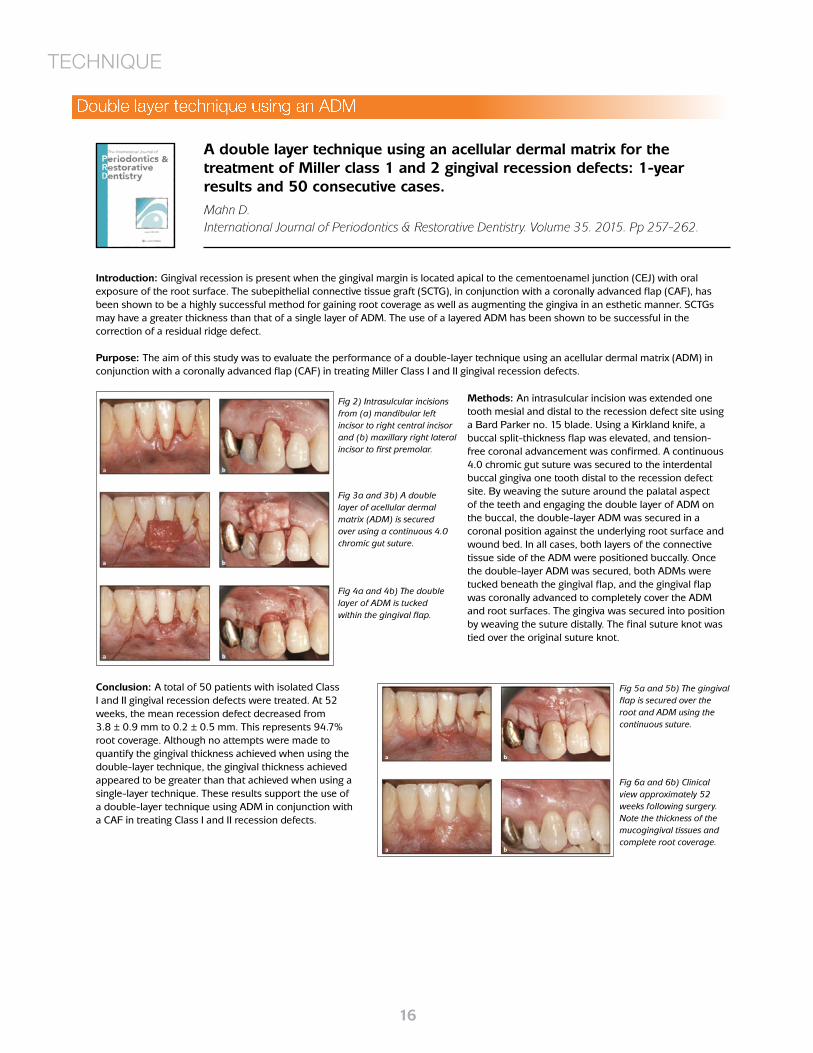
Introduction: Gingival recession is present when the gingival margin is located apical to the cementoenamel junction (CEJ) with oral exposure of the root surface. The subepithelial connective tissue graft (SCTG), in conjunction with a coronally advanced flap (CAF), has been shown to be a highly successful method for gaining root coverage as well as augmenting the gingiva in an esthetic manner. SCTGs may have a greater thickness than that of a single layer of ADM. The use of a layered ADM has been shown to be successful in the correction of a residual ridge defect.
Purpose: The aim of this study was to evaluate the performance of a double-layer technique using an acellular dermal matrix (ADM) in conjunction with a coronally advanced flap (CAF) in treating Miller Class I and II gingival recession defects.
A double layer technique using an acellular dermal matrix for the treatment of Miller class 1 and 2 gingival recession defects: 1-year results and 50 consecutive cases.Mahn D.International Journal of Periodontics & Restorative Dentistry. Volume 35. 2015. Pp 257-262.
Methods: An intrasulcular incision was extended one tooth mesial and distal to the recession defect site using a Bard Parker no. 15 blade. Using a Kirkland knife, a buccal split-thickness flap was elevated, and tension-free coronal advancement was confirmed. A continuous 4.0 chromic gut suture was secured to the interdental buccal gingiva one tooth distal to the recession defect site. By weaving the suture around the palatal aspect of the teeth and engaging the double layer of ADM on the buccal, the double-layer ADM was secured in a coronal position against the underlying root surface and wound bed. In all cases, both layers of the connective tissue side of the ADM were positioned buccally. Once the double-layer ADM was secured, both ADMs were tucked beneath the gingival flap, and the gingival flap was coronally advanced to completely cover the ADM and root surfaces. The gingiva was secured into position by weaving the suture distally. The final suture knot was tied over the original suture knot.
Conclusion: A total of 50 patients with isolated Class I and II gingival recession defects were treated. At 52 weeks, the mean recession defect decreased from 3.8 ± 0.9 mm to 0.2 ± 0.5 mm. This represents 94.7% root coverage. Although no attempts were made to quantify the gingival thickness achieved when using the double-layer technique, the gingival thickness achieved appeared to be greater than that achieved when using a single-layer technique. These results support the use of a double-layer technique using ADM in conjunction with a CAF in treating Class I and II recession defects.
Fig 2) Intrasulcular incisions from (a) mandibular left incisor to right central incisor and (b) maxillary right lateral incisor to first premolar.
Fig 3a and 3b) A double layer of acellular dermal matrix (ADM) is secured over using a continuous 4.0 chromic gut suture.
Fig 4a and 4b) The double layer of ADM is tucked within the gingival flap.
Fig 5a and 5b) The gingival flap is secured over the root and ADM using the continuous suture.
Fig 6a and 6b) Clinical view approximately 52 weeks following surgery. Note the thickness of the mucogingival tissues and complete root coverage.
TECHNIQUE
Double layer technique using an ADM

171717
Introduction: The influence of smoking tobacco on the outcome of root coverage procedures has been previously investigated. Smoking may have a negative impact on root coverage with subepithelial connective tissue graft surgery on a short-term basis.
Purpose: The purpose of the present study was to investigate and compare the stability of combined Acellular Dermal Matrix Graft (ADMG) and Enamel Matrix Derivative (EMD) with ADMG alone for the root coverage of Miller Class I and II gingival recessions in smokers after a 12-month follow-up.
Root Coverage in Smokers with Acellular Dermal Matrix Graft And Enamel Matrix Derivative: A 12-Month Randomized Clinical Trial.Costa P, Alves L, Scombatti de Souza S, Grisi M, Palioto D, Taba, Jr M, Novaes, Jr A. International Journal of Periodontics & Restorative Dentistry. Volume 36. 2016. Pp 525-531.
Methods: A sample of 19 smokers presenting bilateral Miller Class I or II gingival recessions were included. The included patients were adults aged 30 to 50 years, smokers (10 or more cigarettes per day for more than 5 years), with no systemic condition or periodontal pockets associated with the gingival recessions or with adjacent teeth. Each recession was treated in the same surgical session using the extended flap technique in both groups. One side received ADMG + EMD and the other received ADMG alone. Probing depth, clinical attachment level, gingival recession height, keratinized tissue, and root coverage were evaluated.
Results: Both groups had similar-sized defects at baseline. In both groups, a statistically significant reduction was found at the 12-month follow-up for Gingival Recession Height (GRH) and Gingival Recession Width (GRW), along with a gain of RCAL and an increase in Keratinized Tissue Thickness (KTT) and Keratinized Tissue Width (KTW). The mean gain in GRH and percentage of root coverage improved after 12 months in both groups, especially between 3-12 months. This confirms the long-term stability of the treatment.
Discussion: The clinical study was performed to investigate the efficiency of EMD associated with ADMG in achieving more root coverage in smokers when compared with ADMG alone. Clinical data showed that both treatments are useful in treating Miller Class I and II recession defects in smokers. Both groups achieved considerable root coverage and gains in clinical attachment while maintaining the amount of keratinized tissue and shallow PD. Both treatments can serve as an alternative to root coverage of Miller Class I and II gingival recessions in smokers. The association of ADMG and EMD seems to present better clinical performance in smokers after 12 months, confirming the long-term stability of the treatment. However, the cost-benefit ratio associated with adding EMD to ADMG procedure should be carefully evaluated.
LATEST RESEARCH
Fig 1) Representative images taken preoperative, during the surgical procedure, and postoperative. Test Group: (a) Preoperative gingival recession on a maxillary right first premolar. (b) Flap elevated with partial-thickness dissection. (c) EMD application at ADMG-soft tissue interface. (d) Flap coronally sutured covering the entire graft. (e) Postoperative image of the treated area after 1 month. (f) Postoperative image of the treated area after 12 months. Control Group: (g) Preoperative gingival recession on a maxillary left first premolar. (h) Flap elevated with partial-thickness dissection. (i) ADMG sutured in place. (j) Flap coronally sutured covering the entire graft. (k) Postoperative image of the treated area after 1 month. (l) Postoperative image of the treated area after 12 months.
Stability of ADM for root coverage in smokers
181818
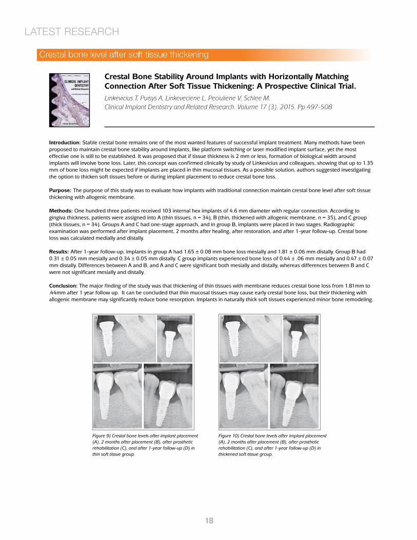
Introduction: Stable crestal bone remains one of the most wanted features of successful implant treatment. Many methods have been proposed to maintain crestal bone stability around implants, like platform switching or laser modified implant surface, yet the most effective one is still to be established. It was proposed that if tissue thickness is 2 mm or less, formation of biological width around implants will involve bone loss. Later, this concept was confirmed clinically by study of Linkevicius and colleagues, showing that up to 1.35 mm of bone loss might be expected if implants are placed in thin mucosal tissues. As a possible solution, authors suggested investigating the option to thicken soft tissues before or during implant placement to reduce crestal bone loss.
Purpose: The purpose of this study was to evaluate how implants with traditional connection maintain crestal bone level after soft tissue thickening with allogenic membrane.
Methods: One hundred three patients received 103 internal hex implants of 4.6 mm diameter with regular connection. According to gingiva thickness, patients were assigned into A (thin tissues, n = 34), B (thin, thickened with allogenic membrane, n = 35), and C group (thick tissues, n = 34). Groups A and C had one-stage approach, and in group B, implants were placed in two stages. Radiographic examination was performed after implant placement, 2 months after healing, after restoration, and after 1-year follow-up. Crestal bone loss was calculated medially and distally.
Results: After 1-year follow-up, implants in group A had 1.65 ± 0.08 mm bone loss mesially and 1.81 ± 0.06 mm distally. Group B had 0.31 ± 0.05 mm mesially and 0.34 ± 0.05 mm distally. C group implants experienced bone loss of 0.44 ± .06 mm mesially and 0.47 ± 0.07 mm distally. Differences between A and B, and A and C were significant both mesially and distally, whereas differences between B and C were not significant mesially and distally.
Conclusion: The major finding of the study was that thickening of thin tissues with membrane reduces crestal bone loss from 1.81mm to .44mm after 1 year follow up. It can be concluded that thin mucosal tissues may cause early crestal bone loss, but their thickening with allogenic membrane may significantly reduce bone resorption. Implants in naturally thick soft tissues experienced minor bone remodeling.
Crestal Bone Stability Around Implants with Horizontally Matching Connection After Soft Tissue Thickening: A Prospective Clinical Trial.Linkevicius T, Puisys A, Linkeveciene L, Peciuliene V, Schlee M.Clinical Implant Dentistry and Related Research. Volume 17 (3). 2015. Pp 497-508
Figure 9) Crestal bone levels after implant placement (A), 2 months after placement (B), after prosthetic rehabilitation (C), and after 1-year follow-up (D) in thin soft tissue group.
Figure 10) Crestal bone levels after implant placement (A), 2 months after placement (B), after prosthetic rehabilitation (C), and after 1-year follow-up (D) in thickened soft tissue group.
LATEST RESEARCH
Crestal bone level after soft tissue thickening
191919
Introduction: The success of implant dentistry has been largely related to the advent of bone augmentation techniques that allow for the regeneration of atrophic alveolar ridges into an ideal ridge form and for placement of implants in their ideal functional and esthetic positions. AlloDerm GBR is a thinner version (thickness, 0.5 to 0.9mm) of the original AlloDerm product, specifically designed for GBR.
Purpose: The purpose of the study was to further evaluate the clinical benefits, or lack thereof, of using an allograft alone or in combination (1:1) with particulate autogenous bone for horizontal ridge augmentation and subsequent implant placement.
Lateral Alveolar Ridge Augmentation Using Tenting Screws, Acellular Dermal Matrix, and Freeze-Dried Bone Allograft Alone or with Particulate Autogenous Bone.Caldwell G, Mills M, Finlayson R, Mealey B.International Journal Periodontics & Restorative Dentistry. Volume 35. 2015. Pp 75-83.
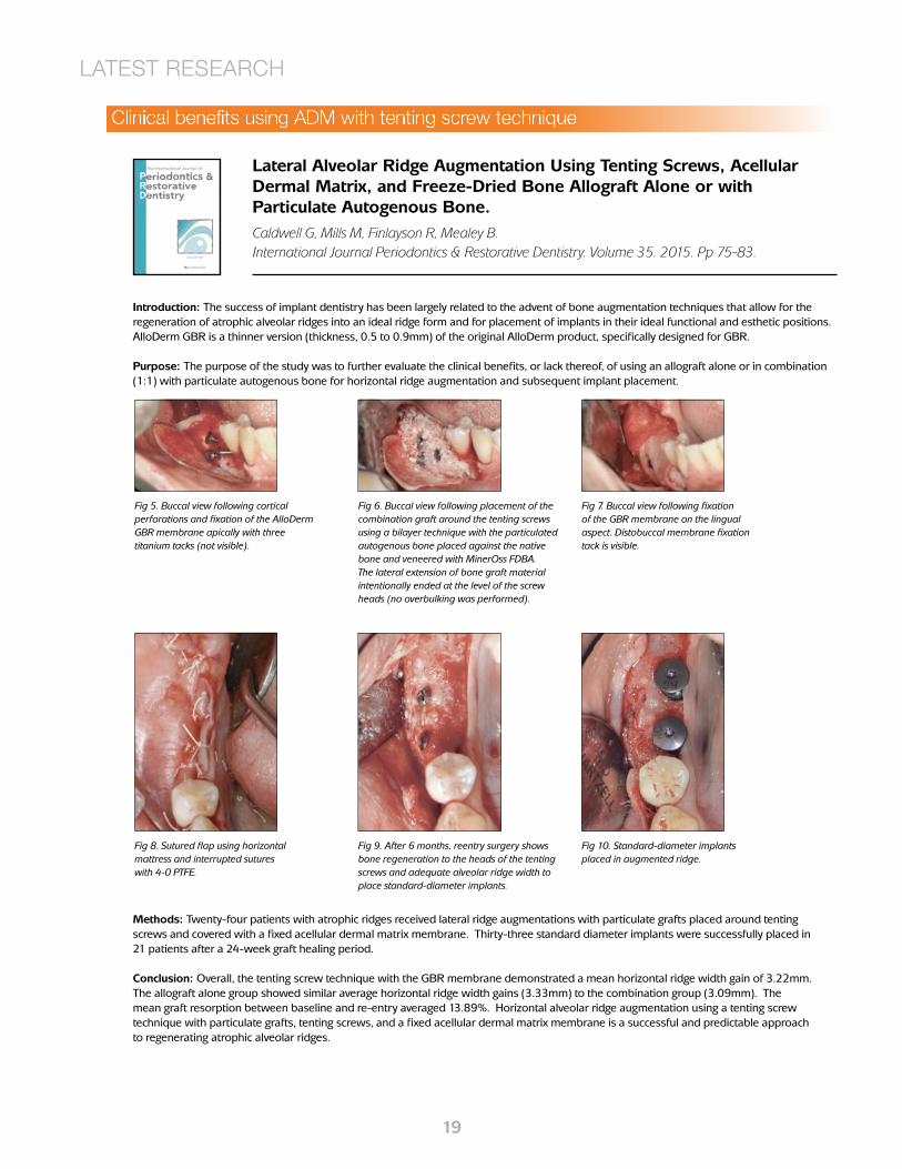
Methods: Twenty-four patients with atrophic ridges received lateral ridge augmentations with particulate grafts placed around tenting screws and covered with a fixed acellular dermal matrix membrane. Thirty-three standard diameter implants were successfully placed in 21 patients after a 24-week graft healing period.
Conclusion: Overall, the tenting screw technique with the GBR membrane demonstrated a mean horizontal ridge width gain of 3.22mm. The allograft alone group showed similar average horizontal ridge width gains (3.33mm) to the combination group (3.09mm). The mean graft resorption between baseline and re-entry averaged 13.89%. Horizontal alveolar ridge augmentation using a tenting screw technique with particulate grafts, tenting screws, and a fixed acellular dermal matrix membrane is a successful and predictable approach to regenerating atrophic alveolar ridges.
Fig 5. Buccal view following cortical perforations and fixation of the AlloDerm GBR membrane apically with three titanium tacks (not visible).
Fig 6. Buccal view following placement of the combination graft around the tenting screws using a bilayer technique with the particulated autogenous bone placed against the native bone and veneered with MinerOss FDBA. The lateral extension of bone graft material intentionally ended at the level of the screw heads (no overbulking was performed).
Fig 7. Buccal view following fixation of the GBR membrane on the lingual aspect. Distobuccal membrane fixation tack is visible.
Fig 8. Sutured flap using horizontal mattress and interrupted sutures with 4-0 PTFE.
Fig 9. After 6 months, reentry surgery shows bone regeneration to the heads of the tenting screws and adequate alveolar ridge width to place standard-diameter implants.
Fig 10. Standard-diameter implants placed in augmented ridge.
LATEST RESEARCH
Clinical benefits using ADM with tenting screw technique
202020
Introduction: Preventing ridge collapse following the extraction of maxillary anterior teeth is vital to a good esthetic restorative outcome. The maintenance of the alveolar bone volume following tooth removal facilitates subsequent placement of dental implants and improves the esthetic and functional prosthodontic result. Alveolar bone resorption after tooth extraction is an inherent condition of the healing process; it is accelerated in the first 6 months after extraction and followed by a gradual remodeling that includes changes in shape and size. Ridge preservation using the guided bone regeneration (GBR) technique has been shown to improve ridge height and width dimensions when compared with tooth extraction alone.
Purpose: The aim of this study was to analyze through clinical and histomorphometric parameters the use of acellular dermal matrix (ADM) with or without mineralized bone allograft (AB) on bone formation in human alveoli after a 6-to 9-month healing period.
Socket Preservation Therapy with Acellular Dermal Matrix and Mineralized Bone Allograft After Tooth Extraction in Humans: A Clinical and Histomorphometric Study. Fernandes P, Reino D, Grisi M, Souza S, Palioto D, Novaes, Jr A.International Journal of Periodontics & Restorative Dentistry. Volume 36. 2016. Pp e16-25.
Methods: A total of 19 patients in need of extraction of the maxillary anterior teeth were selected and randomly assigned to the test group (ADM plus AB) or to the control group (ADM only). Clinical and histomorphometric measurements and histologic analysis were recorded 6 to 8 months after ridge preservation procedures. Clinical parameters and amount of mineralized and nonmineralized tissue were measured and analyzed.
Results: Histologic findings showed higher percentages of mineralized tissue and lower percentages of nonmineralized tissue in the test group when compared with the control group. The present study shows that the technique of GBR with ADM was able to reduce initial bone resorption, since there was resorption of 2.94mm for the test group and 3.18mm for the control group on the horizontal aspect and 1.41mm and 1.97 for the test and control groups, respectively, of the buccal plate.
Discussion: A number of materials, nonresorbable and resorbable, have been used as membranes, with similar results in terms of bone formation. The ideal barrier should be made of material that is less susceptible to membrane exposure or that cannot be significantly colonized by periodontopathogenic bacteria when exposed to the oral cavity. In this randomized controlled clinical and histomorphometric study in humans, acellular dermal matrix in association with mineralized bone allograft reduced alveolar bone loss in the anterior maxillae both in height and width after a follow-up period of 6 to 8 months.
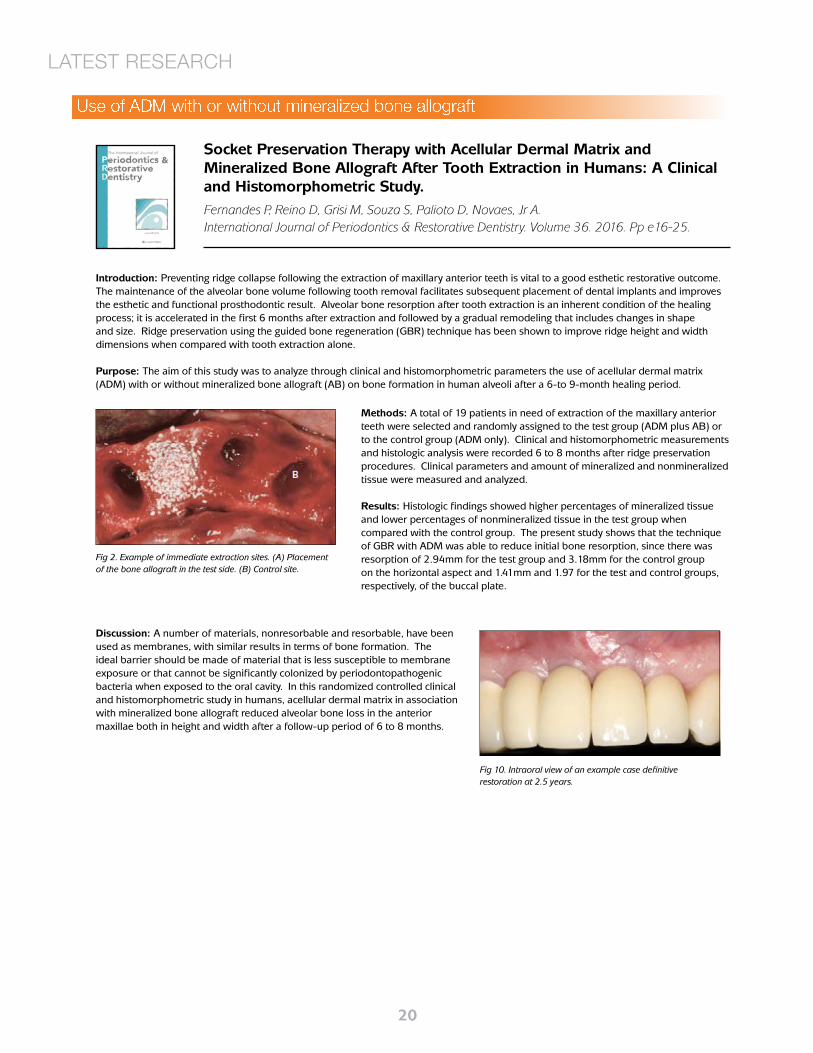
Fig 2. Example of immediate extraction sites. (A) Placement of the bone allograft in the test side. (B) Control site.
Fig 10. Intraoral view of an example case definitive restoration at 2.5 years.
LATEST RESEARCH
Use of ADM with or without mineralized bone allograft

212121
Introduction: Guided bone regeneration (GBR) is an established technique that uses barrier membranes to direct growth of new bone at sites having insufficient bone volumes or dimensions for function and prosthesis placement.
Purpose: The main rationale in guided bone regeneration (GBR) techniques is the creation of space for matrix producing cells if significant volumes of bone are to be achieved. This case report highlights the technique of using allograft and AlloDerm on the principles of GBR technique with satisfactory clinical results.
Guided bone regeneration in anterior maxillary zone: A 3-year case report.Mahesh L, Dhir S, Lahori M.Journal of Interdisciplinary Dentistry. Volume 1. 2011. Issue 1.
Methods: The patient presented with history of avulsed tooth in respect to 21 and 22, and intruded 11 secondary to a road traffic accident. Radiographs were taken. Bone mapping was done and revealed compromised buccolingual width. Average buccolingual soft tissue width of 4 mm and alveolar bone width of 2.2 mm were found. Stage 1 surgery was done with extraction of 11 under local anesthesia to be followed by immediate implant, immediate delayed implant placed in respect to 21, 22, following manufacturer’s protocol. Good primary stability was attained, but all three implants had buccal threads visible. GBR technique was performed using MinerOss and AlloDerm. Nylon suturing was done. Immediate provisional appliance was given.
After an uneventful healing period of 4 months, stage 2 surgery of uncovering of implants was done with tissue punch with a palatal orientation of the punch to maximize the attached tissue remaining in the area critical for prosthetic emergence. Healing abutments were screwed in.
Results: Three-year postoperative radiograph shows good and stable crestal bone levels around the implants.
Discussion: Factors influencing the success of GBR have multiple variables. Maxillary implants show more bone fill (95%) compared to mandible (78%). Insertion of provisional restoration is more favorable. Immediate and immediate delayed implants showed the best results with 92% bone fill when compared with long-term delayed implants with 80% bone fill; early implant placement timings seem to be preferable due to alveolar ridge preservation, more favorable defect morphology. GBR in implant dentistry is very well documented. This is the first case report with AlloDerm and allograft along with the use of tapered implants, in immediate and delayed immediate implant placement. Successful postoperative buccal augmentation was achieved.
HARD TISSUE GRAFTING
Figure 1: Preoperative - buccal view
Figure 2: Preoperative - buccolingual view
Figure 3: Eleven extracted Figure 4: Buccal reflection of flap
Figure 5: BioHorizons tapered internal implants: (a) clinical and (b) radiograph
GBR technique using allograft and AlloDerm

222222
Introduction: To achieve successful osseointegraion, dental implants require maximum bone surface area contact sufficient for implant placement without compromise of the nerve or vascular structures. As a result, bone grafting procedures are frequently used to rebuild the alveolar ridge, to fill extraction defects or to treat peri-implant defects. The bone graft materials used must reproducibly regenerate bone geometry and quality sufficient for implant osseointegration. Conflicting reports concerning the osteoinductivity of demineralized bone matrix (DBM) and historical use of synthetic bone graft substitutes has limited the use of DBM in oral and maxillofacial applications.
Purpose: The purpose of this study was to assess new bone formation with DBM prepared as malleable putty or flexible sheets in a series of patients.
Histologic analysis of implant sites after grafting with demineralized bone matrix putty and sheets. Callan, Salkeld, Scarborough.Implant Dent. 2000;9(1):36-42.
HARD TISSUE GRAFTING
Methods: Bone grafting of the mandible or maxilla was performed to fill extraction sockets and restore ridge structures in a consecutive series of eight patients. DBM prepared as malleable putty or flexible sheets was used. Biopsies were taken at re-entry, and histologic analysis determined the amount of quality of regenerated bone.
Discussion: Clinical experience has shown that, regardless of dental implant design or meticulous surgical technique, osseointegration and subsequent success of the implant are at risk if placed in inadequate or poor-quality bone. Recent studies have demonstrated consistent osteoinductive performance of DBM produced by the D-min process, including Putty and Flex. Clinical factors, such as bacterial or salivary contamination from the oral environment and epithelial cell migration, can limit osteoinduction when DBM is implanted in oral and maxillofacial applications.
Results: In this series, Putty and Flex were well incorporated with extensive new bone as early as 4 months after grafting. The rate of matrix incorporation accommodated the rate of new bone formation, whereas the remodeling of new bone progressed to form mature bone as early as 5 months post grafting. Demineralized bone matrix, in both putty and sheet form, can be used to effectively restore lost alveolar bone facilitating the placement of endosseous dental implants, building support for removable prostheses, and enhancing the cosmetic appearance of fixed prosthesis.
Fig 2. Case 1. A, Preoperative appearance of three periodontally hopeless teeth in a 60-year-old female patient. B, Extraction of the involved teeth from the posterior maxilla was performed. C, Putty was chosen based on its handling characteristics and ability to conform to irregular defect contours. Putty was gently packed into the osseous defects and was used to build out the buccal and lingual surfaces to optimize the ridge geometry for implantation. D, The graft site was reentered at 4 months postgrafting for the placement of endosseous implants. At this time, extensive bony fill and regeneration of the alveolar contours was noted in the area of the extractions.
New bone formation with DBM

232323
Introduction: An ultimate goal of periodontal therapy is the regeneration of periodontal supporting tissues that have been lost as a consequence of periodontitis. Wound healing studies have shown that removal of local bacterial etiology by surgical or nonsurgical therapies results in a resolution of inflammation and an improvement in the clinical signs of periodontitis, but it does not result in the regeneration of a periodontal connective tissue attachment. Defects with two and three bony walls respond more favorably to treatment than do one-wall defects. Attempts to obtain periodontal regeneration also are less successful in patients with poor oral hygiene, in smokers, and in patients who do not comply with periodontal maintenance therapy.
Bone replacement grafts for the treatment of periodontal intrabony defects.PJ Hanes.Oral Maxillofac Surg Clin North Am 2007, Nov 19 (4); 499-512 vi.
Purpose: The purpose of the current study is to review various types of bone replacement graft materials that have shown positive clinical benefits associated with the treatment of periodontal intrabony defects.
Methods: The surgical technique for the treatment of periodontal intrabony defects with bone replacement grafts is essentially the same regardless of the type of graft material being used. Intrasulcular incisions are the common choice, with emphasis on preserving interdental tissue. Flaps are reflected full thickness to expose the underlying osseous defects and allow access for thorough debridement of the defects and meticulous root planing. Once the defect has been debrided of soft tissue and the tooth root surfaces thoroughly planed to remove all deposits of dental plaque and calculus, the bone replacement graft material is packed into the defect to fill the defect to the level of the remaining alveolar bone. Flaps are closed and sutured for primary closure and complete coverage of the bone replacement graft.
Results: The biologic rationale for the treatment of periodontal intrabony defects with DFDBA is based on the assumption that the demineralization of the allograft bone exposes bone morphogenetic proteins, which have been shown to be capable of inducing or enhancing bone regeneration. In terms of the overall resolution of the intrabony defect, however, clinical studies to date have not shown a superiority of DFDBA over FDBA. Most studies have reported an overall 50% to 60% defect resolution with DFDBA and FDBA.
HARD TISSUE GRAFTING
Discussion: Bone replacement grafts, including autogenous grafts from intraoral donor sites, allografts, xenografts, and alloplastic bone substitutes, are the most widely used treatment modalities for the regeneration of periodontal osseous defects. Studies suggest a favorable clinical outcome with the use of these materials in terms of improvements in periodontal probing depths, probing attachment gains, and bone fill. Sites that are treated with bone replacement grafts have been shown to respond at least as well as surgical debridement of the defect alone and usually better particularly in terms of bone fill. Meta-analysis from a recent evidence-based review indicated that bone replacement grafts increase bone level, reduce crestal bone loss, increase clinical attachment level, and reduce probing pocket depths when compared with open flap debridement procedures.
Fig 3. After initial therapy that included oral hygiene instructions, scaling, and root planing and endodontic treatment of the maxillary second premolar, surgical therapy was performed. Full-thickness flaps were elevated facially and palatal, osseous defects were debribed of soft tissue, and root surfaces were thoroughly scaled and root planed. A two-wall intrabony defect was present between the first and second molars and a combination 1-, 2-, and 3-wall defect was present between the first molar and second premolar.
Fig 6. One-year post-treatment radiographs show fill of intrabony defects (A) and a resolution of the angular bony defect between the first molar and second premolar (B).
Treatment of periodontal intrabony defects
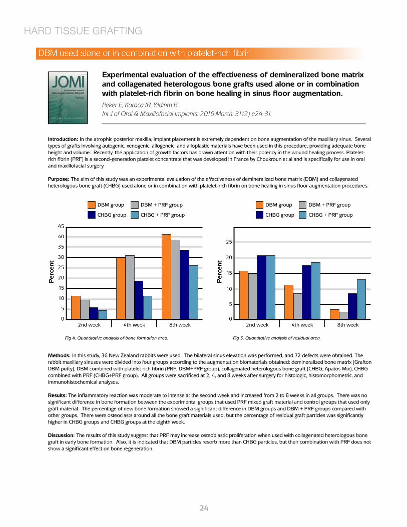
242424
Introduction: In the atrophic posterior maxilla, implant placement is extremely dependent on bone augmentation of the maxillary sinus. Several types of grafts involving autogenic, xenogenic, allogeneic, and alloplastic materials have been used in this procedure, providing adequate bone height and volume. Recently, the application of growth factors has drawn attention with their potency in the wound healing process. Platelet-rich fibrin (PRF) is a second-generation platelet concentrate that was developed in France by Choukroun et al and is specifically for use in oral and maxillofacial surgery.
Purpose: The aim of this study was an experimental evaluation of the effectiveness of demineralized bone matrix (DBM) and collagenated heterologous bone graft (CHBG) used alone or in combination with platelet-rich fibrin on bone healing in sinus floor augmentation procedures.
Experimental evaluation of the effectiveness of demineralized bone matrix and collagenated heterologous bone grafts used alone or in combination with platelet-rich fibrin on bone healing in sinus floor augmentation. Peker E, Karaca IR, Yildirim B.Int J of Oral & Maxillofacial Implants; 2016 March: 31(2) e24-31.
Methods: In this study, 36 New Zealand rabbits were used. The bilateral sinus elevation was performed, and 72 defects were obtained. The rabbit maxillary sinuses were divided into four groups according to the augmentation biomaterials obtained: demineralized bone matrix (Grafton DBM putty), DBM combined with platelet rich fibrin (PRF; DBM+PRF group), collagenated heterologous bone graft (CHBG; Apatos Mix), CHBG combined with PRF (CHBG+PRF group). All groups were sacrificed at 2, 4, and 8 weeks after surgery for histologic, histomorphometric, and immunohistochemical analyses.
Results: The inflammatory reaction was moderate to intense at the second week and increased from 2 to 8 weeks in all groups. There was no significant difference in bone formation between the experimental groups that used PRF mixed graft material and control groups that used only graft material. The percentage of new bone formation showed a significant difference in DBM groups and DBM + PRF groups compared with other groups. There were osteoclasts around all the bone graft materials used, but the percentage of residual graft particles was significantly higher in CHBG groups and CHBG groups at the eighth week.
Discussion: The results of this study suggest that PRF may increase osteoblastic proliferation when used with collagenated heterologous bone graft in early bone formation. Also, it is indicated that DBM particles resorb more than CHBG particles, but their combination with PRF does not show a significant effect on bone regeneration.
HARD TISSUE GRAFTING
45
40
35
30
25
20
15
10
5
02nd week
Fig 4. Quantitative analysis of bone formation area.
Per
cent
DBM group
CHBG group
DBM + PRF group
CHBG + PRF group
4th week 8th week
20
15
10
5
02nd week
Fig 5. Quantitative analysis of residual area.
Per
cent
DBM group
CHBG group
DBM + PRF group
CHBG + PRF group
4th week 8th week
25
DBM used alone or in combination with platelet-rich fibrin

252525
Introduction: Maxillary sinus augmentation is regarded as a predictable bone grafting procedure in cases of alveolar ridge atrophy due to alveolar bone loss, pneumatization of maxillary sinuses, or a combination of them. Sinus augmentation aims at obtaining new bone formation in the maxillary sinus cavity to allow for the placement of implants of an optimal length with primary stability.
Purpose: To document the clinical and histologic outcomes of sinus augmentation using a novel allogenic bone substitute as a sole grafting material.
Clinical and Histologic Outcomes After the Use of a Novel Allograft for Maxillary Sinus Augmentation: A Case Series.Avila G, Neiva R, Misch CE, Galindo-Moreno P, Benavides E, Rudek I, Wang HL.Implant Dentistry. Volume 19. 2010. Pp 330-341.
Methods: Patients in need of sinus augmentation before implant placement were recruited for this study. Sinus augmentation procedures were performed following a lateral approach, using a freeze-dried allograft as the only grafting material. Patients were followed up postoperatively for 6 months. Plaque score, wound healing, and patient discomfort were recorded at each follow-up visit. Implants were placed between 6 and 7 months after sinus augmentation and restored 6 months later. Bone core biopsy specimens were harvested at the time of implant placement and processed for histologic and histomorphometrical analysis. Vital bone, remaining allograft (RA) particles, and nonmineralized tissue percentages were assessed on each sample.
Results: Of the 23 sinus patients, 20 patients underwent sinus augmentation surgery. All patients had satisfactory postoperative healing in the absence of complications. A total of 39 implants were placed. One implant failed and was replaced 3 months later. Histologic analysis revealed the presence of well-organized lamellar bone, in direct contact with RA particles. Mean vital bone was 23.02%, mean RA was 22.25%, and average nonmineralized tissue was 54.73%.
Discussion: A material composed of cortical and cancellous particles was used in this study. The “dual” nature of this material seems to contribute to the attainment of an excellent biological response after grafting. Mineralized cancellous particles may provide a rapidly resorbable, osteoconductive scaffold for the ingrowth of bone cells, and angiogenesis, leading to optimal bone remodeling. On the other hand, mineralized cortical chips may offer an adequate structure to maintain the space due to its size and slower resorption rate. This can be considered a very interesting feature, because space maintenance is a requisite for successful bone augmentation.
HARD TISSUE GRAFTING
Fig. 1. A and B, Pre-operative view of the surgical area. C and D, Elevation of the mucoperiosteal flap. E, Lateral window after Scheneiderian membrane release. F, Sinus cavity filled with the allograft. G, Primary closure achieved after suture. H, Postoperative aspect at 2 months.
Sinus augmentation using a novel allogenic bone substitute
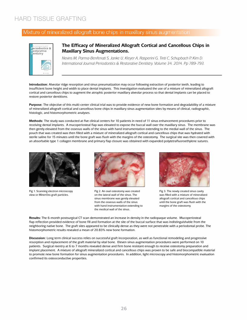
262626
Introduction: Alveolar ridge resorption and sinus pneumatization may occur following extraction of posterior teeth, leading to insufficient bone height and width to place dental implants. This investigation evaluated the use of a mixture of mineralized allograft cortical and cancellous chips to augment the atrophic posterior maxillary alveolar process so that dental implants can be placed to restore posterior dentitions.
Purpose: The objective of this multi-center clinical trial was to provide evidence of new bone formation and degradability of a mixture of mineralized allograft cortical and cancellous bone chips in maxillary sinus augmentation sites by means of clinical, radiographic, histologic, and histomorphometric analyses.
Methods: The study was conducted at five clinical centers for 10 patients in need of 11 sinus enhancement procedures prior to receiving dental implants. A mucoperiosteal flap was elevated to expose the buccal wall over the maxillary sinus. The membrane was then gently elevated from the osseous walls of the sinus with hand instrumentation extending to the medial wall of the sinus. The pouch that was created was then filled with a mixture of mineralized allograft cortical and cancellous chips that was hydrated with sterile saline for 15 minutes until the bone graft was flush with the margins of the osteotomy. The surgical site was then covered with an absorbable type 1 collagen membrane and primary flap closure was obtained with expanded polytetrafluoroethylene sutures.
The Efficacy of Mineralized Allograft Cortical and Cancellous Chips in Maxillary Sinus Augmentations.Nevins M, Parma-Benfenati S, Janke U, Kleyer A, Rasperini G, Tinti C, Schupbach P, Kim D.International Journal Periodontics & Restorative Dentistry. Volume 34. 2014. Pp 789-793.
HARD TISSUE GRAFTING
Results: The 6-month postsurgical CT scan demonstrated an increase in density in the radiopaque volume. Mucoperiosteal flap reflection provided evidence of bone fill and formation at the site of the buccal surface that was indistinguishable from the neighboring native bone. The graft sites appeared to be clinically dense as they were not penetrable with a periodontal probe. The histomorphometric results revealed a mean of 20.83% new bone formation.
Discussion: Long term clinical success relies on successful graft incorporation, as well as functional remodeling and progressive resorption and replacement of the graft material by vital bone. Eleven sinus augmentation procedures were performed on 10 patients. Surgical reentry at 6 to 7 months revealed dense and firm bone resistant enough to receive osteotomy preparation and implant placement. A mixture of allograft mineralized cortical and cancellous chips was proven to be safe and biocompatible material to promote new bone formation for sinus augmentation procedures. In addition, light microscopy and histomorphometric evaluation confirmed its osteoconductive properties.
Fig 1. Scanning electron microscopy view or MinerOss graft particles.
Fig 2. An oval osteotomy was created on the lateral wall of the sinus. The sinus membrane was gently elevated from the osseous walls of the sinus with hand instrumentation extending to the medical wall of the sinus.
Fig 3. The newly created sinus cavity was filled with a mixture of mineralized allograft cortical and cancellous chips until the bone graft was flush with the margins of the osteotomy.
Mixture of mineralized allograft bone chips in maxillary sinus augmentation

272727
Introduction: Rehabilitation of partially or totally edentulous patients with oral implants has become common practice with reliable long-term results. However, unfavorable local conditions of the alveolar ridge, because of atrophy, periodontal disease and trauma may result in insufficient bone volume, which may increase the complexity of implant placement. For cells to progress toward a bone phenotype, it is necessary for them to receive stimuli by growth factors. Platelet-rich fibrin (PRF) derives from a natural and progressive polymerization occurring during centrifugation of blood. It accelerates physiologic wound healing and new bone formation when used in combination with bone grafts and in sinus life procedures, and it improves integration of bone substitution materials during preimplant grafting.
Purpose: To evaluate the effectiveness of platelet-rich fibrin (PRF) and freeze-dried bone allograft (FDBA) in vertical bone augmentation with immediate implant placement using histologic analysis.
Methods: Six Merino sheep received a total of 36 Branemark MKIII implants; three implants were placed supracrestally in each tibia with vertical exposure of four threads. Each implant received one of the three grafting options (MinerOss + PRF or MinerOss or PRF). The grafting materials were covered with a resorbable membrane. Animals were sacrificed at 4 and 8 weeks, respectively, and specimens were prepared and collected for histologic analysis.
Conclusion: The various stages of graft integration into native bone and the implant were observed at different time points, and comparison between the three grafting options was possible. Osteogenic potential with vertical generation of bone was observed in the three groups. At week 4, woven bone formation at the bone graft interface was observed; new bone did not appear to be organized at week 4. At week 8, the graft appeared to be fully replaced by vital mature and well-organized bone arranged in lamellae with osteocytes encapsulated within the bone. The vertical gain at 8 weeks was higher for the PRF + MinerOss group with viable bone extending above the first thread. Both the MinerOss and PRF groups had vertical bone gain extended to the second thread. MinerOss appeared to be effective in vertical bone augmentation with simultaneous implant placement. PRF enhanced vertical bone augmentation when combined with MinerOss.
Assisted Wound Healing and Vertical Bone Regeneration with Simultaneous Implant Placement: A Histologic Pilot Study.Potres Z, Deshpande S, Kloeppel H, Voss K, Klineberg I.International Journal of Oral Maxillofacial Implants. Volume 31. 2016. Pp 45-54.
HARD TISSUE GRAFTING
a-Week 8 of the MinerOss group under low magnification showing implant and surrounding native bone and augmented bone with bone gain to the first thread on one side.Fig 2. (a) Surgical site, showing three implants in the tibia; placement is supracrestal with
four-thread exposure and vertical bone augmentation using (b) PRF, (c) MinerOss, and (d) PRF + MinerOss.
Platelet-rich fibrin and freeze-dried bone allograft in bone augmentation
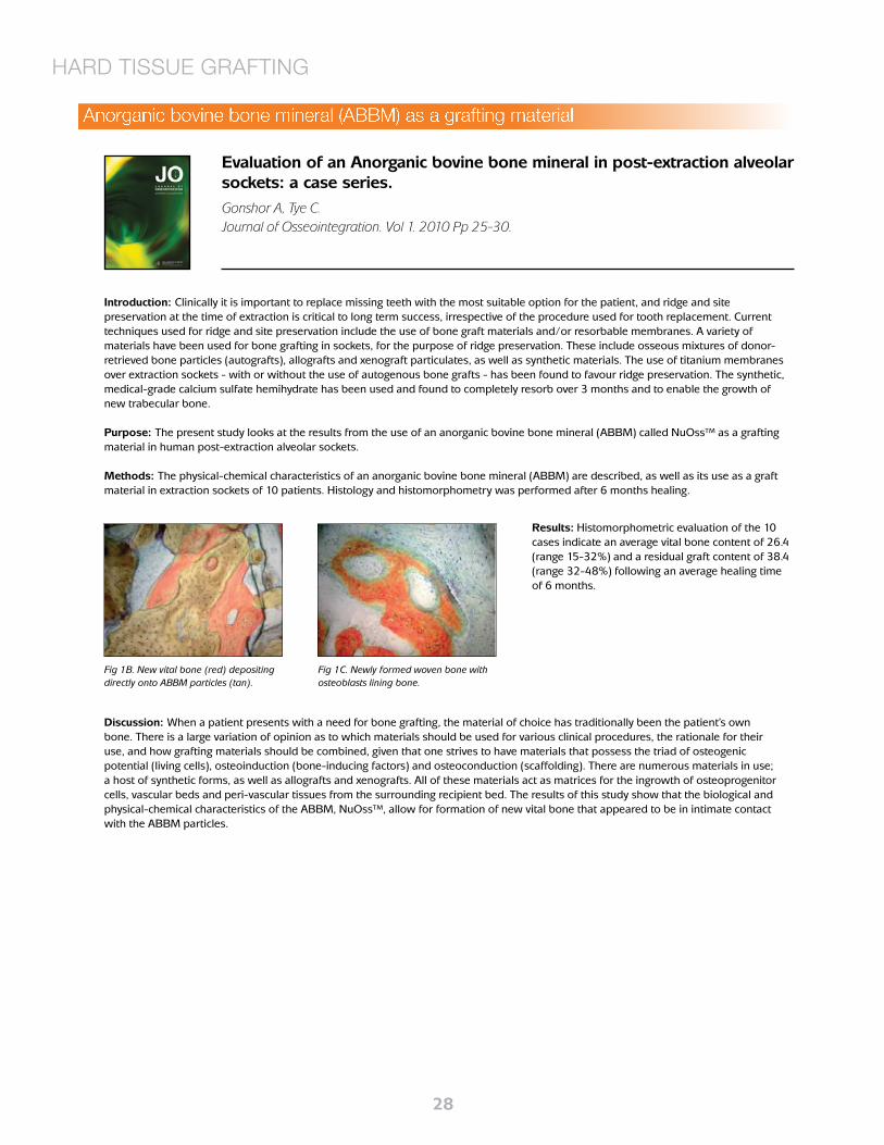
282828
Introduction: Clinically it is important to replace missing teeth with the most suitable option for the patient, and ridge and site preservation at the time of extraction is critical to long term success, irrespective of the procedure used for tooth replacement. Current techniques used for ridge and site preservation include the use of bone graft materials and/or resorbable membranes. A variety of materials have been used for bone grafting in sockets, for the purpose of ridge preservation. These include osseous mixtures of donor-retrieved bone particles (autografts), allografts and xenograft particulates, as well as synthetic materials. The use of titanium membranes over extraction sockets - with or without the use of autogenous bone grafts - has been found to favour ridge preservation. The synthetic, medical-grade calcium sulfate hemihydrate has been used and found to completely resorb over 3 months and to enable the growth of new trabecular bone.
Purpose: The present study looks at the results from the use of an anorganic bovine bone mineral (ABBM) called NuOss™ as a grafting material in human post-extraction alveolar sockets.
Methods: The physical-chemical characteristics of an anorganic bovine bone mineral (ABBM) are described, as well as its use as a graft material in extraction sockets of 10 patients. Histology and histomorphometry was performed after 6 months healing.
Evaluation of an Anorganic bovine bone mineral in post-extraction alveolar sockets: a case series.Gonshor A, Tye C.Journal of Osseointegration. Vol 1. 2010 Pp 25-30.
HARD TISSUE GRAFTING
Results: Histomorphometric evaluation of the 10 cases indicate an average vital bone content of 26.4 (range 15-32%) and a residual graft content of 38.4 (range 32-48%) following an average healing time of 6 months.
Discussion: When a patient presents with a need for bone grafting, the material of choice has traditionally been the patient’s own bone. There is a large variation of opinion as to which materials should be used for various clinical procedures, the rationale for their use, and how grafting materials should be combined, given that one strives to have materials that possess the triad of osteogenic potential (living cells), osteoinduction (bone-inducing factors) and osteoconduction (scaffolding). There are numerous materials in use; a host of synthetic forms, as well as allografts and xenografts. All of these materials act as matrices for the ingrowth of osteoprogenitor cells, vascular beds and peri-vascular tissues from the surrounding recipient bed. The results of this study show that the biological and physical-chemical characteristics of the ABBM, NuOss™, allow for formation of new vital bone that appeared to be in intimate contact with the ABBM particles.
Fig 1B. New vital bone (red) depositing directly onto ABBM particles (tan).
Fig 1C. Newly formed woven bone with osteoblasts lining bone.
Anorganic bovine bone mineral (ABBM) as a grafting material

292929
Introduction: Carbonate Apatite isolated from bovine bone, also known as anorganic bovine bone mineral (e.g. Bio-Oss), has been in commercial distribution for bone grafting applications for more than a decade, particularly in dental surgeries. Porous Carbonate Apatite (PCA) is isolated from the cancellous bone of porcine bone, it possesses the natural pore structure for cell conduction. Further, because the apparent density of PCA is low, the intra-and interparticle space is high for osteo-conduction and new bone deposition.
Purpose: This article summarizes the results of in vitro and in vivo studies conducted for PCA. A commercial product of Carbonate Apatite derived from bovine bone, Bio-Oss, was also characterized for comparison.
Methods: In vitro, the structure of PCA was characterized by X-Ray diffraction (XRD) and Fourier Transform Infrared (FTIR) Spectroscopy, the pore structure of the PCA particles was analyzed from images obtained from scanning electron microscopy (SEM), the particle size distribution as well as the volume fill per unit weight of PCA were also determined. In vivo, two studies were conducted using a rabbit femoral condyle bone defect model and canine intraoral one-wall defect model to evaluate the safety and effectiveness of PCA as a bone grafting material.
Isolation and characterization of a Porous carbonate apatite from porcine cancellous bone.Shu-Tung Li, Hui-Chen Chen, Debbie Yuen.Science, Technology, Innovation, Aug 2014: 1-13.
HARD TISSUE GRAFTING
Figure 1: SEM revealed the porous structure of PCA and Bio-Oss and both showed interconnecting pores, but this was more manifested in PCA than in Bio-Oss. It can be seen from the micrographs that PCA compares favorably against Bio-Oss in terms of pore structure within the particles.
Figure 1a: SEM Micrographs of Porous Carbonate Apatite (PCA)
Conclusion: In summary, in vitro studies demonstrated that PCA fulfills the physical, structural and morphological requirements needed to function as an effective bone grafting material. It was also noted that the surface appears to be rougher in the PCA than in the Bio-Oss, a property favoring the cell adhesion to PCA particles. In vivo studies from rabbit femoral condyle defect model and canine intraoral bone defect model demonstrated that PCA functioned as well as Bio-Oss in both the orthopedic bone defect and the intraoral bone defect as an osteoconductive matrix to support bone regeneration. Based on the results of in vitro and in vivo studies provided in this article, it is anticipated that PCA will function at least as well as Bio-Oss if not better in the clinical environment for intraoral bone grafting applications.
At 4 weeks, the Bio-Oss® defect sites showed a lack of increase in radiodensity, suggesting minimal bone formation as compared to the defect sites treated with PCA. At 8 weeks, the PCA treated sites appeared to have generated a greater interradicular bone pattern, compared to the site treated with Bio-Oss®. By 12 weeks post-treatment, defect sites had become difficult to distinguish radiographically from surrounding bone due to new bone formation within the defect with remodeling and associated article absorption. For PCA treated sites, noticeably increased radiodensity and development of interradicular bone by the 12-week post treatment was observed as compared to previous time points. The Bio-Oss® treated sites as compared to the PCA treated sites, showed less bone development based on overall radiolucencies.
Figure 7a: Representative Radiograph images of PCA Treated Defects in Canine Intraoral Model
Figure 7b: Representative Radiograph images of Bio-Oss® Treated Defects in Canine Intraoral Model
In vitro and in vivo studies conducted for PCA

303030
Introduction: The spontaneous process of bone healing after tooth extraction has been studied in human models. It is characterized by a physiological process of bone remodeling and reabsorption, which occurs rapidly and can determine in the first 6 months the loss of about 40% of the height and 60% of the width of the alveolar bone. Because the best period to preserve the alveolar ridge is at the time of extraction, socket preservation technique has been proposed as a means of counteracting the post-extraction volume loss. Bovine bone-derived is by far the most commonly used and researched xenogeneic bone graft. Porcine derived-bone has been recently also considered as graft biomaterial for bone regeneration.
Purpose: To evaluate and compare histomorphometrically the bone response to two xenografts, one bovine and the other porcine, grafted in adjacent extraction sockets in a human.
Methods: In this case report, two adjacent maxillary premolars were extracted, and the sockets were filled with two different xenogeneic bone substitutes (first premolar with bovine bone, and second premolar with porcine bone) to counteract post-extraction volume loss. Following 6 months bone core specimens were harvested during the placement of implants at the regenerated sites.
Histologic Evaluation of Bone Healing of Adjacent Alveolar Sockets Grafted with Bovine - and Porcine -Derived Bone: A Comparative Case Report in Humans.Guarnieri R, Devilliers P, Grande M.Regenerative Biomaterials. 2017. Pp 1-4.
HARD TISSUE GRAFTING
Conclusion: For bovine xenogeneic the amount of newly formed bone (osteoid), of residual graft, and of connective tissue was 26.85, 17.2 and 48.73%, respectively. For the porcine xenograft newly formed bone (osteoid) represented 32.19%, residual graft material 6.57% and non-mineralized connective tissue 52.99%. The percentages of total trabecular bone were significantly higher in the extraction socket grafted with porcine xenograft than in the extraction socket grafted with bovine xenograft. Moreover, the porcine graft biomaterial showed a higher resorption percentage than the bovine xenograft. The xenogenic biomaterials investigated in this study were found to be biocompatible and osteoconductive.
Figure 3. Histologic section of extraction socket grafted with bovine bone (Trichrome stain x 20): yellow arrow = viable bone, red arrow = newly formed bone (osteoid)
Figure 5. Histologic section of extraction socket grafted with porcine bone (Trichrome stain x 20): yellow arrow = viable bone, red arrow = osteoid
Xenografts grafted in adjacent extraction sockets
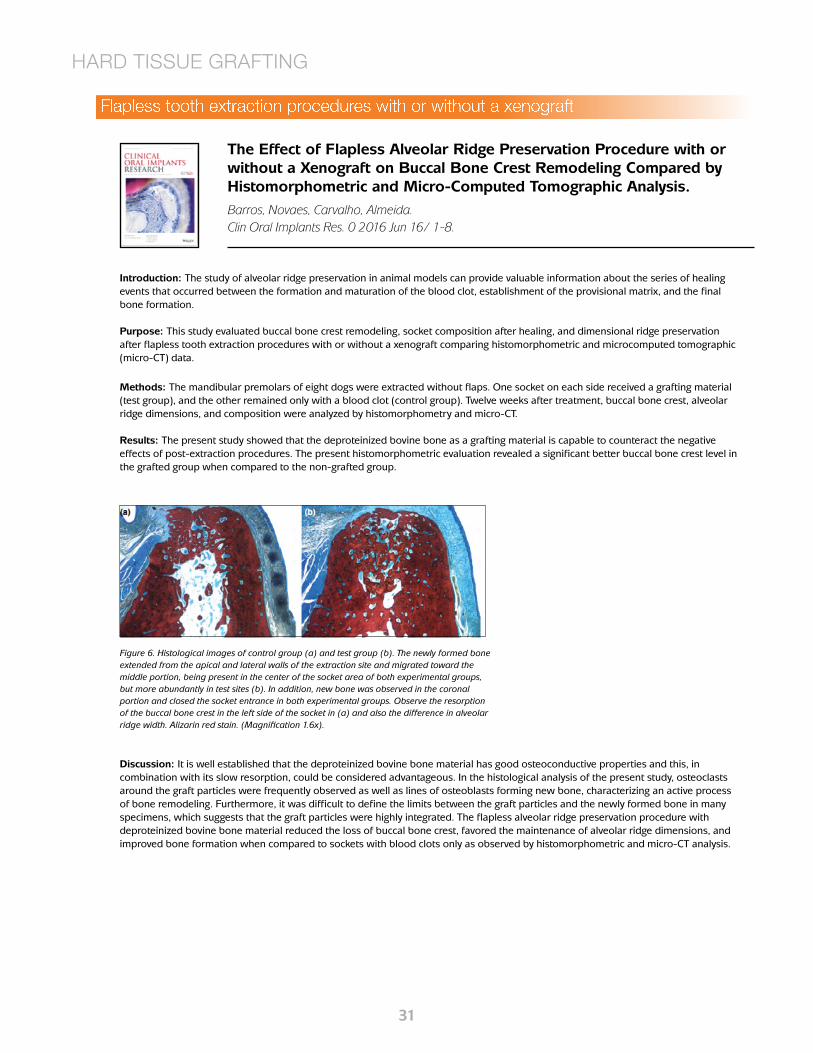
313131
Introduction: The study of alveolar ridge preservation in animal models can provide valuable information about the series of healing events that occurred between the formation and maturation of the blood clot, establishment of the provisional matrix, and the final bone formation.
Purpose: This study evaluated buccal bone crest remodeling, socket composition after healing, and dimensional ridge preservation after flapless tooth extraction procedures with or without a xenograft comparing histomorphometric and microcomputed tomographic (micro-CT) data.
The Effect of Flapless Alveolar Ridge Preservation Procedure with or without a Xenograft on Buccal Bone Crest Remodeling Compared by Histomorphometric and Micro-Computed Tomographic Analysis.Barros, Novaes, Carvalho, Almeida. Clin Oral Implants Res. 0 2016 Jun 16/ 1-8.
Methods: The mandibular premolars of eight dogs were extracted without flaps. One socket on each side received a grafting material (test group), and the other remained only with a blood clot (control group). Twelve weeks after treatment, buccal bone crest, alveolar ridge dimensions, and composition were analyzed by histomorphometry and micro-CT.
Results: The present study showed that the deproteinized bovine bone as a grafting material is capable to counteract the negative effects of post-extraction procedures. The present histomorphometric evaluation revealed a significant better buccal bone crest level in the grafted group when compared to the non-grafted group.
HARD TISSUE GRAFTING
Discussion: It is well established that the deproteinized bovine bone material has good osteoconductive properties and this, in combination with its slow resorption, could be considered advantageous. In the histological analysis of the present study, osteoclasts around the graft particles were frequently observed as well as lines of osteoblasts forming new bone, characterizing an active process of bone remodeling. Furthermore, it was difficult to define the limits between the graft particles and the newly formed bone in many specimens, which suggests that the graft particles were highly integrated. The flapless alveolar ridge preservation procedure with deproteinized bovine bone material reduced the loss of buccal bone crest, favored the maintenance of alveolar ridge dimensions, and improved bone formation when compared to sockets with blood clots only as observed by histomorphometric and micro-CT analysis.
Figure 6. Histological images of control group (a) and test group (b). The newly formed bone extended from the apical and lateral walls of the extraction site and migrated toward the middle portion, being present in the center of the socket area of both experimental groups, but more abundantly in test sites (b). In addition, new bone was observed in the coronal portion and closed the socket entrance in both experimental groups. Observe the resorption of the buccal bone crest in the left side of the socket in (a) and also the difference in alveolar ridge width. Alizarin red stain. (Magnification 1.6x).
Flapless tooth extraction procedures with or without a xenograft

323232
Introduction: To counteract the post-extraction alveolar volume loss, different socket preservation techniques have been proposed. All techniques consist to fill the resulting alveolar socket with different grafting materials with and without sealing the socket with absorbable or non-absorbable membranes. Grafting biomaterials have shown to provide better mechanical support during the healing and remodeling phase compared to spontaneous healing. Moreover, based on their osteogenic, osteoconductive, or osteoinductive properties, graft materials act as stimulants or scaffolds for bone growth.
Purpose: The present paper reports data of a randomized study aimed to analyze and compare the histologic and histomorphometric aspects of bone healing in extraction sites covered with collagen membrane alone or associated with porcine-derived bone graft.
Methods: Thirty patients, with single extraction sockets without severe bone wall defects in the premolar/molar region, were included. Ten extraction sockets were grafted with porcine-derived bone and covered with collagen membrane (group 1), 10 sites were covered with collagen membrane alone (group 2), and 10 sites healed spontaneously (group 3). After 4 months of healing, 26 (8 in group 1, 9 in group 2, and 9 in group 3) bone core specimens were harvested for histologic evaluation, then dental implants were placed.
Bone Healing in Extraction Sockets Covered with Collagen Membrane Alone or Associated with Porcine Derived Bone Graft: a Comparative Histological and Histomorphometric Analysis.Guarnieri R, Testarelli L, Stefanelli L, Angelis F, Mencio F, Pompa G, Di Carlo S.Journal of Oral & Maxillofacial Research. Volume 8. 2017. Pp e4.
HARD TISSUE GRAFTING
Results: Sites in the group 1 and in the group 2 showed similar histologic and histomorphometric results without significant differences in the percentage of vital bone (57.43% vs. 60.01%), and non-mineralized connective tissue 22.99% vs. 18.53%. In group 1 a 16.57% of residual material was found.
Discussion: The present study complements the previously published clinical study on extraction socket preservation techniques by reporting on the histologic and histomorphometric outcomes. Results showed that the use of collagen membrane alone or associated to porcine-derived bone improves the healing bone process, compared to that of extraction sites spontaneously healed. These outcomes are in agreement with data reported by other studies, confirm that porcine-derived bone and reabsorbable membrane are effective in maintaining the post-extractive ridge volume, compared to spontaneously healed extraction sites. Results indicated that extraction sites without severe walls defects and with a vestibular bone thickness > 1.5 mm, treated with a low resorption rate collagen membrane alone, do not need more than 4 months for dental implant insertion.
Figure 2. Histological micrograph of extraction socket treated with collagen membrane and porcine derived-bone (Masson’s trichrome stain, original magnification x20). Porcine bone particles (GR) surrounded by immature bone in a remodelling process. Arrows indicate the osteoid tissue.
Figure 5. Histological micrograph of extraction socket spontaneously healed (trichrome stain, original magnification x20). Presence of vital bone (VB) with thin trabecular organization surrounded by connective tissues (CT).
Aspects of bone healing in extraction sites

333333
Introduction: Over the past decade, resorbable collagen-based membranes have become the standard of care for guided bone regeneration procedures in dental surgeries. To date, there are a number of resorbable collagen-based membranes on the market under various trade names.
Purpose: In this study, a biomechanically strong and yet highly conformable single layer porcine collagen membrane (PCM), developed by Collagen Matrix, Inc., was evaluated in vitro and in vivo and compared to the Bio-Gide® dental membrane.
A Comparative Study of a New Porcine Collagen Membrane to Bio-Gide.Shu-Tung Li, Ph.D., Debbie Yuen, Drew Martin, Natsuyo Shishido Lee. Science, Technology, Innovation, Feb. 1-5, 2015.
Methods: SEM: Samples from both sides of the PCM and Bio-Gide® and their respective cross sections were carbon-coated and micro-graphed using a scanning electron microscope. Suture Pullout Strength: The samples were subjected to continuous loading of 2.5 cm per minute until the suture was pulled out. In Vivo Subcutaneous Implantation in Rats: The explants were evaluated at 4, 8, 12 and 16 weeks after implantation for implant resorption, amount of new collagen deposition, and host tissue response using standard histological techniques.
DENTAL MEMBRANES
Results: The SEM micrographs revealed that both sides of the PCM were smooth compared to Bio-Gide®, which had one fibrous and one smooth side. In using the PCM for guided bone regeneration surgeries, the membrane can be applied without having to distinguish which side should be facing the wound, an advantage over Bio-Gide®. The PCM showed significantly higher average suture pullout strength. The higher suture pullout strength permits the PCM to be firmly anchored to the surrounding tissue with minimal risk of membrane tear or detachment. In vivo total resorption time appeared to be similar for the PCM and Bio-Gide® through the course of the evaluated time points of the subcutaneous implantation study.
Discussion: Bio-Gide® has been on the market for more than a decade and has been well received in the dental surgical community. Its combination of strength and conformability make it a versatile membrane in terms of both application and clinician preference. After substantial in vitro and in vivo pre-clinical testing, it is clear that PCM possesses and, in many cases, exceeds the performance characteristics of Bio-Gide® and thus, will function at least as well as Bio-Gide® in human dental surgical applications with an estimated total resorption time of approximately 12 - 16 weeks with corresponding new host collagen deposition.
Figure 3. SEM micrograph of the PCM (cross-section) at 50x magnification.
Performance/ Characterization
Parameter
Porcine Collagen Membrane Bio-Gide
Hydroxyproline Content (Weight %)
11.7 ± 1.1% 11.8 ± 0.8%
Suture Pull-out Strength
953 ± 110g 330 ± 120g
Evaluation of porcine collagen membrane compared to Bio-Gide
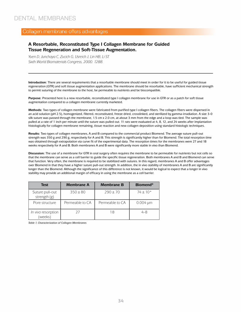
343434
Introduction: There are several requirements that a resorbable membrane should meet in order for it to be useful for guided tissue regeneration (GTR) and soft tissue augmentation applications. The membrane should be resorbable, have sufficient mechanical strength to permit suturing of the membrane to the host, be permeable to nutrients and be biocompatible.
Purpose: Presented here is a new resorbable, reconstituted type I collagen membrane for use in GTR or as a patch for soft tissue augmentation compared to a collagen membrane currently marketed.
Methods: Two types of collagen membrane were fabricated from purified type I collagen fibers. The collagen fibers were dispersed in an acid solution (pH 2.5), homogenized, filtered, reconstituted, freeze dried, crosslinked, and sterilized by gamma-irradiation. A size 3-0 silk suture was passed through the membrane, 1.5 cm x 2.0 cm, at about 3 mm from the edge and a loop was tied. The sample was pulled at a rate of 1 inch per minute until the suture was pulled out. 11 rats were evaluated at 4, 8, 12, and 24 weeks after implantation histologically for collagen membrane remaining, tissue reaction and new collagen deposition using standard histologic techniques.
Results: Two types of collagen membranes, A and B compared to the commercial product Biomend. The average suture pull-out strength was 350 g and 290 g, respectively for A and B. This strength is significantly higher than for Biomend. The total resorption time was obtained through extrapolation via curve fit of the experimental data. The resorption times for the membranes were 27 and 18 weeks respectively for A and B. Both membranes A and B were significantly more stable in vivo than Biomend.
Discussion: The use of a membrane for GTR in oral surgery often requires the membrane to be permeable for nutrients but not cells so that the membrane can serve as a cell barrier to guide the specific tissue regeneration. Both membranes A and B and Biomend can serve that function. Very often, the membrane is required to be stabilized with sutures. In this regard, membranes A and B offer advantages over Biomend in that they have a higher suture pull-out strength. In addition, the in vivo stability of membranes A and B are significantly longer than the Biomend. Although the significance of this difference is not known, it would be logical to expect that a longer in vivo stability may provide an additional margin of efficacy in using the membrane as a cell barrier.
A Resorbable, Reconstituted Type I Collagen Membrane for Guided Tissue Regeneration and Soft-Tissue Augmentation. Yuen D, Junchaya C, Zucich G, Usreich J, Lin HB, Li ST. Sixth World Biomaterials Congress, 2000. 1288.
DENTAL MEMBRANES
Test Membrane A Membrane B Biomend®
Suture pull-out strength (g)
350 ± 80 290 ± 70 74 ± 10*
Pore structure Permeable to CA Permeable to CA 0.004 μm
In vivo resorption (weeks)
27 18 4-8
Table 1. Characterization of Collagen Membranes
Collagen membrane offers advantages

353535
Introduction: Alveolar bone resorption after tooth loss or extraction can lead to insufficient bone volume, which negatively affects the prognosis of dental implants. Traditionally, bony defects have been classified according to anatomical deficiency as follows: horizontal, vertical or combinations. Vertical ridge augmentation has been reported to be successful, but with a low degree of predictability and a rather high complication rate. More predictable results have been obtained with horizontal bone augmentation. In addition, similar clinical and radiological results have been reported for implants placed with bone augmentation compared with those completely placed into pristine bone. Numerous techniques have been described to reconstruct deficient alveolar ridges. In the simultaneous treatment approach, guided bone regeneration (GBR) is associated with superior outcomes when compared to other procedures and has become the treatment of choice to provide optimal bone support for dental implants.
Purpose: The aim of this study was to radiologically assess and clinically investigate the outcome and early resorption of this new GBR technique with a tissue engineering approach.
Methods: This single cohort observational study evaluated the outcome of the leukocyte-and platelet-rich fibrin (L-PRF) Block for horizontal bone augmentation in the maxilla. The L-PRF Block is prepared by mixing a particulate biomaterial with chopped L-PRF membranes at a 50:50 ratio and adding liquid fibrinogen to glue all together. Horizontal augmentation was assessed linearly and volumetrically immediately after surgery and 5–8 months later by matching consecutive cone beam computed tomography (CBCTs).
Leucocyte- and platelet-rich fibrin block for bone augmentation procedure: A proof-of-concept study.Cortellini S, Castro A, Temmerman A, Dessel J, Pinto N, Jacobs R, Quirynen M. Journal of Clinical Periodontology. Vol 45. 2018 Pp. 624-634.
L-PRF APPLICATIONS
L-PRF Block for bone augmentation
Results: Ten patients (mean age of 50.7 years [±17.2]) representing 15 sites with horizontal alveolar deficiencies were included. Superimposition of pre-operative and post healing CBCT scans showed an average linear horizontal bone gain of 4.6 mm (±2.3), 5.3 mm (±1.2) and 4.4 mm (±2.3), measured at 2, 6 and 10 mm from the alveolar crest, respectively. The volumetric gain was 1.05 cm3 (±0.7) on average. The resorption rate after 5–8 months was 15.6% (±6.7) on average. L-PRF Block may be a suitable technique to augment deficient alveolar ridges.
Discussion: This case series demonstrates that the L-PRF Block can be used safely and effectively for horizontal augmentation of resorbed alveolar ridges. A mean horizontal bone gain of 4.7 ± 2 mm was achieved. Some sites gained up to 7–8 mm. Autologous bone blocks are still considered as the gold standard to reconstruct resorbed alveolar ridges. However, the need for a second surgical site evokes a higher patient morbidity. This morbidity further increases when bone is harvested outside the oral cavity. A second drawback is the varying degree of graft resorption during healing. The use of particulate bone grafts instead of bone blocks has been supported in the literature. However, graft instability in particulate grafts can lead to integration failure. The novel technique described in this study combines the properties of bone blocks and particulate grafts reducing the disadvantages of both. The combination with liquid fibrinogen to form the L-PRF Block may increase ease in handling and predictability of the augmentation procedure. It provides a block made out of particulate graft, with increased stability of the augmented area. Furthermore, it excludes the discomfort inherent to the secondary harvesting site.
Graphic representation of leucocyte-and platelet-rich fibrin (L-PRF) Block for horizontal bone augmentation. The small holes in cortical bone guarantee optimal blood supply. The L-PRF Block is adapted to the bony defect and covered with a collagen membrane fixed via membrane tacks. L-PRF membranes are used to cover the augmented site.
Protocol for preparation of L-PRF Block using 0.5 g of biomaterial (BioOss):-Venipuncture: collect 6 tubes (red cap, glass coating) of blood following standard protocol and 2 tubes (white cap, plastic coating), the latter is drawn last and is placed last in centrifuge (2,700 rpm/408 g RCF).-After 3 min interrupt centrifugation, remove both white cap tubes.-Immediately restart the centrifuge with remaining red cap tubes for another 9 min.-Immediately aspirate the yellow fluid (= liquid fibrinogen) in white cap tube with a sterile syringe, get as close as possible to the red cells, but do not aspirate them; the liquid can be kept in the syringe up to 20–30 min.-After full centrifugation of the remaining tubes, remove L-PRF clots and compress gently into membranes.
Preparation of block:-Chop membranes in very small pieces.-Mix chopped membranes and bone substitute in Ti-dish (with a 50:50 ratio), if the mix is too dry, one can add some L-PRF exudate. Get a uniform mix.-Spray 1cc of liquid fibrinogen over the homogeneous mix, and stir gently for ±10 s while shaping it to the desired form.-Fibrinogen will clot into fibrin within a few minutes and trap the biomaterial to form a L-PRF Block.
Table 1: Protocol for the preparation of leucocyte-and platelet-rich fibrin block
363636
Introduction: The classic experimental gingivitis studies in the 60’s demonstrated the direct relation between the accumulation of dental plaque and gingival inflammation. Dental plaque or dental biofilms are defined as a matrix-embedded microbial population, adherent to each other and/or to surfaces or interfaces. Studies on biofilm development in deep periodontal pockets showed that the deepest sites are colonized predominantly by motile species (e.g. spirochetes) and gram-negative bacteria, located adjacent to the epithelial lining of the pocket. Recently, new tissue-engineering techniques have been proposed for regenerative procedures after non-surgical periodontal therapy or for bone augmentation. Leucocyte- and platelet-rich fibrin (L-PRF), a second-generation platelet concentrate, was introduced as an autologous biomaterial that serves as a scaffold for regenerating cells. Two recent systematic reviews have shown the various applications of L-PRF, concluding that favorable effects on hard and soft tissue healing and a decrease of postoperative discomfort could be obtained when L-PRF was used. Several studies have described other biological properties of L-PRF such as the antimicrobial effect against wound bacteria.
Purpose: Given the cellular composition of L-PRF and its bioactive nature, the aim of this study was to evaluate the antimicrobial capacity of an L-PRF membrane and L-PRF exudate against key periodontal pathogens (Porphyromonas gingivalis, Prevotella intermedia, Fusobacterium nucleatum, and Aggregatibacter actinomycetemcomitans).
Methods: The participants in this study (n = 9) were systemically healthy, non-smokers, without a history of periodontal disease and who had not taken any antibiotics for at least 6 months before the study. Four blood samples were collected from each volunteer (n = 9) in a 9 mL glass-coated plastic tube and immediately centrifuged at 408 g for 12 minutes (Intraspin™). The L-PRF clot was compressed and transformed into a standardized membrane of 1 mm in thickness (Xpression® box). During this process, the released exudate was also kept for further use at −80 °C. Due to the low cellular content of L-PRF exudate, we could freeze it without damaging the possible active molecules.
Results: Within the limitations of this study, it can be concluded that an L-PRF membrane has antimicrobial properties against P. gingivalis, whereas the inhibition against P. intermedia, F. nucleatum, and A. actinomycetemcomitans was not statistically significant on agar plates. However, the L-PRF exudate has an antimicrobial effect against P. gingivalis in a dose-dependent way. Two effects on A. actinomycetemcomitans could be observed in this present study. First, there was an effect on the auto-aggregation of these bacteria. Regarding the second effect, bacterial growth stimulation of A. actinomycetemcomitans was detected when it was in contact with the L-PRF exudate.
Discussion: Several studies showed an antimicrobial effect of platelet concentrates against wound bacteria. Only few articles examined oral microorganisms. For instance, Enterococcus faecalis and Candida albicans isolated from the oral cavity were inhibited by pure-platelet-rich plasma (P-PRP), a type of PRP without leucocytes. Yang and co-workers showed inhibition on P. gingivalis and A. actinomycetemcomitans by PRP whereas no inhibition could be observed for F. nucleatum. In this study, the effect of PRP and PRF was compared, concluding that PRP showed superior activity. It should be noted that PRF was prepared by adding calcium chloride to PRP in order to activate the platelets and convert fibrinogen into fibrin, which is not in line with the protocol to prepare PRF. More recently, Kour and co-workers (2018) compared the antibacterial capacity of PRP, PRF, and injectable-PRF (L-PRF) on P. gingivalis and A. actinomycetemcomitans. They concluded that all three platelet concentrates showed some antibacterial activity against both bacteria. However, PRP and L-PRF presented significantly greater inhibition against P. gingivalis compared to PRF. In the present study, limited inhibition could be observed on A. actinomycetemcomitans, which differs from their results. The bacterial strain used and the fact that they incubated A. actinomycetemcomitans anaerobically might have influenced the effect of PRF. P. gingivalis was the most inhibited bacteria strain in this study. However, there is no evidence in the literature showing a direct effect of blood components on P. gingivalis. Proteinase inhibitors constitute 10% of the protein content of human plasma and they can affect the gingipain activity, known as the primary virulence factor of those bacteria. These proteinases also play an important role in the survival of the bacterium within host cells, due to their implication in the cellular invasion and in overcoming the protective defense mechanisms of epithelial cells.
Antimicrobial capacity of Leucocyte-and Platelet Rich Fibrin against periodontal pathogens.Castro A, Herrero E, Slomka V, Pinto N, Teughels W, Quirynen M.Scientific Reports. Vol 9. 2019 Pp 8188.
L-PRF APPLICATIONS
Antimicrobial properties of L-PRF membranes

373737
Background: The use of platelet concentrates in sinus lift procedures, like Leucocyte and Platelet Rich Fibrin (L-PRF), has increased in the last few years. One of the interesting properties of L-PRF is the presence of platelets and their important function in the release of growth factors. It has already been reported that growth factors may stimulate local bone augmentation to various degrees. The use of a L-PRF block in bone augmentation therapies could enhance and improve bone regeneration.
Aim/Hypothesis: This study was designed to evaluate whether the addition of L-PRF can improve the results in bone gain, compared with the golden standard DBBM alone, in lateral sinus floor elevation.
Material and Methods: This study was designed as a prospective, double-blind randomized controlled clinical trial. Subjects in need ofat least one sinus floor elevation for implant placement, where a lateral approach is indicated, were included. After flap elevation andcreation of a bony window, the patients were randomly assigned to the test or control group. For the test group, the sub-sinus cavity wasfilled with L-PRF block and the window covered with L-PRF membranes. For the control group, the sub-sinus cavity was filled with DBBM and the window covered with a collagen membrane. The volumetric gain was evaluated immediately after the augmentation (T1) and at 6 months healing (T2) by a blinded examiner. Measurements were assessed using cone- beam computed tomography. Outcomes were defined as- the gain in ridge width (cm 3 ), the occurrence of any adverse event, and biopsy specimens examined histologically.
Results: Twenty-six patients (14 female, 12 male) with a mean age of 55.5 (± 11.9 years) and presenting 30 sites were treated. One graft had a complication during treatment. Both techniques were effective for two-stage sinus floor elevation. Superimposition of post- operative (T1) and post-healing (T2) CBCT scans revealed an average volumetric gain of 2.56 (± 1.27 cm 3 ) for the control group and 1.65 (± 1.01 cm 3) for the test group. Statistically higher volumetric resorption was reported for the test group. Implant placement, as planned before surgery, was achieved in all the sites in both groups. Histologic analysis showed connection of the DBBM particles with a dense network of newly formed bone with different degrees of maturation.
Conclusion and clinical implications: The L-PRF block seems to be a promising alternative to the use of DBBM alone in sinus floor elevation procedures. However, a higher resorption rate was reported when compared with DBBM. The greater volume loss observed in the sinus grafted with L-PRF block did not hamper the placement of the implants. Within the limitations of this study, the L-PRF block technique can be regarded as a successful technique for sinus floor elevation.
A randomized controlled clinical trail on the use of the L-PRF block compared with DBBM in lateral sinus floor elevation.Cortellini S, Castro A, Temmerman A, Dhondt R, Van Dessel J, Jacobs R, Quirynen M. Clinical Oral Implants Research, abstract
L-PRF APPLICATIONS
L-PRF Block for sinus augmentation

*MLD107* *REV B NOV 2019*MLD107 REV B NOV 2019
s h o p o n l i n e a t
s t o r e . b i o h o r i z o n s . c o m
Direct Offices
DistributorsFor contact information in our 90 countries, visit biohorizons.com
BioHorizons Spain+34 91 713 10 84
BioHorizons Canada866-468-8338
BioHorizons UK+44 (0)1344 752560
BioHorizons USA888-246-8338 or
205-967-7880
BioHorizons Chile+56 (2) 23619519
BioHorizons Italy800-063-040
BioHorizons Mexico800-953-0498
BioHorizons®, Laser-Lok®, MinerOss®, AutoTac®, Mem-Lok® and TeethXpress® are registered trademarks of BioHorizons. Unigrip™ is a trademark of Nobel Biocare AB. Zimmer® Dental ScrewVent® and Tapered ScrewVent® are registered trademarks of Zimmer, Inc. AlloDermTM, AlloDerm GBRTM and NovoMatrixTM are trademarks of LifeCell Corporation, an Allergan affiliate. Grafton® DBM is a registered trademark of Medtronic, Inc. Cytoplast® is a registered trademark of Osteogenics Biomedical, Inc. Puros Dermis is a registered trademark of Zimmer Biomet. Mucograft is a registered trademark of Ed. Geistlich Sogne Ag Fur Chemische Industrie. Symbios PerioDerm is a registered trademark of Dentsply Sirona. Hu-Friedy® is a registered trademark of Hu-Friedy Mfg. Co., LLC. Spiralock® is a registered trademark of Spiralock Corporation. Pomalux® is a registered trademark of Westlake Plastics Co. Locator® is a registered trademark of Zest Anchors, Inc. Delrin® is a registered trademark of E.I. du Pont de Nemours and Company. Bio-Gide® is a registered trademark of Edward Geistlich Sohne AG Fur Chemische Industrie. BioMend® is a registered trademark of Zimmer Biomet Dental. IntraSpin®, L-PRF® and Xpression® are trademarks of Intra-Lock® International Inc. Not all products shown or described in this literature are available in all countries. As applicable, BioHorizons products are cleared for sale in the European Union under the EU Medical Device Directive 93/42/EEC and the tissues and cells Directive 2004/23/EC. We are proud to be registered to ISO 13485:2016, the international quality management system standard for medical devices, which supports and maintains our product licences with Health Canada and in other markets around the globe. Original language is English. ©BioHorizons. All Rights Reserved.