
Clinical Review 2010 - Rocky Mountain Orthodontics
25
CLINICAL Orthodontic a clinical pearl Synergy R ® THE MULTI - FAMILY THE FUNCTIONAL MATRIX a practical solution using FAQ - e-ceph ® Web RMODS ® DIAGNOSIS DIAGNOSIS DIAGNOSIS A Comprehensive Cephalometric Analysis
Transcript of Clinical Review 2010 - Rocky Mountain Orthodontics

CLINICALOrthodontic
a clinical pearlSynergy R®
THE MULTI - FAMILYTHE FUNCTIONAL MATRIX
a practical solution using
FAQ - e-ceph® WebRMODS®
DIAGNOSISDIAGNOSISDIAGNOSISA Comprehensive Cephalometric Analysis

2 Clinical Review
4 lOW FRICtION: tRADItIONAl meChANICS: A peRFeCt FIt By Gary holt D.D.S.
9 SyNeRGy R: A ClINICAl peARl By travis Barr B.S. and Gary holt D.D.S.
12 DIAGNOSIS DIAGNOSIS DIAGNOSIS: A COmpReheNSIve CephAlOmetRIC ANAlySIS By Bradford N. edgren D.D.S., m.S.
32 RmODS / e-Ceph Q & A WIth DR. BuDI KuSNOtO
35 the FuNCtIONAl mAtRIx: A pRACtICAl SOlutION uSING the multI FAmIly By Dr. Franco Bruno
tABle OF CONteNtS
Rocky Mountain® Orthodontics located in Denver Colorado, is The World’s Oldest Synergistic, Bioprogressive®, Breathing Enhancement Orthodontic Company®.
RMO® was founded in 1933 by Colorado orthodontist Dr. Archie Brusse. The last 55 years was led by Martin Brusse whose vision was dedicated to developing continued education and future appliance systems in pursuit of promoting vital oral health for every patient. RMO® remains privately owned and maintains a rich history deeply rooted in Denver culture.
Martin Brusse realized his goals in two very special and capable people he confidently selected to continue guiding RMO® into the future, Tony Zakhem and Jody Hardy.
Rocky Mountain® Orthodontics proudly supports the local community and is honored to design, engineer, and manufacture its premium quality orthodontic products with pride in the U.S.A.
RMO® is proud to be recognized as the longest-running exhibitor at the AAO. Since the company’s inception, Rocky Mountain® O r t h o d o n t i c s has pioneered numerous orthodontic breakthroughs such as pre-formed molar bands and the metal-injection-molding process.
RMO®’s innovations have continued with orthodontic advancements such as RMODS® and e-Ceph® computer aided diagnostic services, interceptive pediatric appliances, and the Straight Wire Low Friction system which includes RMO®’s patented Synergy® bracket line, the Dual-Top temporary anchorage device system, and the RMbond® Indirect Bonding system.
RMO® is dedicated to developing Continuing Education programs designed
to enhance and expand clinicians’ knowledge of various systems, appliances, and biomechanics. RMO® Seminars are conducted throughout the year and around the world with lecture specialists trained in multiple disciplines.
With a world-wide distribution network, a subsidiary division in Europe, and a joint venture operation in Japan, RMO® is truly a global manufacturer. Rocky Mountain® Orthodontics has been awarded twice with The President’s distinguished “E-Star Award for Exports” by the U.S. Secretary of Commerce “For continued outstanding contributions to the Export Expansion Program of the United States of America”. In addition, in 2008 RMO® was awarded the Governor Award for Excellence in Exporting.
Many of RMO®’s great developmental strengths come from valued relationships and the exchange of oral health concepts, innovations, and educational information. Combined, this process allows RMO® to service customers around the world with progressive Synergistic System treatment solutions.
Back Row: (Left to Right) Frank Augustine, Jeff Smith, Adam Pollack, Hugh Carr
Front Row: Jody Hardy, Tony Zakhem
A Road Map to the Future
3Clinical Review
“RMO® is proud of our heritage, history, and legacy. Tony and Jody have recently completed the formation of an entirely new executive management team that will guide the next generation as we move towards the future.”

4 Clinical Review 5Clinical Review
lateral incisor brackets (Synergy® brackets) have a unique passive ligation system when an elastomeric tie is used, but the tie has minimal contact with the wire due to an intelligent design. Clearly, the Synergy R® bracket is the most versatile, active bracket ever. It gives complete control to the doctor to dictate active vs. passive forces, reduces friction dramatically, and total treatment time duration. Some of the highlights of the system include rounded arch slot walls to reduce binding and friction, and offers multiple ligation options—minimal friction ligation or conventional ligation, maximum rotation ligation or minimal rotation ligation.8 The bracket has rounded slot walls and bosses on the bracket tie wings to minimize the possible contact surface with the arch wire and prevent the ligation force from exertion on the arch wire.9
Friction is typically the enemy in two areas of orthodontic treatment—leveling and aligning as well as space closure because frictional forces generated between bracket and arch wire have a significant effect on tooth movement.10 The low friction bracket systems seek to reduce friction compared to conventional orthodontic bracket systems. There is evidence that these brackets offer
lower frictional resistance (FR) values than conventional brackets when coupled with small round arch wires.11,12 To reduce friction in the mouth some authors have recommended the use of low friction brackets, small initial wires, and less stiff wires.13 The benefit of lower friction is more rapid alignment of teeth, quicker leveling of arches, and progression into bigger arch wires sooner in treatment. This allows the doctor to start anterior-posterior changes sooner, i.e., start using Class II elastics. The Synergy® system is unique in that it can be used with your current anterior-posterior mechanics: you can use a Wilson® Distalizing Arch, Pendulum, or any other distalizing arch. You can use other inter-arch mechanics such as a Forsus, Herbst, AdvanSync, etc. We have noted rapid treatment times for Class II cases when we couple the leveling and alignment efficiency of the Synergy R® with the concurrent Class II correction using AdvanSync. The point is you’re in complete control and don’t need to change bio-mechanics to conform to the bracket, but rather the bracket will support your current mechanics.
With lower frictional forces, the space closing phase of orthodontic treatment can be accomplished quite quickly. The Synergy R® bracket supports your current space closing technique. If you prefer to distalize canines into Class I with Energy
The orthodontic profession has three major technologies or trends that are evolving and offering new and exciting ways to practice according to the editor of the Journal of Clinical Orthodontics.1 These are 3-D cone beam computed tomography (CBCT), mini implants or temporary anchorage devices (TADs) and low friction bracket systems. At the forefront of the orthodontic profession right now is the question of low friction systems or passive self-ligating bracket systems and how they may benefit the orthodontist. One needs to look no further than a recent issue of American Journal of Orthodontics to discover that low friction brackets are a hot button topic.2 In this particular issue there were two impassioned letters to the editor expressing polar views on the topic. In fact, the editor of AJO, Dr. David Turpin, recently penned an editorial urging more in-vivo studies of self-ligation, low friction brackets and urged prudence when investigating these brackets. 3
Why the interest in low friction brackets? Orthodontists are trying to minimize total treatment time, reduce the patient burden, expedite each adjustment appointment, increase appointment intervals while providing superior results and many doctors are examining the bracket system as a means to achieve these goals. This is nothing new. In the 1930s the Russell bracket was introduced and reported to do just that. This bracket would produce more comfort, fewer office visits, and shorter overall treatment time.4 Other examples of the early self-ligation brackets were the Ormco Edgelok (1972), Forestadent Mobil-Lock (1980), Orec SPEED (1980), and A Company Activa (1986).5 The self- ligation concept was given a big boost when Dr. Dwight Damon entered his namesake bracket in 1998 and has continued to enjoy a resurgence in popularity since that time.6,7 The Damon system was interesting because it was a passive bracket that had a “fourth wall” (door) that was comparable to a buccal tube. There is another bracket on
the market that is truly passive and acts like a buccal tube—Synergy R® from Rocky Mountain® Orthodontics. This novel bracket system has a removable cover over the arch slot on the cuspids, first bicuspids, and second bicuspids that enable the bracket to function similar to a buccal tube during the initial leveling and aligning treatment stages. However, Synergy R® differs from every passive self-ligating bracket currently on the market because it converts, while bonded to the tooth, to a traditional active bracket with full ligation capabilities for space closure and finishing during the later treatment stages.
Note the novel 6 tie wing design and hook. Note the rounded walls and funnel shape tube for easy entry of wire.
The wire is simply thread through the tubes on the 3s, 4s, and 5s. The central and
Low Friction: traditional mechanics:
a perfect fitBy Gary holt
D.D.S.Denver, CO
FSCF R I C T I O N S E L E C T I O N C O N T R O L
CONVENTIONAL CONTROL
MAXIMUM CONTROL
REDUCED FRICTION
MAXIMUM ROTATION
MODERATE ROTATION
Figure “FSC”
“The point is you are in complete control and don’t need to change your bio-mechanics to conform to the bracket, but rather
the bracket will support your current mechanics.”
Dr. Gary Holt graduated Magna Cum Laude from the University of Maryland Dental School and then completed his orthodontic residency at the University of Missouri-Kansas City. He has completed the training to be Dawson Level I certified. His interests are efficient treatment with attention to detailed occlusion, the use of TADs to improve treatment time and effectiveness, and the use of Diode Lasers in the orthodontic practice. He has completed three Ironman races and lives in Littleton, CO with his wife and three children.

6 Clinical Review 7Clinical Review
ChainTM, then that is exactly what you do with Synergy R®. The Energy ChainTM is placed in the same manner as you place it with a conventional bracket. If you like to distalize the canines into Class I using a Ni-Ti coil spring then that is exactly what you do with Synergy R®. The brackets have a hook in the middle of the bracket for easy access and bio-mechanic advantage. Once the canines are Class I and you want complete space closure you can chain 6-6 or you can place a crimpable hook on the arch wire and slide with a Ni-Ti coil spring. The low friction system lends itself to sliding mechanics and space closure is accomplished very quickly.
One concern with self-ligating systems is the loss of torque control, especially in the maxillary anterior. To many orthodontists, the desire to maintain careful 3D control of the maxillary incisors is a very important aspect of orthodontic treatment.14 Enter the Synergy R® bracket. This bracket has the ability to allow the doctor to dictate the necessary friction in the maxillary and mandibular incisors. The clinician can dial in the bracket / arch wire friction to fit his / her specific treatment needs. If the doctor wants passive ligation in the anterior, that can be accomplished with the use of an elastomeric tie just around the center tie wings. If he / she desires more detailed rotation control, then he / she can tie only the mesial or distal tie wings. If the doctor wants complete 3D control of the bracket then the doctor can place the ligatures around all wings. This bracket system takes advantage of a completely passive system from the cuspids to the molars, but allows for more control in the anterior. This bracket offers some of the same advantages as a Giannelly bidimensional system without the bracket dimensions needing to be different. The bracket can be passive early in treatment, but can be made to have complete 3D control at any point in time.
As many orthodontists say, “It is not how you start the case, but how you finish the case.” That is indeed the truth. The attention to detail in the finished cases is what separates us as specialists. Another concern with low friction systems is the inability to finish cases as desired. The Synergy R® has overcome this weakness of other bracket systems. Detailing and finishing of the orthodontic case is usually accomplished by either repositioning the
bracket or placing bends into the arch wire. Synergy R® supports both methods. The bracket is very durable because it is manufactured using the Metal Injection Molding (MIM) process and gives the strongest appliance available. Thus, you can simply debond the bracket, clean the tooth, clean the bracket pad and rebond the same bracket into the desired position. If you prefer to bend the arch wire to finish and detail the case then you place the desired bend into the arch wire and you simply convert the 3, 4, or 5 brackets by removing the cap. You don’t have to convert all the brackets, just the teeth where the bend is placed. After converting the bracket, the arch wire is tied in with an elastomeric ligature or steel ligature. In this manner you can utilize the passive, low friction benefits during the initial leveling and alignment phase and then you can finish the case with the detail you desire. This is a big advantage of the Synergy R® system.
13 week follow up photos aligned with .018 x .018 arch wires. The patient was ready to proceed into the working mechanics phase of treatment.
CASE 3
CASE 2
After 12 weeks of treatment the vertical correction of the cuspid was almost completed without affecting other aspects of the arch form.
Synergy R® Cap Remover Pliers - T01200
► Uses joint plier transer to shear off convertible caps effortlessly
► Easy access the buccal region with little obstruction
► Can be used on any convertible buccal tubes and convertible brackets
After 12 weeks of treatment and expansion the mandibular bicuspids were improved.
After 15 weeks of using a low friction bracket, the cuspid was in occlusion, and the anterior segment 2-2 had not been negatively affected.
Note: Maxillary bicuspids Maxillary retroclined incisors Maxillary left lateral
Note: Mandibular rotations incisors Mandibular rotations biscuspids
At initial bonding note the blocked out maxillary cuspid and high irregularity in the lower arch.
After 12 weeks of treatment space had been created for the upper right cuspid and the lower arch alignment had improved dramatically.
CASE 4
CASE 1
CASE 5
Patient presented with Class II division 2, deep bite, and retroclined incisors. The treatment plan was to level the Curve of Spee, align the teeth, followed by Class II elastics.
Patient presented as Class I crowded with blocked out maxillary right cuspid and severe crowding in mandibular arch. Treatment plan was to open space for UR3 and level and align the lower arch.
Patient presented with a Class III tendency, open bite, and high maxillary left cuspid. The treatment plan was to bring the cuspid into occlusion without impact to the anterior segment.
Patient presented with a Class II deep bite, posterior cross-bite, and rotations in the lower arch. The treatment plan was to correct the cross-bite with an RPE and then level and align the arches with Synerg y R.®
Patient presented with a Class II malocclusion. The treatment plan was to bring the cuspid into the maxillary arch as quickly as possible. Then proceed into the working wires and initiate Class II mechanics. The low friction brackets aided in the vertical alignment of the high cuspid without impact to the other anterior segments.

8 Clinical Review 9Clinical Review
Synergy R®
a clinical pearl
1. Keim RG. Editor’s corner: orthodontic megatrends. J Clin Orthod 2005;39:345-6.
2. Am J Orthod Dentofacial Orthop 2009;136:756-8.
3. Turpin DL. In-vivo studies offer best measure of self-ligation. Am J Orthod Dentofacial Orthop 2009;136:141-2.
4. Stolzenberg J. The Russell attachment and its improved advantages. Int J Orthod Dent Child 1935;21:837-40.
5. Rinchuse DJ, Miles PG. Self-ligating backets: Present and future. Am J Orthod Dentofacial Orthop 2007;132:216-22.
6. Rinchuse Daniel J, Rinchuse Donald J. Developmental occlusion, orthodontic interventions, and orthognathic surgery for adolescents. Dent Clin N Am 2006;50:69-86.
7. Damon DH. The Damon low-friction bracket: a biologically compatible straight-wire system. J Clin Orthod 1998;32:670-80.
8. RMO (Rocky Mountain Orthodontics) Product Catalog 2009; p. 95: www.rmortho.com.
9. Thorstenson GA, Kusy RP. Effects of ligation type and method on the resistance to sliding of novel orthodontic brackets with second-order angulation in the dry and wet states. Angle Orthod 2003;73:418-30.
10. Tidy DC. Frictional forces in fixed appliances. Am J Orthod Dentofacial Orthop 1989;96:249-54.
11. Henao SP, Kusy RP. Evaluation of the frictional resistance of conventional and self-ligating bracket designs using standardized archwires and dental typodonts. Angle Orthod 2004;74:202-11.
12. Redlich M, Mayer Y, Harari D, Lewinstein I. In vitro study of frictional forces during sliding mechanics of “reduced-friction” brackets. Am J Orthod Dentofacial Orthop. 2003;124:69-73.
13. Materese G, et al. Evaluation of frictional forces during dental alignment: An experimental model with 3 nonleveled brackets. Am J Orthod Dentofacial Orthop 2008;133:708-15.
14. Sinclair PM. Reader’s corner. J Clinic Orthod 1993;27:221-23.
References
In conclusion, I would like to comment on a patient that re-visited the practice recently and caused me to reflect on brackets. My office had seen this patient several years ago for an initial orthodontic consultation and the family elected to go with another orthodontist in the area. I had thought nothing more about the case until they recently showed up at my practice. The patient has been in appliances for over two years and there has been little progress. The patient was bonded with a leading self-ligating bracket and as you can see there has been minimal progress over the course of a two year treatment.
Why do I bring this up? Because the bracket is not the doctor. The bracket can’t diagnose, can’t treatment plan, and can’t treat the case. The patient should not be asking for a specific bracket, nor should the marketing of a specific bracket be the place of any practice. Even a fantastic bracket is worth little if the doctor lacks the knowledge or skill to treat the case. The bracket should be a tool to aid the doctor in accomplishing the goal of moving the teeth in a faster, easier, and more comfortable and convenient way. That is our job. We are still the doctor. Synergy R® can make all these things easier and can help treatment progress faster. Synergy R® can aid in the A-P, vertical, and transverse correction and Synergy R® can aid in the detailing and finishing of the case, but remember that you are still the doctor and every case still deserves the personalized attention to detail that Synergy R® can provide.
RMO®
’s Synergy R® bracket System is a new and unique frictionless bracket system utilizing covered slots on all cuspids and bicuspids (figure 1) as well as a frictionless anterior ligature tie setup using Synergy R® brackets (figure 2). Synergy R® brackets offer a frictionless design without the hassle of doors while still providing patients with the much loved ligature colors at the later treatment stages. However, as with all new and improved technology come challenges. With the Synergy R® bracket the challenge is presented at the initial bonding, when placing the first archwire. As with most orthodontic cases, the interbracket mesial to distal distance can be very small, and/or have rotational angles that exceed 45 degrees, and/or have a height difference of several millimeters (figure 2). Using Synergy R® brackets to treat these cases works well when full wire engagement in the brackets occurs. Complete wire engagement in Synergy R® brackets requires the “threading” of the wire between and through each bracket (figure 2).
In this article we describe a technique that utilizes the natural flexibility of Ni-Ti to fully engage the archwire. This technique results in complete expression of the wire and best utilizes the frictionless environment provided by Synergy R® brackets.
Figure 2. Example of full arch wire engagement using Synerg y R® brackets. Also shows the slotted cover on cuspids/biscupid brackets as well as the frictionless anterior lateral to lateral setup.
Procedure
Starting the wire sequence with a .014 Thermaloy® Plus archwire is preferred for the material property benefits. The .014 Thermaloy® Plus wire works well due to its flexibility, ability to regain its initial shape after placement, and adequate force level.
The focus of this technique is wire insertion/threading through cuspid and bicuspid brackets, because the greatest challenge is to “thread” the wire from 1st to 2nd bicuspid, and/or from 2nd bicuspid to 1st molar. The following four-step sequence describes this process:
“The bracket should be a tool to aid the doctor in
accomplishing the goal of moving the teeth in a faster,
easier, and more comfortable and convenient way...”
After 16 weeks of treatment, the arch forms were significantly improved and the patient was ready to move into working wires and Class II mechanics.
Two years of treatment- self ligating
“Synergy R® can make all these things easier...”
Article written in by Travis Barr B.S. and Gary Holt D.D.S.
Step 1. Push the wire through the bracket until you can see it coming out the distal part of the bracket.
Step 2. Place a scalar on the distal part of the bracket behind the wire and grab an anterior part of the wire with a Hemostat.
Step 3. Push the wire buccally with the scalar while simultaneously pushing distally on the wire with the Hemostat. This will allow the wire to come through the slot. Push an ample amount of wire through; this will be your working wire. Usually the length of two bicuspids is enough.
Step 4. Grab the wire with the Hemostat and thread it through the next tube. The wire will curl back around on itself. The extra wire allows for flexibility and if the wire is damaged during this step you can remove the damaged area.
“Synergy R® brackets offer a
frictionless design”
Figure 1. Shows the slot and slot cover for the RMO Synerg y R® bracket.
CASE 6Patient presented with a Class II, division 2 malocclusion, deep bite, rotations, and a poor arch form. The treatment plan was to open the bite by leveling the Curve of Spee, improve the arch form using Synerg y R, and then move into Class II elastics.

10 Clinical Review
Figure 3. Instrumentation used for wire placement; Clinical photo showing the rotational challenges often encountered.
Discussion
This simple four-step procedure works well in most cases to allow full wire engagement in the most difficult bracket placements (figure 3). However, if there is less than 2 mm interbracket distance, the technique is not as effective. This is due to either not having enough wire flexibility to complete the threading or not having enough free movement to allow the torque built up in the wire twisting to be released. A semi-permanent curl can result in the wire (figure 4) until more room is available.
Blueporscheredpurple red GreeNoraNGeBlack clearyellow Graylt. piNk
MediumReducedClosed Narrow
To order, please contact your RMO® Sales Representative or call 800.525.6375
The World’s OldestSynergistic, Bioprogressive®,
Breathing EnhancementOrthodontic Company.®
Move teeth rapidly and efficiently with rMo®'s energy chain™e n e r g y c h a i n ™
Patented formula provides light continuous forces for weeks
Independently tested and clinically proven performance may reduce appointment intervals and save valuable chair time
Less stress decay and less elongation over time compared to virtually all other elastic chains available
Stain resistant and latex-free
Light-protective spool containers can extend shelf-life, and snap together for stacking and storage efficiency
Available in 4 sizes and a variety of colors – plus Gray and Clear
All Energy Chain™ colors perform similarly to Gray and Clear
Figure 4. Demonstration of a curled wire that was unable to release the torque build-up until further room was made between the brackets.
Another challenge that occurs at initial bonding is when the distal bracket slot is pressed against the adjacent tooth, not allowing room for the wire to slide through the slot. This can easily be overcome with bracket placement and a reposition later in treatment.
Conclusion
By following a simple procedure, full arch wire engagement is achieved in Synergy R® brackets unless there is an extreme case of anatomy misalignment. The full functionality of the frictionless Synergy R® bracket system is expressed at the initial bonding.
Take control of your treatment with FSC®. Combined with SWLF Synergy R®’s integrated convertible cap, FSC® modes
deliver maximum tooth-by-tooth control throughout the entire course of treatment. Plus, clinicians can still satisfy color
requests even during unconverted bracket stages by ligating the center wings without compromising performance.
(Ligatures illustrated using original Synergy® bracket.)
THE BEST JUST GOT BETTER
RMO®’s SWLF (Straight Wire Low Friction) Synergy R® bracket
represents the latest development in Conver Technology: Passive
when you want it, total control when you need it. No clips, no doors,
and no failures. SWLF Synergy R® combines the simplicity and ease
of self-ligating bracket design with the flexibility and advanced
performance of Synergy®’s Friction Selection Control® (FSC®) modes.
SWLF Synergy R® provides minimal friction and rapid wire change-
out, with cuspid and bicuspid brackets that can be converted into
traditional Synergy®-style brackets at any time during treatment.
Clinically tested and proven effective, SWLF Synergy R® is designed,
engineered, and manufactured with pride in the USA.
S W L F S Y N E R G Y R ®
For more information or to order call:
1.800.525.6375the world’s oldest synergistic®, Bioprogressive®, Breathing enhancement orthodontic company.™
Features and benefits include:
• cuspid and bicuspid brackets feature an integrated convertible cap
• can reduce treatment time and appointment intervals
• no moving parts—no broken clips, doors, or slides
• large flared lead-ins reduce kinking and binding
• low profile—comfortable for your patient
• convert to a standard Synergy®-style bracket at any time for advanced FSC® modes
CONVENTIONAL CONTROL MAXIMUM CONTROLREDUCED FRICTION MAXIMUM ROTATIONMODERATE ROTATION
®
FSCF R I C T I O N S E L E C T I O N C O N T R O L®
11Clinical Review
12 Clinical Review
DIAGnoSIS
DIAGnoSIS
DIAGnoSIS
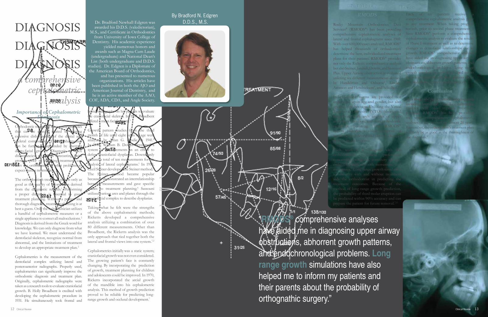
By Bradford N. edgren D.D.S., m.S.
Greeley, CO
Dr. Bradford Newhall Edgren was awarded his D.D.S. (valedictorian),
M.S., and Certificate in Orthodontics from University of Iowa College of
Dentistry. His academic experience yielded numerous honors and
awards such as Magna Cum Laude (undergraduate) and National Dean’s List (both undergraduate and D.D.S.
studies). Dr. Edgren is a Diplomate of the American Board of Orthodontics,
and has presented to numerous organizations. His articles have
been published in both the AJO and American Journal of Dentistry, and he is an active member of the AAO,
COF, ADA, CDA, and Angle Society.
Importance of Cephalometric Diagnosis
Orthodontic treatment faces many obstacles that can be directly related to existing excessive disharmonies of the dental and skeletal components. These disharmonies can be further compounded by aberrant dento-facial growth. Diagnosis of such discrepancies, as well as forecasting facial growth, prior to initiating treatment can alert the orthodontist of what problems to expect during treatment.
The orthodontist’s treatment plan is only as good as the quality of information derived from the diagnostic records.1 Performing a proper diagnosis is essential to good treatment planning. Without a proper and thorough diagnosis, treatment planning is at best a guess. Only the naïve clinician utilizes a handful of cephalometric measures or a single appliance to correct all malocclusions.2 Diagnosis is derived from the Greek word for knowledge. We can only diagnose from what we have learned. We must understand the dentofacial skeleton, recognize normal from abnormal, and the limitations of treatment to develop an appropriate treatment plan.2
Cephalometrics is the measurement of the dentofacial complex utilizing lateral and posteroanterior radiographs. Properly used, cephalometrics can significantly improve the orthodontic diagnosis and treatment plan. Originally, cephalometric radiographs were taken as a research tools to evaluate craniofacial growth. B. Holly Broadbent is credited with developing the cephalometric procedure in 1931. He simultaneously took frontal and
lateral radiographs on his patients to evaluate the craniofacial skeleton. In 1937, Broadbent offered a mean facial pattern.3
Growth pattern studies from the third month of life until eight years of age were analyzed by Allan G. Brodie in 1941.4 In 1948, William B. Downs developed a system of measurements in an effort to define craniofacial dysplasias. Downs only utilized a total of ten measurements for his analysis of lateral cephalograms.1 In 1953, Cecil Steiner developed the Steiner method.5
The Steiner method became popular because it demonstrated an interrelationship between measurements and gave specific guides to treatment planning.6 Sassouni utilized various arcs and planes through the craniofacial complex to describe dysplasias.
Taking what he felt were the strengths of the above cephalometric methods; Ricketts developed a comprehensive analysis utilizing a combination of over 80 different measurements. Other than Broadbent, the Ricketts analysis was the only approach that tied together both the lateral and frontal views into one system.7,2
Cephalometrics initially was a static system; craniofacial growth was not even considered. The growing patient’s face is constantly changing. By incorporating the prediction of growth, treatment planning for children and adolescents could be improved. In 1970, Ricketts incorporated the arcial growth of the mandible into his cephalometric analysis. This method of growth prediction proved to be reliable for predicting long-range growth and occlusal development.7
a comprehensive cephalometric
analysis
13Clinical Review
RMODS
Rocky Mountain Orthodontics Data Services® (RMODS®) has been providing comprehensive cephalometric analyses of lateral and frontal cephalograms since 1969. With over 600,000 cases analyzed, RMODS® has helped thousands of orthodontists determine the best, individualized treatment plans for their patients. RMODS® provides not only the Ricketts comprehensive analysis but also Steiner, Jarabak, Downs, and Sassouni Plus. Upper Airway obstruction is evaluated utilizing six different measurements devised by Handelman and Osborne8, Linder-Aronson and Hendrickson9, and Schulhof.10
Individualized norms are provided not only based upon age and gender, but also upon ethnic heritage. Utilizing the Visual Treatment Objective (VTO) (short and long term) with arch analysis of upper and lower dentition, assists in orthodontic treatment planning. The RMODS® system provides a visual blueprint of recommended dental and skeletal changes specific to each patient.
Each work-up is designed with the orthodontist’s treatment preferences concerning extraction, convexity change, esthetics, limits of tooth movement, and mechanics. Long range growth simulation to maturity with and without treatment aids the orthodontist in predicting the treatment outcomes. Because of the method of long-range growth prediction, the probability of third molar eruption can be predicted within 90% accuracy and can prepare the patient for future removal.7,11
Each of my patients receives a comprehensive cephalometric analysis prior to any treatment. When taking progress records, prior to second phase treatment, I have RMODS® perform a comprehensive cephalometric analysis to evaluate the results of Phase I treatment as well as to determine changes in dentofacial relationships due to growth. RMODS® comprehensive analyses have aided me in diagnosing upper airway obstructions, abhorrent growth patterns, and endochronological problems. Long range growth simulations have also helped me to inform my patients and their parents about the probability of orthognathic surgery.
Conclusion
A thorough and proper orthodontic diagnosis including lateral and frontal cephalometric analyses will only improve treatment planning. RMODS® has been providing me with comprehensive cephalometric analyses of my patients for almost 20 years. Remember, the orthodontist’s treatment plan will only be as good as the thoroughness of the diagnosis.6
“RMODS® comprehensive analyses have aided me in diagnosing upper airway obstructions, abhorrent growth patterns, and endochronological problems. Long range growth simulations have also helped me to inform my patients and their parents about the probability of orthognathic surgery.”

14 Clinical Review 15Clinical Review
This case is a good example of upper airway obstruction and a poor facial growth pattern.
She had a history of snoring, mouth breathing, food allergies, and asthma. Her comprehensive cephalometric analysis demonstrated the following:
1. Class II canine
2. Severe skeletal Class II due to both jaws
3. Skeletal open bite due to the Mandible
4. Possible excessive mandibular growth
5. Adenoid blockage of the airway
6. Skeletal buccal cross bite pattern due to the mandible
7. Mandibular arch wide compared to jaw
8. Possible low tongue position
Because of her short porion location, high cranial base deflection and forward ramus position, she is more likely to grow a lower jaw that is too large relative to the upper face. As a result of the upper airway obstruction and poor growth characteristics, this patient was referred to an Otolaryngologist for evaluation of upper airway obstruction. The tonsils and adenoids were removed prior to the start of orthodontic treatment. Following maxillary expansion with a bonded RME (Rapid Maxillary Expander), the upper and lower arches were leveled and aligned.
► Unilateral or bilateral posterior cross-bites
► Tonsil or adenoids present or history of respiratory problems
► Open-bite
► Tongue thrust upon swallowing
► Mouth breathing
► Functional cross-bite with deflection of the mandible to one side or possibly deflected anteriorly producing a pseudo-Class I condition.
Many orthodontists are surprised to learn that the size of the adenoid, tonsil, and nasopharyngeal airway can be evaluated on the lateral cephalogram. Linder-Aronson and Henrickson9, Schulhof10, Handelman and Osborne8, and Ricketts19 have all devised airway measurements of adenoidal enlargement relative to the nasopharyngeal airway. Radiographic analysis in the lateral and posteroanterior aspects provides a systematic means of evaluating airway dimensions, the morphogenetic factors affecting lower facial heights, bimaxillary morphology and dentofacial growth in mouth breathers. Individuals with inherent vertical facial growth characteristics are the most significantly impacted by mouthbreathing.20
RMODS® uses the Schulhof10 analysis of adenoid enlargement which includes the two linear measurements by Linder-Aronson and Hendrickson9, a linear measurement by Ricketts19, the airway percentage in an epipharyngeal trapezoidal area described by Handelman and Osborne9, and the craniofacial angles N-S-Ba and BA-S-PNS. RMODS® analyzes each case for the potential adenoid obstruction of the mesopharyngeal airway. Adenoid blockage of the mesopharyngeal airway is deemed to be present if three or more measurements are one or more standard deviations from the norm.10 If the patient is a mouth breather and the analysis indicates that the adenoid is too large for the airway21, the orthodontist can make a referral to an otolaryngologist for further evaluation and appropriate treatment.
RMODS ® Mandibular Growth Awareness Form alerts the orthodontist to possible abhorrent dentofacial growth.
Diagnostic Intraoral Photographs
Diagnostic Panoramic Radiograph
Case Study I Airway Obstruction and Poor Facial Growth Patterns
Mouth breathing has been identified as a cause for a number of orthodontic problems including cross bites, low tongue positions, and vertical dysplasias.12-15 Children who have a genetic predisposition towards a narrow, dolichocephalic facial pattern, and having airway compromise are particularly at risk to developing long face syndrome. Moreover, children with a genetic propensity to developing mandibular prognathism, possessing tonsillar hypertrophy and who are chronic mouth-breathers are at particular risk for developing advanced mandibular prognathism.16
Mouth breathing should also be regarded as an obstacle to successful orthodontic treatment and is likely to result in orthodontic relapse if not treated. It is imperative that the existence of mouth breathing, as well as its etiology, be recognized as soon as possible and ideally before orthodontic treatment has been attempted.7 Since anteroposterior and vertical dentofacial discrepancies are linked to growth, interceptive measures should be initiated around age seven. To wait until age 12, when 90% of a dentofacial deformity has already been established, before instituting orthodontic treatment is not consistent with today’s preventative philosophy.17 The earlier the re-establishment of normal oropharyngeal function and nasal respiration, the more likely normal dentofacial development will occur. Oral breathing may persist for a year or more after the airway has been restored while the original chronic mouth-breathing habit is “unlearned”.18
Ricketts described a condition associated with upper airway obstruction; he labeled it the Respiratory Obstruction Syndrome.18 Clinically, Ricketts found the following characteristics generally associated with the presence of enlarged adenoids and tonsils:

16 Clinical Review 17Clinical Review
The following progress records were taken after 24 months of treatment, prior to banding the second molars and Class II correction. This patient no longer snores and her respiration is now nasal. Note that her low tongue position and forward head posture to open her airway has improved. Her dental overbite has been maintained.
Progress Intraoral Photos
“This patient no longer snores and her respiration is now nasal.”
This patient presented with a Class I malocclusion, a tendency for a skeletal open bite, possible excessive lower jaw growth and a significant arch length discrepancy with ectopic maxillary canines.
Cephalometric analysis also revealed a skeletal lingual cross bite pattern due to both the maxilla and mandible; as well as possible excessive mandibular growth.
This patient’s treatment plan included rapid maxillary expansion and fixed appliances. The result was a nicely treated Class I occlusion.
Case Study IIFrontal Analysis
The frontal cephalometric analysis is often overlooked by most orthodontists. Asymmetries, dental cross bites, skeletal cross bites, maxillary and mandibular dental arch widths, nasal widths, turbinate enlargement, deviated nasal septums, and facial proportions can all be evaluated from the posteroanterior cephalogram. Many orthodontists think of the maxilla as being the only culprit of dental or skeletal lingual cross bite patterns. However, many times the width of the mandible can be the major contributor to skeletal lingual cross bite patterns.
Dental compensations can hide overt hypo-plastic maxillary and hyper-plastic mandibular transverse discrepancies. Rapid maxillary expansion can improve skeletal lingual cross bite patterns, but without a posteroanterior cephalogram, it is impossible to diagnose them. The affect of the excessive mandibular width may not be clinically evident until late adolescence, when rapid maxillary expansion may be more difficult. Taking a posteroanterior cephalogram on patients is simple and the benefits to the patient are immeasurable. Furthermore, with the development of cone beam computed tomography, all patients that have a CBCT scan will have both lateral and frontal images readily available for analysis with a single scan.
Diagnostic Panoramic RadiographProgress i-CAT ® panoramic report
Progress RMODS® Tracing
Case Study I continued

18 Clinical Review 19Clinical Review
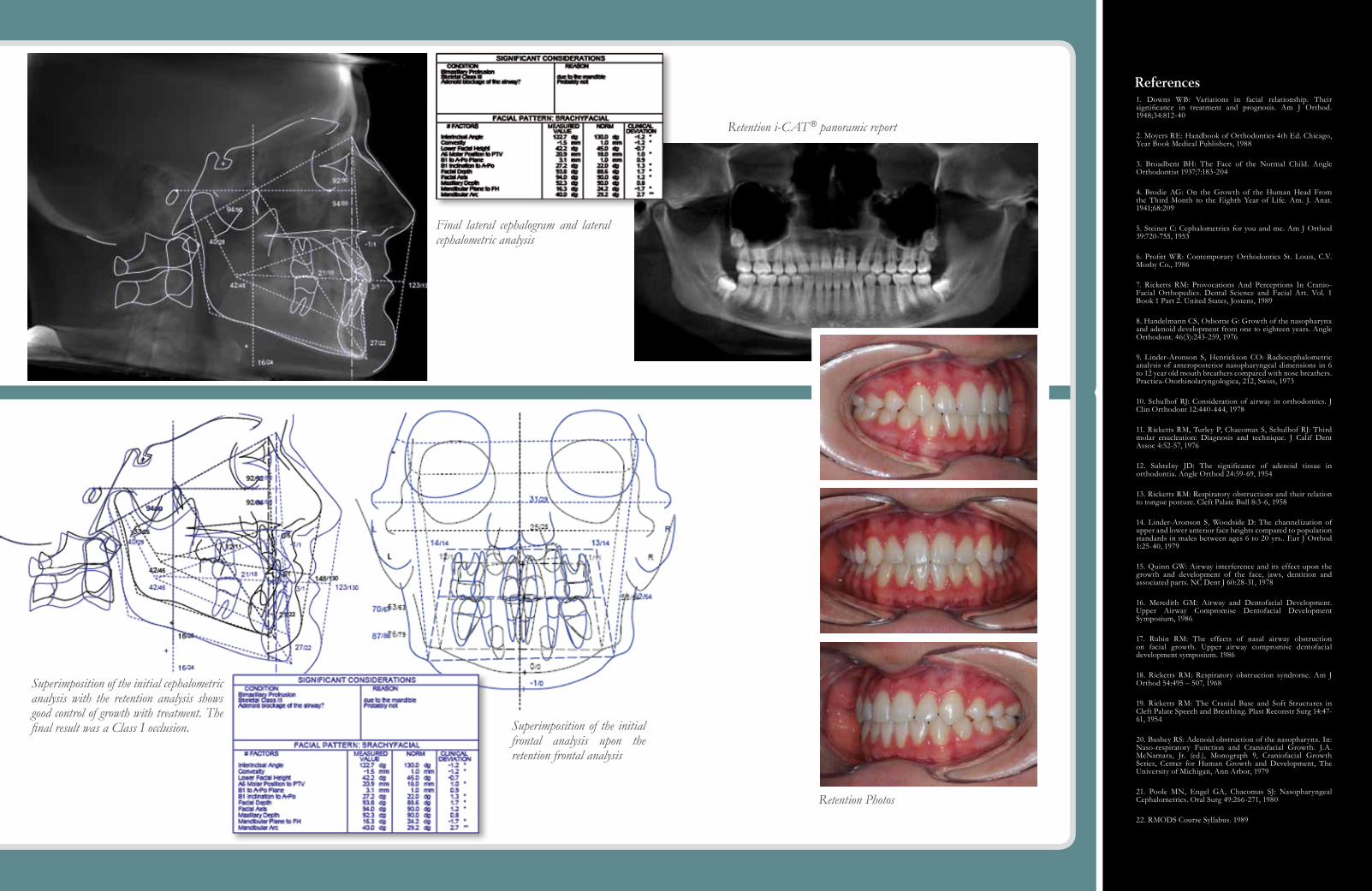
Superimposition of the initial vs. the final lateral cephalometric analysis demonstrates both significant horizontal and vertical mandibular growth, as predicted in RMODS® initial comprehensive analysis.
This patient presented with the following problems:
1. Class II malocclusion due to the upper right first molar2. Severe Overjet
3. Severe Class II Skeletal Malocclusion due to the mandible and maxilla
4. Open Bite
5. Tendency for Skeletal Open bite due to the mandible and maxilla6. Wide mandibular arch compared to jaw
7. Midline asymmetry
Superimposition of the current lateral cephalometric tracing over the growth to maturity without treatment demonstrates probable significant growth of both jaws, especially the mandible. However, despite the mandibular growth, the class II molar relationship does not improve without treatment. Treatment designed to take advantage of the remaining mandibular growth, while maintaining upper molar position would be of advantage to improve the class II malocclusion. An orthodontist has more control over the dentition than the skeletal component.7
Long Range Growth Forecasting (CASE III, CASE IV, CASE V, CASE VI)
As previously stated, the ability to forecast the facial growth of a patient to maturity is of great benefit. Regardless of how thorough a cephalometric analysis is devised to evaluate a growing patient’s present state, that technique will be insufficient for treatment planning because of future growth and dentofacial development. Incorporation of craniofacial growth into the method of diagnosis can only result in improved treatment planning. The craniofacial relationships seen even two years after the start of treatment in a growing child may not be the same at maturity. A case treated to suitable balance at age 12 may prove to be a failed result at age 25 due to continued growth. This is especially true in those patients that demonstrate abnormally large amounts of lower jaw growth during their late teenage years and early twenties.22
RMODS® computer performs growth simulations by combining the following four growth curves with individual average directions and amounts of change per year for approximately 200 cephalometric landmarks.
These four different growth curves are: ► Total body height ► Soft tissue ► Cranial base ► Mandibular growth
Each curve is subdivided by race, gender, and skeletal age (this final subdivision is used to classify which patients are normal growers vs. late and advanced growth categories). When treatment planning for a growing patient, it is important to consider how much growth will or will not occur within the treatment time. Skeletal age can be extremely valuable in determining remaining growth in late adolescence. Moreover, the most significant factor in evaluating growth is not absolute amount, but relative amount. It is important, that the relative growth of the maxilla and mandible be normal. Deviations of growth between the jaws within 20% can generally be tolerated, but those deviations greater than 50% will result in a considerable deformity.22
Case Study III
Superimposition of the initial cephalometric vs. the final frontal cephalometric analysis on the occlusal plane shows improvement in the cant of the maxilla. Rapid maxillary expansion of the maxilla has also successfully corrected the skeletal lingual cross bite pattern and eliminated dental crowding, demonstrating the logic in a non-extraction treatment plan.
Diagnostic Panoramic RadiographSuperimposition of the initial frontal analysis upon the visual norm
Diagnostic Intraoral Photographs

21Clinical Review
This patient was treated with rapid maxillary expansion, straight-pull headgear and fixed appliances during Phase I treatment. Superimposition of the initial lateral cephalometric analysis upon the progress cephalometric analysis, prior to initiation of Phase II treatment, shows significant improvement to a Class I molar relationship. The upper molar position was maintained within the maxilla, forward movement of the lower molar and growth of the mandible helped in the correction of the class II malocclusion.
Retention Intraoral Photographs
Progress Intraoral Photographs
Superimposition of the retention frontal analysis upon the visual norm demonstrates that rapid maxillary expansion during Phase I treatment reduced the probable skeletal lingual cross bite pattern due to additional mandibular transverse growth.
Retention i-CAT ® Panoramic Report
“The RMODS ® computer performs growth
simulations by combining the following four growth curves.”
These four different growth curves are:
► Total body height ► Soft tissue ► Cranial base ► Mandibular growth
Final superimposition of the initial and retention cephalometric analyses demonstrates the Class II to Class I correction. Taking advantage of the mandibular growth as forecasted at the beginning treatment resulted in a nice Class I result for this patient.
20 Clinical Review
Case Study III continued

23Clinical Review
Until recently, most diagnostic systems were located and
maintained in- office and the practitioner was responsible for upgrades, upkeep and
maintenance.
Today, e-Ceph® Web can deliver the latest orthodontic
diagnostics right to your web browser!
e-Ceph® Web provides an easy two step process for sending
patient data and getting diagnostic results. Step one
enables users to digitize x-rays directly through their web
browser, or to submit files of patient records to our analysts for evaluation. Step two allows
you to receive your results through the same web interface.
So now you can enjoy the thoroughness and accuracy of
the RMO Data Service combined with the convenience and
flexibility of an in-office system.
The e-Ceph® Web diagnostic workup delivers the same quality you’ve come to expect from us.
e ceph®
Diagnostic Intraoral Photos
Growth to Maturity without Treatment
The frontal cephalometric analysis reveals a skeletal lingual cross bite pattern due to the maxilla and the mandible.
The growth forecast also illustrates no improvement in the Class II malocclusion, further upright of the lower incisors and deepening of the bite without orthodontic treatment. Maintaining upper molar position and taking advantage of future mandibular growth will aid in orthodontic correction.
Superimposition of the initial cephalometric analysis upon the progress cephalometric analysis demonstrates forward growth of the mandible, as forecasted.
Case Study IVThis is the case of a Class II malocclusion with the potential for excessive lower jaw growth. Superimposition of the lateral cephalometric upon the growth to maturity forecast shows the potential for significant lower jaw growth.
Superimposition of the initial frontal analysis upon the progress frontal analysis.
This patient now has a nice final Class I occlusion with the help of the growth prediction.
Retention lateral cephalometric analysis
22 Clinical Review

24 Clinical Review
Diagnostic Intraoral Photos
Retention records demonstrating Class II to a solid Class I correction.
Retention Intraoral PhotosRetention panelipse
Case Study VThis patient presented with a Class II malocclusion. The growth forecast to maturity demonstrated strong lower jaw growth in a horizontal direction. Maintaining the upper molar position and allowing for the forecasted lower jaw growth will help in correcting the class II malocclusion.
Wilson® 3D®The Wilson® 3D® system comprises a series of interrelated fixed/removable intraoral modules that simplify and improve treatment. Wilson® 3D® appliances can be used to supplement all techniques while delivering practical and simple solutions to both typical and extraordinary movement challenges. RMO® sponsors numerous CE events that teach the skills needed to incorporate Wilson® 3D® concepts and materials into your present technique. Please call RMO® or visit our website for additional information about the legendary Wilson® 3D® system.
• Time tested and proven
• Over 100 different movements possible, including: expansion, contraction, distalization, space maintenance, bilateral, and unilateral
• Does not replace your current technique – the Wilson® system simply complements your current system
• First phase, early treatment, mixed dentition, and adults
• Preconfigured sizes to fit all patient dental ranges
• Fixed for the patient and easily removable by the clinician for rapid chairside adjustments
The World’s Oldest Synergistic, Bioprogressive®, Breathing Enhancement Orthodontic Company.®
For more information, please call 800.525.6375 or visit our website at www.rmortho.com.
25Clinical Review

26 Clinical Review 27Clinical Review
Progress photos
Case Study VIThe following patient had a severe Class III malocclusion.
Superimposition of the initial lateral cephalometric analysis upon the growth to maturity forecast demonstrates the potential for significant additional mandibular growth. Treatment designed to address this possible excessive growth will improve overall treatment success.
Superimposition of the initial frontal analysis upon the visual norm
Diagnostic intraoral photos
Superimposition of the progress lateral cephalometric analysis upon the initial cephalometric analysis demonstrating how early treatment involving fixed appliances along with the growth forecast aided in improving this patient’s malocclucion.
Superimposition of the lateral cephalometric analysis upon the visual norm illustrates
the significant mandibular prognathism.
Progress Panelipse
28 Clinical Review 29Clinical Review
1. Downs WB: Variations in facial relationship. Their significance in treatment and prognosis. Am J Orthod. 1948;34:812-40
2. Moyers RE: Handbook of Orthodontics 4th Ed. Chicago, Year Book Medical Publishers, 1988
3. Broadbent BH: The Face of the Normal Child. Angle Orthodontist 1937;7:183-204
4. Brodie AG: On the Growth of the Human Head From the Third Month to the Eighth Year of Life. Am. J. Anat. 1941;68:209
5. Steiner C: Cephalometrics for you and me. Am J Orthod 39:720-755, 1953
6. Profitt WR: Contemporary Orthodontics St. Louis, C.V. Mosby Co., 1986
7. Ricketts RM: Provocations And Perceptions In Cranio-Facial Orthopedics. Dental Science and Facial Art. Vol. 1 Book 1 Part 2. United States, Jostens, 1989
8. Handelmann CS, Osborne G: Growth of the nasopharynx and adenoid development from one to eighteen years. Angle Orthodont. 46(3):243-259, 1976
9. Linder-Aronson S, Henrickson CO: Radiocephalometric analysis of anteroposterior nasopharyngeal dimensions in 6 to 12 year old mouth breathers compared with nose breathers. Practica-Otorhinolaryngologica, 212, Swiss, 1973
10. Schulhof RJ: Consideration of airway in orthodontics. J Clin Orthodont 12:440-444, 1978
11. Ricketts RM, Turley P, Chacomas S, Schulhof RJ: Third molar enucleation: Diagnosis and technique. J Calif Dent Assoc 4:52-57, 1976
12. Subtelny JD: The significance of adenoid tissue in orthodontia. Angle Orthod 24:59-69, 1954
13. Ricketts RM: Respiratory obstructions and their relation to tongue posture. Cleft Palate Bull 8:3-6, 1958
14. Linder-Aronson S, Woodside D: The channelization of upper and lower anterior face heights compared to population standards in males between ages 6 to 20 yrs.. Eur J Orthod 1:25-40, 1979
15. Quinn GW: Airway interference and its effect upon the growth and development of the face, jaws, dentition and associated parts. NC Dent J 60:28-31, 1978
16. Meredith GM: Airway and Dentofacial Development. Upper Airway Compromise Dentofacial Development Symposium, 1986
17. Rubin RM: The effects of nasal airway obstruction on facial growth. Upper airway compromise dentofacial development symposium. 1986
18. Ricketts RM: Respiratory obstruction syndrome. Am J Orthod 54:495 – 507, 1968
19. Ricketts RM: The Cranial Base and Soft Structures in Cleft Palate Speech and Breathing. Plast Reconstr Surg 14:47-61, 1954
20. Bushey RS: Adenoid obstruction of the nasopharynx. In: Naso-respiratory Function and Craniofacial Growth. J.A. McNamara, Jr. (ed.), Monograph 9, Craniofacial Growth Series, Center for Human Growth and Development, The University of Michigan, Ann Arbor, 1979
21. Poole MN, Engel GA, Chacomas SJ: Nasopharyngeal Cephalometrics. Oral Surg 49:266-271, 1980
22. RMODS Course Syllabus. 1989
References
Final lateral cephalogram and lateral cephalometric analysis
Superimposition of the initial cephalometric analysis with the retention analysis shows good control of growth with treatment. The final result was a Class I occlusion. Superimposition of the initial
frontal analysis upon the retention frontal analysis
Retention Photos
Retention i-CAT ® panoramic report

why iNdirect BoNdiNG?rMo®’s rMBond® indirect Bonding system provides clinicians a simple and
consistent solution for maximizing practice efficiency. the rMBond®
indirect Bonding (idB) system delivers a step-by-step process that
allows doctors to fundamentally reduce the amount of chair
time involved when bonding appliances to a patient. this
results in a greatly improved patient experience also, as the
idB process significantly reduces the patient’s chair time
and discomfort during bonding. the rMbond® system
allows for extremely accurate bracket placement
under convenient setup conditions working on a
study model, and most of the procedures can
be conducted by staff persons with modest
training. the rMbond® start-up kit is a turnkey
system that includes all of the materials
necessary to begin indirect Bonding your
patients immediately.
uNique coMpoNeNts iN the rMBoNd® iNdirect BoNdiNG systeM iNclude:
RMbond® InneR TRay MaTeRIal:
• provides predictable and reliable working time, with excellent flow characteristics for complete encapsulation of appliances
• clear material visibility during bracket transfer assures accurate seating and rapid light curing
• provides an ideal tear strength when removing inner tray Material – no debonds and minimal cleanup
• eliminates the need for block outs around hooks and undercuts
RMbond® lC Flowable adhesIve:
• precise dispensing system with needle tip
• ideal viscosity
• reduces flash
• excellent bond strength
systeM hiGhliGhts• reduces chair time
• significantly more comfortable bonding experience for patient
• convenient and more precise final appliance placement on a study model at doctor’s leisure
• reduces clinician neck and back pain by minimizing time bent over a patient during bonding procedure
• No need for two models – study model also functions as idB model
precise bracket placement on a study model
transfer tray fabrication - inner tray Material fully encapsulates all appliances
rapid patient bonding process - light curing directly through transfer tray
30 Clinical Review 31Clinical Review
Inner Tray Material
Tray Finishdispensing Gun
Model storage boxseparating Medium
lC bonding ResinRound Rope wax
lC Flowable adhesivelC Turbo Material

32 Clinical Review 33Clinical Review
®
e ceph®RMoDS®/ Q & A with Dr. Budi Kusnoto
Dr. Budi Kusnoto is a tenured full time associate professor in the Department of Orthodontics, University of Illinois at Chicago. His computer science background and knowledge in biomechanics as well as management of craniofacial deformities are complimentary to his teaching in the field of orthodontic diagnosis and treatment planning. He also has been actively involved in clinical research in the area of temporary anchorage devices, invisible orthodontic appliances, computerized orthognathic-craniofacial surgical imaging, 3D imaging-computerized treatment simulation, and longitudinal digital data mining project. Currently Dr. Kusnoto also maintains a private practice and clinic directorship at the Department of Orthodontics, College of Dentistry University of Illinois at Chicago. He is an active member of American Dental Association, Illinois Society of Orthodontists, Chicago Dental Society, American Association of Orthodontists, and is a Diplomate of American Board of Orthodontics.
Budi Kusnoto, D.D.S., M.S.
Department of Orthodontics
University of Illinois at Chicago
e ceph®
Dr. Kusnoto has been using RMODS ® services for the past 5 years for his research in validating computerized cephalometric prediction treatment outcome, he is also constantly involved in evaluating many other cephalometric imaging software in the market.
Q: How long does it take for me to receive my results?
A: On average results will be returned within 3-5 minutes, depending on the complexity of the analysis requested and Internet speed. If you have submitted your records to RMODS® for the analysts to digitize, results should be returned within 3 days.
Q: Is there tech suppor t ava ilable?
A: Yes, well trained analysts and technical support is available Monday through Friday during business hours.
Q: Why would I want to digit ize a frontal?
A: Much more data, that can influence our treatment objectives and eventually treatment mechanics, can be gathered by simply adding frontal analysis. Often clinicians tend to skip looking at skeletal/dental asymmetry in the transverse dimension or possible airway obstruction which can be quantified using the frontal analysis.
Q: Why do I need to digitize the lower arch and what kind of information will it provide me?A: Digitizing the lower dental arch will give the clinician much more information (about occlusion, tooth size discrepancy, dental development) as it relates to the skeletal and facial structures which were derived from lateral and frontal cephalometric radiographs. The digitized information from the lower arch is required by the RMODS® program to produce the treatment planning segments of the results. It provides a 3rd dimension of the view of the patient.
Q: Why do I d ig it ize the upper arch and what kind of informat ion will it supply me?
A: By adding the upper arch you will be provided with the Bolton Analysis as well as a more complete view of the patient’s current situation.
Q: What is a Visual Norm? Where does it come f rom?
A: e-ceph® Web is one of the extremely few cephalometric software programs currently available in the market that has the ability to accurately produce a Visual Norm (graphical representation of a NORM) which can be used as a template while treating the case (to guide clinicians in designing their orthodontic mechanics to move teeth/bone in space).
Q: What is a Visual Treatment Objec t ive ( V TO) and how does it help me in my diagnost ics?
A: By using the VTO, we can map our treatment into a moving target (in a growing individuals) as well as graphically represent our treatment goal in terms of where should we position the teeth at the end of treatment. Clinicians can also utilize the VTO to improve the accuracy of their treatment. We have the ability to design how much certain parts of the occlusion should be moved, whether it is dental or skeletal, in order to achieve the optimal stable occlusion for the patient.
Q: Can I get just a he ight pred ic t ion? What informat ion is required for this?
A: Yes, all that is required is the patient’s date of birth and their present height. If you would like improved accuracy you can include the skeletal age from the current hand wrist film.
Q: Can you prov ide us with an over v iew of R MODS ® and e - ceph ® Web?
A: e-ceph® Web can be summarized as a web-portal (Internet virtual meeting place) to various cephalometric analyses, growth simulations, data/image management, and case management tools to aid in developing excellent treatment objectives/plans. It can also be a web-portal for potential inter-institutional as well as inter-clinician world wide exchange of study cases.
Q: Why use e - ceph ® Web?
A: e-ceph® Web is purely web based, meaning it is not installed on a computer. It is easily accessible through any terminal connected to the Internet. No updates or maintenance will ever be needed, as this is done automatically
through the e-ceph® Web RMODS® server. All data can be securely stored in the RMODS® server facilities and are easily accessible from anywhere on the planet with a high speed Internet connection.
Q: What is the bene f it of e - ceph ® Web?
A: e-ceph® Web functions as cephalometric digitizing software, and also gives you the flexibility of being able to send your records directly to RMODS® where well trained and highly experienced personnel will digitize them and return the results to you.
Q: How is e - ceph ® Web bet ter than the sof tware that I would have in my of f i ce?
A: It is the only cephalometric analysis software in the market that can actually produce interpretation of the cephalometric numbers and its parameters which can lead to formulating treatment objectives, thus coming up with suggested treatment plans and treatment mechanics including treatment sequence and timing.
Q: What d if ferent t ypes of analyses does e - ceph ® Web of fer?
A: e-ceph® Web offers the same cephalometric tools and analyses as the RMODS® service; Ricketts, Downs, Steiner, Sassouni Plus, and Jarabak.
Q: Is any spec ial equipment r equired?
A: A computer with standard high speed Internet (such as DSL or cable) running standard web-browser will be sufficient to run e-ceph® Web application.
Q: What i f I don’t have t ime to d ig it ize my case?
A: If you would like the RMODS® analysts to digitize your case, you can simply click on the “PROCESS by RMODS®” option after uploading all the necessary radiographs/digital images and patient information into the e-ceph® Web system. The final result will be sent back to you by email.

34 Clinical Review 35Clinical Review
a practical solution using The Multi-Family
By Dr. Franco BrunoItaly
ThE FunCTIonAL
MATRIx: Buccal
BumperMidlinecorrection
Defined tooth channels
Can be sterilized andand/or disinfected
Lingualenvelope
Raised OcclusalPlane
Multi-Family
Functional Education
M u l t i T r a i n e rMulti-T®
M u l t i T r a i n e rMulti-T®
Multi-TB™
Multi Trainer for Braces
Multi-S™
Multi Start
diminishes the effect of
labial forces
acts to insure the correct
positioning of the midline
repositions the tongue in the maxilla
Multi-Family Appliances
The Multi-Family Appliances are an integrated system of appliances that allow the orthodontists to choose the ideal appliance according to the age and the malocclusion of the patient.
Multi-P ®
Multi Purpose
Dr. Franco Bruno received his Medical Degree from the University of Pavia, Italy. His Orthodontic Specialty degree was awarded at the University of Cagliari, Italy.
Postgraduate Degrees include Straight Wire Therapy and and TMJ Therapy from the University of Milan and Lingual Orthodontics from the University of Varese.
Dr. Bruno completed the 2 year Zerobase Bioprogressive Course and is the Chair of Bioprogressive Philosophy at the University of Cagliari. He is also Head of the
Bioprogressive Department, Dental Clinic, at the same institution.
Dr. Bruno has a Private Practice Limited to Orthodontics, which he opened in 1986.
INTRODUCTION
A long-term goal in orthodontics has been to understand the interaction between the Functional Matrix and malocclusion. Research in this area began in the early 19th century and, to date, there is no definitive understanding. Contemporary orthodontics recognizes two opposing views. The “functionalists” believe that the Functional Matrix, especially that of a muscular nature, is the determinant principle of malocclusion. Contrary to this belief is the “mechanistics” view, whose proponents say that muscular dysfunctions are a result of malocclusion. Unfortunately, the latter have yet to submit a theory on the etiology of malocclusion. There are various positions between these two extremes that, to a greater or lesser degree, recognize the influence of the functional matrix on malocclusion. It is difficult for the clinician to address malocclusion both in etiological terms and long-term stability. A primary issue is the probability of relapse after orthodontic treatment. If the Functional Matrix is the cause of malocclusion, and it is not neutralized during treatment, there will be a greater possibility of relapse. However, if the dysfunction is a result of the malocclusion, only its complete resolution will guarantee stability of the case. From our perspective, this ideological dualism is irrelevant.
The philosophy of “Self Confident Orthodontics” views the interaction between the Functional Matrix and malocclusion as a continuous exchange of information between the two components and, therefore, foresees a therapeutic protocol that aims at correcting both parts of the system in order to find the most appropriate solution for long-term stability. The main therapeutic idea is to work on each component at different treatment times. In the absence of definitive scientific evidence, the clinician must develop his/her own viewpoint and objectives to best resolve the patient’s problems and reach a clinical outcome that will be stable over time.
Our therapeutic protocol calls for a three-step treatment sequence to address the Functional Matrix:
1. Preparation Stage: use myofunctional orthodontics at an early age, from 4-5 up to 10-12 years of age, while waiting for the appropriate time to start treatment with conventional orthodontic mechanics.
2. Treatment Stage: use myofunctional appliances in association with conventional fixed appliance therapy.
3. Retention Stage: use myofunctional orthodontics at the end of treatment to promote adaptation of the Functional Matrix to the new occlusion.
This approach is based on simple considerations. If alterations of the Functional Matrix are the cause of malocclusions, its neutralization guarantees simpler active treatment. If, however, the dysfunctions are the result of a malocclusion its treatment will be more complex; therefore, neutralization of the Functional Matrix would allow faster and more simplified treatment. Lastly, if the resolution of the malocclusion is decisive for correction of the dysfunction, control during active treatment allows a quicker adaptation of the Functional Matrix to the new occlusion. Therefore, the guideline is to act on both components without certain knowledge of which is the cause and effect. Simplified therapeutic protocols will produce a better and more stable result.
Based on these concepts we have tried to find a solution to patient treatment with a simple, economical, and easy to use myofunctional approach that can be utilized at any age and at all stages of orthodontic treatment.
The appliances of the MULTI SYSTEM respond very well to these characteristics and therefore are included in the “Self Confident Orthodontics” philosophy of treatment.

36 Clinical Review 37Clinical Review
type age sizes holes lip-Bumper effectMulti- s 5-8 1 yes yesMulti-t 6-10 1 yes yesMulti-p 9-13 multiple yes noMulti-tB all 1 no yes
type GuidanceMulti- s incisorsMulti-t incisors and caninesMulti-p incisors, canines and BicuspidsMulti-tB No guidance
References1: Meyer PG. Tongue lip and jaw differentiation and its relationship to orofacial myofunctional treatment. Int J Orofacial Myology. 2008 Nov;34:36-45. PubMed PMID:19545089.
2: Paskay LC. Instrumentation and measurement procedures in orofacial myology. Int J Orofacial Myology. 2008 Nov;34:15-35. PubMed PMID: 19545088.
3: Giuca MR, Pasini M, Pagano A, Mummolo S, Vanni A. Longitudinal study on a rehabilitative model for correction of atypical swallowing. Eur J Paediatr Dent. 2008 Dec;9(4):170-4. PubMed PMID: 19072004.
4: Felicio CM, Ferreira CL. Protocol of orofacial myofunctional evaluation with scores. Int J Pediatr Otorhinolaryngol. 2008 Mar;72(3):367-75. Epub 2008 Jan 9. PubMed PMID: 18187209.
5: Grabowski R, Kundt G, Stahl F. Interrelation between occlusal findings and orofacial myofunctional status in primary and mixed dentition: Part III: Interrelation between malocclusions and orofacial dysfunctions. J Orofac Orthop. 2007 Nov;68(6):462-76. English, German. PubMed PMID: 18034287.
6: Verrastro AP, Stefani FM, Rodrigues CR, Wanderley MT. Occlusal and orofacial myofunctional evaluation in children with anterior open bite before and after removal of pacifier sucking habit. Int J Orthod Milwaukee. 2007 Fall;18(3):19-25.PubMed PMID: 17958262.
7: Stahl F, Grabowski R, Gaebel M, Kundt G. Relationship between occlusal findings and orofacial myofunctional status in primary and mixed dentition. Part
II: Prevalence of orofacial dysfunctions. J Orofac Orthop. 2007 Mar;68(2):74-90. English, German. PubMed PMID: 17372707.
8: Fraser C. Tongue thrust and its influence in orthodontics. Int J Orthod Milwaukee. 2006 Spring;17(1):9-18. PubMed PMID: 16617883.
9: Korbmacher HM, Schwan M, Berndsen S, Bull J, Kahl-Nieke B. Evaluation of a new concept of myofunctional therapy in children. Int J Orofacial Myology. 2004 Nov;30:39-52. PubMed PMID: 15832861.
10: Usumez S, Uysal T, Sari Z, Basciftci FA, Karaman AI, Guray E. The effects of early preorthodontic trainer treatment on Class II, division 1 patients. Angle Orthod. 2004 Oct;74(5):605-9. PubMed PMID: 15529493.
11: Jefferson Y. Orthodontic diagnosis in young children: beyond dental malocclusions. Gen Dent. 2003 Mar-Apr;51(2):104-11. Review. PubMed PMID: 15055681.
12: Zardetto CG, Rodrigues CR, Stefani FM. Effects of different pacifiers on the primary dentition and oral myofunctional strutures of preschool children. Pediatr Dent. 2002 Nov-Dec;24(6):552-60. PubMed PMID: 12528948.
13: Meyer PG. Tongue lip and jaw differentiation and its relationship to orofacial myofunctional treatment. Int J Orofacial Myology. 2000 Nov;26:44-52. Review. PubMed PMID: 11307348.
14: Bacha SM, Rìspoli CF. Myofunctional therapy: brief intervention. Int J Orofacial Myology. 1999 Nov;25:37-47. PubMed PMID: 10863453.
15: Klocke A, Korbmacher H, Kahl-Nieke B. Influence of orthodontic appliances on myofunctional therapy. J Orofac Orthop. 2000;61(6):414-20. English, German. PubMed PMID: 11126016.
16: Reinicke C, Obijou N, Tränkmann J. The palatal shape of upper removable appliances. Influence on the tongue position in swallowing. J Orofac Orthop. 1998;59(4):202-7. English, German. PubMed PMID: 9713176.
17: Tallgren A, Christiansen RL, Ash M Jr, Miller RL. Effects of a myofunctional appliance on orofacial muscle activity and structures. Angle Orthod. 1998 Jun;68(3):249-58. PubMed PMID: 9622762.
18: Pierce RB. The effectiveness of oral myofunctional therapy in improving patients’ ability to swallow pills. Int J Orofacial Myology. 1997;23:50-1. PubMed PMID: 9487830.
19: Benkert KK. The effectiveness of orofacial myofunctional therapy in improving dental occlusion. Int J Orofacial Myology. 1997;23:35-46. PubMed PMID: 9487828.
20: Umberger FG, Johnston RG. The efficacy of oral myofunctional and
coarticulation therapy. Int J Orofacial Myology. 1997;23:3-9. Review. PubMed PMID: 9487825.
21: Thiele E. Timing in myofunctional training. Int J Orofacial Myology. 1996 Nov;22:28-31. PubMed PMID: 9487823.
22: Marchesan IQ, Krakauer LR. The importance of respiratory activity in myofunctional therapy. Int J Orofacial Myology. 1996 Nov;22:23-7. PubMed PMID:9487822.
23: Annunciato NF. Plasticity of the nervous system. Int J Orofacial Myology. 1995 Nov;21:53-60. Review. PubMed PMID: 9055672.
24: Gommerman SL, Hodge MM. Effects of oral myofunctional therapy on swallowing and sibilant production. Int J Orofacial Myology. 1995 Nov;21:9-22. PubMed PMID: 9055666.
25: Sergl HG, Zentner A. Theoretical approaches to behavior change in myofunctional therapy. Int J Orofacial Myology. 1994 Nov;20:32-9. Review. PubMed PMID: 9055662.
26: Seminara R, Seminara G. Cephalometrics and oral myofunctional impairment. N Y State Dent J. 1994 Oct;60(8):53-7. PubMed PMID: 7970420.
27: Stavridi R, Ahlgren J. Muscle response to the oral-screen activator. An EMG study of the masseter, buccinator, and mentalis muscles. Eur J Orthod. 1992 Oct;14(5):339-49. PubMed PMID: 1397072.
28: Winchell B. Orofacial myofunctional therapy for adult patients. Int J Orofacial Myology. 1989 Mar;15(1):14-8. PubMed PMID: 2599777.
29: Bergersen EO. The eruption guidance myofunctional appliance in the consecutive treatment of malocclusion. Gen Dent. 1986 Jan-Feb;34(1):24-9. PubMed PMID: 3456331.
30: Garliner D. The current status of myofunctional therapy in dental medicine. Int J Orthod. 1982 Mar;20(1):21-5. PubMed PMID: 6953051.
31: Garliner D. The modern myofunctional therapeutic concept. Int J Orthod. 1980 Jun;18(2):21-3. PubMed PMID: 6930367.
32: Hanson ML. Oral myofunctional therapy. Am J Orthod. 1978 Jan;73(1):59-67. PubMed PMID: 271473.
33: Leone KJ. Myofunctional therapy in specialty as well as general practice. Int J Orthod. 1977 Sep-Dec;15(3-4):10-32. PubMed PMID: 271634.
34: Haas AJ. Let’s take a rational look at myofunctional therapy. Int J Oral Myol. 1977 Jul;3(3):24-7. PubMed PMID: 275226.
35: Gottlieb EL. Orthodontics vs myofunctional therapy. J Clin Orthod. 1977 Feb;11(2):83-5. PubMed PMID: 273609.
36: Proffit WR, Brandt S. Dr. William R. Proffit on the proper role of myofunctional therapy. J Clin Orthod. 1977 Feb;11(2):101-5. PubMed PMID: 273603.
37: Wildman AJ. The motor system: a clinical appraisal. Dent Clin North Am. 1976 Oct;20(4):691-705. PubMed PMID: 1067201.
38: Kaye SR. A rational approach to myofunctional therapy. Quintessence Int Dent Dig. 1976 Aug;7(8):51-4. PubMed PMID: 1076571.
39: Cottingham LL. Myofunctional therapy. Orthodontics--tongue thrusting--speech therapy. Am J Orthod. 1976 Jun;69(6):679-87. PubMed PMID: 775999.
THE MULTI SYSTEM OF ORTHODONTICS
The MULTI SYSTEM of Orthodontics represents an integrated series of myofunctional appliances that allow the orthodontist to utilize the device that is most suitable based on the age and characteristics of the patient’s malocclusion.
The MULTI series of appliances are primarily myofunctional in nature and, as such, each appliance is designed for specific functions. All appliances in the series have various characteristics in common, although each has unique features rendering them case specific for various stages of treatment.
THE COMMON CHARACTERISTICS OF MULTI SYSTEM APPLIANCES
Like all myofunctional devices, these appliances have a monoblock shape in order to simultaneously work on both dental arches. The mandibular position protrudes with respect to a edge to edge incisor position. Moreover, the appliances have a raised occlusal plane. This positioning promotes an immediate mechanical unlocking of the TMJ in association with the functional unlocking of muscles.
In addition, all of the appliances have a large vestibular shield which serves to activate the perioral muscles; the shield is adequately extended in order to provoke stretching and activation of the musculature although not arriving up to the fornix given that it is preformed and not customized for the patient. Lingually, the appliance has a frontal lingual ramp for the re-teaching of lingual posture and two lateral wings which increase the re-education effect of the frontal elevator.
In summary, the specific design characteristics of the MULTI SYSTEM are:
a. Vestibular Shield
b. Lingual Elevator
c. Lateral Wings
d. Occlusal Plane
e. Mandibular Protrusion
SPECIFIC CHARACTERISTICS OF THE MULTI SYSTEM APPLIANCES
The MULTI appliances, MULTI-S, MULTI-T, MULTI-P, are designed to be used independent of other orthodontic devices. As part of their design, dental tooth eruption/positioning guides are included as innovative additions to myofunctional therapy. The extent of the guides vary among the appliances to follow the development of tooth eruption with age. MULTI-S contains a guide only for the incisors; MULTI-T contains guides for the incisors and canines; MULTI-P has additional guides for premolars. MULTI-
TB, was designed to be used in combination with conventional orthodontic treatment, and therefore does not have any dental guides.
All of the appliances, with the exception of the MULTI-TB, have 3 holes in the front of the appliance to allow for partial oral respiration. These holes, which have the effect of increasing the elasticity of the frontal plane, permit a greater elastic response during closing exercises and, therefore, a more effective intervention on anterior teeth in cases of deep-bite.
MULTI-S, MULTI-T and MULTI-TB utilize the shield to create a thickening in the anterior segment designed to increase the effect of the lip-bumper.
MULTI-S, MULTI-T and MULTI-TB are available only in one size.
MULTI-P is available in two models: low and high volume, that is, with a different frontal thickness of the occlusal lift.
The low volume MULTI-P is available in 13 different sizes.
The high volume MULTI-P is available in 11 different sizes.
The sizes, easily identified by a special measuring instrument, differ in the mesial thickness of the incisors.
BASIC INSTRUCTIONS FOR USE
Based on the specific characteristics of the malocclusions, it is relatively easy for the orthodontist to make an accurate determination as to what appliance is appropriate for the case at hand.
MULTI-S is indicated for younger patients and is applicable starting from 5 up to 7-8 years of age.
Following eruption of the first permanent molars it is often preferable to utilize MULTI-T that is applicable from 6 to 9-10 years of age.
MULTI-P is used after the exchange of the lower canines or first upper bicuspids (depending on the patient’s pattern of exchange) up to 13-14 years of age with braces/myofunctional orthodontics.
MULTI-P has specific indications for use for each of its two models. The low volume model is designed for mesofacial or brachyfacial patients; the high volume method is designed for a dolichofacial patients.
Beyond age 13-14, it is advisable to use MULTI-TB in association with conventional orthodontics.
When should the MULTI series of appliances be used? As previously discussed, these are primarily myofunctional devices. They are designed to stretch the lateral and periodontal muscles to generate strength in order to modify the skeletal and/or dental relationship. As per classical myofunctional therapy, their main use is in Class II and certain Class I cases and they possess three principal functions:
a. UPPER RIDGE: Dental tipping and guide for tooth eruption.
b. SKELETAL: Possible interference with the growth of the jaw bone; increase of lower jaw growth; remodelling and modification of the TMJ.
c. MODIFICATION OF THE FUNCTIONAL MATRIX ACTIVITY: MULTI family appliances do not require impressions or the need for a dental laboratory. This is very important because most patients would prefer to avoid having impressions taken, and initiating orthodontic treatment without the need for impressions may incline the patient and parents to be more comfortable with their orthodontist. In addition, when the dental laboratory is by-passed, the MULTI SYSTEM becomes exclusively an in-office procedure without a costly laboratory fee.
Multi-S
Multi-T
Multi-P
Multi-TB

38 Clinical Review 39Clinical Review
Treatment Plan: Multi-T for correcting the cross-bite, reshaping the arches, and correcting the deep-bite. Quad-Helix for gaining space and mesio-distal rotation of upper first molars.
Fig. 1
Cephalometric Tracing
After 7 months of Multi-T, ready for Quad-Helix phase
Before treatment
AFTERCASE # 1:
Roberto; age 7Class 1, Crowding upper and lower, Cross-Bite, Deep-Bite
BEFORE In summary, the specific design characteristics of the MULTI SYSTEM are:a. Vestibular Shieldb. Lingual Elevatorc. Lateral Wingsd. Occlusal Planee. Mandibular Protrusion

40 Clinical Review 41Clinical Review
Class II, Open-Bite, Thumb Sucking
Treatment Plan: 2 Phase Treatment
Phase # 1: Habit correction, Facial Axis Control: Multi-S and Re-education
Phase #2: Class II Correction, smile analysis and gummy smile correction: Fixed Appliances
After phase 1 treatment
Superimposition before and after: Ba-Na on CC Facial Axis controlled
AFTER
BEFORE
CASE # 2: Ivan; age 6
Figure 1
Before treatment
Superimposition before and after: Xi-Pm on Pm mandible unlocked, over-jet correction with lower incisor movement to lingual
Our therapeutic protocol calls for a three-step treatment sequence to address the Functional Matrix:
1. Preparation Stage: use myofunctional orthodontics at an early age, from 4-5 up to 10-12 years of age, while waiting for the appropriate time to start treatment with conventional mechanical orthodontics.
2. Mechanical Stage: use myofunctional appliances in association with conventional fixed appliance therapy.
3. Retentive Stage: use myofunctional orthodontics at the end of mechanical treatment to promote adaptation of the Functional Matrix to the new occlusion.

42 Clinical Review
CASE # 3 : Erica; age 7
Treatment Plan: 2 Phase Treatment
Phase # 1: Deep-Bite correction, crowding correction, Facial Axis control: Multi-P Low Volume for 13 months
Phase #2: Class II correction, Occlusal Plane inclination correction: Fixed Appliances
43Clinical Review
BEFORE
AFTER
AFTER
Class II, Upper and Lower anterior crowding, Deep-Bite
Before treatment
Before treatment
After treatment
10 Months after treatment without any retention: the case is stable
Superimposition Palatal Plane on ANE
Real intrusion of upper incisors
Superimposition Xi-Pm on Pm
No advancement or inclination of the lower incisors

44 Clinical Review 45Clinical Review
APPENDIX I
Orthodontic Literature Review: Muscular Function
We have searched the Pubmed index from 1960 to 2008 to analyze interest in muscle action/interaction in orthodontics over this time period.
Papers (110) were divided into two groups:
Group A, Meta analysis or Theories
Group B: Clinical Trials
As shown in Graph 1, interest in the study of muscular function in orthodontics increased during this time period.
Color recess for simple quadrant identification
Instrument notches for secure grip and
easy positioning
Anatomical notch provides optimum orientation
and positioningEnhanced opening
simplifies wire insertion
Expanded exit port reduces friction and wire impingement
FLIS E R I E S TUBES
B U C C A L
RMO’®
’’’’’s NEW FLI Series Buccal Tubes deliver superior performance in an extremely small package. With Metal-Injection-Molded (MIM) construction for smooth comfortable contours, the ultra-low profile design features enhanced mesial openings that make wire insertion a snap. Combined with RMO®’s anatomical bases, the FLI Series is ideal for both direct bond and molar band applications.
RMO®’s FLI Series Buccal Tubes are designed, engineered, and manufactured with pride in the USA.
TM
Graph 1
AnchorAge where And when you need iT.
For More information or to order, please contact your rMo
representative or call 800.525.6044
The World’s Oldest Synergistic, Bioprogressive,® Breathing Enhancement Orthodontic Company.™
TAD System Storage Block
Hand Driver & Attachments
Ni-TiCoil Springs
CrimpableHooks
CrimpableHook Pliers
Wilson®
Accessories
rMo’s dual-Top Temporary Anchorage device (TAd) system provides efficient and flexible biomechanics. dual-Top TAds significantly enhance treatment capabilities and can be extremely effective in reducing treatment time, surgeries, and extractions. Appliances can be inserted chairside by the doctor and loaded immediately. experience the next generation of appliances: rMo’s dual-Top TAds.
Dual-TopTAD System• Self drilling and self tapping
• no pilot hole, tissue punch, incision, or flap necessary
• 100% Biocompatible - Titanium Alloy
• Low profile - comfortable for your patient
• Force loads rated up to 500 grams
• Available in 1.4mm, 1.6mm, and 2.0mm diameters with 6mm, 8mm, and 10mm lengths
Green: number of papers in Group A
Red: number of papers in Group B
An increasing interest on muscular function and muscle interaction in orthodontics supports our analyzing the effects of myofunctional appliances in our patients. The MULTI Appliances represent a modern and complete system to apply the increased focus on muscular function to clinical orthodontics.

46 Clinical Review P00748 Rev. -
TM
TechnologyUtilization of CAD / CAM simulations resulting in 50% improved fracture strength
Material Polycrystalline ceramic - 99.99% pure alumina oxide for maximum strength
Ligation Tie wings incorporate undercuts and downdraft for easy and secure ligation
Reduced Friction Smooth rounded flared archslot lead-ins for reduced friction and improved sliding mechanics
Finish Fully transparent for unmatched aesthetics without glare or reflections
Finish Smooth rounded
surface is fully polished for exceptional patient comfort
Contoured base design provides optimum adaptation to tooth surfaces
Patented mushroom-style base rails deliver superior mechanical
bonding with reliable and consistent debonding
Base
The World’s OldestSynergistic, Bioprogressive®,
Breathing EnhancementOrthodontic Company.®
Design Extremely low profile ensures maximum patient comfort
To order, please contact your RMO® Sales Representative or call 800.525.6375To order, please contact your RMO® Sales Representative or call 800.525.6375
The World’s OldestSynergistic, Bioprogressive®,
Breathing EnhancementOrthodontic Company.®
The World’s OldestSynergistic, Bioprogressive®,
Breathing EnhancementOrthodontic Company.®
INSTRUMENT LUBRICATION
Needle Holder Small (Mathieu) • Precision made ligating instrument lip-safe beaks• With ratchet lock handle• Free sliding inner spring opens beaks when lock is released• Tips are serrated and carbide coated for positive gripping of ligature wires and modules; also to reduce wear• 5 1/4” (13cm) long• Ligature wire up to .016" (0.406mm) i00030
Mosquito Hemostat • 4.75” (12cm) long• With locking handle• Serrated tips for positive gripping
i00124
Posterior Band Removing Plier • Long chisel tip with carbide insert• Facilitates removal of posterior bands• With replaceable occlusal nylon pads• Stainless steel• Will stay sharp• Box joint
i00347Replacement pads (pkg of 10) i00357
Direct Bond Removing Plier• Wedges between both edges of the base and the tooth
surface and lifts off with virtually no stress• Grips firmly for occlusal-gingival or mesial-distal use• Hard tool steel insert
Narrow i00545Wide i00546
Utility Scaling and Band Seating Instrument (Schure) • Extra hard stainless steel• Precision knurled handle• Sharp scaling shape on one end and serrated band seating
tip on the other• Tips are heat-treated to maintain sharp edge for extended use• New narrower scaller tip i00349
Band Pusher/Scaler (Guequierre) • Extra hard stainless steel• Dual scaling surface• One end has sharp curved scaling head, and serrated tip on the other i00358
Cap Removal Instrument • To remove caps from convertible tubes• Replaceable blade provides leverage when removing cap• Tip of blade fits securely in arch slot of tube• With 10 blades• Stainless steel handle and blade i00557Replacement blades (pkg of 5) i00559
Modular Omega Plier (RMO®)• For adjusting the expansion or contraction of the Omega
loop on the Bimetric Distalizing Arch (Wilson®)• Hook on round end to hold Omega loop
i00548 WReplacement tips (pkg of 2) i00549 W
Crimpable Hook Plier• For attaching crimpable hooks securely to arch wires with
minimum pressure• Stainless steel
i00129
RMO Instrument Lubricant • Technical grade white oil that can be applied after drying and prior to sterilization• Improves the life of the instrument• Proves a fast and economical solution for preventing rust and protecting cutting edges• Regular treatment reduces the danger of oxidation
J00201
BAND AND BRACKET REMOVAL/SCALERS
SPECIALIZED INSTRUMENTS
WARRANTY INFORMATION
• All RMO®
SCHWEICKHARDT Instruments are fully guaranteed against corrosion and separation of cutting inserts from the instruments for 5 years from the purchase date.
• All RMO®
SCHWEICKHARDT Instruments are fully guaranteed for the life of the instrument to be free of defects in materials and workmanship.
• Any Instruments subjected to misuse, abuse, or improper care and/or maintenance will void all warranty claims
RETURN• All RMO
® SCHWEICKHARDT Instruments which are unused, unopened, and in the original
package may be returned for full credit of the purchase price within 90 days of invoice.
• All RMO®
SCHWEICKHARDT Instruments, if defective and not misused or abused in any manner may be return to RMO® within 90 days for repair, replacement, or refund of the purchase price.
REPAIRFor RMO
®
SCHWEICKHARDT Instruments outside the warranty, please contact RMO®
customer service or your sales representative fro repair recommendations.
CARE AND MAINTENANCELUBRICATION (RMO® Instrument Lubricant - #J00201) – RMO® recommends weekly lubrication of all instruments (depending on use). RMO® has chosen a hi-tech material for its standard plier tip inserts which provides the most superior properties for orthodontic applications. This ferrous material is extremely strong, resistant to abrasion, flexible without fracturing in thin cross sections, and exceptionally durable. These materials combined with box-jointed stainless steel forgings give you excellent value for your instrument dollar. The materials require your participation in proper care however, in order to provide maximum potential to your practice.
H2O - One of the most important factors in the safe and effective cleaning and sterilization of your valuable instrument investment is water quality.
Water in many municipal supplies can contain high levels of chlorine, chloramines, iron, sulfur, plus other trace elements that can damage your pliers. Tap water used to dilute cleaning solutions and for rinsing pliers prior to sterilization can cause severe damage. Chemicals in tap water can also neutralize rust inhibitors, causing a corrosive effect on plier tips even when you are spending the time and money to use the right materials. We highly recommend that your office use distilled, R/O, or filtered water for mixing your cleaning solutions, combined with a no-rinse formula for cleaning solutions.
STERILIZATIONRMO® STRONGLY RECOMMENDS DRY HEAT STERILIZATION FOR INSERTED ORTHODONTIC PLIERS AS THE OPTIMUM METHOD OF INFECTION CONTROL. Other methods of sterilization can be adapted to the non-stainless materials used in many inserted orthodontic pliers, but the following guidelines must be followed carefully.
DRY HEAT STERILIZATIONBecause of the non-stainless characteristics of a vast majority of orthodontic plier tip materials, Rapid Dry Heat Sterilization became the most widely used method of infection control in this dental specialty. Between rapid cycle turn-around and large load capacity, it was the logical choice for clinicians seeking to protect their substantial instrument investment. Despite advances in materials technology, Dry Heat remains one of the most sensible choices for safety and efficacy in a busy orthodontic practice.
• Clean in an ultrasonic unit for 10 minutes, with a no-rinse general purpose solution that includes a rust inhibitor. Keep tips open during cleaning.
• Dry instruments with a compressed air blast, towel, or allow to drain for 5 minutes if using a no-rinse solution.
• Place pliers on rack or cassette and load sterilizer according to manufacturer’s instructions. Loading method should allow plier tips to remain open during sterilization cycle.
• After sterilization cycle is complete, lubricate pliers with a silicon (non-petroleum) lubricant. DO NOT USE TAP WATER during any sterilization process, and always dry instruments whenever they are rinsed.
CHEMCLAVE – (Unsaturated Chemical Vapor)• Clean in an ultrasonic unit for 10 minutes with a no-rinse general purpose solution that includes a rust inhibitor. Keep tips open during cleaning.
• Dry instruments with a compressed air blast, towel, or allow to drain for 5 minutes if using a no-rinse solution.
• Load pliers on tray, placing layers of paper towels between instruments. Loading method should allow plier tips to remain open during sterilization cycle.
• Sterilize according to manufacturer’s instructions.
• After sterilization cycle is complete, depressurize equipment and allow pliers to cool.
• Remove instruments and make sure they are dry prior to storage.
• Lubricate pliers with a silicon (non-petroleum) lubricant. DO NOT USE TAP WATER during any sterilization process and always dry instruments whenever they are rinsed.
AUTOCLAVEBecause of the high levels of moisture in the autoclave process, this method can be damaging to ferrous plier tips and is not generally recommended unless instruments are 100% stainless steel or tungsten carbide inserted.
• Clean in an ultrasonic unit for 10 minutes with a no-rinse general purpose solution that includes a rust inhibitor. Keep tips open during cleaning.
• Dry or drain instruments and dip in instrument milk.
• Load pliers on tray. Loading method should allow plier tips to remain open during sterilization cycle.
• Sterilize according to manufacturer’s instructions.
• After sterilization cycle is complete, depressurize equipment and allow pliers to cool.
• Remove instruments and make sure they are dry prior to storage.
• Lubricate pliers with a silicon (non-petroleum) lubricant. DO NOT USE TAP WATER during any sterilization process and always dry instruments whenever they are rinsed.
COLD STERILIZATIONCDC and ADA guidelines federally mandate the use of heat sterilization for instruments used in dental care. Many professional offices use cold sterilization/high level disinfectants for holding solutions and processing of heat sensitive items. If your office uses these types of products, please observe the following recommendations for avoiding damage to your pliers:
• Always ultrasonically clean prior to immersion in high level disinfectants and cold sterile solutions using the same guidelines as specified in heat sterilization methods.
• If using glutaraldehyde solutions, use only those that are non-acidic in composition and include a rust inhibitor. • Avoid contact with quarternary ammonium compounds and iodophors.
• Keep plier tips open in liquid. Avoid immersion overnight in these chemical solutions.
• If solutions require dilution, DO NOT USE TAP WATER. Use distilled, R/O, or filtered water free of errant chemicals.
• Dry instruments immediately after rinsing.
• Lubricate frequently with a silicon (non-petroleum) lubricant.
CARE AND MAINTENANCE / STERILIZATION / WARRANTY
SUPERIOR QUALITY MAXIMIZES SATISFACTION.
RMO®’s premium Schweickhardt instruments represent the finest quality available at any price. Each Schweickhardt instrument is precisely manufactured to our specifications in Germany with hardened inserts that can be sharpened or replaced, resulting in a more economical product over time than disposable instruments. All Schweickhardt instruments are crafted of 100% surgical stainless steel, are forged, finished by hand, and carry a superb warranty.• Premium quality instruments result in a more satisfying experience because they allow for a more precise and ergonomic work process day to day, year over year.
• RMO® Schweickhardt inserts are made from a special alloy and are applied to the plier with a highly sophisticated soldering technique. Schweickhardt insert alloy combines hardness (around 62 HRc) with high corrosion resistance.
• All Schweickhardt beaks and inserts are milled on high precision machines and finished by hand by expert craftsmen. In addition, all beaks are protected with a tungsten carbine coating for
improved wire grip and maximum reliability. • All box locks and screw joints are produced with exacting care to ensure a smooth and precise action throughout the entire working angle.
• All edges are carefully chamfered for increased safety - (no pinching or wounding of soft tissue).
• Because the instruments are not chrome plated, they can be subjected to a variety of sterilization methods such as ultrasonic, dry heat, chemclave, autoclave, and cold sterilization.
Extremely accurate precision beaks guarantee perfect holding, bending, and cutting results
Ergonomic design delivers safety and comfort through careful chamfering of all edges
High corrosion resistance without chrome plating
Optimum guidance of working ends through precision box locks and screw joints provide consistent action over time
schweickhardt
P00695 Rev. B

The World’s Oldest Synergistic, Bioprogressive®, Breathing Enhancement Orthodontic Company.®
p.O. Box 17085 Denver, Colorado 80217-0085
http://www.rmortho.com/
Rocky Mountain Orthodontics
Check out our website www.rmortho.com for updated product information and resources