Clinical presentation of a patient with cutis laxa with ... · Clinical presentation of a patient...
6
Rom J Morphol Embryol 2015, 56(3):1205–1210 ISSN (print) 1220–0522 ISSN (on-line) 2066–8279 CASE REPORT Clinical presentation of a patient with cutis laxa with systemic involvement: a case report DOINA-ECATERINA TOFOLEAN 1) , LAURA MAZILU 2) , FLORICA STĂNICEANU 3) , LILIANA MOCANU 4) , ANDRA-IULIA SUCEVEANU 1) , RADU OCTAVIAN BAZ 5) , RALUCA-IRINEL PAREPA 6) , ADRIAN-PAUL SUCEVEANU 1) , SIMONA BONDARI 7) , DAN BONDARI 8) , FELIX VOINEA 9) 1) Department of Internal Medicine, Faculty of Medicine, “Ovidius” University, Constanta, Romania 2) Department of Oncology, Faculty of Medicine, “Ovidius” University, Constanta, Romania 3) Department of Pathology, Colentina Clinical Hospital, Bucharest, Romania 4) Department of Pathology, “St. Apostle Andrew” Emergency County Hospital, Constanta, Romania 5) Department of Radiology and Medical Imaging, Faculty of Medicine, “Ovidius” University, Constanta, Romania 6) Department of Cardiology, Faculty of Medicine, “Ovidius” University, Constanta, Romania 7) Department of Radiology and Medical Imaging, University of Medicine and Pharmacy of Craiova, Romania 8) Department of Management and Public Health, University of Medicine and Pharmacy of Craiova, Romania 9) Department of Urology, Faculty of Medicine, “Ovidius” University, Constanta, Romania Abstract Cutis laxa (CL) or elastolysis is a rare inherited or acquired connective tissue disorder in which the skin becomes inelastic and hangs loosely in folds (Mitra et al., 2013). The clinical presentation and the type of inheritance show considerable heterogeneity (Shehzad et al., 2010). We aimed to present the atypical case of a young male patient diagnosed at 36-year-old with CL with systemic involvement. The complex medical history, with a suspected but unconfirmed progeria at nine months, repeated lung and urinary infections, complicated inguinoscrotal hernia, prostatic hypertrophy, bilateral entropion, colorectal diverticula and heart failure, suggested a systemic genetic disease, but the absence of family history made the diagnosis of CL difficult. The skin biopsy and the characteristic features discovered during anatomopathological exam made possible the positive and differential diagnosis, creating the link between the various organ involvement and CL diagnosis. Because of the age of our patient, of normal growth and mental development, and negative family history, we suspected an autosomal dominant form of CL with early onset and severe manifestation. Of course, we cannot exclude a recessive form, due to the heterogeneity of this disease. Keywords: inherited, cutis laxa, elastase, progeroid syndrome, systemic involvement. Introduction Over the last years, the field of hereditary connective tissue disorders has changed consistently. The vast under- lying molecular network connecting these disorders opened a vast area of research. Identification and clinical or molecular characterization of new phenotypes within the connective tissue disorder spectrum is often the key to further discover the pathways involved in connective tissue biology and rises up the hypothesis of possible targeted therapies [1]. One of the main representatives of this category is cutis laxa (CL) or elastolysis, a rare inherited or acquired connective tissue disorder in which the skin becomes inelastic and hangs loosely in folds [2]. The clinical presentation and the type of inheritance show considerable heterogeneity [3]. Acquired form is less common and aggressive than the congenital ones, and can involve only the skin [2, 4]. Autosomal dominant, autosomal recessive and X-linked recessive patterns have been noted in inherited forms. The heritable forms of CL predominantly begin at birth, but it may be delayed until adolescence or third decade of life, with extracuta- neous manifestations including pulmonary emphysema, umbilical and inguinal hernias, and gastrointestinal and vesicourinary tract diverticula. The acquired forms can be less aggressive, some of them involving only the skin, localized or generalized. An acquired form of the disease occurs in young adults, after arthropod stings or drug administration (D-Penicillamine, Isoniazid) and can be preceded by allergic reaction [4]. Case report A 36-year-old non-smoking male patient was admitted to the Emergency Unit of “St. Apostle Andrew” Emergency County Hospital of Constanţa, Romania, in April 2013, for aggravating dyspnea, palpitations and collapse. The EKG (electrocardiography) made in emergency conditions showed an atrial flutter with 2:1 block, with hemodynamic impairment. Therefore, the patient was referred to the Department of Cardiology, where he was converted to sinus rhythm using electric shock (50 J, 100 J) and anti- arrhythmic drugs (Amiodarone) (Figure 1). After two days of hospitalization in Intensive Care Unit, Department of Cardiology, during which the patient was stabilized regarding the cardiac rhythm, he was transferred to the Department of Pneumology, due to the persistent dyspnea associated with fever (38.9 0 C), chills, right anterior chest pain, cough, mucopurulent sputum, severe leukocytosis (23×10 3 elements/mm 3 ) with neutrophilia (20×10 3 elements/ mm 3 ), and a chest X-ray showing a heterogeneous opacity in the lower third of the right hemithorax. R J M E Romanian Journal of Morphology & Embryology http://www.rjme.ro/
Transcript of Clinical presentation of a patient with cutis laxa with ... · Clinical presentation of a patient...

Rom J Morphol Embryol 2015, 56(3):1205–1210
ISSN (print) 1220–0522 ISSN (on-line) 2066–8279
CCAASSEE RREEPPOORRTT
Clinical presentation of a patient with cutis laxa with systemic involvement: a case report
DOINA-ECATERINA TOFOLEAN1), LAURA MAZILU2), FLORICA STĂNICEANU3), LILIANA MOCANU4), ANDRA-IULIA SUCEVEANU1), RADU OCTAVIAN BAZ5), RALUCA-IRINEL PAREPA6), ADRIAN-PAUL SUCEVEANU1), SIMONA BONDARI7), DAN BONDARI8), FELIX VOINEA9)
1)Department of Internal Medicine, Faculty of Medicine, “Ovidius” University, Constanta, Romania 2)Department of Oncology, Faculty of Medicine, “Ovidius” University, Constanta, Romania 3)Department of Pathology, Colentina Clinical Hospital, Bucharest, Romania 4)Department of Pathology, “St. Apostle Andrew” Emergency County Hospital, Constanta, Romania 5)Department of Radiology and Medical Imaging, Faculty of Medicine, “Ovidius” University, Constanta, Romania 6)Department of Cardiology, Faculty of Medicine, “Ovidius” University, Constanta, Romania 7)Department of Radiology and Medical Imaging, University of Medicine and Pharmacy of Craiova, Romania 8)Department of Management and Public Health, University of Medicine and Pharmacy of Craiova, Romania 9)Department of Urology, Faculty of Medicine, “Ovidius” University, Constanta, Romania
Abstract Cutis laxa (CL) or elastolysis is a rare inherited or acquired connective tissue disorder in which the skin becomes inelastic and hangs loosely in folds (Mitra et al., 2013). The clinical presentation and the type of inheritance show considerable heterogeneity (Shehzad et al., 2010). We aimed to present the atypical case of a young male patient diagnosed at 36-year-old with CL with systemic involvement. The complex medical history, with a suspected but unconfirmed progeria at nine months, repeated lung and urinary infections, complicated inguinoscrotal hernia, prostatic hypertrophy, bilateral entropion, colorectal diverticula and heart failure, suggested a systemic genetic disease, but the absence of family history made the diagnosis of CL difficult. The skin biopsy and the characteristic features discovered during anatomopathological exam made possible the positive and differential diagnosis, creating the link between the various organ involvement and CL diagnosis. Because of the age of our patient, of normal growth and mental development, and negative family history, we suspected an autosomal dominant form of CL with early onset and severe manifestation. Of course, we cannot exclude a recessive form, due to the heterogeneity of this disease.
Keywords: inherited, cutis laxa, elastase, progeroid syndrome, systemic involvement.
Introduction
Over the last years, the field of hereditary connective tissue disorders has changed consistently. The vast under-lying molecular network connecting these disorders opened a vast area of research. Identification and clinical or molecular characterization of new phenotypes within the connective tissue disorder spectrum is often the key to further discover the pathways involved in connective tissue biology and rises up the hypothesis of possible targeted therapies [1]. One of the main representatives of this category is cutis laxa (CL) or elastolysis, a rare inherited or acquired connective tissue disorder in which the skin becomes inelastic and hangs loosely in folds [2]. The clinical presentation and the type of inheritance show considerable heterogeneity [3]. Acquired form is less common and aggressive than the congenital ones, and can involve only the skin [2, 4]. Autosomal dominant, autosomal recessive and X-linked recessive patterns have been noted in inherited forms. The heritable forms of CL predominantly begin at birth, but it may be delayed until adolescence or third decade of life, with extracuta-neous manifestations including pulmonary emphysema, umbilical and inguinal hernias, and gastrointestinal and vesicourinary tract diverticula. The acquired forms can be less aggressive, some of them involving only the skin,
localized or generalized. An acquired form of the disease occurs in young adults, after arthropod stings or drug administration (D-Penicillamine, Isoniazid) and can be preceded by allergic reaction [4].
Case report
A 36-year-old non-smoking male patient was admitted to the Emergency Unit of “St. Apostle Andrew” Emergency County Hospital of Constanţa, Romania, in April 2013, for aggravating dyspnea, palpitations and collapse. The EKG (electrocardiography) made in emergency conditions showed an atrial flutter with 2:1 block, with hemodynamic impairment. Therefore, the patient was referred to the Department of Cardiology, where he was converted to sinus rhythm using electric shock (50 J, 100 J) and anti-arrhythmic drugs (Amiodarone) (Figure 1).
After two days of hospitalization in Intensive Care Unit, Department of Cardiology, during which the patient was stabilized regarding the cardiac rhythm, he was transferred to the Department of Pneumology, due to the persistent dyspnea associated with fever (38.90C), chills, right anterior chest pain, cough, mucopurulent sputum, severe leukocytosis (23×103 elements/mm3) with neutrophilia (20×103 elements/ mm3), and a chest X-ray showing a heterogeneous opacity in the lower third of the right hemithorax.
R J M ERomanian Journal of
Morphology & Embryologyhttp://www.rjme.ro/
Doina-Ecaterina Tofolean et al.
1206
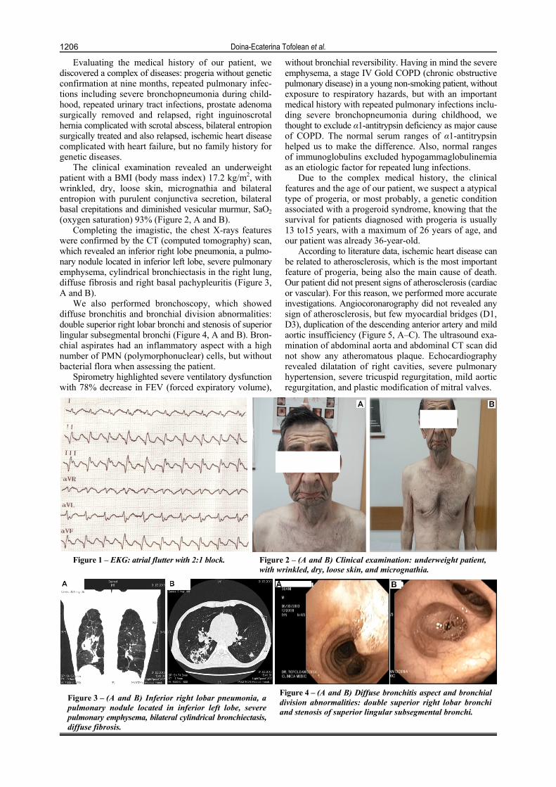
Evaluating the medical history of our patient, we discovered a complex of diseases: progeria without genetic confirmation at nine months, repeated pulmonary infec-tions including severe bronchopneumonia during child-hood, repeated urinary tract infections, prostate adenoma surgically removed and relapsed, right inguinoscrotal hernia complicated with scrotal abscess, bilateral entropion surgically treated and also relapsed, ischemic heart disease complicated with heart failure, but no family history for genetic diseases.
The clinical examination revealed an underweight patient with a BMI (body mass index) 17.2 kg/m2, with wrinkled, dry, loose skin, micrognathia and bilateral entropion with purulent conjunctiva secretion, bilateral basal crepitations and diminished vesicular murmur, SaO2 (oxygen saturation) 93% (Figure 2, A and B).
Completing the imagistic, the chest X-rays features were confirmed by the CT (computed tomography) scan, which revealed an inferior right lobe pneumonia, a pulmo-nary nodule located in inferior left lobe, severe pulmonary emphysema, cylindrical bronchiectasis in the right lung, diffuse fibrosis and right basal pachypleuritis (Figure 3, A and B).
We also performed bronchoscopy, which showed diffuse bronchitis and bronchial division abnormalities: double superior right lobar bronchi and stenosis of superior lingular subsegmental bronchi (Figure 4, A and B). Bron-chial aspirates had an inflammatory aspect with a high number of PMN (polymorphonuclear) cells, but without bacterial flora when assessing the patient.
Spirometry highlighted severe ventilatory dysfunction with 78% decrease in FEV (forced expiratory volume),
without bronchial reversibility. Having in mind the severe emphysema, a stage IV Gold COPD (chronic obstructive pulmonary disease) in a young non-smoking patient, without exposure to respiratory hazards, but with an important medical history with repeated pulmonary infections inclu-ding severe bronchopneumonia during childhood, we thought to exclude α1-antitrypsin deficiency as major cause of COPD. The normal serum ranges of α1-antitrypsin helped us to make the difference. Also, normal ranges of immunoglobulins excluded hypogammaglobulinemia as an etiologic factor for repeated lung infections.
Due to the complex medical history, the clinical features and the age of our patient, we suspect a atypical type of progeria, or most probably, a genetic condition associated with a progeroid syndrome, knowing that the survival for patients diagnosed with progeria is usually 13 to15 years, with a maximum of 26 years of age, and our patient was already 36-year-old.
According to literature data, ischemic heart disease can be related to atherosclerosis, which is the most important feature of progeria, being also the main cause of death. Our patient did not present signs of atherosclerosis (cardiac or vascular). For this reason, we performed more accurate investigations. Angiocoronarography did not revealed any sign of atherosclerosis, but few myocardial bridges (D1, D3), duplication of the descending anterior artery and mild aortic insufficiency (Figure 5, A–C). The ultrasound exa-mination of abdominal aorta and abdominal CT scan did not show any atheromatous plaque. Echocardiography revealed dilatation of right cavities, severe pulmonary hypertension, severe tricuspid regurgitation, mild aortic regurgitation, and plastic modification of mitral valves.
Figure 1 – EKG: atrial flutter with 2:1 block. Figure 2 – (A and B) Clinical examination: underweight patient, with wrinkled, dry, loose skin, and micrognathia.
Figure 3 – (A and B) Inferior right lobar pneumonia, a pulmonary nodule located in inferior left lobe, severe pulmonary emphysema, bilateral cylindrical bronchiectasis, diffuse fibrosis.
Figure 4 – (A and B) Diffuse bronchitis aspect and bronchial division abnormalities: double superior right lobar bronchi and stenosis of superior lingular subsegmental bronchi.
Clinical presentation of a patient with cutis laxa with systemic involvement: a case report
1207
Figure 5 – (A–C) Angiocoronarography: myocardial bridges (D1, D3), duplication of the descending anterior artery and mild aortic insufficiency; no signs of atherosclerosis.
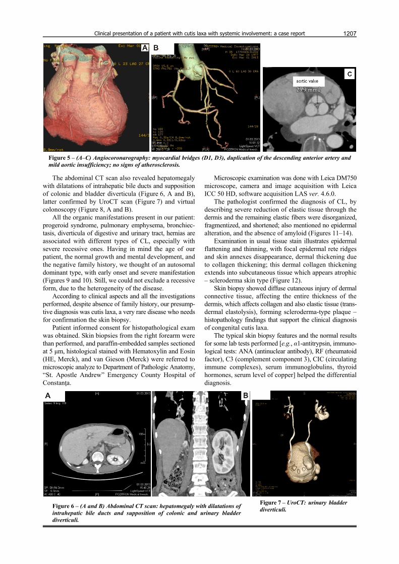
The abdominal CT scan also revealed hepatomegaly with dilatations of intrahepatic bile ducts and supposition of colonic and bladder diverticula (Figure 6, A and B), latter confirmed by UroCT scan (Figure 7) and virtual colonoscopy (Figure 8, A and B).
All the organic manifestations present in our patient: progeroid syndrome, pulmonary emphysema, bronchiec-tasis, diverticula of digestive and urinary tract, hernias are associated with different types of CL, especially with severe recessive ones. Having in mind the age of our patient, the normal growth and mental development, and the negative family history, we thought of an autosomal dominant type, with early onset and severe manifestation (Figures 9 and 10). Still, we could not exclude a recessive form, due to the heterogeneity of the disease.
According to clinical aspects and all the investigations performed, despite absence of family history, our presump-tive diagnosis was cutis laxa, a very rare disease who needs for confirmation the skin biopsy.
Patient informed consent for histopathological exam was obtained. Skin biopsies from the right forearm were than performed, and paraffin-embedded samples sectioned at 5 μm, histological stained with Hematoxylin and Eosin (HE, Merck), and van Gieson (Merck) were referred to microscopic analyze to Department of Pathologic Anatomy, “St. Apostle Andrew” Emergency County Hospital of Constanţa.
Microscopic examination was done with Leica DM750 microscope, camera and image acquisition with Leica ICC 50 HD, software acquisition LAS ver. 4.6.0.
The pathologist confirmed the diagnosis of CL, by describing severe reduction of elastic tissue through the dermis and the remaining elastic fibers were disorganized, fragmentized, and shortened; also mentioned no epidermal alteration, and the absence of amyloid (Figures 11–14).
Examination in usual tissue stain illustrates epidermal flattening and thinning, with focal epidermal rete ridges and skin annexes disappearance, dermal thickening due to collagen thickening; this dermal collagen thickening extends into subcutaneous tissue which appears atrophic – scleroderma skin type (Figure 12).
Skin biopsy showed diffuse cutaneous injury of dermal connective tissue, affecting the entire thickness of the dermis, which affects collagen and also elastic tissue (trans-dermal elastolysis), forming scleroderma-type plaque – histopathology findings that support the clinical diagnosis of congenital cutis laxa.
The typical skin biopsy features and the normal results for some lab tests performed [e.g., α1-antitrypsin, immuno-logical tests: ANA (antinuclear antibody), RF (rheumatoid factor), C3 (complement component 3), CIC (circulating immune complexes), serum immunoglobulins, thyroid hormones, serum level of copper] helped the differential diagnosis.
Figure 6 – (A and B) Abdominal CT scan: hepatomegaly with dilatations of intrahepatic bile ducts and supposition of colonic and urinary bladder diverticuli.
Figure 7 – UroCT: urinary bladder diverticuli.

Doina-Ecaterina Tofolean et al.
1208
Figure 8 – (A and B) Virtual colonoscopy: colonic diverticuli. Figure 9 – Our patient at the age of six
weeks (A), and 2.9 years (B).
Figure 10 – Our patient at the age of nine years (A), 29 years (B), and 32 years (C).
Figure 11 – Examination in usual tissue stain illustrates epidermal flattening and thinning , with focal epidermal rete ridges and skin annexes disappearance, dermal thickening due to collagen thickening; this dermal collagen thickening extends into subcutaneous tissue, which appears atrophic – scleroderma skin type. HE staining, ×40.
Figure 12 – Homogenous thickened collagen fibers; dermal collagen thickening extends into subcutaneous tissue, which appears atrophic – scleroderma skin type. Van Gieson staining, ×40.
Figure 13 – Discrete lympho-monocytic perivascular inflammatory infiltrate. Van Gieson staining, ×200.
Figure 14 – Thickened and shortened collagen fibers; elastic fibers are thin, fragmented, distorted, and low in numbers. Van Gieson staining, ×400.

Clinical presentation of a patient with cutis laxa with systemic involvement: a case report
1209
Discussion
Cutis laxa (CL), or elastolysis, is a rare, inherited or acquired connective tissue disorder in which the skin becomes inelastic and hangs loosely in folds [2, 3]. Recent molecular studies have provided many new insights into the causes of CL and revealed greater genetic heterogeneity than previously appreciated [5].
In inherited CL, an abnormal synthesis of extracellular matrix proteins occurs due to genetic defects coding for diverse extracellular matrix components. Recently, other different inborn errors of metabolism, such as P5CS, ATP6V0A2-CDG and PYCR1 defects have been found to be associated with CL [6, 7]. Autosomal dominant, autosomal recessive and X-linked recessive patterns have been noted in inherited forms. A serine to proline amino acid substitution in the fibulin 5 (FBLN5) gene has been associated with problems in normal elastogenesis, resulting in a recessive form of CL in humans [8–10]. Autosomal recessive CL is a genetically heterogeneous condition [11]. A combined disorder of N- and O-linked glycosylation has been described in children with conge-nital CL in association with severe central nervous system involvement, brain migration defects, seizures, and hearing loss. The X-linked form is currently classified in the group of copper transport diseases. The precise cause is unknown, but it may be due to abnormal elastin metabolism resulting in markedly reduced dermal elastin content. Autosomal dominant congenital CL (ADCL) is genetically hetero-geneous and shows clinical variability. Mutations in the elastin gene (ELN) have been described [12].
According to Reed et al. [13] hypothesis, trough which due to an unknown cause, the mast cells release mediators of inflammation causing a infiltration of neutrophils and eosinophils around the blood vessels in the papillary dermis and between the collagen fibers in the mid- and reticular dermis. The neutrophils release elastase and elastosis is primarily seen in areas with neutrophilic infil-tration around the blood vessels, and between collagen fibers, and then it covers the entire dermis. These features could be localized or generalized, as we found in our patient.
In inherited type, the internal organs are frequently involved. The literature data reported 209 patients at the end of 2012 [14]. Regarding acquired CL, Turner–Stokes et al. described pulmonary emphysema as a manifestation of CL in two teenage patients [15], and Reed et al. wrote about a 17-year-old boy with CL and emphysema diag-nosed after he had presented with an urticarial eruption [13]. Our patient presented multiple internal organs invol-vement (lung, heart, colon, urinary bladder), and personal history excluded acquired form of CL. Also, skin mani-festation were present from birth, which is consistent with an inherited, not an acquired form of CL.
CL with pulmonary emphysema is usually related to recessive, more severe CL, and the manifestation occurs early from few days–weeks of life with severe respiratory distress and death before age of two years. Our patient was diagnosed with pulmonary emphysema at 27-year-old without urticarial eruption, and due to the long evolution of the diseases, we excluded the severe recessive form of CL.
From skin biopsy, no specific histological abnormality was seen on routine HE staining. On Weigert–Van Gieson staining elastic fibers stains, we could see a reduction in
the number of elastic fibers throughout the dermis, with remaining fibers shortened, clumped, granular, or frag-mented.
In severe cases, no elastic fibers may be present, but only fine, dust-like particles scattered throughout the dermis can be seen. In cases preceded by an inflammatory eruption, such as urticaria or vesicles, the inflammatory infiltrate may be mononuclear (lymphocytes and histio-cytes) or mixed, containing neutrophils [16]. When vesicles are present, they are subepidermal, with papillary collect-ions of neutrophils and eosinophils mimicking dermatitis herpetiformis [17]. Electron microscopic examination reveals degenerative changes in the elastic fibers, which are variable from case to case. However, the most signi-ficant finding is the presence of electron-dense amorphous or granular aggregates that are irregularly distributed near the elastic fibers.
The skin biopsy confirmed the diagnosis of CL for our patient by describing severe reduction of number, disorganization and fragmentation of elastic fibers, and was negative for amyloid. The positive skin biopsy explains skin and lung involvement, colonic and urinary bladder diverticula, and right inguinoscrotal hernia (19-year-old), all present in our patient.
Genetic tests are not mandatory for positive diagnosis of CL, comparing with progeria in which they must be done for diagnosis (LMNA gene mutation) [18].
Differential diagnoses includes all progeroid syndromes and other diseases that affect skin aspect like acroder-matitis chronica atrophicans, anetoderma, atrophoderma of Pasini and Pierini, Costello syndrome, cutaneous T-cell lymphoma, De Barsy syndrome, Ehlers–Danlos syndrome, focal dermal hypoplasia syndrome, geroderma osteo-dysplasticum hereditaria, Lenz–Majewski hyperostotic dwarfism, lipodystrophy, pseudoxanthoma elasticum, SCARF (skeletal abnormalities, cutis laxa, craniostenosis, ambiguous genitalia, retardation, facial abnormalities) syndrome, Wrinkly skin syndrome and others [19–32].
No treatment exists to prevent disease progression, although Dapsone can be used to control swelling in persons with acquired CL (elastolysis). Penicillamine and Doxycycline are ineffective. Surgical correction of redundant skin folds, prolapses, or hernias may be under-taken. However, surgery often produces only temporary benefit. Botulinum toxin injections are being considered for improving the aged appearance and facial defects seen in persons with CL [2].
The most serious complications, which may be life threatening, are cor pulmonale resulting from severe pro-gressive pulmonary emphysema and respiratory failure.
Conclusions
CL is a rare genetic disease involving the damage of connective tissue, hard to diagnose and accompanied by important morbidity and mortality. The typical skin biopsy features of CL made possible the positive diagnosis of a patient without a family history of genetic diseases, with a complex medical history, admitted in our clinic with severe lung and heart disease and other organ involvement. The skin biopsy proved to be an important link between the various organ dysfunctions and the genetic condition causing the improper elastogenesis and connective tissue disorder.

Doina-Ecaterina Tofolean et al.
1210
Conflict of interests The authors declare that they have no conflict of
interests.
Author contribution First two authors have equal scientific contribution.
References [1] Vanakker O, Callewaert B, Malfait F, Coucke P. The genetics
of soft connective tissue disorders. Annu Rev Genomics Hum Genet, 2015, 16:229–255.
[2] Mitra S, Agarwal AA, Das JK, Gangopadhyay A. Cutis laxa: a report of two interesting cases. Indian J Dermatol, 2013, 58(4):328.
[3] Shehzad A, Amir F, Qaseem MK. Cutis laxa with systemic involvement. J Pak Assoc Dermatol, 2010, 20(4):238–242.
[4] Gverić T, Barić M, Bulat V, Situm M, Pusić J, Huljev D, Zdilar B, Gverić-Ahmetasević S, Tomas D. Clinical presentation of a patient with localized acquired cutis laxa of abdomen: a case report. Dermatol Res Pract, 2010, 2010:402093.
[5] Berk DR, Bentley DD, Bayliss SJ, Lind A, Urban Z. Cutis laxa: a review. J Am Acad Dermatol, 2012, 66(5):842.e1–842.e17.
[6] Mohamed M, Voet M, Gardeitchik T, Morava E. Cutis laxa. Adv Exp Med Biol, 2014, 802:161–184.
[7] Mohamed M, Kouwenberg D, Gardeitchik T, Kornak U, Wevers RA, Morava E. Metabolic cutis laxa syndromes. J Inherit Metab Dis, 2011, 34(4):907–916.
[8] Loeys B, Van Maldergem L, Mortier G, Coucke P, Gerniers S, Naeyaert JM, De Paepe A. Homozygosity for a missense mutation in fibulin-5 (FBLN5) results in a severe form of cutis laxa. Hum Mol Genet, 2002, 11(18):2113–2118.
[9] Nascimento GM, Nunes CS, Menegotto PF, Raskin S, Almeida Nd. Cutis laxa: case report. An Bras Dermatol, 2010, 85(5):684–686.
[10] Hucthagowder V, Sausgruber N, Kim KH, Angle B, Marmorstein LY, Urban Z. Fibulin-4: a novel gene for an autosomal recessive cutis laxa syndrome. Am J Hum Genet, 2006, 78(6):1075–1080.
[11] Morava E, Lefeber DJ, Urban Z, de Meirleir L, Meinecke P, Gillessen Kaesbach G, Sykut-Cegielska J, Adamowicz M, Salafsky I, Ranells J, Lemyre E, van Reeuwijk J, Brunner HG, Wevers RA. Defining the phenotype in an autosomal recessive cutis laxa syndrome with a combined congenital defect of glycosylation. Eur J Hum Genet, 2008, 16(1):28–35.
[12] Graul-Neumann LM, Hausser I, Essayie M, Rauch A, Kraus C. Highly variable cutis laxa resulting from a dominant splicing mutation of the elastin gene. Am J Med Genet A, 2008, 146A(8):977–983.
[13] Reed WB, Horowitz RE, Beighton P. Acquired cutis laxa. Primary generalized elastolysis. Arch Dermatol, 1971, 103(6): 661–669.
[14] ***. Prevalence and incidence of rare diseases: bibliographic data. Prevalence, incidence or number of published cases listed by diseases (in alphabetical order). Orphanet Report Series, Rare Diseases Collection, July 2015, No. 1, http:// www.orpha.net/orphacom/cahiers/docs/GB/Prevalence_of_rare_diseases_by_alphabetical_list.pdf.
[15] Turner-Stokes L, Turton C, Pope FM, Green M. Emphysema and cutis laxa. Thorax, 1983, 38(10):790–792.
[16] Turner RB, Haynes HA, Granter SR, Miller DM. Acquired cutis laxa following urticarial vasculitis associated with IgA myeloma. J Am Acad Dermatol, 2009, 60(6):1052–1057.
[17] Lewis FM, Lewis-Jones S, Gipson M. Acquired cutis laxa with dermatitis herpetiformis and sarcoidosis. J Am Acad Dermatol, 1993, 29(5 Pt 2):846–848.
[18] Kamat AK, Rocchi M, Smith DI, Miller OJ. Lamin A/C gene and a related sequence map to human chromosomes 1q12.1-q23 and 10. Somat Cell Mol Genet, 1993, 19(2):203–208.
[19] Smetanick MT, Zellis SL, Ermolovich T. Acrodermatitis chro-nica atrophicans: a case report and review of the literature. Cutis, 2010, 85(5):247–252.
[20] Patrizi A, Neri I, Virdi A, Misciali C, D’Acunto C. Familial anetoderma: a report of two families. Eur J Dermatol, 2011, 21(5):680–685.
[21] Handler MZ, Alshaiji JM, Shiman MI, Elgart GW, Schachner LA. Congenital idiopathic atrophoderma of Pasini and Pierini. Dermatol Online J, 2012, 18(4):4.
[22] Davies SJ, Hughes HE. Costello syndrome: natural history and differential diagnosis of cutis laxa. J Med Genet, 1994, 31(6):486–489.
[23] Vonderheid EC, Bernengo MG, Burg G, Duvic M, Heald P, Laroche L, Olsen E, Pittelkow M, Russell-Jones R, Takigawa M, Willemze R; ISCL. Update on erythrodermic cutaneous T-cell lymphoma: report of the International Society for Cutaneous Lymphomas. J Am Acad Dermatol, 2002, 46(1):95–106.
[24] Leao-Teles E, Quelhas D, Vilarinho L, Jaeken J. De Barsy syndrome and ATP6V0A2-CDG. Eur J Hum Genet, 2010, 18(5):526; author reply 526.
[25] Castori M. Ehlers–Danlos syndrome, hypermobility type: an underdiagnosed hereditary connective tissue disorder with mucocutaneous, articular, and systemic manifestations. ISRN Dermatol, 2012, 2012:751768.
[26] Lombardi MP, Bulk S, Celli J, Lampe A, Gabbett MT, Ousager LB, van der Smagt JJ, Soller M, Stattin EL, Mannens MA, Smigiel R, Hennekam RC. Mutation update for the PORCN gene. Hum Mutat, 2011, 32(7):723–728.
[27] Rajab A, Kornak U, Budde BS, Hoffmann K, Jaeken J, Nürnberg P, Mundlos S. Geroderma osteodysplasticum hereditaria and wrinkly skin syndrome in 22 patients from Oman. Am J Med Genet A, 2008, 146A(8):965–976.
[28] Majewski F. Lenz-Majewski hyperostotic dwarfism: reexami-nation of the original patient. Am J Med Genet, 2000, 93(4): 335–338.
[29] Herranz P, de Lucas R, Pérez-España L, Mayor M. Lipodys-trophy syndromes. Dermatol Clin, 2008, 26(4):569–578, ix.
[30] Neldner KH. Pseudoxanthoma elasticum. Int J Dermatol, 1988, 27(2):98–100.
[31] Koppe R, Kaplan P, Hunter A, MacMurray B. Ambiguous genitalia associated with skeletal abnormalities, cutis laxa, craniostenosis, psychomotor retardation, and facial abnorma-lities (SCARF syndrome). Am J Med Genet, 1989, 34(3):305–312.
[32] Al-Gazali LI, Sztriha L, Skaff F, Haas D. Gerodermia osteo-dysplastica and wrinkly skin syndrome: are they the same? Am J Med Genet, 2001, 101(3):213–220.
Corresponding author Laura Mazilu, Lecturer, MD, PhD, Department of Oncology, “St. Apostle Andrew” Clinical Emergency Hospital, “Ovidius” University, 145 Tomis Avenue, 900591 Constanţa, Romania; Phone +40241–503 482, Fax +40241–503 482, e-mail: [email protected] Received: May 28, 2014
Accepted: November 30, 2015