Clinical Practice Guidelines for the Prevention, Diagnosis ...
2010 Clinical Practice Guidelines Osteoporosis: Background and Technical Report
Page 1
Clinical Practice Guidelines for the Diagnosis and Management of Osteoporosis in Canada: Background and Technical Report
Authors: Alexandra Papaioannou MD MSc1, Suzanne Morin MD MSc2, Angela M. Cheung MD PhD3, Stephanie Atkinson PhD4, Jacques P. Brown MD5, Sidney Feldman MD6, David A. Hanley MD7, Anthony Hodsman MD8, Sophie A. Jamal MD PhD9, Robert G. Josse MD BS10, Stephanie M. Kaiser MD11, Brent Kvern MD12, Kerry Siminoski MD13, William D. Leslie MD MSc14; for the Scientific Advisory Council of Osteoporosis Canada
Correspondence to:
A. Papaioannou St. Peter's Hospital Alexander Pavilion, Juravinski Research Centre 88 Maplewood Hamilton Ontario L8M 1W9 Phone: 905-525-9140 Extension:77715 Fax 905-318-2654 [email protected] 1 Professor, Department of Medicine, McMaster University 2 Associate Professor, Department of Medicine, Division of General Internal Medicine, McGill University
3 Associate Professor, Departments of Medicine and Medical Imaging, University of Toronto 4 Professor, Department of Pediatrics and Biomedical Sciences, McMaster University 5 Clinical Professor, Department of Medicine, Laval University 6 Assistant Professor, Department of Family & Community Medicine, University of Toronto

2010 Clinical Practice Guidelines Osteoporosis: Background and Technical Report
Page 2
7 Professor, Departments of Medicine, Community Health Sciences, and Oncology, University of Calgary 8 Professor, Department of Medicine, University of Western Ontario 9 Associate Professor, Faculty of Medicine, University of Toronto
10 Professor, Department of Medicine, University of Toronto 11 Associate Professor, Division of Endocrinology and Metabolism, Dalhousie University
12 Associate Professor, Department of Family Medicine, University of Manitoba 13 Associate Professor, Departments of Radiology & Medicine, University of Alberta 14 Professor, Departments of Medicine and Radiology, University of Manitoba
2010 Clinical Practice Guidelines Osteoporosis: Background and Technical Report
Page 3
ABSTRACT
Summary: Since the publication of the 2002 Osteoporosis Canada guidelines, there has
been a paradigm shift in the prevention and treatment of osteoporosis and fractures. This
background document contains the technical reviews that were used to inform the
development of the 2010 Clinical Practice Guidelines for the Diagnosis and Management
of Osteoporosis in Canada.
Introduction: The focus is now on preventing fragility fractures and their negative
consequences rather than treating low bone mineral density (BMD), which is viewed as
only one of several risk factors for fracture. Current data suggests that many patients
with fractures are not appropriately assessed or treated.
Results: Systematic reviews of the literature were conducted to update our knowledge in
two key areas: 1) fracture risk assessment and 2) therapies for osteoporosis. Additional
topics included were identified as important for the management of osteoporosis.
Discussion: The management of osteoporosis should be guided by an assessment of the
patient’s absolute risk of osteoporosis-related fractures. Given that certain clinical factors
increase fracture risk independent of BMD, it is important to take an integrated approach
and base treatment decision on the absolute risk of fracture.
2010 Clinical Practice Guidelines Osteoporosis: Background and Technical Report
Page 4
INTRODUCTION
Since the publication of the 2002 Osteoporosis Canada guidelines1, there has been a
paradigm shift in the prevention and treatment of osteoporosis and fractures.2 This
background document contains the evidence and technical reviews that were used to
inform the development of the 2010 Clinical Practice Guidelines for the Diagnosis and
Management of Osteoporosis in Canada.3 The guidelines summary was published in the
Canadian Medical Association Journal in November of 2010 and can be viewed online at
www.cmaj.ca/cgi/content/full/182/17/1864
The World Health Organization (WHO) has defined osteoporosis as a systemic skeletal
disease characterized by low bone mass and microarchitectural deterioration of bone
tissue, with a consequent increase in bone fragility and susceptibility to fracture. Based
on epidemiological data linking low bone mass with increased fracture risk, a WHO
Study Group developed a bone mineral density (BMD) definition of osteoporosis as a
BMD T-score 2.5 or more standard deviations below peak bone mass.4 Using this BMD
definition, the Canadian Multicentre Osteoporosis Study (CaMos) estimated the
prevalence of osteoporosis in those over age 50 to be 21.3% in women and 5.5% in men.5
Since the publication of the last Osteoporosis Canada guidelines in 20021 there has been a
paradigm shift in fracture risk assessment and treatment decisions. In 2005, Osteoporosis
Canada adopted a system for ten-year absolute fracture risk assessment to be used in
BMD reporting.6 Our new guidelines focus on the clinical impact of fragility fractures;
assessment and management of women and men at high risk for fragility fracture; and
integrate a new absolute risk assessment model into an overall management approach.

2010 Clinical Practice Guidelines Osteoporosis: Background and Technical Report
Page 5
Detailed background information and methods can be found in the Appendix 1, available
at www.cmaj.ca/cgi/content/full/cmaj.100771/DC1.
DEVELOPMENT OF THE PRACTICE GUIDELINES The development of these guidelines followed the Appraisal of Guidelines, Research and
Evaluation (AGREE) framework (Appendix 1, Development of Guidelines and
Methods).7 Key stakeholders were surveyed to identify priorities for these guidelines.
Based on these priorities, systematic reviews of the literature were conducted to update
our knowledge in two key areas: 1) fracture risk assessment and 2) therapies for
osteoporosis. Additional topics included were identified by experts and primary care
clinicians as important for the management of osteoporosis (Appendix 1, Tables A1-A5).
We convened a Best Practice Guidelines Committee consisting of participants from
across Canada with methodological and content expertise. Literature searches in eight
electronic databases were performed: Medline, EMBASE, Cochrane Database of
Systematic Reviews, Database of reviews of Effectiveness (DARE), Controlled Trials
Register (CENTRAL), ACP Journal Club, Health Technology Assessment Database, and
NHS Economic Evaluation Database (Appendix 1, Table A6). We developed search
strategies based on systematic reviews by the Cochrane Musculoskeletal Group, the
PRESS (Peer Reviewed Electronic Search Strategy) checklist8 and the Cochrane
Collaboration Handbook. 9 The committee identified 35 papers for assessment of fracture
risk, published from January 1990 to December 2009. To maintain currency, we
incorporated further relevant data up to Sept. 19, 2010. We used the systematic review of
osteoporosis therapies of MacLean and colleagues,10 who included 76 randomized trials
and 24 meta-analyses, supplemented with data from 30 randomized controlled trials
2010 Clinical Practice Guidelines Osteoporosis: Background and Technical Report
Page 6
published since 2008. The PRISMA flow diagram for reporting purposes was used
(Appendix 1, Figures A2, A3). We abstracted all papers, graded them for quality of
evidence and assigned a level of evidence using established criteria (Appendix 1, Tables
A15-A23). The committee then developed and graded initial recommendations.
Recommendations were graded according to the system used to grade recommendations
for the 2002 guidelines1, which incorporates both level of evidence and expert consensus
(Appendix 1, Table A4). Recommendations were assigned a grade of D when they were
based only on committee consensus in the absence of clear supporting evidence or when
evidence was weak.
An expert panel, consisting of members of the Osteoporosis Canada Scientific Advisory
Council, members of stakeholder organizations, family physicians and experts from
across Canada, met to discuss the initial recommendations (Appendix 1, Table A5). The
group used a modified RAND/University of California, Los Angeles Delphi method for
developing consensus to ensure clinical relevance and applicability.11 The Guidelines
Committee and the Executive Committee of the Osteoporosis Canada Scientific Advisory
Council then reviewed the recommendations. The revised recommendations (presented in
this report with grades in square brackets) are based on the feedback provided and were
endorsed by the expert panel.
The target population of these guidelines is women and men 50 years and older and
consequently the systematic reviews focused on this population. Although we
acknowledge the importance of other populations with elevated risks for fracture (for
2010 Clinical Practice Guidelines Osteoporosis: Background and Technical Report
Page 7
example, individuals with chronic kidney disease), in-depth reviews of these conditions
were beyond the scope of these guidelines.
FRAGILITY FRACTURES The most serious manifestation of osteoporosis is a fragility fracture, defined as a fracture
occurring spontaneously or following minor trauma such as a fall from standing height or
less.12-14 Fragility fractures (which exclude craniofacial, hand, ankle and foot fractures)
represent 80% of all fractures occurring in postmenopausal women age 50 years and
older. 14 A fracture remains one of the most significant risk factors for predicting future
fractures.15, 16 Forty percent of women who experience a fracture have a history of prior
fracture.12 The risk of experiencing another clinical fracture in the year following a hip
fracture is 5-10%17, 18 and there is a 20% risk of having a second vertebral fracture in the
year following of a vertebral fracture.19
Falls are major risk factors for subsequent fractures, with 5-10% of falls resulting in a
fracture.20 Of those who reported a fractured hip in the 2005 Canadian Community
Health Survey, 92% occurred after a fall.21 Over 80% of falls-related admissions to
hospitals in Canadian seniors are due to fracture; 56% are of the femur, pelvis, hip or
thigh, and 24% are of the upper or lower limb.22
The Significance of Fragility Fractures The consequences of fracture include increased mortality, morbidity, institutionalization
and economic costs.23, 24 An individual with a hip fracture has a 25% risk of death in the
year following the fracture and this excess risk continues into the second year
2010 Clinical Practice Guidelines Osteoporosis: Background and Technical Report
Page 8
independent of age and co-morbidity.25 For those residing in long-term care , the
mortality one year post-hip fracture rises to 39%.17 Women with vertebral fractures are
at increased risk of death in the first year of follow-up (adjusted HR 3.7, 95% CI 1.1–
12.8) as well as the second year (adjusted HR 3.2, 95% CI 1.2–8.1).25 Post-fracture
mortality and institutionalization rates are even higher for men than women.26 The
annual cost of hip fractures alone in Canada was estimated at $650 million in 1993 and is
expected to increase to $2.4 billion by 2041.24
When compared to other chronic diseases in a population-based study of Canadians,
osteoporosis was rated as having a greater impact on quality of life than chronic
obstructive pulmonary disease (COPD), diabetes mellitus, or heart disease.27 Loss of
confidence and fear of falling have been reported with all types of fractures and less than
40% of those who experience a hip fracture return to their prior walking abilities.28, 29 In
women, clinical vertebral fractures negatively affect self-care and mobility and are
associated with chronic pain.30
FRAGILITY FRACTURES
Clinical Recommendation:
1. Individuals over age 50 who have experienced a fragility fracture should be assessed
[grade A].
2010 Clinical Practice Guidelines Osteoporosis: Background and Technical Report
Page 9
Care Gaps Despite the high rate of fracture in the Canadian population, less than 20% of individuals
receive therapies to reduce future fracture within the year following fracture.14, 31 A
number of Canadian and international studies have identified similar diagnostic and
therapeutic care gaps in postfracture care.14, 32-35 The therapeutic care gap is even wider in
men; less than 10% of Canadian men with fragility fractures receive any osteoporosis
therapy.36 Furthermore, treatment rates following a fracture are lower for those
individuals who reside in long-term care.37 This is in stark contrast to myocardial
infarction which overcame a significant care gap over the past 15 years; 75% of
individuals now receive beta blockers to help prevent recurrent myocardial
infarction.38, 39
Those who receive a BMD diagnosis of osteoporosis are more likely to be treated, as
most physicians now regard BMD as the main criterion for initiation of therapy.14, 31, 33, 36,
40 However, many individuals who experience a fracture (and even multiple fractures)
have BMD scores in the low bone mass (formerly called osteopenia) range. (T-score
between –1 and -2.5). These individuals may not be appropriately identified as being at
high risk of future fractures, and often do not receive osteoporosis therapy.41, 42 Thus,
over-reliance on BMD results is a missed opportunity to prevent future fractures. The
additive impact of non-BMD risk factors (especially prior fracture and older age) on
future fracture risk has not been widely appreciated, and underscores the value of a more
comprehensive approach to fracture risk assessment as described below.
2010 Clinical Practice Guidelines Osteoporosis: Background and Technical Report
Page 10
CLINICAL APPROACH TO OSTEOPOROSIS Osteoporosis has no clinical manifestation until a fracture occurs. A history and physical
examination should be performed with several objectives: 1) to identify factors (some of
which may be reversible) that may be contributing to bone loss, 2) to identify factors that
may be predictive of future fractures, and 3) to exclude secondary causes of
osteoporosis1, 43, 44 (Table 1).
History A history of dietary calcium intake and physical activity helps to tailor bone health
strategies. Risk factors for fracture in those over age 50 should be assessed including: a
fragility fracture after age 40; parental history of hip fracture; lifestyle factors such as
smoking, excessive alcohol, and physical inactivity; weight loss since age 25 of greater
than 10%, poor nutrition; and premature menopause.45-48 Glucocorticoid use greater than
3 months in the prior year at a prednisone equivalent dose of greater than 7.5 mg daily is
a major risk factor for fracture as early as 3-6 months after starting glucocorticoids.49
Integrating osteoporosis and falls risk assessment is critical in reducing the risk of
fracture in the older adult, at both the individual and health system level. A history of
falls in the last year is one of the most significant risk factors for predicting future falls,50
as well as the inability to rise from a chair without using the arms and walk a few steps
and return (Get up and Go test).20, 50-54 Dementia and poor physical function have also
been found to be associated with falls and fractures in older adults.47, 51-53
2010 Clinical Practice Guidelines Osteoporosis: Background and Technical Report
Page 11
Physical Examination
Height and weight should be measured, as low weight and body mass index (BMI) are
predictors of low BMD and fractures.31, 46-48, 55, 56 Vertebral fracture is the most common
manifestation of osteoporosis.19 Two thirds of vertebral fractures are seemingly
asymptomatic, but nonetheless associated with chronic back pain and decreased
activity.57 Because vertebral fractures are associated with an increased risk of future
fractures, it is important that the clinicians identify patients with unrecognized vertebral
fractures through a targeted physical examination.58 Vertebral fractures can produce
kyphosis, height loss, and reduced rib-pelvis distance.49 Historical height loss of 6 cm
(difference between the tallest recalled height and current measured height)59, 60 or
measured height loss of 2 cm (from two or more office visits within 3 years of each
other)61-63 are associated with the presence of vertebral fractures. If these height loss
criteria are met, vertebral fracture should be investigated by means of a lateral spine
radiograph (Table 1). Risk for fall and fracture can further be assessed by performing the
Get Up and Go test or by simply asking the patient to get up from a chair without using
their arms.51-53, 64 A multifactorial falls assessment including environmental and
functional assessment is recommended for those who have fallen (Appendix 1, Figure
A4).20
Radiologic Investigations Height loss should trigger further investigations including a lateral thoracic and lumbar
spine radiograph. Unfortunately, a Canadian study of emergency department radiographs
found that only 55% of vertebral fractures were mentioned in the radiology report, so it is
very important for the ordering physician to specify that the radiograph is being ordered
2010 Clinical Practice Guidelines Osteoporosis: Background and Technical Report
Page 12
to look for compression fractures.65 Osteoporotic vertebral fractures are best recognized
on radiograph as 25% or greater vertebral height loss with end-plate disruption. 66
Radiographic examinations of the spine that may be helpful for investigation of height
loss and vertebral fracture detection are presented in Appendix 1, Table A9.
Screening for Secondary Causes of Osteoporosis In primary care the prevalence of secondary osteoporosis is unknown, but is probably less
than 20% in women66, 67, and possibly as high as 50% in men.68 Many diseases that
contribute to low BMD have specific therapies and it is appropriate to assess for and treat
these conditions before making a diagnosis of osteoporosis solely on the basis of low
BMD.1, 69
Simple biochemical screening should be considered in all patients with documented
osteoporosis prior to initiating pharmacologic treatment (Table 2). Recently published
Osteoporosis Canada guidelines for vitamin D have emphasized the high prevalence of
vitamin D insufficiency in the population and the importance of recommending
supplements to ensure optimal vitamin D status. Vitamin D insufficiency should be
considered in any patient with osteoporosis, particularly when there are recurrent
fractures, bone loss despite therapy or when co-morbid conditions such as celiac disease
or gastric bypass that affect vitamin D absorption or action are present. In individuals
receiving pharmacologic therapy for osteoporosis, measurement of serum 25-OH-D
should follow 3-4 months of an adequate supplementation dose and should not be
repeated if optimal level (>75 nmoles/liter) is achieved.70
2010 Clinical Practice Guidelines Osteoporosis: Background and Technical Report
Page 13
Among patients in whom a specific secondary cause of osteoporosis is identified (such as
hyperparathyroidism, liver disease, celiac disease, multiple myeloma), blood and urine
studies should be obtained before starting therapy. Some examples of additional testing
that could be ordered based on clinical assessment are presented in Table 3. Routine
measurement of testosterone in men who do not have signs or symptoms of
hypogonadism is not recommended due to variability in the assay, lack of clarity
concerning which assay to use (bioavailable, total, free), and the fact that testosterone
levels are not consistently associated with increased fracture risk.71
CLINICAL ASSESSMENT
Summary Statements:
1. There is an important osteoporosis care gap in Canada14, 32-34, 72 (Level 1).
2. A history of a fall in the past year is predictive of future falls20, 50-54 (Level 1).
CLINICAL ASSESSMENT Clinical Recommendations: 1. Individuals over age 50 who have experienced a fragility fracture should be assessed
[grade A]. Measure height annually, and assess for the presence of vertebral fractures
[grade A].
2. Assess history of falls in the past year. If there has been such a fall, a multifactorial
risk assessment should be conducted, including the ability to get out of a chair
without using arms [grade A].
3. Perform additional biochemical testing to rule out secondary causes of osteoporosis in
selected patients, on the basis of the clinical assessment [grade D].
2010 Clinical Practice Guidelines Osteoporosis: Background and Technical Report
Page 14
4. Measure serum level of 25-hydroxyvitamin D in individuals who will receive
pharmacologic therapy for osteoporosis, those who have sustained recurrent fractures
or have bone loss despite osteoporosis treatment, and those with co-morbid conditions
that affect absorption or action of vitamin D [grade D].
5. Measure Serum 25-hydroxyvitamin D after three to four months of adequate
supplementation and do not repeat if an optimal level (75 nmol/L) is
achieved [grade B].
6. Serum 25-hydroxyvitamin D should not be measured in healthy adults at low risk of
vitamin D deficiency, i.e., without osteoporosis or conditions affecting the absorption
or action of vitamin D [grade D].
7. Perform lateral thoracic and lumbar spine radiography or vertebral fracture
assessment by dual energy x-ray absorptiometry if clinical evidence is suggestive of a
vertebral fracture [grade A].
FRACTURE RISK ASSESSMENT
Systematic Review of Risk Assessment Models
The systematic review of Risk Assessment Models identified and compared existing
models for defining fracture risk and examined the level of evidence that supports the use
of these models in Canada. The search identified 327 papers (prospective cohorts, meta-
analyses, systematic reviews, and RCTs where the control arm was analyzed for fracture
risk assessment). After removal of duplicates and screening of the abstracts, 35 papers
were retained and examined in full text for data abstraction. Further analysis resulted in
18 papers excluded for the following possible reasons: it was the wrong study design6, 73-
2010 Clinical Practice Guidelines Osteoporosis: Background and Technical Report
Page 15
75,or population76, 77; it did not describe a clinical risk assessment system78, 79, it did not
evaluate clinical risk factors80, 81 or the system did not report absolute risk or fracture
outcomes82-85, it covered the wrong risk variable (such as the use of ultrasound)86-88, or
because it was a duplicate report.89 The final review included 17 studies of absolute
fracture risk assessment systems as summarized in Appendix 1, Figure A2.
The clinical risk factors included in each of the risk assessment models are summarized
in Appendix 1, Table A17. This review focused on the following general principles for
developing and validating risk prediction models90:
● Independence - “Was the model validated in a population other than the one in
which it was initially derived?”
● Discrimination - “How well did the model perform in terms of risk
stratification?”
● Calibration - “Was the observed fracture risk consistent with the predicted
fracture risk?”
Since fracture rates vary markedly between different populations and countries91, 92, and
are also changing over time in Canada, 93 it is important to ensure that results from a risk
assessment model can be applied to the Canadian population (Appendix 1, Figure A5).
Some assessment systems, such as FRAX, must be specifically calibrated to the country
in which it is going to be used. Therefore, Appendix 1, Table A15 separates those
systems that have been directly tested in the Canadian population (candidates for clinical
adoption and therefore graded) from those that have been evaluated in other populations
(requiring additional Canadian testing before adoption and therefore not graded). There

2010 Clinical Practice Guidelines Osteoporosis: Background and Technical Report
Page 16
are important similarities and differences between risk assessment systems, and the risk
factors that are most consistently associated with fractures which may be of additional
value in clinical decision making for individuals who are categorized as moderate risk.
Studies were identified in which the absolute risk of future osteoporotic fracture was
predicted over a discrete time interval, usually five to 10 years, or as a fracture rate per
1000 person-years. Papers providing only relative or proportional risk models were not
considered. Studies were done on populations in several parts of the world, including
Canada16, 94, 95, the USA94, 96-99, Europe77, 100-103 Australia89, 104, and Japan.76 FRAX was
based upon pooling individual-level data from nine primary derivation cohorts
(N=46,340 men and women) and included 9,101 Canadian participants from CaMos.
Gradient of risk and receiver operating characteristic (ROC) area under the curve were
similar in the original derivation cohorts and in an even larger pooled analysis from
eleven validation cohorts (N=230,486).105 Most studies recruited white postmenopausal
women16, 77, 89, 94, 96, 98, 99, 104, 106-108 although other ethnic groups76, 94, 99 and men16, 77, 103-
105 were included in some reports.
While most authors have studied large cohorts of women, two studies comprised fewer
than 2,000 women.89, 104 Most models determined risk for the four major fragility
fractures typical of osteoporosis, including fractures of the vertebra (clinical and/or
radiographic), hip, forearm, and proximal humerus.16, 76, 77, 100, 103-108 Four reports were
limited to an assessment of hip fracture risk.89, 97, 99, 101
2010 Clinical Practice Guidelines Osteoporosis: Background and Technical Report
Page 17
Eighteen papers were included in the analysis, covering 14 separate models. Thirty
different variables were used in one or more of these models (Appendix 1, Table A17).
Aside from BMD, the most commonly used clinical variables were age and gender (both
used in all models), prior history of fracture (11 models), BMI or weight (seven models),
parental history of hip fracture or osteoporosis (six models), and smoking history (six
models). Four studies included the use of corticosteroids, three studies included the
ability to rise from a chair without use of the arms, and the level of physical activity.
Height or height loss, weight loss, fall history, self-reported health, and number of prior
fractures were each used in two models. A number of variables were used in only one
model, including rheumatoid arthritis, alcohol intake, walking speed, hip fracture in a
sister, use of long-acting benzodiazepines, pulse rate, caffeine intake, anticonvulsant use,
hyperthyroidism, depth perception, visual contrast sensitivity, vertebral fracture severity,
energy level , grip strength, diabetes, race/ethnic group, and family history of fracture in a
first-degree relative.
Of the 30 different variables used in one or more of the previously reviewed absolute risk
assessment models, only the following were evaluated in four or more studies: age, sex,
prior history of fracture, BMI (or weight), parental history of hip fracture or osteoporosis,
smoking history and corticosteroid use. Age and gender are not amenable to further risk
stratification. BMI (or weight) and smoking are not included in the CAROC system.
WHO meta-analyses have shown that they are relatively weak risk factors for
osteoporotic fractures after adjustment for age and BMD (risk ratio [RR] for BMI
category from 0.91 to 1.07, RR 1.13 for current smoking).109, 110 Family history of
fracture is also not included in the CAROC system. A WHO meta-analysis found that
2010 Clinical Practice Guidelines Osteoporosis: Background and Technical Report
Page 18
parental hip fracture was predictive of future osteoporotic fractures (BMD adjusted RR
1.54 [95% CI 1.25–1.88]) while any parental fracture was a weak risk factors only (RR
1.22 [95% CI 1.08–1.38]).15 A subsequent analysis from CaMos found minimal gain in
fracture prediction when parental hip fracture was added to prediction based upon age,
BMD and prior fractures (RR 2.01 [95% CI 1.81–2.25] and AUC 0.69 versus 2.06 [95%
CI 1.85–2.31] and AUC 0.70).(24) At the present time, fall history is not considered by
either the FRAX or CAROC risk assessment systems. Therefore, fracture risk will be
underestimated in those at risk for recurrent falls.
Changes in Risk Assessment
In 1994, the World Health Organization (WHO) expert panel set the operational
definition of osteoporosis in postmenopausal white women as a bone mineral density
(BMD) T-score of 2.5 or more standard deviations (SD) below the normal BMD for
young healthy white women.111 The WHO Collaborating Centre has recently provided
guidance on the diagnosis of osteoporosis in older white and non-white women and men,
designating BMD measurement made at the femoral neck with DXA as the reference
standard.112 The recommended reference range is the NHANES III reference database
for femoral neck measurements in white women aged 20-29 years using a similar cut-off
value for both men and women (BMD T-score 2.5 SD or more below the average for
young adult women). The WHO position remains controversial and other groups
advocate sex-matched reference data.106, 113, 114 A recent report from CaMos supports the
WHO position, and therefore this is now the recommendation for BMD reporting in
Canada.115

2010 Clinical Practice Guidelines Osteoporosis: Background and Technical Report
Page 19
BMD assessment with dual energy x-ray absorptiometry (DXA) is well established for
the diagnosis of osteoporosis and for fracture risk assessment in postmenopausal women
and men (see Table 4 for indications).116, 117 Currently, a diagnosis of osteoporosis is
made in older women and men who have a BMD T-score 2.5 or more SD below the
normal BMD for young healthy white women,4 with BMD measurement made at the
femoral neck from DXA as the reference standard (see frequency of clinical risk factors
included in the risk assessment models in Appendix 1, Table A17).112 It is appropriate to
consider a clinical diagnosis of osteoporosis in individuals who have sustained fragility
fracture(s) even if BMD is not in the osteoporotic range, as the majority of fragility
fractures occur in those who have a T-score above -2.5.41
Prior to age 50, the WHO T-score system is not appropriate, and age- and sex-matched Z-
scores are preferred. For Z-scores, a value of -2.0 or lower is considered below the
expected range for age and a value above -2.0 is considered within the expected range for
age (Table 5).118 Similarly, the models for fracture risk prediction discussed below
should not be applied to individuals younger than age 50. Risk assessment and
osteoporosis therapy considerations are complex in individuals less than age 50,
particularly those with medical conditions that may have adverse skeletal consequences
(Table 5), and often benefit from consultation with a specialist.
Since the 2002 Osteoporosis Canada guidelines, the importance of using multiple risk
factors to predict quantitative (absolute) fracture risk has been recognized. Bone density
T-scores are difficult for many patients to understand, and as outlined above, do not
identify the majority of patients suffering fragility fractures. Calculating an absolute
2010 Clinical Practice Guidelines Osteoporosis: Background and Technical Report
Page 20
10-year fracture risk may contribute to a more meaningful patient-physician dialogue
over the risks and benefits of treatment, and was preferred to T-scores in a survey of
physicians .119 Accordingly, in 2005, Osteoporosis Canada adopted 10-year absolute
fracture risk assessment as the preferred method for risk assessment and BMD reporting
in women and men age 50 and older.6 The original risk assessment model was developed
as a collaboration of the Canadian Association of Radiologists and Osteoporosis Canada
(referred to as the CAROC system). Since publication of the 2005 recommendations,
several other risk assessment models have been developed, most notably the WHO
fracture risk assessment tool (FRAX) as discussed below.77 A systematic review was
performed and forms the basis of guidelines regarding the most suitable risk assessment
models for use in Canada. The clinical risk factors included in each of the risk assessment
models, together with key methodological considerations and outcomes, are summarized
in Appendix 1, Tables A15, A16.
Many clinicians are unaware of the large differences in osteoporotic fracture rates
between countries (more than ten-fold)91, 92, and the fact that fracture rates are changing
over time in Canada and elsewhere.93 Although it is beyond the scope of this document
to explore the possible reasons behind these differences, it is important to ensure that
results from a risk assessment model can be applied to the Canadian population.
Therefore, our recommendations only consider those systems that have been directly
tested and validated in the Canadian population.
2010 Clinical Practice Guidelines Osteoporosis: Background and Technical Report
Page 21
Risk Assessment Systems Validated in Canada WHO Fracture Risk Assessment (FRAX) tool: The WHO Collaborating Centre has
identified clinical risk factors which, in addition to age and sex, contribute to fracture risk
independently of BMD.120 The fracture risk assessment (FRAX) tool, released in 2008,
computes 10-year probability of major osteoporotic fracture (composite of hip, vertebra
forearm and humerus) from sex, age, BMI, prior fracture, parental hip fracture, prolonged
glucocorticoid use, rheumatoid arthritis (or secondary causes of osteoporosis), current
smoking, alcohol intake (3 or more units daily) and femoral neck BMD.121 Although
FRAX also computes 10-year probability of hip fracture alone, the primary designation
of risk for clinical decision-making should be the global assessment of major osteoporotic
fracture probability. The online FRAX calculator and more details on how it is used can
be found at: www.shef.ac.uk/FRAX.
As fracture rates are known to vary by more than an order of magnitude worldwide,91
calibration for the FRAX tool is population/country specific (Appendix 1, Table A5).
Using national fracture data, a FRAX model for Canada was recently constructed for the
prediction of hip fracture risk and major osteoporotic fracture risk with and without use
of BMD.95, 122 Performance of this system was independently assessed in CaMos (4,778
women and 1,919 men) and a clinical cohort from Manitoba (36,730 women and 2,873
men).123, 124 The Canadian FRAX tool generated fracture risk predictions that were
generally consistent with observed fracture rates across a wide range of risk
categories.123-125 Fracture discrimination using FRAX with BMD was better than FRAX
without BMD or BMD alone, as has been seen in other cohorts.105
2010 Clinical Practice Guidelines Osteoporosis: Background and Technical Report
Page 22
Canadian Association of Radiologists/Osteoporosis Canada (CAROC): This risk
assessment model provides a semi-quantitative (ordinal risk category) method for
estimating 10-year absolute risk of a major osteoporotic fracture in postmenopausal
women and men over age 50.6 An individual’s 10-year absolute fracture risk (combined
risk for fractures of the proximal femur, vertebra [clinical], forearm, and proximal
humerus) is stratified into three 10-year absolute fracture risk zones designated low risk
(less than 10%), moderate risk (10-20%), and high risk (over 20%), similar to the
absolute risk categories already used for cardiovascular risk assessment126 (Figure 1).
Other fractures attributable to osteoporosis (e.g., pelvic fractures and undiagnosed
vertebral fractures) are not reflected in the CAROC or FRAX predictions, which will
therefore underestimate the total osteoporotic fracture burden. Underestimation of
fracture risk using CAROC and FRAX also occurs if the patient has suffered more than
one fragility fracture, which markedly increases the 10-year risk.
An initial (basal) risk category is obtained from age, sex, and T-score at the femoral neck.
The spine BMD is not considered in the initial risk assessment for either CAROC or
FRAX. However, when determining the risk category, a patient with a T-score of the
spine or hip ≤-2.5 should not be considered low risk (i.e. should be classified having at
least moderate risk). Certain clinical factors increase fracture risk independently of
BMD, the most important being: fragility fractures after age 40 (especially vertebral
compression fractures)66, 127and recent prolonged systemic glucocorticoid use (e.g., at
least 3 months cumulative during the preceding year at a prednisone equivalent dose
greater than 7.5 mg daily).127 The presence of either of these factors substantially
elevates fracture risk independent of the basal risk category (estimated from age, sex and
2010 Clinical Practice Guidelines Osteoporosis: Background and Technical Report
Page 23
BMD) and their effect is put into use by increasing the risk categorization to the next
level: from low risk to moderate risk, or from moderate risk to high risk. When both
factors are present (i.e., fragility fractures and prolonged systemic glucocorticoid use),
the patient is considered to be at high fracture risk regardless of the BMD result. These
clinical risk factors have been shown to enhance fracture prediction in Canadian women
independent of age and BMD alone.128 Recently, CAROC has been recalibrated using
Canadian hip fracture data with an online tool that can be downloaded (Figure 1). The
updated version of CAROC (2010 version) has been validated in two large Canadian
cohorts and replaces the previous 2005 version of CAROC95 The updated CAROC
system shows a high overall degree of concordance in risk categorization (approaching
90% agreement) with the Canadian FRAX system.95
Summary: Appropriate utilization of interventions to prevent fractures is predicated on
accurate identification of those at risk (presumed to be amenable to therapeutic
intervention) and therefore most likely to benefit from treatment.13, 129 Observed and
predicted fracture rates under the Canadian FRAX or CAROC systems are generally in
close agreement for women and men from the general population and also in those
clinically referred for BMD testing123. FRAX is based upon a more complete set of
clinical risk factors and can be used even without BMD results, but the calculations
require access to the FRAX software or website. CAROC is less complete but captures
the major risk factors for fracture, and is easy to apply using the tools provided in this
document. Therefore, the choice of using FRAX or CAROC is largely a matter of
personal preference and convenience.
2010 Clinical Practice Guidelines Osteoporosis: Background and Technical Report
Page 24
Laboratory and Radiographic Risk Factors for Fracture The preceding discussion concentrated on clinical risk factors that can be combined with
BMD to assess absolute fracture risk. The potential value of laboratory measures,
specifically bone turnover markers (BTM), and radiographic imaging of the spine
including vertebral fracture assessment (VFA), was not systematically reviewed. The
recommendations for clinical assessment can be found in Appendix 1, Table A7. These
were recent topics of Osteoporosis Canada position statements.
Bone Turnover Markers (BTM)
The potential clinical role for BTMs was the subject of a joint review between
Osteoporosis Canada, medical biochemists and clinical chemists. A number of
prospective population-based studies have reported that increased levels of BTMs are
associated with an approximately two-fold increased risk of fracture (vertebral and
nonvertebral) compared to those with normal BTM levels, both in women 65 years of age
or older130, 131 and in those younger than 65 years.132 The ability of BTMs to predict
fracture was largely independent of, and complementary to, BMD. In estimating the 10-
year absolute risk of hip fracture, the combination of an elevated resorption marker
(urinary C-terminal telopeptide) with an osteoporotic BMD or a history of previous
fracture resulted in a 70-100% higher risk than from BMD alone133 (Appendix 1, Table
A10). The value of BTMs in estimating future risk of fracture in individual patients needs
further research. As a result, BTMs have not yet been integrated in any fracture risk
assessment system.
2010 Clinical Practice Guidelines Osteoporosis: Background and Technical Report
Page 25
Vertebral Fracture Assessment (VFA)
Vertebral fracture recognition and reporting by radiologists were the subject of a recent
review by Osteoporosis Canada and the Canadian Association of Radiologists66
(Appendix 1, Table A9). VFA is an available scanning and software option on bone
densitometers which use a fan-beam scanning technology, and will identify moderate
(>25% compression) or severe (>40%) vertebral deformities. Unequivocal vertebral
fractures (>25% height loss with end-plate disruption) unrelated to trauma are associated
with a 5-fold increased risk for recurrent vertebral fractures. Therefore, a fracture
detected by VFA or radiograph (a morphometric vertebral fracture) should be considered
a prior fracture under the FRAX or CAROC system. However, mild spinal deformities
(<25% height loss without definite end-plate fracture) are not as strong predictors of
future osteoporotic fractures or low bone density.66 Canadian centres have been slow to
adopt VFA technology despite its potential clinical value in identifying patients with
previously unrecognized vertebral fractures. Like radiographic fractures, VFA-detected
fractures predict future osteoporotic and hip fractures independently of age, weight, and
BMD.66, 134
FRACTURE RISK ASSESSMENT AFTER AGE 50
Summary Statements:
1. Clinical risk factors (especially age, prior fragility fracture and prolonged
glucocorticoid exposure) enhance fracture prediction independent of BMD alone105,
108, 135, 136 [Level 1].
2. The Canadian FRAX tool and CAROC are well calibrated for prediction of major
osteoporotic fracture risk95, 123 [Level 1].
2010 Clinical Practice Guidelines Osteoporosis: Background and Technical Report
Page 26
3. The CAROC model shows a high overall degree of concordance in risk categorization
with the Canadian FRAX system136 [Level 1].
FRACTURE RISK ASSESSMENT AFTER AGE 50 Clinical Recommendations: 1. Assessment of the absolute risk of fracture should be based on established factors,
including age, bone mineral density, prior fragility fractures and glucocorticoid use
[grade A].
2. The 2010 version of the Canadian Association of Radiologists and Osteoporosis
Canada tool and the Canadian version of the WHO Fracture Risk Assessment tool
should be used in Canada, because they have been validated in the Canadian
population [grade A].
3. For purposes of reporting bone mineral density, the 2010 version of the Canadian
Association of Radiologists and Osteoporosis Canada tool is currently the preferred
national risk assessment system [grade D].
4. Only the T-score for the femoral neck (derived from the reference range for white
women of the National Health and Nutrition Education Survey III) should be used to
calculate risk of future osteoporotic fractures under either system [grade D].
5. Individuals with a T-score for the lumbar spine or total hip ≤ –2.5 should be
considered to have at least moderate risk [grade D].
6. Multiple fractures confer greater risk than a single fracture. In addition, prior fractures
of the hip and vertebra carry greater risk than fractures at other sites [grade B].
2010 Clinical Practice Guidelines Osteoporosis: Background and Technical Report
Page 27
STRATEGIES FOR FRACTURE PREVENTION
There are many non-pharmacologic interventions available to promote bone health and
pharmacologic therapies to reduce fracture risk. Available therapeutic options can reduce
the risk of future fractures in high-risk individuals by up to 40-60% but are dependent on
the site of fracture and nature of the treatment.70
Lifestyle Modifications
Several lifestyle interventions promote bone health including: appropriate dietary intake
and where necessary, supplementation of calcium and vitamin D, exercise, fall prevention
and avoidance of behaviours detrimental to bone health such as smoking and excessive
alcohol consumption. Many of these interventions apply to other chronic diseases and the
individual elements can be integrated into disease management and/or self-management
programs.137 For a summary of the studies on vitamin D and calcium reviewed for the
development of the guidelines, see Appendix 1, Table A19.
Vitamin D There is evidence that vitamin D supplementation is associated with increases in bone
mineral density138-140 and reductions in fractures141, particularly when combined with
adequate calcium intake.142 A meta-analysis that combined data from five trials
(N=9,829) that used 17.5-20 µg (700-800 IU) of vitamin D3 reported a 23% reduction in
nonvertebral fractures. A fracture risk reduction was associated with higher serum 25-
OH-D levels, particularly when these exceeded 75 nmol/L. 141 An update of this meta-
analysis found that the combined relative risk from six trials (N= 45,509) of vitamin D3
(10-20 µg [400-800 IU]) combined with calcium was 0.82 (95% CI, 0.71, 0.94),
2010 Clinical Practice Guidelines Osteoporosis: Background and Technical Report
Page 28
consistent with an 18% (95% CI, 6-29) reduction in hip fractures.143 Greater treatment
effects are noted in institutionalized elderly patients where there is supervision of
medications. 144
A recent review and guideline statement from Osteoporosis Canada70 recommends
increased vitamin D supplementation for low risk adults (under age 50 without
osteoporosis or conditions affecting vitamin D absorption) from 10 µg (400 IU) daily to
10-25 µg (400-1,000 IU) daily. In adults over age 50, and those at high risk for adverse
outcomes from vitamin D insufficiency (e.g., recurrent fractures or osteoporosis and co-
morbid conditions that affect vitamin D absorption) recommendations have been
increased from 20 µg (800 IU)/day to 20-50 µg (800-2,000 IU) daily; some of these
patients need doses higher than 50 µg (2000 IU) daily, and monitoring of the serum 25-
OH-D response is appropriate. The optimal level of serum 25OH-D for musculoskeletal
benefits is estimated to be at least 75 nmol/L.70 Supplemental vitamin D of at least 700
IU daily has also been found to reduce falls risk by 19% in both community and
institutionalized elderly.141 The risk of hip and nonvertebral fractures was also reduced
when vitamin D was given daily in combination with calcium.141, 144
Serum 25-OH-D should only be measured in situations where deficiency is suspected, or
would affect response to therapy, e.g. individuals with impaired intestinal absorption, or
in patients with osteoporosis requiring pharmacologic therapy. The half-life of 25-OH-D
in the body is 15-20 days145 and the serum 25-OH-D response to standard-dose
supplementation plateaus after 3-4 months.146 Therefore, serum 25-OH-D should be
checked no sooner than 3 months after commencing standard-dose supplementation in
2010 Clinical Practice Guidelines Osteoporosis: Background and Technical Report
Page 29
patients who have osteoporosis. Monitoring of routine supplement use, and routine
testing of otherwise healthy individuals as a screening procedure, are not necessary.70
Calcium Dietary calcium exerts a mild suppressive effect on bone turnover and this has a
beneficial impact on BMD.147, 148 In a meta-analysis it was concluded that calcium with
or without vitamin D resulted in fewer fractures.142 However, there is controversy
regarding the potential adverse effects of high-dose calcium supplementation on renal
calculi and cardiovascular events in older women147, 149, 150 and prostate cancer in older
men. Health Canada defines adequate calcium intake (from diet and supplements) as
1200 mg daily with an upper tolerable level of 2500 mg per day for adults age 50 and
older.151 The upper tolerable levels were derived from historical concerns over the
development of milk-alkali syndrome in individuals who consumed large doses of
calcium. High doses of calcium supplements are difficult to achieve as individuals
experience gastrointestinal symptoms such as constipation. These symptoms may have
contributed to compliance rates of 40% or less in the majority of randomized controlled
trials (RCTs) on calcium supplementation.147, 152
VITAMIN D AND CALCIUM
Summary Statements:
1. Vitamin D3 with calcium supplementation increases bone density in postmenopausal
women and men over age 50 138-140 and reduces the risk of fractures142 (Level 1).
2010 Clinical Practice Guidelines Osteoporosis: Background and Technical Report
Page 30
2. Vitamin D3 at daily doses of 20 µg (800 IU) with calcium (1000 mg) reduces the risk
of hip and nonvertebral fractures in elderly populations in institutions141, 142, 153 (Level
1). The evidence in community-dwelling individuals is less strong154 (Level 2).
3. There is evidence that daily 20 µg (800 IU) vitamin D3 reduces fall risk, particularly
in trials that adequately ascertained falls154 (Level 2).
4. A daily intake of 25 µg vitamin D3 (1000 IU) - a commonly available safe dose - will
raise serum 25-OH-D level on average by 15-25 nmol/L146 (Level 2).
VITAMIN D AND CALCIUM Clinical Recommendation: 1. The total daily intake of elemental calcium (through diet and supplements) for
individuals over age 50 should be 1200 mg [grade B].
2. For healthy adults at low risk of vitamin D deficiency, routine supplementation with
400–1000 IU (10–25 µg) vitamin D3 daily is recommended [grade D].
3. For adults over age 50 at moderate risk of vitamin D deficiency, supplementation
with 800–1000 IU (20–25 µg) vitamin D3 daily is recommended. To achieve optimal
vitamin D status, daily supplementation with more than 1000 IU (25 µg) may be
required. Daily doses up to 2000 IU (50 µg) are safe and do not necessitate
monitoring [grade C]. For individuals receiving pharmacologic therapy for
osteoporosis, measurement of serum 25-hydroxyvitamin D should follow three to
four months of adequate supplementation and should not be repeated if an optimal
level (75 nmol/L) is achieved [grade D].
2010 Clinical Practice Guidelines Osteoporosis: Background and Technical Report
Page 31
Exercise and Falls Prevention Exercise is often recommended for individuals with osteoporosis. Programs that are at
least one year in duration and include aerobic exercises and strength training have
demonstrated positive effects on BMD but have limited evidence for fracture reduction.
A systematic review found these programs ranged from 2 to 5 days a week with session
durations from 20 to 60 minutes, and included strength training for the extremities and
trunk, jumping, aerobic exercise (such as walking), stretching and balance.155 A meta-
analysis of cohort studies has demonstrated that moderate to vigorous exercise has
demonstrated reduced hip fractures and supports the importance of healthy lifestyle
promotion for bone health156.
Thoracic kyphosis may be reduced by a program that includes muscle strengthening,
range of motion, and postural alignment exercises.155 Quality of life associated with
exercise has been shown to improve in those with osteoporosis, with and without
fractures, particularly in the domains of physical function, pain and vitality.157 Refer to
Appendix 1, Table A13 for exercise advice to patients.
An integrated approach to osteoporosis treatment and falls interventions is also beneficial
for exercise interventions. In a systematic review, exercise-focused interventions reduced
falls for community-dwelling older people.158 Tai chi, gait and balance training were
effective in reducing falls.159-161 Home safety assessment was only effective in those with
severe visual impairment and in others at high risk for falls.159 Removal of the first
cataract has been demonstrated to reduce falls.159
2010 Clinical Practice Guidelines Osteoporosis: Background and Technical Report
Page 32
Hip protectors have been shown to be ineffective for those older adults residing in the
community.162, 163 A modest reduction in hip fractures was demonstrated in two meta-
analyses of elderly long-term care residents.162, 164 A recent Canadian analysis found hip
protectors were cost effective in reducing hip fractures in long-term care.165 Compliance
with wearing hip protectors poses a challenge and may be responsible for the
ineffectiveness of this intervention.162 A subsequent RCT found no protective effect with
a type of hip protector that is not used in clinical practice.166
OTHER NON-PHARMACOLOGIC THERAPIES
Summary Statements:
1. Exercises for individuals with osteoporosis should include weight bearing, balance
and strengthening exercises156, 167, 168 (Level 2).
2. Exercise-focused interventions improve balance and reduce falls in community-
dwelling older people159, 169 (Level 2).
3. Hip protectors may reduce the risk of hip fractures in long-term care residents,
however compliance with their use may pose a challenge for the older adult162, 164
(Level 2).
OTHER NON-PHARMACOLOGIC THERAPIES Clinical Recommendations: 1. Exercises involving resistance training appropriate for the individual’s age and
functional capacity and/or weight-bearing aerobic exercises are recommended for
those with osteoporosis or at risk for osteoporosis [grade B].
2010 Clinical Practice Guidelines Osteoporosis: Background and Technical Report
Page 33
2. Exercises to enhance core stability and thus to compensate for weakness or postural
abnormalities are recommended for individuals who have had vertebral fractures
[grade B].
3. Exercises that focus on balance, such as tai chi, or on balance and gait training should
be considered for those at risk of falls [grade A]. Use of hip protectors should be
considered for older adults residing in long-term care facilities who are at high risk
for fracture [grade B].
Pharmacologic Therapy For Fracture Prevention When deciding to initiate pharmacologic therapy, the clinician should take into
consideration the benefit to harm ratio, particularly in patients at low risk. When
choosing between therapies, the patient’s individual risk, co-morbid conditions,
preferences and lifestyle should be considered. First-line osteoporosis therapies with
evidence for fracture prevention are summarized in Appendix 1, Table A11.
A systematic review of 76 randomized trials and 24 meta-analyses graded the quality of
the evidence for various osteoporosis therapies.10 A number of therapies demonstrated
good evidence for fracture prevention in high risk groups which included individuals with
≥1 fracture at baseline, BMD in the osteoporotic range, transplant populations, and those
with neuromuscular impairment (stroke, Alzheimer’s disease). Subsequently, several
other systematic reviews have been published and are summarized in Appendix 1, Table
A19. Although the reviews differ in their inclusion criteria, a T-score above -2.0 was
generally used to define lower risk, while a T-score below -2.0 and/or prior vertebral
fractures was considered higher risk. Thirty more RCTs have been published since the
2010 Clinical Practice Guidelines Osteoporosis: Background and Technical Report
Page 34
last systematic review search date and are summarized in Appendix 1, Table A18. The
results of the RCTs are consistent with those previously reported.10
For vertebral fracture prevention, the following agents have good evidence to support
their use for individuals at high risk of fracture: alendronate, risedronate, etidronate,
zoledronic acid, denosumab, teriparatide, raloxifene and estrogen. There is fair evidence
for the use of calcitonin in vertebral fracture prevention. For hip fracture prevention, the
following therapies have good evidence: alendronate, risedronate, zoledronic acid,
denosumab and estrogen. For nonvertebral fracture prevention, there is good evidence for
alendronate, zoledronic acid, risedronate, denosumab, teriparatide, and estrogen.10 Both
calcitonin and teriparatide may decrease the pain associated with vertebral fractures.170,
171
Because vertebral and hip fractures are associated with increased risk of mortality, one
might expect that the clinical trials of osteoporosis drugs would show a reduction in
mortality. However, most subjects recruited in clinical trials are recruited on the basis of
good health except for the presence of increased fracture risk. The only clinical trial
providing evidence that fracture prevention can reduce mortality was in participants
receiving zoledronic acid within 90 days of hip fracture; mortality was analyzed as a
secondary outcome and biases may have limited the validity of the results (e.g., not all
participants were followed for the entire 36 months).172 However, a recent meta-analysis
also reported a 10% reduction in mortality in older individuals at high risk of fractures
treated with osteoporosis therapies.150 Prescribing information for osteoporosis
pharmacologic agents is summarized in Table 6.
2010 Clinical Practice Guidelines Osteoporosis: Background and Technical Report
Page 35
Antiresorptive Agents
Most pharmacologic agents used in osteoporosis prevention and therapy reduce bone
resorption or slow the overall rate of bone turnover. These include bisphosphonates,
denosumab, calcitonin, estrogen, and the selective estrogen receptor modulators.
Bisphosphonates A meta-analysis of 11 studies representing 12,068 postmenopausal women with
osteoporosis receiving at least one year of alendronate 173 showed significant reductions
in vertebral fractures (RR 0.55, 95% CI 0.43–0.69) across the range of fracture risk
whether women were at low or high risk of fractures based on bone mineral density and
the presence of clinical risk factors174 for 5 years of treatment (For number needed to
treat, NNT, see Appendix 1, Table A19). Significant reductions were also found for the
secondary prevention of nonvertebral fractures (RR 0.77 95% CI, 0.64 to 0.92), wrist
fractures (RR 0.50, 95% CI, 0.34 to 0.73) and hip fractures (RR 0.47 95% CI, 0.26 to
0.85).
Etidronate demonstrated a relative risk reduction of 41% for vertebral fractures across
eight studies (RR 0.59, 95% CI 0.36 to 0.96) and greater efficacy in secondary prevention
trials (RR 0.47, 95% CI 0.32 to 0.87), there were no significant reductions for
nonvertebral fractures (RR 0.98, 95% CI 0.68 to 1.42), hip fractures (RR 1.20, 95% CI
0.37 to 3.88) or wrist fractures (RR 0.87, 95% CI: 0.32 to 2.36).175 It was concluded that
cyclical etidronate is beneficial in the secondary prevention of vertebral fractures.
Similarly, a meta-analysis assessing the efficacy of risedronate in the prevention of
osteoporotic fracture in postmenopausal women found that 5 mg per day was associated
with a 39% relative risk reduction (RR: 0.61, 95% CI, 0.50 to 0.76), 5% ARR for
2010 Clinical Practice Guidelines Osteoporosis: Background and Technical Report
Page 36
secondary prevention of vertebral fractures versus an overall reduction of 37% (CI 0.51
to 0.77) for vertebral fractures when primary and secondary prevention trials were
combined. For nonvertebral fractures, risedronate demonstrated a 20% relative risk
reduction (RR: 0.80, 95% CI, 0.72 to 0.90), 2% ARR, and 26% relative risk reduction
(RR: 0.74, 95% CI, 0.59 to 0.94), 1% ARR for hip fractures, but no significant risk
reduction for wrist fractures.176 In 2 trials with zoledronic acid there was evidence of
vertebral ( RR 0.33, CI 0.274 to 0.4), nonvertebral (RR 0.75, CI 0.66 to 0.85) and hip
fracture ( RR 0.62, CI 0.47 to 0.83) reduction.176
Other Antiresorptives Hormone therapy (HT) was found to reduce overall fractures with a relative risk
reduction of 30%. Benefit was seen for vertebral fractures (RR 0.67, CI 0.48 to 0.93),
nonvertebral fractures (0.73, CI 0.64 to 0.81) and hip fractures (RR 0.60 CI 0.42 to
0.93).177 A number of organizations have recommended that the primary indication for
HT is moderate to severe vasomotor symptoms and should be used at the lowest effective
dose. However, low dose HT( < 0.625 conjugated estrogen) has not been demonstrated to
reduce fractures. In those individuals who have adverse effects and/or are intolerant of
other osteoporotic therapies, continuation of HT may be an option after discussion of
risks and benefits.178, 179 Raloxifene (RR 0.64, CI 0.54 to 0.78)180 and calcitonin (RR
0.65, CI 0.48 to 0.88) were found to reduce the risk of vertebral fractures, but not
nonvertebral fractures.177
Denosumab is a human monoclonal antibody to the receptor activator of nuclear factor-
kappa B ligand (RANKL) that blocks its binding to RANK, inhibiting the development
and activation of osteoclasts. In an RCT of 7868 women, denosumab given twice yearly
2010 Clinical Practice Guidelines Osteoporosis: Background and Technical Report
Page 37
reduced the risk of hip fracture by 40% compared to placebo (hazard ratio, 0.60; 95% CI,
0.37 to 0.97; ARR 0.5%).181 Denosumab also reduced the risk of nonvertebral fracture by
20% (hazard ratio, 0.80; 95% CI, 0.67 to 0.95; ARR 1.5%).181
Anabolic Agents
Osteoporosis Canada completed a systematic review of the efficacy of the human
parathyroid hormone product, teriparatide (hPTH 1-34), and found good evidence that its
use reduced the risk of vertebral fractures; there was insufficient evidence that
teriparatide prevented hip or wrist fractures.182, 183 A more recent meta-analysis177
included additional trials and concluded that both vertebral fractures (RR 0.36, CI 0.23 to
0.57) and nonvertebral fractures (RR 0.49, CI 0.27 to 0.87) were reduced by teriparatide.
Combination Therapy The combination of therapies such as HT or raloxifene with a bisphosphonate 184-189 have
demonstrated a greater improvement in BMD. However, there are no RCTs
demonstrating additional benefit in reduction of fractures. The combination of
antiresorptive agents is not recommended for fracture reduction.
Testosterone and Men There is no evidence to date that testosterone reduces fractures in men, 10
nor is there evidence that hypogonadal men respond differently than eugonadal men to
bisphosphonate therapy in the presence of osteoporosis.118 77
2010 Clinical Practice Guidelines Osteoporosis: Background and Technical Report
Page 38
In an RCT in which one-third of men were hypogonadal, defined by low serum free
testosterone, the BMD response from alendronate was similar regardless of baseline
testosterone level.190 In a meta-analysis of alendronate therapy, men with
hypogonadism responded to treatment with a lower odds ratio for incident
vertebral fractures of 0.44 (95% CI 0.23, 0.83)191 with similar response to eugonadal
men. Studies to date have not been powered to determine efficacy of testosterone in
reducing nonvertebral fractures in eugonadal or hypogonadal men.
Length of Therapy There is very little evidence to support any recommendation regarding the questions of
how long to treat, use of drug holidays, and the effectiveness of resuming treatment after
discontinuation of therapy. There have been no studies comparing the effects of various
drug holiday regimens and holiday lengths, and no studies have examined the
effectiveness of resuming therapy after a holiday. The possible benefits of a drug holiday
include reduction of potential adverse events and costs. 192, 193
In the FLEX (Fracture Intervention Trial Long-Term Extension) trial 194, after five years
of treatment with alendronate, participants either continued on alendronate for five
additional years, or were randomized to placebo for five years. At the end of the
extension phase, the 5-year clinical vertebral fracture rates were decreased by 55% in
those who continued on alendronate (for a total of 10 years) compared to those
randomized to placebo (i.e., received five years alendronate and five years placebo).
There were no differences in nonvertebral fractures or radiographic vertebral fractures.194
2010 Clinical Practice Guidelines Osteoporosis: Background and Technical Report
Page 39
In an RCT with risedronate, participants who had been on treatment for three years
(risedronate or placebo) discontinued their study medication and continued on calcium
and vitamin D for an additional year. At the end of one year off treatment, BMD
decreased in those who had been on risedronate previously, but remained higher than
baseline in placebo treated subjects. 195
Discontinuation of HT results in BMD loss of 3-6% during the first year with fracture
risk similar to those who have never been prescribed HT.196
Adverse Events Adverse events have been noted in RCTs that assess treatment efficacy for all currently
available osteoporotic drugs.10 Evidence from RCTs, systematic reviews, and case reports
on adverse events are found in Appendix 1, Tables A21-A23. Oral bisphosphonate
therapy, has been shown to be associated with upper gastrointestinal events.10 Flu-like
symptoms, reported in up to 10% of patients following zoledronic acid infusion, are most
prominent after the first dose and are self limited.174 Major adverse events associated with
raloxifene include an increased risk of pulmonary embolism, and an increased risk of
thromboembolic events.10
Adverse events from RCTs and postmarketing surveillance include reports of
osteonecrosis 197, 198 of the jaw and atypical femur fractures associated with
bisphosphonates199 (Appendix 1, Table A23). It is important to note that the adverse
events reported outside of the pivotal trials should be interpreted with caution.

2010 Clinical Practice Guidelines Osteoporosis: Background and Technical Report
Page 40
PHARMACOLOGIC THERAPIES
Summary Statements:
1. Alendronate prevents vertebral, nonvertebral, hip, and wrist fractures in
postmenopausal women173, 200 (Level 1).
2. Cyclical etidronate prevents vertebral fractures, but has not demonstrated risk-
reductions for other nonvertebral fracture types175 (Level 1).
3. Risedronate prevents vertebral, nonvertebral, and hip fractures in postmenopausal
women176 (Level 1).
4. Zoledronic acid prevents vertebral, nonvertebral, hip in men and women177 (Level 1).
5. Hormone therapy prevents vertebral, nonvertebral, and hip fractures, but is
recommended for women with moderate to severe vasomotor symptoms194 (Level 1).
6. Raloxifene and calcitonin reduce vertebral fractures, but have not demonstrated risk-
reductions for nonvertebral fractures180 (Level 1).
7. Teriparatide reduces vertebral and nonvertebral fractures182, 183 (Level 1).
8. Combination of osteoporosis therapies does not show greater fracture reduction than a
single agent184-188 (Level 1).
9. Denosumab reduces vertebral, nonvertebral fractures and hip fractures in
postmenopausal women181 (Level 1).
PHARMACOLOGIC THERAPIES
Clinical Recommendations:
1. For menopausal women requiring treatment of osteoporosis, alendronate, risedronate,
zoledronic acid and denosumab can be used as first-line therapies for prevention of
hip, nonvertebral and vertebral fractures [grade A].

2010 Clinical Practice Guidelines Osteoporosis: Background and Technical Report
Page 41
2. For menopausal women requiring treatment of osteoporosis, raloxifene can be used as
a first-line therapy for prevention of vertebral fractures [grade A].
3. For menopausal women requiring treatment of osteoporosis in combination with
treatment for vasomotor symptoms, hormone therapy can be used as first-line therapy
for prevention of hip, nonvertebral and vertebral fractures [grade A].
4. For menopausal women intolerant of first-line therapies, calcitonin or etidronate can
be considered for prevention of vertebral fractures [grade B].
5. For men requiring treatment of osteoporosis, alendronate, risedronate and zoledronic
acid can be used as first-line therapies for prevention of fractures [grade D].
6. Testosterone is not recommended for the treatment of osteoporosis in men [grade B].
7. The potential benefits and risks of the prescribed agents should be discussed before
therapy is initiated, to support informed decision-making [grade D].
Special Groups It is beyond the scope of these guidelines to address all special groups at risk of
osteoporosis. However, a number of key co-morbidities and relevant RCTs evaluating
osteoporosis therapies have demonstrated a fracture reduction.
Patients with Long-Term Glucocorticoid Use Osteoporosis therapies are often initiated in patients on long-term glucocorticoid therapy
to prevent fractures.10 Long-term use of glucocorticoids (≥ 3 months) has resulted in 30-
50% incidence of fractures, particularly in those over the age of 40 and those using high
doses.49 Both alendronate 201, 202 and risedronate203, 204 have demonstrated a reduction in
morphometric vertebral fractures compared to placebo in patients who are treated with

2010 Clinical Practice Guidelines Osteoporosis: Background and Technical Report
Page 42
glucocorticoids. There is evidence that etidronate is protective against bone loss at the
spine but fracture prevention was only seen in sub-group analysis.10, 205 A non-inferiority
study comparing zoledronic acid to risedronate demonstrated a greater improvement in
lumbar spine BMD with zoledronic acid, however the study was not powered to detect
differences in fracture reduction.206
Other therapeutic options include teriparatide and calcitonin. Teriparatide treatment
resulted in fewer new radiographic vertebral fractures compared to those receiving
alendronate (ARR 5.5%); although the incidence of nonvertebral fractures was not
significantly different between the groups.207 A meta-analysis of trials with calcitonin
compared to placebo did not find a significant effect for the prevention of vertebral or
nonvertebral fractures for individuals treated with glucocorticoids.10 There was evidence
that calcitonin prevented bone loss at the spine but not at the hip compared to placebo.201,
208
Patients with Breast or Prostate Cancer Women with breast cancer receiving aromatase inhibitor (AI) therapy may have
increased BMD loss and fractures.209-211 Zoledronic acid, denosumab212, 213, and
risedronate have been demonstrated to reduce AI-associated BMD loss.214 Up-front
zoledronic acid prevented AI-associated BMD loss with early breast cancer more
effectively than delaying therapy until BMD loss or fracture occurs.215 As well, the
addition of zoledronic acid to adjuvant endocrine therapy improves disease-free survival
in premenopausal patients with estrogen-responsive early breast cancer.216 For patients
taking adjuvant anastrozole for early breast cancer, risedronate resulted in significant
increase in lumbar spine and total hip BMD.217
2010 Clinical Practice Guidelines Osteoporosis: Background and Technical Report
Page 43
Men who receive androgen deprivation therapy (ADT) for prostate cancer are at higher
risk for fracture.218, 219 and should be assessed for pharmacologic therapy. 220 There was
insufficient fracture data in studies with bisphosphonates and SERMs; however,
denosumab showed a decreased cumulative incidence of new vertebral fractures at 36
months (ARR 2.4%).213 .
SPECIAL GROUPS
Summary Statements:
1. Osteoporosis therapies including alendronate, risedronate, and teriparatide reduce the
risk of vertebral fractures and maintain BMD in those prescribed glucocorticoids > 3
months10, 201-204 ( Level 1).
2. Etidronate, zoledronic acid and calcitonin maintain BMD in those prescribed
glucocorticoids > 3 months10, 201, 205, 206, 208 (Level 2).
3. Bisphosphonates maintain BMD in women prescribed aromatase inhibitors and men
prescribed androgen deprivation therapy209-211, 213-215 (Level 2).
SPECIAL GROUPS Clinical Recommendations: 1. For individuals over age 50 who are on long-term glucocorticoid therapy (three
months cumulative therapy during the preceding year at a prednisone-equivalent dose
> 7.5 mg daily), a bisphosphonate (alendronate, risedronate, zoledronic acid) should
be initiated at the outset and should be continued for at least the duration of the
glucocorticoid therapy [grade A].
2010 Clinical Practice Guidelines Osteoporosis: Background and Technical Report
Page 44
2. Teriparatide should be considered for those at high risk for fracture who are taking
glucocorticoids (three months cumulative therapy during the preceding year at a
prednisone-equivalent dose > 7.5 mg daily) [grade A].
3. For long-term glucocorticoid users who are intolerant of first-line therapies,
calcitonin or etidronate may be considered for preventing loss of bone mineral density
[grade B].
4. Women who are taking aromatase inhibitors and men who are undergoing androgen-
deprivation therapy should be assessed for fracture risk, and osteoporosis therapy to
prevent fractures should be considered [grade B].
Testosterone and Men There is no evidence to date that testosterone reduces fractures in men,10 nor is there
evidence that hypogonadal men respond differently than eugonadal men to
bisphosphonate therapy in the presence of osteoporosis.77, 118
In an RCT in which one-third of men were hypogonadal, defined by low serum free
testosterone, the BMD response from alendronate was similar regardless of baseline
testosterone level.190 In a meta-analysis of alendronate therapy, men with
hypogonadism responded to treatment with a lower odds ratio for incident
vertebral fractures of 0.44 (95% CI 0.23, 0.83)191 with similar response to eugonadal
men. Studies to date have not been powered to determine efficacy of testosterone in
reducing nonvertebral fractures in eugonadal or hypogonadal men.
2010 Clinical Practice Guidelines Osteoporosis: Background and Technical Report
Page 45
TESTOSTERONE IN MEN Summary Statement: 1. Testosterone maintains BMD in hypogonadal men but has not been shown to reduce
the risk of fractures10 (Level 2).
INTEGRATED MANAGEMENT
An integrated risk assessment and treatment model is desirable to ensure that there is a
consistent approach to overall management. This should involve a participatory approach
to clinical decision-making, with patient and health care provider reviewing the patient’s
risk for osteoporotic fracture and health care preferences, leading to the formulation of an
individualized care plan (Figure 2).
General Principles of Therapy To achieve the most dramatic reduction in future fracture rates and orthopaedic health
care costs, healthcare providers must first target those patients who have already
fractured because they are the ones at highest risk for more fractures (Figure 3).
The integrated model emphasizes three fracture risk categories that are in general
alignment with treatment requirements: low risk (usually not requiring pharmacologic
treatment), moderate risk (consider additional clinical risk factors to determine need for
pharmacologic treatment) and high risk (should be considered for pharmacologic
treatment). Under the FRAX or CAROC risk assessment systems, these categories are
2010 Clinical Practice Guidelines Osteoporosis: Background and Technical Report
Page 46
determined from sex, age, femoral neck BMD and a set of clinical risk factors.6, 120 For
those at moderate fracture risk, it may be helpful to consider additional clinical risk
factors that are not already considered in the risk assessment system to refine assessment
of risk within that category (Appendix 1, Table A12).
General nutrition includes optimizing total (dietary and supplements) calcium and
vitamin D intake, regular weight bearing, balance and strengthening exercises, and
smoking cessation. In older patients, falls prevention should be considered, including a
multifactorial assessment for contributing causes. In general, these measures are
sufficient for individuals at low fracture risk who do not already have low BMD or risk
factors for rapid BMD loss.
Pharmacologic Therapy There is consistent evidence from randomized clinical trials for vertebral fracture
prevention in individuals with osteoporosis as defined by a T-score <-2.5, and some (but
not all) interventions have also been shown to prevent nonvertebral and/or hip fractures
as discussed elsewhere.221 Patients with prior low trauma fractures affecting vertebrae or
hip benefit from pharmacologic intervention.222, 223 For fractures involving a site other
than vertebrae or hip (e.g., wrist fracture), there is inconsistent evidence for benefit from
pharmacologic therapy in those who do not also have osteoporotic T-scores.
Pharmacologic therapy should be offered to patients at high absolute risk (>20%
probability for major osteoporotic fracture over 10 years). Post hoc analysis from two
clinical trials found greater fracture reduction at higher FRAX
2010 Clinical Practice Guidelines Osteoporosis: Background and Technical Report
Page 47
fracture probabilities.224, 225. The US (National Osteoporosis Foundation) has identified a
10-year risk of a major osteoporotic of >20% as a cost-effective intervention point.226, 227
Additional Considerations in Decision-Making
For those with moderate fracture risk and no other risk factors, treatment should be
individualized and may include pharmacologic therapy, or basic bone health with
monitoring. Patient preference and additional clinical risk factors that are not already
incorporated in the risk assessment system will also help to guide management decisions.
Practical considerations limit the complexity and number of factors included in a risk
assessment system. Although some of these additional factors appear to add little in
terms of fracture prediction at the population level, they may still have important effects
on fracture risk for the individual. In individuals at moderate fracture risk, refining the
risk assessment based on consideration of additional features of risk factors within an
existing model (e.g., number and site of prior fractures, glucocorticoid dose) or additional
risk factors not included in that model (e.g., recurrent falls or spine T-score in the FRAX
or CAROC systems) will help to guide the clinician in treatment decisions.
History of fracture and glucocorticoid use are considered as dichotomous (yes/no) under
the FRAX or CAROC systems, but they have been shown to have dose-dependent
effects. Multiple fractures confer greater risk than a single fracture,89 104 and in particular
multiple vertebral fractures confer a stronger risk than a single vertebral fracture.66
Individuals with more than one low-trauma fracture should therefore be regarded as at
particularly high risk for future fracture. In addition, prior fractures of the hip and
2010 Clinical Practice Guidelines Osteoporosis: Background and Technical Report
Page 48
vertebra carry greater risk than other fracture sites.108, 183 Notably, in the Canadian
Multicentre Osteoporosis Study, radiographic vertebral fractures were strongly associated
with future osteoporotic fractures independently of prior clinical fractures.16 Together
these findings emphasize that more than one low-trauma fracture, or a low-trauma
fracture of hip or vertebra, justifies a recommendation for pharmacotherapy. As noted
elsewhere, vertebral fracture assessment (VFA) predicts future osteoporotic and hip
fractures independent of age, weight, and BMD.40, 41 Lateral radiographs or VFA of the
thoracolumbar spine to diagnose unrecognized vertebral compression fractures will also
assist in further stratifying risk and clinical decision making.
A WHO meta-analysis of corticosteroid use did not have sufficient details to stratify
according to dose or duration42, but other large studies have confirmed that higher dose
(prednisone or equivalent at 15 mg daily or greater) and recent exposure (within the last
3-6 months) indicate a higher risk subgroup. Corticosteroid-induced bone loss is
believed to be most rapid in the first few months of treatment, especially within the spine.
In 244,235 oral corticosteroid users and 244,235 controls (average age 57 years) from the
UK General Practice Research Database (GPRD) the adverse effect of corticosteroids
appeared to develop quickly (within 3-6 months) with a rapid decline in fracture risk
toward baseline after cessation, and increased risk was seen with prednisone doses as low
as 2.5-7.5 mg daily.43 From the same GPRD cohort of oral corticosteroid users aged 40
years and older, a simplified scoring system was developed for absolute 5-year and 10-
year fracture risk prediction.44 Osteoporotic fractures were independently predicted by
corticosteroid dose (but not duration), age, gender, fall history, fracture history, BMI,
smoking, specific medical diagnosis, indication for corticosteroid treatment, other
2010 Clinical Practice Guidelines Osteoporosis: Background and Technical Report
Page 49
medications and recent hospitalizations (information on BMD was not available).
Adverse effects of glucocorticoids on bone develop quickly (within 3-6 months), with
increased fracture risk for prednisone doses as low as 2.5-7.5 mg daily, although fracture
risk rapidly declines toward baseline after cessation.49 These findings justify intervention
in individuals recently started on therapeutic long-term or repeated systemic
glucocorticoids (oral or parenteral), even before they meet the conventional criteria for
prolonged systemic glucocorticoid use (e.g., at least 3 months cumulative during the
preceding year at a prednisone equivalent dose greater than 7.5 mg daily). This
recommendation does not apply to the use of glucocorticoid therapy for appropriate
physiologic adrenal glucocorticoid replacement.
Individualized Decision-Making
Cost-effectiveness models and guidelines typically do not consider personal preferences
and health priorities. It has been suggested that integration of individual-specific with
population-specific factors could ideally lead to ‘‘individualized intervention
thresholds’’, thus aiding clinicians to maximize benefits to patients and society.228
Monitoring The major objective of follow-up testing is to identify individuals with continued BMD
loss, despite appropriate osteoporosis treatment. Measurement error must be considered
when interpreting serial BMD assessments in order to determine whether the change is
real and not simply random fluctuation or artifact. Each centre should determine its
precision error in order to estimate the least significant change (LSC) (i.e., the change in
BMD required to have 95% confidence that the change is real).229 Continued BMD loss
2010 Clinical Practice Guidelines Osteoporosis: Background and Technical Report
Page 50
exceeding the LSC may reflect poor adherence to therapy, failure to respond to therapy or
previously unrecognized secondary causes of osteoporosis (e.g., vitamin D insufficiency).
However, most osteoporosis therapies do not cause large increases in BMD, and the
antifracture effect of treatment is only partly explained by the relatively small changes in
BMD.16 Stable BMD is consistent with successful treatment.
Once a decision to initiate osteoporosis drug therapy has been made, the expectation is
that patients will experience antifracture benefits similar to those reported in clinical
trials. Therapeutic benefit is reduced or eliminated if there is suboptimal adherence to the
regimen, including frequently missed doses, failing to take the medication correctly to
optimize absorption and action, or discontinuation of therapy.230-232 Compliance rates at
one year in the range 25-50% with oral osteoporosis agents are commonly reported, and
are only marginally better with less frequent dosing regimens.230, 233
Several approaches can be considered to ensure that patients are adherent to therapy and
to confirm treatment response. These include a combination of reminders, information,
counseling, simplifying dosing regimen, and self-monitoring. The most effective
monitoring strategy and the role of BMD in facilitating adherence are uncertain.234 In
part, this reflects conflicting data on the usefulness of BMD change as an independent
risk factor for fracture232, 235, relatively low sensitivity to identify short-term BMD
changes236, and a variable relationship between fracture risk reduction and BMD change
(i.e. an increase in BMD during antiresorptive therapy accounts for a relatively small
proportion of the observed reduction in the risk for fractures).79, 193, 237, 238
2010 Clinical Practice Guidelines Osteoporosis: Background and Technical Report
Page 51
Measurement of BMD is commonly done to monitor the response to a pharmacologic
therapy or to document the stability of bone density in untreated patients at risk for bone
loss. No randomized trials have directly assessed the value of repeat BMD testing on
persistence with medication or fracture reduction. Notwithstanding the lack of
conclusive data, many patients and clinicians find value in an objective measurement that
documents the effect of treatment.193 If used correctly, serial BMD testing can be a
helpful clinical tool.6 Depending on the clinical situation, BMD scans are usually
repeated every 1 to 3 years, with a decrease in testing once therapy is shown to be
effective. In those at low risk without additional risk factors for rapid BMD loss, a longer
testing interval (5-10 years) may be sufficient.239, 240 As noted in “Risk Assessment”,
individuals with a T-score of the spine or hip ≤-2.5 should be considered as having at
least moderate risk and a repeat BMD measurement should be obtained after 1-3 years to
monitor for rapid bone loss. If BMD is stable then less frequent monitoring can be
considered.
BMD monitoring can be used to guide initiation of osteoporosis drug therapy in those at
moderate fracture risk undergoing basic care. Some, but not all, studies show that more
rapid BMD loss in untreated individuals is an independent risk for fracture.5, 241; others
have questioned the value of repeated BMD measurements to determine the rate of loss
and suggest that it is the actual BMD level at any given time that predicts fracture risk
rather than the rate of bone loss.232, 242, 243
Bone turnover markers have the potential to provide evidence of treatment effect much
earlier than BMD (within the first 3-6 months), though further confirmation in clinical
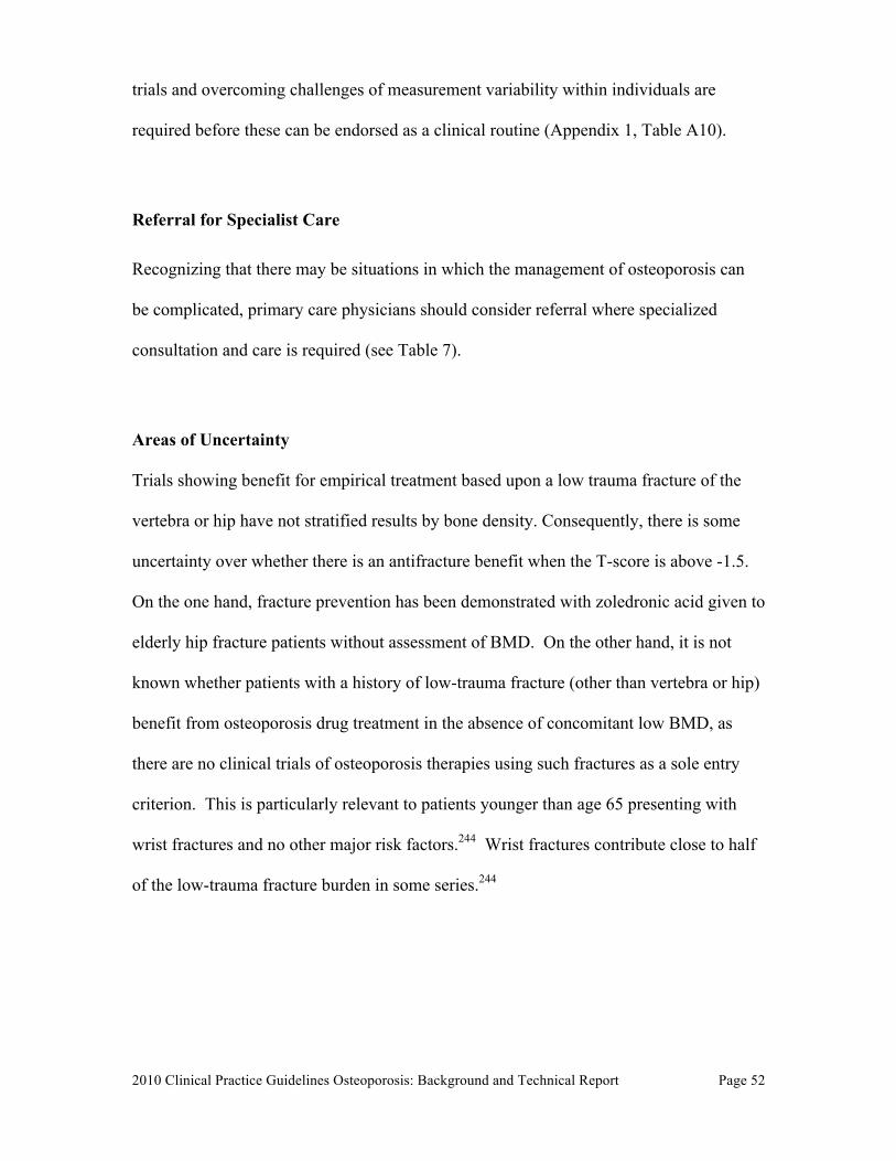
2010 Clinical Practice Guidelines Osteoporosis: Background and Technical Report
Page 52
trials and overcoming challenges of measurement variability within individuals are
required before these can be endorsed as a clinical routine (Appendix 1, Table A10).
Referral for Specialist Care
Recognizing that there may be situations in which the management of osteoporosis can
be complicated, primary care physicians should consider referral where specialized
consultation and care is required (see Table 7).
Areas of Uncertainty
Trials showing benefit for empirical treatment based upon a low trauma fracture of the
vertebra or hip have not stratified results by bone density. Consequently, there is some
uncertainty over whether there is an antifracture benefit when the T-score is above -1.5.
On the one hand, fracture prevention has been demonstrated with zoledronic acid given to
elderly hip fracture patients without assessment of BMD. On the other hand, it is not
known whether patients with a history of low-trauma fracture (other than vertebra or hip)
benefit from osteoporosis drug treatment in the absence of concomitant low BMD, as
there are no clinical trials of osteoporosis therapies using such fractures as a sole entry
criterion. This is particularly relevant to patients younger than age 65 presenting with
wrist fractures and no other major risk factors.244 Wrist fractures contribute close to half
of the low-trauma fracture burden in some series.244
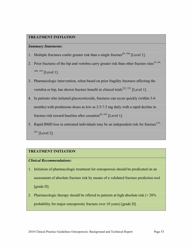
2010 Clinical Practice Guidelines Osteoporosis: Background and Technical Report
Page 53
TREATMENT INITIATION
Summary Statements:
1. Multiple fractures confer greater risk than a single fracture89, 104 [Level 1].
2. Prior fractures of the hip and vertebra carry greater risk than other fracture sites16, 66,
108, 183 [Level 1].
3. Pharmacologic intervention, when based on prior fragility fractures affecting the
vertebra or hip, has shown fracture benefit in clinical trials222, 223 [Level 1].
4. In patients who initiated glucocorticoids, fractures can occur quickly (within 3-6
months) with prednisone doses as low as 2.5-7.5 mg daily with a rapid decline in
fracture risk toward baseline after cessation49, 245 [Level 1].
5. Rapid BMD loss in untreated individuals may be an independent risk for fracture235,
241 [Level 2].
TREATMENT INITIATION Clinical Recommendations: 1. Initiation of pharmacologic treatment for osteoporosis should be predicated on an
assessment of absolute fracture risk by means of a validated fracture prediction tool
[grade D].
2. Pharmacologic therapy should be offered to patients at high absolute risk (> 20%
probability for major osteoporotic fracture over 10 years) [grade D].
2010 Clinical Practice Guidelines Osteoporosis: Background and Technical Report
Page 54
3. Individuals over age 50 who have had a fragility fracture of the hip or vertebra and
those who have had more than one fragility fracture are at high risk for future
fractures, and such individuals should be offered pharmacologic therapy [grade B].
4. For those at moderate risk of fracture, patient preference and additional risk factors
(Appendix 1, Table A12) should be used to guide pharmacologic therapy [grade C].
5. Individuals at high risk for fracture should continue osteoporosis therapy without a
drug holiday [grade D].
6. Clinicians should avoid simultaneously prescribing more than one antiresorptive
agent for fracture reduction [grade D].
KNOWLEDGE TRANSLATION Despite a number of osteoporosis updates and position papers since the 2002
Osteoporosis Canada guidelines were published, 6, 45, 46, 66, 183, 246-248 an osteoporosis care
gap remains. Translation of evidence to improve clinical care in osteoporosis needs to be
addressed, with a particular emphasis on those at high risk for fracture.
Educational Strategies Targeting Patients and Health Care Professionals A systematic review of osteoporosis disease management tools found that interventions
which targeted both the physician and patient and that were multifaceted, such as
reminders, education and risk assessment in either paper or electronic format, improved
both appropriate use of BMD and treatment.249 Reminders in conjunction with education,
targeted to physicians and patients, have demonstrated an increase in BMD testing (RR
range 1.43 to 8.67) and osteoporosis medication use (RR range 1.60 to 8.67) and in one
study reduced fractures.250 Point-of-care tools that are evidenced-based and facilitate
2010 Clinical Practice Guidelines Osteoporosis: Background and Technical Report
Page 55
diagnosis and treatment at the time of care have also been demonstrated to improve care.
These include computer-based algorithms, access to diagnostic tools, guidelines (either
electronic or printed copies). 251 However, there is limited research with point of care
tools and osteoporosis management.249 In an educational strategy targeting over 3000
primary care physicians, the Canadian quality circles44, 252 involved a multifaceted
program that included tools to assist in applying guidelines.253 The outcome of this
program was an improvement in the appropriate use of BMD and in the management of
high risk individuals.252
Chronic Disease Management Models Patients’ perceptions of future fracture risk are influenced by whether or not they believe
they have osteoporosis. Furthermore, up to 46% of individuals who had experienced a
fragility fracture did not believe that they were at an increased risk for a future fracture.
Other barriers to postfracture care include lack of integration between those providers
who deliver fracture care, such as orthopedic surgeons, and those who provide
osteoporosis and falls management care. To address these barriers, Ontario Ministry of
Health and Long-Term Care in partnership with Osteoporosis Canada has developed an
integrated care delivery model to improve postfracture care -- one of the first
comprehensive strategies in the world (logic model and tools available at
http://www.osteostrategy.on.ca).
Several Canadian RCTs have demonstrated the effectiveness of multi-faceted approaches
using case managers to co-ordinate care. In an economic analysis, compared with usual
care, the case management strategy was dominant: for every 100 patients case managed,
2010 Clinical Practice Guidelines Osteoporosis: Background and Technical Report
Page 56
six fractures (four hip fractures) were prevented, four quality-adjusted life-years were
gained, and CAD$260,000 was saved by the health care system.254
KNOWLEDGE TRANSLATION
Summary Statements:
1. Educational initiatives targeting both physicians and patients improve osteoporosis
management for those individuals who experienced a fragility fracture255, 256 (Level
2).
2. Case managers associated with high volume orthopedic clinics are cost effective in
improving appropriate management for patients who have experienced a fragility
fracture254 (Level 1).
KNOWLEDGE TRANSLATION Clinical Recommendations: 1. Following a fragility fracture, an educational initiative should be targeted at both the
patient and the primary care physician [grade B].
2. Case management is recommended as an effective approach to post-fracture care, to
improve both the diagnosis and the management of osteoporosis [grade A].
3. Point-of-care tools and other targeted strategies are recommended to support the
implementation of osteoporosis guidelines in clinical practice [grade B].
2010 Clinical Practice Guidelines Osteoporosis: Background and Technical Report
Page 57
COMPARISON WITH OTHER GUIDELINES
The use of an absolute 10-year fracture risk assessment system as a guide for treatment
intervention is a recent development in osteoporosis management. The US National
Osteoporosis Foundation (NOF) and the UK National Osteoporosis Guideline Group
(NOGG) describe two different approaches to using fracture risk estimates from FRAX to
determine treatment intervention thresholds. Both derive from cost-effectiveness
analyses.
The NOF Clinician’s Guide states that postmenopausal women or men over 50 with a T-
score of <-2.5 at the hip or spine, should be treated, regardless of prior fracture status.61, 62
Similarly, patients with a prior hip or spine fracture should be treated regardless of BMD.
In addition, based on risk calculations from the US FRAX tool, patients with low bone
mass (T-score between -1.0 and -2.5 at the femoral neck, total hip or spine) should be
treated when there is a 10-year probability of hip fracture that is ≥3% or a 10-year
probability of a major osteoporosis related fracture that is ≥20%.
NOGG suggests an age-dependent intervention threshold which varies from a 10-year
probability of a major osteoporotic fracture of 7.5% at age 50 years to 30% at the age of
80 years.63 Assessment thresholds for testing individuals with BMD are also proposed by
NOGG and would apply to 6–9% of the population at the age of 50 years, rising to 18–
36% at the age of 80 years. The overall use of the NOGG thresholds in a case-finding
strategy was projected to identify 6–20% women as eligible for BMD testing and 23–
46% as eligible for treatment, depending on age.63
2010 Clinical Practice Guidelines Osteoporosis: Background and Technical Report
Page 58
The NOF approach would treat a much larger proportion of the population than under the
NOGG guidelines, even after the recent downwards re-calibration in the US FRAX tool
risk estimates; thus NOF guidelines are estimated to treat 40.5% of white women over
age 50, rising to 67.9-90.8% after age 70.64, 65 For comparison, a women age 68 or older
with femoral neck T-score -2.4 and no other risk factors would be recommended for
treatment under the NOF guidelines but not under the NOGG guidelines.
It is unclear which approach would be better suited to the Canadian context. Cost-
effectiveness studies using Canadian cost data and the currently proposed Canadian
FRAX and CAROC tools are required. The choice of an intervention threshold of 20%
risk of major osteoporotic fractures under the CAROC system is therefore an evolving
target that may change as additional information emerges.
2010 Clinical Practice Guidelines Osteoporosis: Background and Technical Report
Page 59
Notes: Competing interests: All authors received consulting fees and travel support from Osteoporosis Canada during the preparation of this article. In addition, Alexandra Papaioannou has been an advisory board member for Amgen, Eli Lilly, Merck Frosst, Novartis and Procter & Gamble; has served as a consultant to Amgen, Aventis Pharma, Eli Lilly, Lundbeck Canada Inc., Merck Frosst, Novartis, Procter & Gamble, Servier, Warner Chillcott and Wyeth-Ayerst; has received unrestricted research grants from Amgen, Eli Lilly, Merck Frosst, Procter & Gamble and Sanofi-Aventis; has received clinical trial grants from Novartis and Pfizer; has received a research grant from the Ontario Ministry of Health and Long-Term Care; and has served as a member of the Continuing Medical Education Steering Committee of the Ontario College of Family Physicians. Suzanne Morin has been an advisory board member for Amgen, Eli Lilly, Novartis and Warner-Chilcott and has received speaker’s honoraria from Amgen, Novartis and Merck. Angela M. Cheung has been an advisory board member for Amgen and Eli Lilly; has served as a consultant for Merck; and has received speaker’s honoraria from Amgen, Eli Lilly, Merck, Novartis and Warner Chilcott. Stephanie Atkinson has served as a consultant to Pfizer and Wyeth Nutritionals and has participated in a multisite clinical trial funded by Novartis. Jacques P. Brown has been an advisory board member for Amgen, Eli Lilly, Merck, Novartis and Warner Chilcott; has served as a consultant for Amgen, Eli Lilly, Merck, Novartis and Warner Chilcott; has received grants from Abbott, Amgen, Eli Lilly, GlaxoSmithKline, Merck, Novartis, Pfizer, Roche, Sanofi-Aventis, Servier and Warner Chilcott; and has received speaker’s honoraria from Amgen, Eli Lilly, Merck, Novartis and Warner Chilcott. David A. Hanley has served as an advisory board member for Amgen Canada, Eli Lilly Canada, Novartis Canada, NPS Pharmaceuticals, Servier Canada and Warner Chilcott; has participated in clinical trials funded by Amgen, Eli Lilly, Novartis, NPS Pharmaceuticals, Pfizer, Servier and Wyeth Ayerst; and has received speaker’s honoraria from Amgen Canada, Eli Lilly Canada, Novartis Canada, NPS Pharmaceuticals and Servier Canada. Anthony Hodsman has been an advisory board member for Amgen Canada, Novartis Canada, Procter & Gamble Canada, Shire Pharmaceuticals Canada and Warner-Chilcott Canada; has served as a consultant to Cytochroma Canada; and has received speaker’s honoraria from McGill University and Novartis Canada. Stephanie M. Kaiser has served as an advisory board member for Amgen, AstaZeneca, Bristol Myers Squibb, Eli Lilly Canada, Merck Frosst/Schering, Novartis and Servier; has received speaker’s honoraria from Amgen, AstraZeneca, Eli Lilly, Merck Frosst/Schering Plough, Novartis, Procter and Gamble (now Warner Chilcott/Aventis), and Servier Canada; has received payment for development of educational presentations from Eli Lilly Canada Inc.; and has received travel funds for activities unrelated to this paper from Amgen Canada. Brent Kvern has been an advisory board member for the Alliance for Better Bone Health (sponsored by SanofiAventis and Warner) and for Amgen Canada; has served as a consultant for Servier Canada; has received honoraria from the Alliance for Better Bone Health, Amgen Canada, Eli Lilly, Merck Frosst Canada and Servier Canada; and has received payment for development of educational presentations from the Alliance for Better Bone Health, Amgen Canada, Eli Lilly, Merck Frosst Canada and Servier Canada. William D. Leslie has been an advisory board member for Amgen, Genzyme and Novartis; has received unrestricted research grants from Amgen, Genzyme, Merck Frosst, Procter & Gamble and Sanofi-Aventis; has received speaker’s fees from Amgen and Merck Frosst; and has
2010 Clinical Practice Guidelines Osteoporosis: Background and Technical Report
Page 60
received travel funds for activities unrelated to this paper from Genzyme. No additional competing interests declared for Sidney Feldman, Sophie Jamal and Kerry Siminoski. Contributors: The Clinical Practice Guidelines for the Diagnosis and Management of Osteoporosis in Canada were created with input from more than 30 experts and stakeholders in the field of osteoporosis. Dr. Papaioannou, chair of the Therapies Working Group, and Dr. Leslie, chair of the Risk Assessment Working Group, were the overall project leaders. The members of the two working groups (Alexandra Papaioannou, Suzanne Morin, Angela M. Cheung, Stephanie Atkinson, Sophie A. Jamal, Stephanie M. Kaiser and Brent Kvern for the Therapies Working Group and William D. Leslie, Angela M. Cheung, Jacques P. Brown, Sidney Feldman, David A. Hanley, Anthony Hodsman and Kerry Siminoski for the Risk Assessment Working Group) participated in the design and development of the guidelines, including analysis and interpretation of data and writing and editing of the various sections in the full guidelines document. A smaller writing group (Alexandra Papaioannou, Suzanne Morin, Angela M. Cheung and William D. Leslie) was responsible for developing the current summary version of the guidelines, which was reviewed and approved by all members of the two working groups. Funding: Alexandra Papaioannou is supported by a Canadian Institutes of Health Research (CIHR)/Eli Lilly Chair in Women’s Health, Angela M. Cheung by a CIHR/Institute of Gender and Health Senior Scientist Award and Sophie A. Jamal by a CIHR New Investigator Award. Osteoporosis Canada provided financial support for the face-to-face meetings and for the project coordinator and research assistants who worked under the direction of the guidelines co-chairs. Acknowledgements: The authors thank their support team for their dedication to the guidelines project and acknowledge their contributions: Best Practices Project Coordinator Elke Henneberg of ebmed Inc. (www.ebmed.ca); researchers Loretta Hillier MA, Christina Lacchetti MHSc, Tamara Rader MLIS and Hajera Khaja MSc; and consumer representative Marg Mac-Donell. We also acknowledge the support of Famida Jiwa DC MHSc, Farah Samji DC MHSc and Donna Spafford RN BA of Osteoporosis Canada. We thank the members of the expert panel for their contributions to the guidelines: Brian Lentle MD (moderator), Jacques Levesque MD, Sumit R. Majumdar MD, Heather Frame MD, Lynn Nash MD, Michel Fortier MD, Earl Bogoch MD, David Goltzman MD, Robert Josse MBBS, Colleen Metge BSc(Pharm) PhD, Louis-Georges Ste-Marie MD, Diane Theriault MD and Anne Marie Whelan PharmD. In addition, we gratefully acknowledge Gillian Hawker MD, Jonathan D. Adachi MD, Heather McDonald-Blumer MD and Irene Polidoulis MD for reviewing materials related to the manuscript.
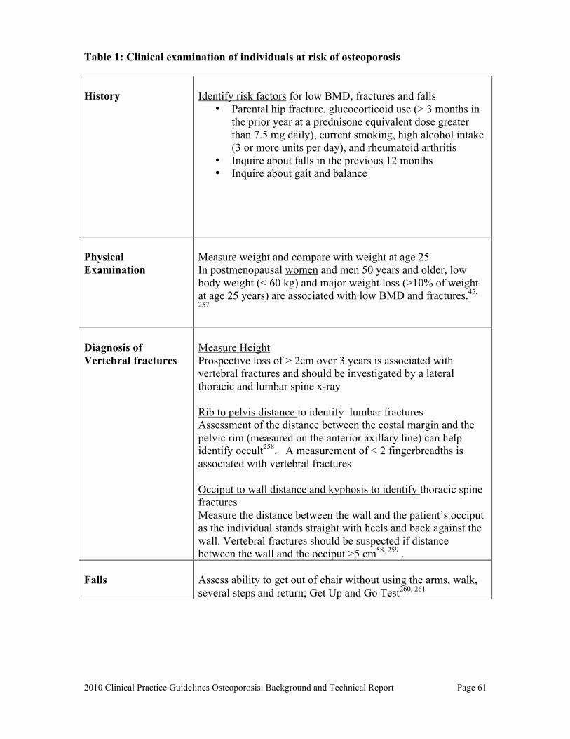
2010 Clinical Practice Guidelines Osteoporosis: Background and Technical Report
Page 61
Table 1: Clinical examination of individuals at risk of osteoporosis History
Identify risk factors for low BMD, fractures and falls
• Parental hip fracture, glucocorticoid use (> 3 months in the prior year at a prednisone equivalent dose greater than 7.5 mg daily), current smoking, high alcohol intake (3 or more units per day), and rheumatoid arthritis
• Inquire about falls in the previous 12 months • Inquire about gait and balance
Physical Examination
Measure weight and compare with weight at age 25 In postmenopausal women and men 50 years and older, low body weight (< 60 kg) and major weight loss (>10% of weight at age 25 years) are associated with low BMD and fractures.45,
257
Diagnosis of Vertebral fractures
Measure Height Prospective loss of > 2cm over 3 years is associated with vertebral fractures and should be investigated by a lateral thoracic and lumbar spine x-ray Rib to pelvis distance to identify lumbar fractures Assessment of the distance between the costal margin and the pelvic rim (measured on the anterior axillary line) can help identify occult258. A measurement of < 2 fingerbreadths is associated with vertebral fractures Occiput to wall distance and kyphosis to identify thoracic spine fractures Measure the distance between the wall and the patient’s occiput as the individual stands straight with heels and back against the wall. Vertebral fractures should be suspected if distance between the wall and the occiput >5 cm58, 259 .
Falls
Assess ability to get out of chair without using the arms, walk, several steps and return; Get Up and Go Test260, 261
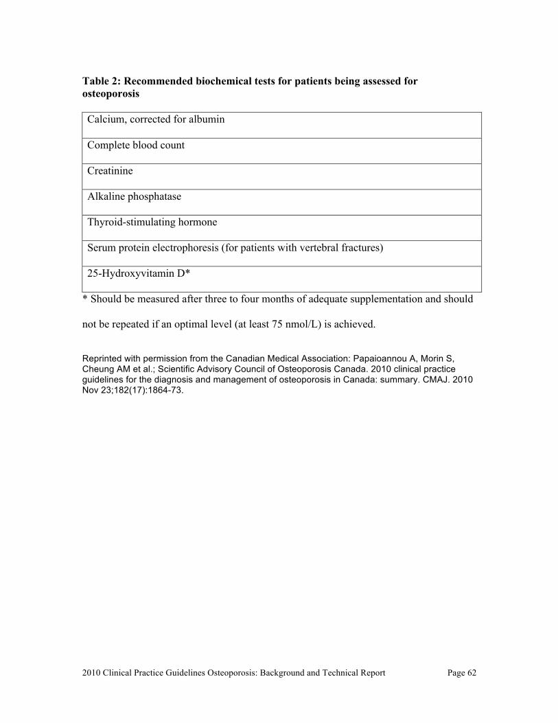
2010 Clinical Practice Guidelines Osteoporosis: Background and Technical Report
Page 62
Table 2: Recommended biochemical tests for patients being assessed for osteoporosis Calcium, corrected for albumin
Complete blood count
Creatinine
Alkaline phosphatase
Thyroid-stimulating hormone
Serum protein electrophoresis (for patients with vertebral fractures)
25-Hydroxyvitamin D*
* Should be measured after three to four months of adequate supplementation and should
not be repeated if an optimal level (at least 75 nmol/L) is achieved.
Reprinted with permission from the Canadian Medical Association: Papaioannou A, Morin S, Cheung AM et al.; Scientific Advisory Council of Osteoporosis Canada. 2010 clinical practice guidelines for the diagnosis and management of osteoporosis in Canada: summary. CMAJ. 2010 Nov 23;182(17):1864-73.
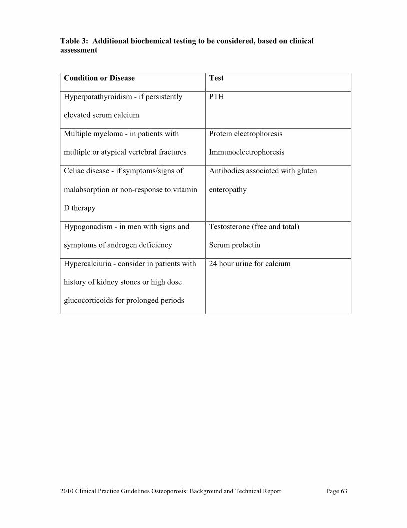
2010 Clinical Practice Guidelines Osteoporosis: Background and Technical Report
Page 63
Table 3: Additional biochemical testing to be considered, based on clinical assessment Condition or Disease Test
Hyperparathyroidism - if persistently
elevated serum calcium
PTH
Multiple myeloma - in patients with
multiple or atypical vertebral fractures
Protein electrophoresis
Immunoelectrophoresis
Celiac disease - if symptoms/signs of
malabsorption or non-response to vitamin
D therapy
Antibodies associated with gluten
enteropathy
Hypogonadism - in men with signs and
symptoms of androgen deficiency
Testosterone (free and total)
Serum prolactin
Hypercalciuria - consider in patients with
history of kidney stones or high dose
glucocorticoids for prolonged periods
24 hour urine for calcium
2010 Clinical Practice Guidelines Osteoporosis: Background and Technical Report
Page 64
Table 4: Indications for measuring bone mineral density
Other disorders strongly associated with osteoporosis include:
• Primary hyperparathyroidism • Type I diabetes • Osteogenesis imperfecta in adults • Untreated long-standing hyperthyroidism, hypogonadism or premature
menopause (<45 years) • Cushing’s disease • Chronic malnutrition or malabsorption • Chronic liver disease • Chronic inflammatory conditions (e.g., inflammatory bowel disease).
Reprinted with permission from the Canadian Medical Association: Papaioannou A, Morin S, Cheung AM et al.; Scientific Advisory Council of Osteoporosis Canada. 2010 clinical practice guidelines for the diagnosis and management of osteoporosis in Canada: summary. CMAJ. 2010 Nov 23;182(17):1864-73.
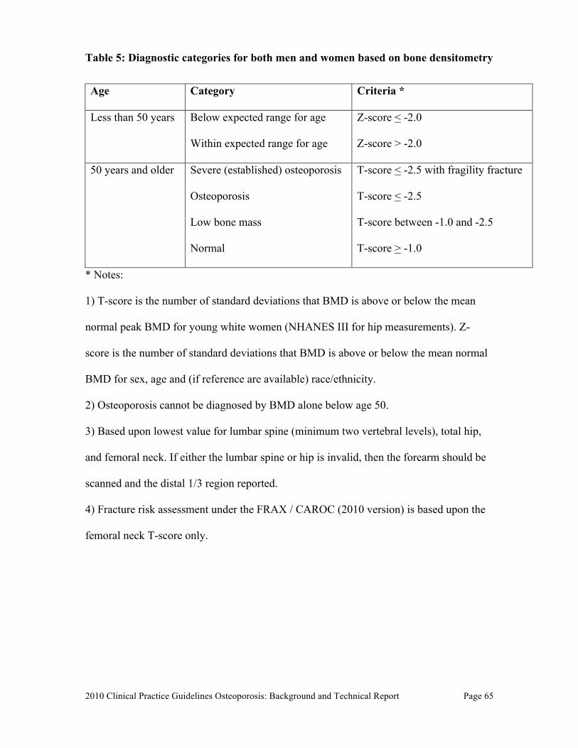
2010 Clinical Practice Guidelines Osteoporosis: Background and Technical Report
Page 65
Table 5: Diagnostic categories for both men and women based on bone densitometry
Age Category Criteria *
Less than 50 years Below expected range for age
Within expected range for age
Z-score < -2.0
Z-score > -2.0
50 years and older Severe (established) osteoporosis
Osteoporosis
Low bone mass
Normal
T-score < -2.5 with fragility fracture
T-score < -2.5
T-score between -1.0 and -2.5
T-score > -1.0
* Notes:
1) T-score is the number of standard deviations that BMD is above or below the mean
normal peak BMD for young white women (NHANES III for hip measurements). Z-
score is the number of standard deviations that BMD is above or below the mean normal
BMD for sex, age and (if reference are available) race/ethnicity.
2) Osteoporosis cannot be diagnosed by BMD alone below age 50.
3) Based upon lowest value for lumbar spine (minimum two vertebral levels), total hip,
and femoral neck. If either the lumbar spine or hip is invalid, then the forearm should be
scanned and the distal 1/3 region reported.
4) Fracture risk assessment under the FRAX / CAROC (2010 version) is based upon the
femoral neck T-score only.
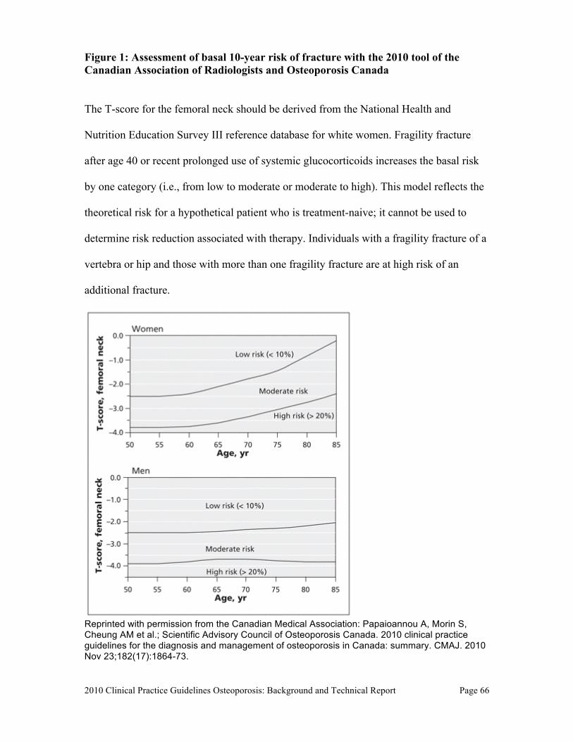
2010 Clinical Practice Guidelines Osteoporosis: Background and Technical Report
Page 66
Figure 1: Assessment of basal 10-year risk of fracture with the 2010 tool of the Canadian Association of Radiologists and Osteoporosis Canada
The T-score for the femoral neck should be derived from the National Health and
Nutrition Education Survey III reference database for white women. Fragility fracture
after age 40 or recent prolonged use of systemic glucocorticoids increases the basal risk
by one category (i.e., from low to moderate or moderate to high). This model reflects the
theoretical risk for a hypothetical patient who is treatment-naive; it cannot be used to
determine risk reduction associated with therapy. Individuals with a fragility fracture of a
vertebra or hip and those with more than one fragility fracture are at high risk of an
additional fracture.
Reprinted with permission from the Canadian Medical Association: Papaioannou A, Morin S, Cheung AM et al.; Scientific Advisory Council of Osteoporosis Canada. 2010 clinical practice guidelines for the diagnosis and management of osteoporosis in Canada: summary. CMAJ. 2010 Nov 23;182(17):1864-73.
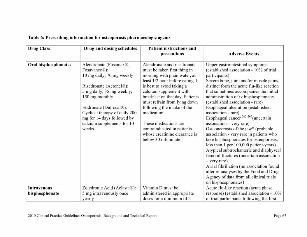
2010 Clinical Practice Guidelines Osteoporosis: Background and Technical Report
Page 67
Table 6: Prescribing information for osteoporosis pharmacologic agents Drug Class
Drug and dosing schedules
Patient instructions and precautions
Adverse Events
Oral bisphosphonates Alendronate (Fosamax®,
Fosavance®): 10 mg daily, 70 mg weekly Risedronate (Actonel®): 5 mg daily, 35 mg weekly, 150 mg monthly Etidronate (Didrocal®): Cyclical therapy of daily 200 mg for 14 days followed by calcium supplements for 10 weeks
Alendronate and risedronate must be taken first thing in morning with plain water, at least 1/2 hour before eating. It is best to avoid taking a calcium supplement with breakfast on that day. Patients must refrain from lying down following the intake of the medication. These medications are contraindicated in patients whose creatinine clearance is below 30 ml/minute
Upper gastrointestinal symptoms (established association - 10% of trial participants) Severe bone, joint and/or muscle pains, distinct form the acute flu-like reaction that sometimes accompanies the initial administration of iv bisphosphonates (established association - rare) Esophageal ulceration (established association - rare) Esophageal cancer 262-265(uncertain association – very rare) Osteonecrosis of the jaw* (probable association - very rare in patients who take bisphosphonates for osteoporosis, less than 1 per 100,000 patient-years) Atypical subtrochanteric and diaphyseal femoral fractures (uncertain association – very rare) Atrial fibrillation (no association found after re-analyses by the Food and Drug Agency of data from all clinical trials on bisphosphonates)
Intravenous bisphosphonate
Zoledronic Acid (Aclasta®): 5 mg intravenously once yearly
Vitamin D must be administered in appropriate doses for a minimum of 2
Acute flu-like reaction (acute phase response) (established association - 10% of trial participants following the first
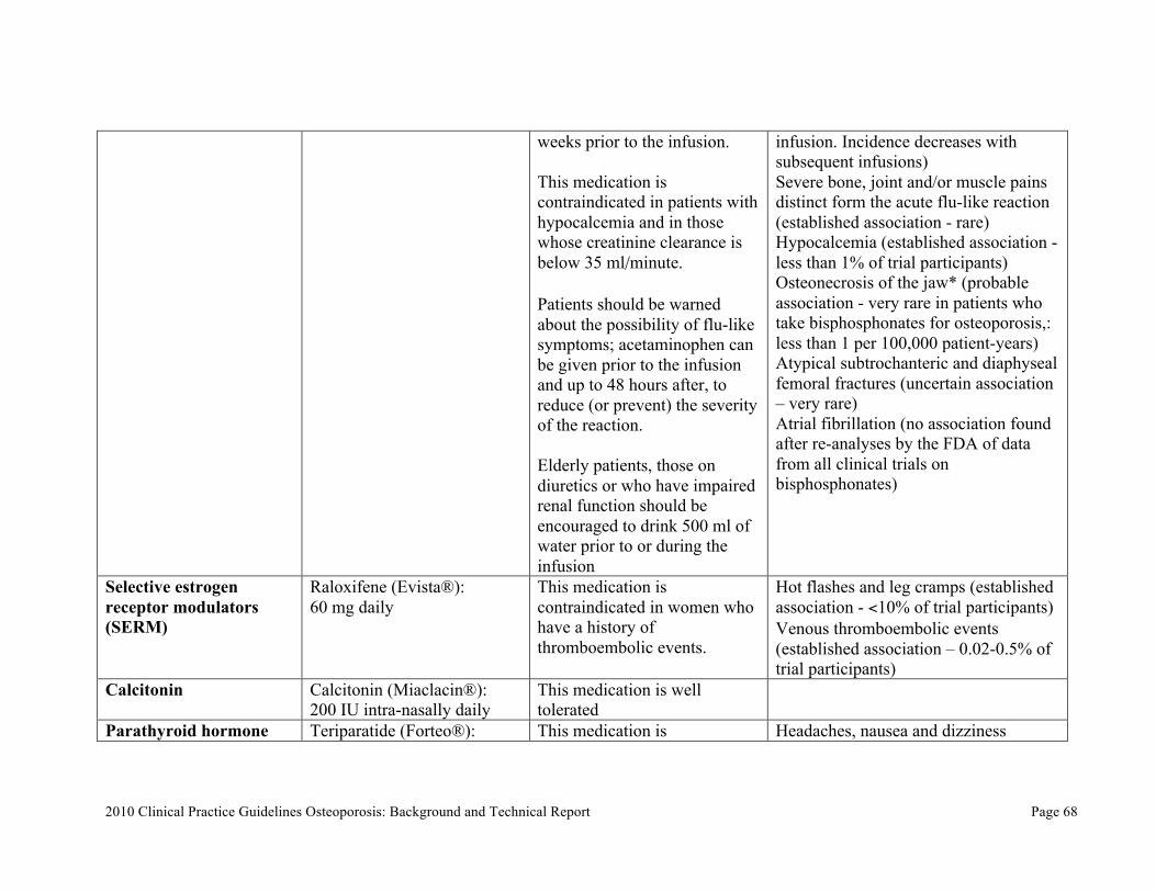
2010 Clinical Practice Guidelines Osteoporosis: Background and Technical Report
Page 68
weeks prior to the infusion. This medication is contraindicated in patients with hypocalcemia and in those whose creatinine clearance is below 35 ml/minute. Patients should be warned about the possibility of flu-like symptoms; acetaminophen can be given prior to the infusion and up to 48 hours after, to reduce (or prevent) the severity of the reaction. Elderly patients, those on diuretics or who have impaired renal function should be encouraged to drink 500 ml of water prior to or during the infusion
infusion. Incidence decreases with subsequent infusions) Severe bone, joint and/or muscle pains distinct form the acute flu-like reaction (established association - rare) Hypocalcemia (established association - less than 1% of trial participants) Osteonecrosis of the jaw* (probable association - very rare in patients who take bisphosphonates for osteoporosis,: less than 1 per 100,000 patient-years) Atypical subtrochanteric and diaphyseal femoral fractures (uncertain association – very rare) Atrial fibrillation (no association found after re-analyses by the FDA of data from all clinical trials on bisphosphonates)
Selective estrogen receptor modulators (SERM)
Raloxifene (Evista®): 60 mg daily
This medication is contraindicated in women who have a history of thromboembolic events.
Hot flashes and leg cramps (established association - <10% of trial participants) Venous thromboembolic events (established association – 0.02-0.5% of trial participants)
Calcitonin Calcitonin (Miaclacin®): 200 IU intra-nasally daily
This medication is well tolerated
Parathyroid hormone Teriparatide (Forteo®): This medication is Headaches, nausea and dizziness
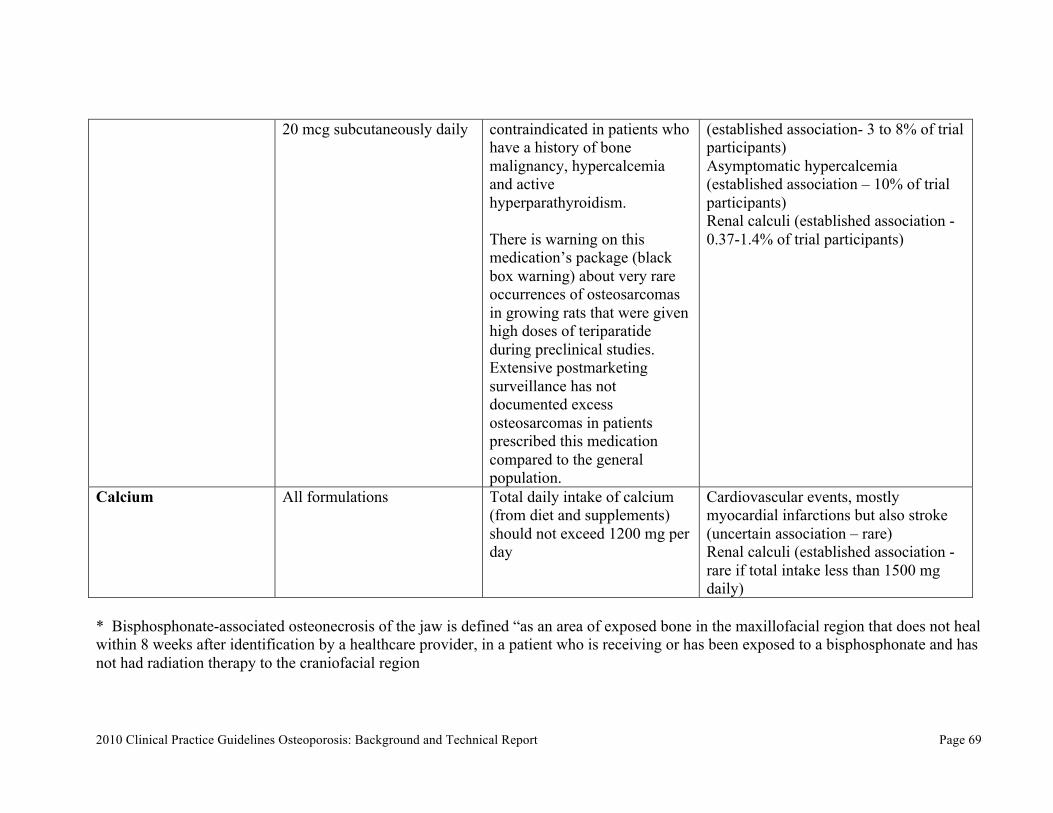
2010 Clinical Practice Guidelines Osteoporosis: Background and Technical Report
Page 69
20 mcg subcutaneously daily contraindicated in patients who have a history of bone malignancy, hypercalcemia and active hyperparathyroidism. There is warning on this medication’s package (black box warning) about very rare occurrences of osteosarcomas in growing rats that were given high doses of teriparatide during preclinical studies. Extensive postmarketing surveillance has not documented excess osteosarcomas in patients prescribed this medication compared to the general population.
(established association- 3 to 8% of trial participants) Asymptomatic hypercalcemia (established association – 10% of trial participants) Renal calculi (established association - 0.37-1.4% of trial participants)
Calcium All formulations Total daily intake of calcium (from diet and supplements) should not exceed 1200 mg per day
Cardiovascular events, mostly myocardial infarctions but also stroke (uncertain association – rare) Renal calculi (established association - rare if total intake less than 1500 mg daily)
* Bisphosphonate-associated osteonecrosis of the jaw is defined “as an area of exposed bone in the maxillofacial region that does not heal within 8 weeks after identification by a healthcare provider, in a patient who is receiving or has been exposed to a bisphosphonate and has not had radiation therapy to the craniofacial region
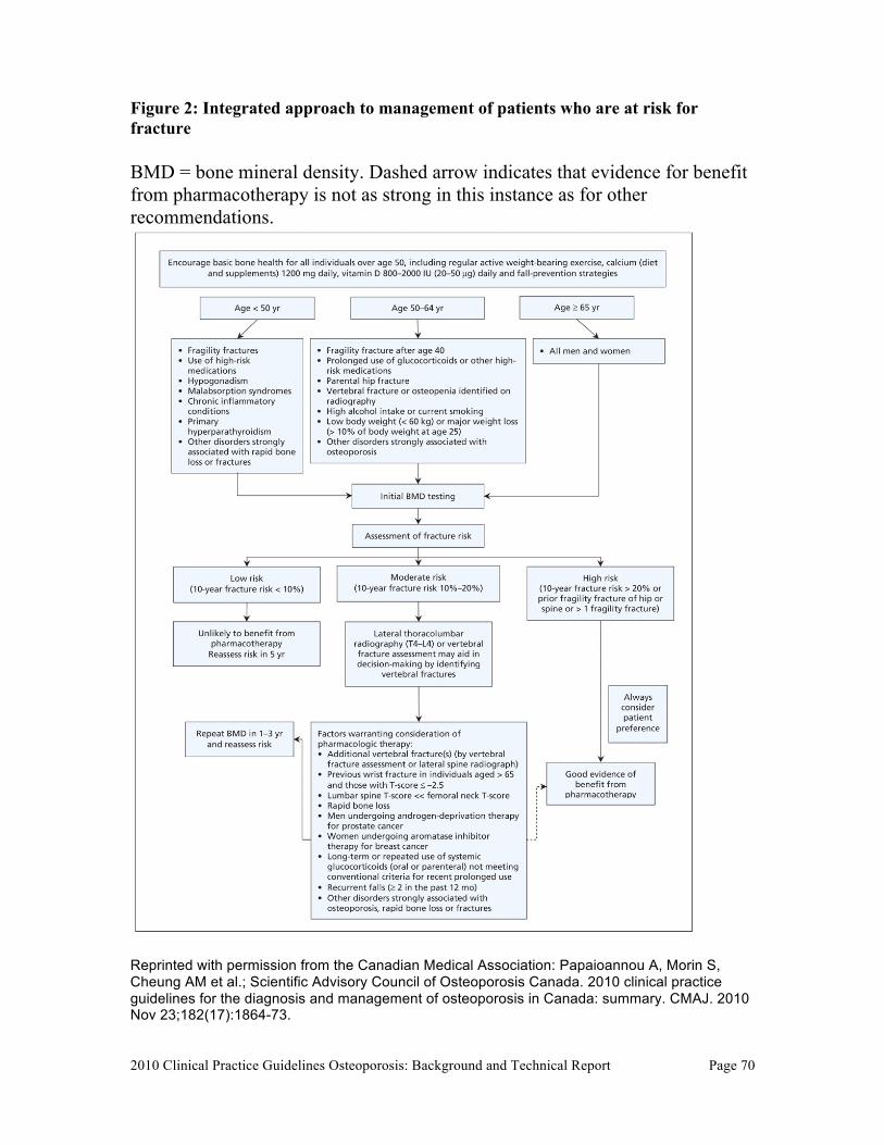
2010 Clinical Practice Guidelines Osteoporosis: Background and Technical Report
Page 70
Figure 2: Integrated approach to management of patients who are at risk for fracture BMD = bone mineral density. Dashed arrow indicates that evidence for benefit from pharmacotherapy is not as strong in this instance as for other recommendations.
Reprinted with permission from the Canadian Medical Association: Papaioannou A, Morin S, Cheung AM et al.; Scientific Advisory Council of Osteoporosis Canada. 2010 clinical practice guidelines for the diagnosis and management of osteoporosis in Canada: summary. CMAJ. 2010 Nov 23;182(17):1864-73.
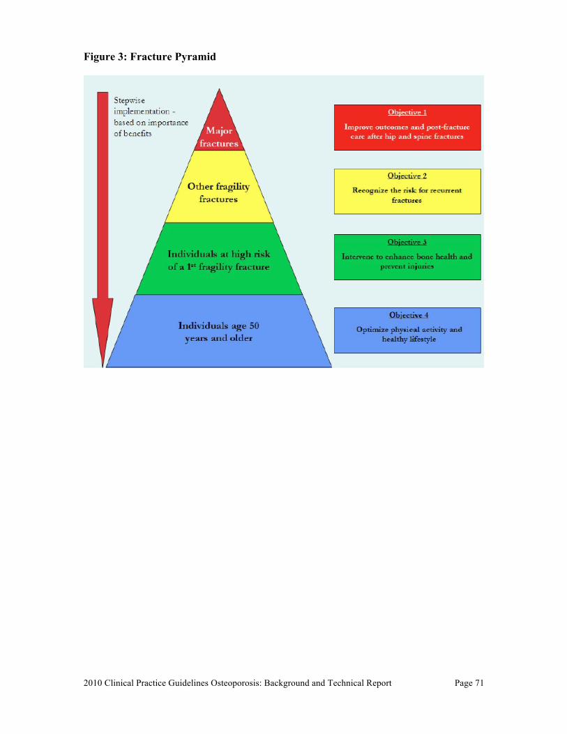
2010 Clinical Practice Guidelines Osteoporosis: Background and Technical Report
Page 71
Figure 3: Fracture Pyramid
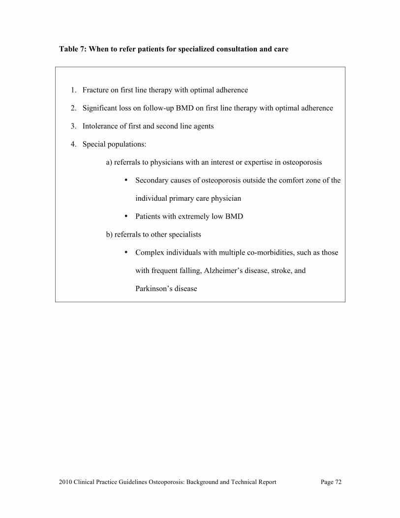
2010 Clinical Practice Guidelines Osteoporosis: Background and Technical Report
Page 72
Table 7: When to refer patients for specialized consultation and care
1. Fracture on first line therapy with optimal adherence
2. Significant loss on follow-up BMD on first line therapy with optimal adherence
3. Intolerance of first and second line agents
4. Special populations:
a) referrals to physicians with an interest or expertise in osteoporosis
• Secondary causes of osteoporosis outside the comfort zone of the
individual primary care physician
• Patients with extremely low BMD
b) referrals to other specialists
• Complex individuals with multiple co-morbidities, such as those
with frequent falling, Alzheimer’s disease, stroke, and
Parkinson’s disease
2010 Clinical Practice Guidelines Osteoporosis: Background and Technical Report
Page 73
REFERENCES
1. Brown JP, Josse RG. 2002 clinical practice guidelines for the diagnosis and management of osteoporosis in Canada. CMAJ 2002;167:S1-34. 2. Cheung AM, Detsky AS. Osteoporosis and fractures: missing the bridge? JAMA 2008;299:1468-70. 3. Papaioannou A, Morin S, Cheung AM, et al. 2010 clinical practice guidelines for the diagnosis and management of osteoporosis in Canada: summary. CMAJ 2010;182:1864-73. 4. Assessment of fracture risk and its application to screening for postmenopausal osteoporosis. Report of a WHO Study Group. World Health Organ Tech Rep Ser 1994;843:1-129. 5. Berger C, Goltzman D, Langsetmo L, et al. Peak bone mass from longitudinal data: implications for the prevalence, pathophysiology, and diagnosis of osteoporosis. J Bone Miner Res 2010;25:1948-57. 6. Siminoski K, Leslie WD, Frame H, et al. Recommendations for bone mineral density reporting in Canada. Can Assoc Radiol J 2005;56:178-88. 7. Development and validation of an international appraisal instrument for assessing the quality of clinical practice guidelines: the AGREE project. Qual Saf Health Care 2003;12:18-23. 8. Sampson M, McGowan J, Cogo E, Grimshaw J, Moher D, Lefebvre C. An evidence-based practice guideline for the peer review of electronic search strategies. J Clin Epidemiol 2009;62:944-52. 9. JPT JH, Green S, Collaboration TC. Cochrane Handbook for Systematic Reviews of Interventions 2011;Version 5.1.0 [updated March 2011]. 10. MacLean C, Newberry S, Maglione M, et al. Systematic review: comparative effectiveness of treatments to prevent fractures in men and women with low bone density or osteoporosis. Ann Intern Med 2008;148:197-213. 11. Fitch K, Bernstein S, Aguilar M, al. e. The RAND/UCLA appropriateness method user’s manual. Santa Monica: The RAND Corporation; 2001. 12. Tenenhouse A, Joseph L, Kreiger N, et al. Estimation of the prevalence of low bone density in Canadian women and men using a population-specific DXA reference standard: the Canadian Multicentre Osteoporosis Study (CaMos). Osteoporos Int 2000;11:897-904. 13. Kanis JA, Oden A, Johnell O, Jonsson B, de Laet C, Dawson A. The burden of osteoporotic fractures: a method for setting intervention thresholds. Osteoporos Int 2001;12:417-27. 14. Bessette L, Ste-Marie LG, Jean S, et al. The care gap in diagnosis and treatment of women with a fragility fracture. Osteoporos Int 2008;19:79-86.
2010 Clinical Practice Guidelines Osteoporosis: Background and Technical Report
Page 74
15. Kanis JA, Johansson H, Oden A, et al. A family history of fracture and fracture risk: a meta-analysis. Bone 2004;35:1029-37. 16. Chen P, Krege JH, Adachi JD, et al. Vertebral fracture status and the World Health Organization risk factors for predicting osteoporotic fracture risk. J Bone Miner Res 2009;24:495-502. 17. Papaioannou A, Wiktorowicz M, Adachi J, al. e. Mortality, independence in living, and re-fracture, one year following hip fracture in Canadians. Journal of the Society of Obstetrics and Gynaecology Canada 2000;22:591-7. 18. Colon-Emeric C, Kuchibhatla M, Pieper C, et al. The contribution of hip fracture to risk of subsequent fractures: data from two longitudinal studies. Osteoporos Int 2003;14:879-83. 19. Lindsay R, Silverman SL, Cooper C, et al. Risk of new vertebral fracture in the year following a fracture. JAMA 2001;285:320-3. 20. Tinetti ME. Clinical practice. Preventing falls in elderly persons. N Engl J Med 2003;348:42-9. 21. V Scott, M Pearce, Pengelly. C. Technical report: Injury resulting from falls among Canadians age 65 and over on the analysis of data from the Canadian Community Health Survey Cycle 2.1. 2005; 2009. 22. Report on Senior's Falls in Canada: Public Health Agency of Canada; 2005. 23. Papaioannou A, Adachi JD, Parkinson W. Lengthy hospitalization associated with veretebral fractures despite control for comorbid conditions. Osteoporos Int 2001;12:870-4. 24. Wiktorowicz ME, Goeree R, Papaioannou A, Adachi JD, Papadimitropoulos E. Economic implications of hip fracture: health service use, institutional care and cost in Canada. Osteoporos Int 2001;12:271-8. 25. Ioannidis G, Papaioannou A, Hopman W. Relation between fractures and mortality: results from the Canadian Multicentre Osteoporosis study. CMAJ 2009;181:265-71. 26. Tosteson AN, Gottlieb DJ, Radley DC, Fisher ES, Melton LJ, 3rd. Excess mortality following hip fracture: the role of underlying health status. Osteoporos Int 2007;18:1463-72. 27. Sawka AM, Thabane L, Papaioannou A, et al. Health-related quality of life measurements in elderly Canadians with osteoporosis compared to other chronic medical conditions: a population-based study from the Canadian Multicentre Osteoporosis Study (CaMos). Osteoporos Int 2005;16:1836-40. 28. Cranney AB, Coyle D, Hopman WM, Hum V, Power B, Tugwell PS. Prospective evaluation of preferences and quality of life in women with hip fractures. J Rheumatol 2005;32:2393-9. 29. Pasco JA, Sanders KM, Hoekstra FM, Henry MJ, Nicholson GC, Kotowicz MA. The human cost of fracture. Osteoporos Int 2005;16:2046-52. 30. Papaioannou A, Kennedy CC, Ioannidis G, et al. The impact of incident fractures on health-related quality of life: 5 years of data from the Canadian Multicentre Osteoporosis Study. Osteoporos Int 2009;20:703-14. 31. Papaioannou A, Giangregorio L, Kvern B, Boulos P, Ioannidis G, Adachi JD. The osteoporosis care gap in Canada. BMC Musculoskelet Disord 2004;5:11.
2010 Clinical Practice Guidelines Osteoporosis: Background and Technical Report
Page 75
32. Giangregorio L, Papaioannou A, Cranney A, Zytaruk N, Adachi JD. Fragility fractures and the osteoporosis care gap: an international phenomenon. Semin Arthritis Rheum 2006;35:293-305. 33. Elliot-Gibson V, Bogoch ER, Jamal SA, Beaton DE. Practice patterns in the diagnosis and treatment of osteoporosis after a fragility fracture: a systematic review. Osteoporos Int 2004;15:767-78. 34. Haaland DA, Cohen DR, Kennedy CC, Khalidi NA, Adachi JD, Papaioannou A. Closing the osteoporosis care gap: increased osteoporosis awareness among geriatrics and rehabilitation teams. BMC Geriatr 2009;9:28. 35. Bessette L, Ste-Marie LG, Jean S, et al. Recognizing osteoporosis and its consequences in Quebec (ROCQ): background, rationale, and methods of an anti-fracture patient health-management programme. Contemp Clin Trials 2008;29:194-210. 36. Papaioannou A, Kennedy CC, Ioannidis G, et al. The osteoporosis care gap in men with fragility fractures: the Canadian Multicentre Osteoporosis Study. Osteoporos Int 2008;19:581-7. 37. Giangregorio L, Jantzi M, Papaioannou A. Osteoporosis management among residents living in long-term care. Osteoporos Int 2009. 38. Austin PC, Tu JV, Ko DT, Alter DA. Factors associated with the use of evidence-based therapies after discharge among elderly patients with myocardial infarction. CMAJ 2008;179:901-8. 39. The state of healthcare quality. Industry trends and analysis. 2008. (Accessed March, 2009, 2009, at www ncqa org/Portals/0/Newsroom/SOHC/SOHC_8 pdf ) 40. Cranney A, Tsang JF, Leslie WD. Factors predicting osteoporosis treatment initiation in a regionally based cohort. Osteoporos Int 2009;20:1621-5. 41. Cranney A, Jamal SA, Tsang JF, Josse RG, Leslie WD. Low bone mineral density and fracture burden in postmenopausal women. CMAJ 2007;177:575-80. 42. Langsetmo L, Goltzman D, Kovacs CS, et al. Repeat low-trauma fractures occur frequently among men and women who have osteopenic BMD. J Bone Miner Res 2009;24:1515-22. 43. Hanley DA, Brown JP, Tenenhouse A, et al. Associations among disease conditions, bone mineral density, and prevalent vertebral deformities in men and women 50 years of age and older: cross-sectional results from the Canadian Multicentre Osteoporosis Study. J Bone Miner Res 2003;18:784-90. 44. Ioannidis G, Papaioannou A, Thabane L, et al. Family physicians' personal and practice characteristics that are associated with improved utilization of bone mineral density testing and osteoporosis medication prescribing. Popul Health Manag 2009;12:131-8. 45. Papaioannou A, Kennedy CC, Cranney A, et al. Risk factors for low BMD in healthy men age 50 years or older: a systematic review. Osteoporos Int 2009;20:507-18. 46. Waugh EJ, Lam MA, Hawker GA, et al. Risk factors for low bone mass in healthy 40-60 year old women: a systematic review of the literature. Osteoporos Int 2009;20:1-21. 47. Cummings SRN. Risk factors for hip fracture in white women. New England Journal of Medicine 1995;332:23.
2010 Clinical Practice Guidelines Osteoporosis: Background and Technical Report
Page 76
48. Papaioannou A, Joseph L, Ioannidis G, et al. Risk factors associated with incident clinical vertebral and nonvertebral fractures in postmenopausal women: the Canadian Multicentre Osteoporosis Study (CaMos). Osteoporos Int 2005;16:568-78. 49. van Staa T, Leufkens HG, Abenhain L. Use of oral corticosteroids and risk of fractures. J Bone Miner Res 2000;15:993-1000. 50. Ganz DA, Bao Y, Shekelle PG, Rubenstein LZ. Will my patient fall? JAMA 2007;297:77-86. 51. Guideline for the prevention of falls in older persons. American Geriatrics Society, British Geriatrics Society, and American Academy of Orthopaedic Surgeons Panel on Falls Prevention. J Am Geriatr Soc 2001;49:664-72. 52. Bensen R, Adachi JD, Papaioannou A, et al. Evaluation of easily measured risk factors in the prediction of osteoporotic fractures. BMC Musculoskelet Disord 2005;6:47. 53. Cawthon PM, Fullman RL, Marshall L, et al. Physical performance and risk of hip fractures in older men. J Bone Miner Res 2008;23:1037-44. 54. Gates S, Fisher JD, Cooke MW, Carter YH, Lamb SE. Multifactorial assessment and targeted intervention for preventing falls and injuries among older people in community and emergency care settings: systematic review and meta-analysis. BMJ 2008;336:130-3. 55. Kanis J, Johnell O, Gullberg B, et al. Risk factors for hip fracture in men from southern Europe: the MEDOS study. Mediterranean Osteoporosis Study. Osteoporos Int 1999;9:45-54. 56. Morin S, Tsang JF, Leslie WD. Weight and body mass index predict bone mineral density and fractures in women aged 40 to 59 years. Osteoporos Int 2009;20:363-70. 57. Nevitt MC, Ettinger B, Black DM, et al. The association of radiographically detected vertebral fractures with back pain and function: a prospective study. Ann Intern Med 1998;128:793-800. 58. Green AD, Colón-Emeric CS, Bastian L, Drake MT, Lyles KW. Does This Woman Have Osteoporosis? JAMA: The Journal of the American Medical Association 2004;292:2890-900. 59. Siminoski K, Warshawski RS, Jen H, Lee K. The accuracy of historical height loss for the detection of vertebral fractures in postmenopausal women. Osteoporosis International 2006;17:290-6. 60. Briot K, Legrand E, Pouchain D, Monnier S, Roux C. Accuracy of patient-reported height loss and risk factors for height loss among postmenopausal women. CMAJ 2010;182:558-62. 61. Moayyeri A, Luben RN, Bingham SA, et al. Measured height loss predicts fractures in middle-aged and older men and women: the EPIC-Norfolk prospective population study. Journal of Bone & Mineral Research 2008;23:425-32. 62. Siminoski K, Jiang G, Adachi JD, et al. Accuracy of height loss during prospective monitoring for detection of incident vertebral fractures. Osteoporosis International 2005;16:403-10. 63. Kaptoge SA. When should the doctor order a spine X-ray? Identifying vertebral fractures for osteoporosis care: Results from the European Prospective Osteoporosis Study (EPOS). Journal of Bone and Mineral Research 2004;19:Dec.
2010 Clinical Practice Guidelines Osteoporosis: Background and Technical Report
Page 77
64. Cummings SR, Black D. Bone mass measurements and risk of fracture in Caucasian women: a review of findings from prospective studies. [Review] [26 refs]. American Journal of Medicine 1995;98:24S-8S. 65. Majumdar SR, Kim N, Colman I, et al. Incidental Vertebral Fractures Discovered With Chest Radiography in the Emergency Department: Prevalence, Recognition, and Osteoporosis Management in a Cohort of Elderly Patients. Arch Intern Med 2005;165:905-9. 66. Lentle BC, Brown JP, Khan A, et al. Recognizing and reporting vertebral fractures: reducing the risk of future osteoporotic fractures. Can Assoc Radiol J 2007;58:27-36. 67. Harper KD, Weber TJ. Secondary osteoporosis. Diagnostic considerations. Endocrinol Metab Clin North Am 1998;27:325-48. 68. Pye SR, Adams KR, Halsey JP, et al. Frequency and causes of osteoporosis in men. Rheumatology 2003;42:811-2. 69. Liu H, Paige NM, Goldzweig CL, et al. Screening for osteoporosis in men: a systematic review for an American College of Physicians guideline. Ann Intern Med 2008;148:685-701. 70. Hanley DA, Cranney A, Jones G, et al. Vitamin D in adult health and disease: a review and guideline statement from Osteoporosis Canada. CMAJ 2010;182:E610-8. 71. LeBlanc ES, Nielson CM, Marshall LM, et al. The Effects of Serum Testosterone, Estradiol, and Sex Hormone Binding Globulin Levels on Fracture Risk in Older Men. J Clin Endocrinol Metab 2009;94:3337-46. 72. Hajcsar EE, Hawker G, Bogoch ER. Investigation and treatment of osteoporosis in patients with fragility fractures. CMAJ 2000;163:819-22. 73. Kanis JA, Burlet N, Cooper C, et al. European guidance for the diagnosis and management of osteoporosis in postmenopausal women.[erratum appears in Osteoporos Int. 2008 Jul;19(7): 1103-4.]. [Review] [166 refs]. Osteoporosis International 2008;19:399-428. 74. Durosier CH. Prediction and Discrimination of Osteoporotic Hip Fracture in Postmenopausal Women. Journal of Clinical Densitometry 2006;9:Oct. 75. Tucker GM. The importance of calculating absolute rather than relative fracture risk. Bone 2007;41:Dec. 76. Fujiwara S, Nakamura T, Orimo H, et al. Development and application of a Japanese model of the WHO fracture risk assessment tool (FRAX). Osteoporosis International 2008;19:429-35. 77. Kanis JA, Johnell O, Oden A, Dawson A, De Laet C, Jonsson B. Ten year probabilities of osteoporotic fractures according to BMD and diagnostic thresholds. Osteoporosis International 2001;12:989-95. 78. Haentjens PJ. Evidence from data searches and life-table analyses for gender-related differences in absolute risk of hip fracture after Colles' or spine fracture: Colles' fracture as an early and sensitive marker of skeletal fragility in white men. Journal of Bone and Mineral Research 2004;19:Dec. 79. Sarkar S RJYCGGD-P. Relationship between changes in biochemical markers of bone turnover and BMD to predict vertebral fracture risk. Journal of bone and mineral research : the official journal of the American Society for Bone and Mineral Research 2004;19:394-401.
2010 Clinical Practice Guidelines Osteoporosis: Background and Technical Report
Page 78
80. Marshall D, Johnell O, Wedel H. Meta-analysis of how well measures of bone mineral density predict occurrence of osteoporotic fractures.[see comment]. BMJ 1996;312:1254-9. 81. Centre for R, Dissemination. Vertebral assessment using dual-energy X-ray absorptiometry for osteoporotic fracture risk assessment (Provisional record). Database of Abstracts of Reviews of Effects 2008;Issue 4, 2008. 82. A simple clinical tool that included age, weight, and estrogen use helped to select women for bone densitometry. ACP Journal Club 2001;v134:37. 83. Garnero P, Dargent-Molina P, Hans D, et al. Do markers of bone resorption add to bone mineral density and ultrasonographic heel measurement for the prediction of hip fracture in elderly women? The EPIDOS prospective study. Osteoporosis International 1998;8:563-9. 84. Siris ES, Genant HK, Laster AJ, Chen P, Misurski DA, Krege JH. Enhanced prediction of fracture risk combining vertebral fracture status and BMD. Osteoporosis International 2007;18:761-70. 85. Colon-Emeric CSP. Can historical and functional risk factors be used to predict fractures in community-dwelling older adults? Development and validation of a clinical tool. Osteoporosis International 2002;13:01. 86. Durosier CH. Defining Risk Thresholds for a 10-Year Probability of Hip Fracture Model That Combines Clinical Risk Factors and Quantitative Ultrasound: Results Using the EPISEM Cohort. Journal of Clinical Densitometry 2008;11:July. 87. Hans DD. Assessment of the 10-year probability of osteoporotic hip fracture combining clinical risk factors and heel bone ultrasound: The EPISEM prospective cohort of 12,958 elderly women. Journal of Bone and Mineral Research 2008;23:Jul. 88. Gonnelli S, Cepollaro C, Gennari L, et al. Quantitative ultrasound and dual-energy X-ray absorptiometry in the prediction of fragility fracture in men. Osteoporosis International 2005;16:963-8. 89. Nguyen NDF. Development of a nomogram for individualizing hip fracture risk in men and women. Osteoporosis International 2007;18:Aug. 90. Steyerberg E. Clinical prediction models: A practical approach to development, validation, and updating. New York: Springer; 2008. 91. Kanis JA, Johnell O, De Laet C, Jonsson B, Oden A, Ogelsby AK. International Variations in Hip Fracture Probabilities: Implications for Risk Assessment. Journal of Bone and Mineral Research 2002;17:1237-44. 92. Melton LJ, 3rd. Epidemiology worldwide. Endocrinol Metab Clin North Am 2003;32:1-13, v. 93. Leslie WD, O'Donnell S, Jean S, et al. Trends in hip fracture rates in Canada. JAMA 2009;302:883-9. 94. Ettinger B, Hillier TA, Pressman A, et al. Simple computer model for calculating and reporting 5-year osteoporotic fracture risk in postmenopausal women. Journal of Women's Health 2005;14:159-71. 95. Leslie WD, Berger C, Langsetmo L, et al. Construction and validation of a simplified fracture risk assessment tool for Canadian women and men: results from the CaMos and Manitoba cohorts. Osteoporos Int 2011;22:1873-83. 96. Black DM, Steinbuch M, Palermo L, et al. An assessment tool for predicting fracture risk in postmenopausal women. Osteoporosis International 2001;12:519-28.
2010 Clinical Practice Guidelines Osteoporosis: Background and Technical Report
Page 79
97. Ensrud KE, Ewing SK, Taylor BC, et al. Comparison of 2 frailty indexes for prediction of falls, disability, fractures, and death in older women. Archives of internal medicine 2008;168:382-9. 98. Melton Iii LJA. Evaluation of a prediction model for long-term fracture risk. Journal of Bone and Mineral Research 2005;20:Apr. 99. Robbins JA. Factors associated with 5-year risk of hip fracture in postmenopausal women. Journal of the American Medical Association 2007;298:28. 100. Abrahamsen B, Vestergaard P, Rud B, et al. Ten-year absolute risk of osteoporotic fractures according to BMD T score at menopause: the Danish Osteoporosis Prevention Study. Journal of Bone & Mineral Research 2006;21:796-800. 101. Ahmed LA, Schirmer H, Fonnebo V, et al. Validation of the Cummings' risk score; how well does it identify women with high risk of hip fracture: the Tromso Study. European Journal of Epidemiology 2006;21:815-22. 102. Bagger YZ, Tanko LB, Alexandersen P, et al. The long-term predictive value of bone mineral density measurements for fracture risk is independent of the site of measurement and the age at diagnosis: results from the Prospective Epidemiological Risk Factors study. Osteoporosis International 2006;17:471-7. 103. Kanis JA, Johnell O, Oden A, Johansson H, McCloskey E. FRAX and the assessment of fracture probability in men and women from the UK. Osteoporosis International 2008;19:385-97. 104. Nguyen ND, Frost SA, Center JR, Eisman JA, Nguyen TV. Development of prognostic nomograms for individualizing 5-year and 10-year fracture risks. Osteoporosis International 2008;19:1431-44. 105. Kanis JA, Oden A, Johnell O, et al. The use of clinical risk factors enhances the performance of BMD in the prediction of hip and osteoporotic fractures in men and women. [Review] [79 refs]. Osteoporosis International 2007;18:1033-46. 106. Siminoski K, Leslie WD, Frame H, et al. Recommendations for bone mineral density reporting in Canada. Canadian Association of Radiologists Journal 2005;56:178-88. 107. Leslie WD, Tsang JF, Lix LM. Validation of ten-year fracture risk prediction: a clinical cohort study from the Manitoba Bone Density Program. Bone 2008;43:667-71. 108. Leslie WD, Tsang JF, Lix LM. Simplified system for absolute fracture risk assessment: clinical validation in Canadian women. J Bone MinerRes 2009;24:353-60. 109. De Laet C, Kanis JA, Oden A, et al. Body mass index as a predictor of fracture risk: a meta-analysis. Osteoporos Int 2005;16:1330-8. 110. Kanis JAJ. Smoking and fracture risk: A meta-analysis. Osteoporosis International 2005;16:Feb. 111. Kanis JA. Assessment of fracture risk and its application to screening for postmenopausal osteoporosis: synopsis of a WHO report. WHO Study Group. Osteoporos Int 1994;4:368-81. 112. Kanis JA, McCloskey EV, Johansson H, Oden A, Melton Iii LJ, Khaltaev N. A reference standard for the description of osteoporosis. Bone 2008;42:467-75. 113. Binkley NC, Schmeer P, Wasnich RD, Lenchik L. What are the criteria by which a densitometric diagnosis of osteoporosis can be made in males and non-Caucasians? J Clin Densitom 2002;5 Suppl:S19-27.
2010 Clinical Practice Guidelines Osteoporosis: Background and Technical Report
Page 80
114. Lewiecki EM, Watts NB, McClung MR, et al. Official positions of the international society for clinical densitometry. J Clin Endocrinol Metab 2004;89:3651-5. 115. Langsetmo L, Leslie WD, Zhou W, et al. Using the same bone density reference database for men and women provides a simpler estimation of fracture risk. J Bone Miner Res 2010;25:2108-14. 116. Cummings SR, Bates D, Black DM. Clinical Use of Bone Densitometry. JAMA: The Journal of the American Medical Association 2002;288:1889-97. 117. Johnell O, Kanis JA, Oden A, et al. Predictive value of BMD for hip and other fractures. J Bone Miner Res 2005;20:1185-94. 118. Leslie WD, Adler RA, El-Hajj Fuleihan G, et al. Application of the 1994 WHO Classification to Populations Other Than Postmenopausal Caucasian Women: The 2005 ISCD Official Positions. Journal of Clinical Densitometry;9:22-30. 119. Leslie WD. Absolute fracture risk reporting in clinical practice: a physician-centered survey. Osteoporos Int 2008;19:459-63. 120. Kanis JA, Johansson H, Oden A, McCloskey EV. Assessment of fracture risk. European Journal of Radiology 2009;71:392-7. 121. Kanis JA, Oden A, Johnell O, et al. The use of clinical risk factors enhances the performance of BMD in the prediction of hip and osteoporotic fractures in men and women. Osteoporos Int 2007;18:1033-46. 122. Leslie WD, Lix LM, Langsetmo L, et al. Construction of a FRAX(R) model for the assessment of fracture probability in Canada and implications for treatment. Osteoporos Int 2011;22:817-27. 123. Leslie WD, Lix LM, Johansson H, Oden A, McCloskey E, Kanis JA. Independent clinical validation of a Canadian FRAX tool: fracture prediction and model calibration. J Bone Miner Res 2010;25:2350-8. 124. Fraser LA, Langsetmo L, Berger C, et al. Fracture prediction and calibration of a Canadian FRAX(R) tool: a population-based report from CaMos. Osteoporos Int 2011;22:829-37. 125. Leslie WD, Lix LM, Johannsen HG, Oden A, Mc Closkey E, Kanis J. Independent clinical validation of a Canadian FRAX® Tool: Fracture prediction and model calibration. J Bone Miner Res 2010. 126. McPherson R, Frohlich J, Fodor G, Genest J, Canadian Cardiovascular S. Canadian Cardiovascular Society position statement--recommendations for the diagnosis and treatment of dyslipidemia and prevention of cardiovascular disease. Can J Cardiol 2006;22:913-27. 127. Kanis JA, Johansson H, Oden A, et al. A meta-analysis of prior corticosteroid use and fracture risk. J Bone Miner Res 2004;19:893-9. 128. Leslie WD, Tsang JF, Lix LM. Simplified system for absolute fracture risk assessment: clinical validation in Canadian women. J Bone Miner Res 2009;24:353-60. 129. Kanis JA, Borgstrom F, De Laet C, et al. Assessment of fracture risk. Osteoporosis International 2005;16:581-9. 130. Ross PD, Kress BC, Parson RE, Wasnich RD, Armour KA, Mizrahi IA. Serum bone alkaline phosphatase and calcaneus bone density predict fractures: a prospective study. Osteoporos Int 2000;11:76-82.
2010 Clinical Practice Guidelines Osteoporosis: Background and Technical Report
Page 81
131. Garnero P, Sornay-Rendu E, Chapuy M-C, Delmas PD. Increased bone turnover in late postmenopausal women is a major determinant of osteoporosis. Journal of Bone and Mineral Research 1996;11:337-49. 132. Garnero P, Sornay-Rendu E, Claustrat B, Delmas PD. Biochemical Markers of Bone Turnover, Endogenous Hormones and the Risk of Fractures in Postmenopausal Women: The OFELY Study. Journal of Bone and Mineral Research 2000;15:1526-36. 133. Johnell O, Oden A, De Laet C, Garnero P, Delmas PD, Kanis JA. Biochemical indices of bone turnover and the assessment of fracture probability. Osteoporos Int 2002;13:523-6. 134. McCloskey EV, Vasireddy S, Threlkeld J, et al. Vertebral Fracture Assessment (VFA) With a Densitometer Predicts Future Fractures in Elderly Women Unselected for Osteoporosis. Journal of Bone and Mineral Research 2008;23:1561-8. 135. Leslie WD, Goltzman D, Hanley D, Adachi JD, Langsetmo L, Berger C. Canadian FRAX model calibration summary: CaMos and Manitoba BMD cohort comparisons. Technical Report: Osteoporosis Canada; 2010. 136. Leslie WD, Goltzman D, Hanley D, Adachi JD, Langsetmo L, Berger C. CAROC (version 2.0) derivation and validation: CaMos and Manitoba BMD cohort comparisons (Technical Report, Osteoporosis Canada). ; 2009. 137. Chodosh J, Morton SC, Mojica W, et al. Meta-analysis: chronic disease self-management programs for older adults. Ann Intern Med 2005;143:427-38. 138. Dawson-Hughes B, Harris SS, Krall EA, Dallal GE. Effect of Calcium and Vitamin D Supplementation on Bone Density in Men and Women 65 Years of Age or Older. New England Journal of Medicine 1997;337:670-6. 139. Grados F, Brazier M, Kamel S, et al. Effects on bone mineral density of calcium and vitamin D supplementation in elderly women with vitamin D deficiency. Joint Bone Spine 2003;70:203-8. 140. Jackson RD, LaCroix AZ, Gass M, et al. Calcium plus Vitamin D Supplementation and the Risk of Fractures. New England Journal of Medicine 2006;354:669-83. 141. Bischoff-Ferrari HA, Willett WC, Wong JB, Giovannucci E, Dietrich T, Dawson-Hughes B. Fracture Prevention With Vitamin D Supplementation. JAMA: The Journal of the American Medical Association 2005;293:2257-64. 142. Tang BM, Eslick GD, Nowson C, Smith C, Bensoussan A. Use of calcium or calcium in combination with vitamin D supplementation to prevent fractures and bone loss in people aged 50 years and older: a meta-analysis. Lancet 2007;370:657-66. 143. Boonen SL. Need for additional calcium to reduce the risk of hip fracture with vitamin D supplementation: Evidence from a comparative metaanalysis of randomized controlled trials. Journal of Clinical Endocrinology and Metabolism 2007;92:Apr. 144. Cranney A, Horsley T, O'Donnell S, et al. Effectiveness and safety of vitamin D in relation to bone health. [Review] [282 refs]. Evidence Report/Technology Assessment 2007:1-235. 145. Jones G. Pharmacokinetics of vitamin D toxicity. The American journal of clinical nutrition 2008;88:582S-6S. 146. Heaney RP, Davies KM, Chen TC, Holick MF, Barger-Lux MJ. Human serum 25-hydroxycholecalciferol response to extended oral dosing with cholecalciferol. Am J Clin Nutr 2003;77:204-10.
2010 Clinical Practice Guidelines Osteoporosis: Background and Technical Report
Page 82
147. Reid IR, Bolland MJ, Grey A. Effect of calcium supplementation on hip fractures. Osteoporos Int 2008;19:1119-23. 148. Moschonis G, Katsaroli I, Lyritis GP, Manios Y. The effects of a 30-month dietary intervention on bone mineral density: The Postmenopausal Health Study. British Journal of Nutrition 2010;104:100-7. 149. Bolland MJ, Barber PA, Doughty RN, et al. Vascular events in healthy older women receiving calcium supplementation: randomised controlled trial. BMJ 2008;336:262-6. 150. Bolland MJ, Grey AB, Gamble GD, Reid IR. Effect of Osteoporosis Treatment on Mortality: A Meta-Analysis. J Clin Endocrinol Metab 2010;95:1174-81. 151. Dietary reference intakes. Health Canada, 2005. (Accessed 26-10-2009, 2009, at http://www.hc-sc.gc.ca/fn-an/nutrition/reference/table/ref_elements_tbl-eng.php ) 152. Prince RL, Devine A, Dhaliwal SS, Dick IM. Effects of Calcium Supplementation on Clinical Fracture and Bone Structure: Results of a 5-Year, Double-blind, Placebo-Controlled Trial in Elderly Women. Arch Intern Med 2006;166:869-75. 153. Boonen S, Lips P, Bouillon R, et al. Need for additional calcium to reduce the risk of hip fracture with vitamin d supplementation: evidence from a comparative metaanalysis of randomized controlled trials. Journal of Clinical Endocrinology & Metabolism 2007;92:1415-23. 154. Porthouse J, Cockayne S, King C, et al. Randomised controlled trial of calcium and supplementation with cholecalciferol (vitamin D3) for prevention of fractures in primary care. BMJ 2005;330:1003. 155. de Kam D, Smulders E, Weerdesteyn V, Smits-Engelsman BC. Exercise interventions to reduce fall-related fractures and their risk factors in individuals with low bone density: a systematic review of randomized controlled trials. Osteoporos Int 2009;20:2111-25. 156. Moayyeri A. The association between physical activity and osteoporotic fractures: a review of the evidence and implications for future research. Ann Epidemiol 2008;18:827-35. 157. Li WC, Chen YC, Yang RS, Tsauo JY. Effects of exercise programmes on quality of life in osteoporotic and osteopenic postmenopausal women: a systematic review and meta-analysis. Clin Rehabil 2009;23:888-96. 158. Gillespie LD, Robertson MC, Gillespie WJ, et al. Interventions for preventing falls in older people living in the community. Cochrane Database Syst Rev 2009:CD007146. 159. Gillespie LD, Robertson MC, Gillespie WJ, et al. Interventions for preventing falls in older people living in the community. GillespieLesleyD, RobertsonM Clare, GillespieWilliamJ, LambSarahE, GatesSimon, CummingRobert G, RoweBrianHInterventions for preventing fallsin olderpeoplelivingin the communityCochrane Database of Systematic Reviews: Reviews 2009 Issue 2009. 160. Cameron ID, Murray GR, Gillespie LD, et al. Interventions for preventing falls in older people in nursing care facilities and hospitals. Cochrane Database Syst Rev 2010:CD005465. 161. McClure R, Turner C, Peel N, Spinks A, Eakin E, Hughes K. Population-based interventions for the prevention of fall-related injuries in older people. Cochrane Database Syst Rev 2005:CD004441.
2010 Clinical Practice Guidelines Osteoporosis: Background and Technical Report
Page 83
162. Sawka AMB. Hip protectors decrease hip fracture risk in elderly nursing home residents: a Bayesian meta-analysis. Journal of Clinical Epidemiology 2007;60:Apr. 163. Parker MJ, Gillespie WJ, Gillespie LD. Effectiveness of hip protectors for preventing hip fractures in elderly people: systematic review. BMJ 2006;332:571-4. 164. Oliver D, Connelly JB, Victor CR, et al. Strategies to prevent falls and fractures in hospitals and care homes and effect of cognitive impairment: systematic review and meta-analyses. BMJ 2007;334:82. 165. Health CAfDaTi. Hip Protectors in Long-Term Care Policy Guidance and Implementation. Ottawa: CADTH Health Technology Inquiry Service (HTIS); 2010. 166. Kiel DPM. Efficacy of a hip protector to prevent hip fracture in nursing home residents: The HIP PRO randomized controlled trial. Journal of the American Medical Association 2007;298:25. 167. Gregg EW, Cauley JA, Seeley DG, Ensrud KE, Bauer DC. Physical Activity and Osteoporotic Fracture Risk in Older Women. Annals of internal medicine 1998;129:81-8. 168. Silman AJ, O'Neill TW, Cooper C, Kanis J, Felsenberg D. Influence of Physical Activity on Vertebral Deformity in Men and Women: Results from the European Vertebral Osteoporosis Study. Journal of Bone and Mineral Research 1997;12:813-9. 169. Hamilton CJ, Swan VJ, Jamal SA. The effects of exercise and physical activity participation on bone mass and geometry in postmenopausal women: a systematic review of pQCT studies. Osteoporos Int 2010;21:11-23. 170. Knopp JA, Diner BM, Blitz M, Lyritis GP, Rowe BH. Calcitonin for treating acute pain of osteoporotic vertebral compression fractures: a systematic review of randomized, controlled trials. Osteoporos Int 2005;16:1281-90. 171. Langdahl BL, Rajzbaum G, Jakob F, et al. Reduction in fracture rate and back pain and increased quality of life in postmenopausal women treated with teriparatide: 18-month data from the European Forsteo Observational Study (EFOS). Calcif Tissue Int 2009;85:484-93. 172. Duque G. Intravenous zoledronic acid reduced new clinical fractures and deaths in patients who had recent surgery for hip fracture. ACP J Club 2008;148:40. 173. Wells GA. Alendronate for the primary and secondary prevention of osteoporotic fractures in postmenopausal women. Cochrane Database of Systematic Reviews 2008;3, 2008. 174. Rahmani P, Morin S. Prevention of osteoporosis-related fractures among postmenopausal women and older men. CMAJ 2009;181:815-20. 175. Wells GA. Etidronate for the primary and secondary prevention of osteoporotic fractures in postmenopausal women. Cochrane Database of Systematic Reviews 2008;3, 2008. 176. Wells G. Risedronate for the primary and secondary prevention of osteoporotic fractures in postmenopausal women. Cochrane Database of Systematic Reviews 2008;3, 2008. 177. Excellence NIfHaC. Osteoporosis: assessment of fracture risk and the prevention of osteoporotic fracture in individuals at high risk. National Collaborating Centre for Nursing and Supportive Care 2008. 178. Reid R, Blake J, B A. Menopause and Osteoporosis Update 2009. Journal of Obstetrics and Gynaecology Canada. 2009;31:S1-S52.
2010 Clinical Practice Guidelines Osteoporosis: Background and Technical Report
Page 84
179. Estrogen and progestogen use in postmenopausal women: 2010 position statement of The North American Menopause Society. Menopause 2010;17:242-55. 180. Ensrud KES. Effects of raloxifene on fracture risk in postmenopausal women: The raloxifene use for the heart trial. Journal of Bone and Mineral Research 2008;23:Jan. 181. Cummings SR, San Martin J, McClung MR, et al. Denosumab for prevention of fractures in postmenopausal women with osteoporosis. N Engl J Med 2009;361:756-65. 182. Cranney A, Papaioannou A, Zytaruk N, et al. Parathyroid hormone for the treatment of osteoporosis: a systematic review. CMAJ 2006;175:52-9. 183. Hodsman A, Papaioannou A, Ann C. Clinical practice guidelines for the use of parathyroid hormone in the treatment of osteoporosis. CMAJ 2006;175:48. 184. Evio S, Tiitinen A, Laitinen K, Ylikorkala O, Valimaki MJ. Effects of alendronate and hormone replacement therapy, alone and in combination, on bone mass and markers of bone turnover in elderly women with osteoporosis. J Clin Endocrinol Metab 2004;89:626-31. 185. Lindsay R, Cosman F, Lobo RA, et al. Addition of alendronate to ongoing hormone replacement therapy in the treatment of osteoporosis: a randomized, controlled clinical trial. J Clin Endocrinol Metab 1999;84:3076-81. 186. Greenspan SL, Resnick NM, Parker RA. Combination therapy with hormone replacement and alendronate for prevention of bone loss in elderly women: a randomized controlled trial. JAMA 2003;289:2525-33. 187. Bone HG, Greenspan SL, McKeever C, et al. Alendronate and estrogen effects in postmenopausal women with low bone mineral density. Alendronate/Estrogen Study Group. J Clin Endocrinol Metab 2000;85:720-6. 188. Harris ST, Eriksen EF, Davidson M, et al. Effect of combined risedronate and hormone replacement therapies on bone mineral density in postmenopausal women. J Clin Endocrinol Metab 2001;86:1890-7. 189. Johnell O, Scheele WH, Lu Y, Reginster J-Y, Need AG, Seeman E. Additive Effects of Raloxifene and Alendronate on Bone Density and Biochemical Markers of Bone Remodeling in Postmenopausal Women with Osteoporosis. J Clin Endocrinol Metab 2002;87:985-92. 190. Orwoll E, Ettinger M, Weiss S, et al. Alendronate for the Treatment of Osteoporosis in Men. New England Journal of Medicine 2000;343:604-10. 191. Sawka AM, Papaioannou A, Adachi JD, Gafni A, Hanley DA, Thabane L. Does alendronate reduce the risk of fracture in men? A meta-analysis incorporating prior knowledge of anti-fracture efficacy in women. BMC Musculoskelet Disord 2005;6:39. 192. Watts NB, Chines A, Olszynski WP, et al. Fracture risk remains reduced one year after discontinuation of risedronate. Osteoporos Int 2008;19:365-72. 193. Lewiecki EM, Watts NB. Assessing response to osteoporosis therapy. Osteoporos Int 2008;19:1363-8. 194. Black DM, Schwartz AV, Ensrud KE, et al. Effects of continuing or stopping alendronate after 5 years of treatment: the Fracture Intervention Trial Long-term Extension (FLEX): a randomized trial. JAMA 2006;296:2927-38. 195. Watts NB, Chines A, Olszynski WP, et al. Fracture risk remains reduced one year after discontinuation of risedronate. Osteoporosis International 2008;19:365-72.
2010 Clinical Practice Guidelines Osteoporosis: Background and Technical Report
Page 85
196. Schneider DL, Barrett-Connor EL, Morton DJ. Timing of postmenopausal estrogen for optimal bone mineral density. The Rancho Bernardo Study. JAMA 1997;277:543-7. 197. Khosla S, Burr D, Cauley J, et al. Bisphosphonate-associated osteonecrosis of the jaw: report of a task force of the American Society for Bone and Mineral Research. J Bone Miner Res 2007;22:1479-91. 198. Grbic JT, Landesberg R, Lin SQ, et al. Incidence of osteonecrosis of the jaw in women with postmenopausal osteoporosis in the health outcomes and reduced incidence with zoledronic acid once yearly pivotal fracture trial.[see comment]. Journal of the American Dental Association 2008;139:32-40. 199. Shane E, Burr D, Ebeling PR, et al. Atypical subtrochanteric and diaphyseal femoral fractures: report of a task force of the American Society for Bone and Mineral Research. J Bone Miner Res 2010;25:2267-94. 200. Jamal SA, Bauer DC, Ensrud KE, et al. Alendronate treatment in women with normal to severely impaired renal function: an analysis of the fracture intervention trial. Journal of Bone & Mineral Research 2007;22:503-8. 201. Adachi JD, Saag KG, Delmas PD, et al. Two-year effects of alendronate on bone mineral density and vertebral fracture in patients receiving glucocorticoids: a randomized, double-blind, placebo-controlled extension trial. Arthritis Rheum 2001;44:202-11. 202. Saag KG, Emkey R, Schnitzer TJ, et al. Alendronate for the prevention and treatment of glucocorticoid-induced osteoporosis. Glucocorticoid-Induced Osteoporosis Intervention Study Group. N Engl J Med 1998;339:292-9. 203. Wallach S, Cohen S, Reid DM, et al. Effects of risedronate treatment on bone density and vertebral fracture in patients on corticosteroid therapy. Calcif Tissue Int 2000;67:277-85. 204. Reid DM, Hughes RA, Laan RF, et al. Efficacy and safety of daily risedronate in the treatment of corticosteroid-induced osteoporosis in men and women: a randomized trial. European Corticosteroid-Induced Osteoporosis Treatment Study. J Bone Miner Res 2000;15:1006-13. 205. Qaseem A, Snow V, Shekelle P, Hopkins R, Jr., Forciea MA, Owens DK. Pharmacologic treatment of low bone density or osteoporosis to prevent fractures: a clinical practice guideline from the American College of Physicians. Ann Intern Med 2008;149:404-15. 206. Reid DM, Devogelaer JP, Saag K, et al. Zoledronic acid and risedronate in the prevention and treatment of glucocorticoid-induced osteoporosis (HORIZON): a multicentre, double-blind, double-dummy, randomised controlled trial. Lancet 2009;373:1253-63. 207. Saag KG, Shane E, Boonen S, et al. Teriparatide or alendronate in glucocorticoid-induced osteoporosis.[see comment]. New England Journal of Medicine 2007;357:2028-39. 208. Cranney A, Welch V, Adachi JD, et al. Calcitonin for the treatment and prevention of corticosteroid-induced osteoporosis. Cochrane Database Syst Rev 2000:CD001983. 209. Saad F, Adachi JD, Brown JP, et al. Cancer treatment-induced bone loss in breast and prostate cancer. J Clin Oncol 2008;26:5465-76.
2010 Clinical Practice Guidelines Osteoporosis: Background and Technical Report
Page 86
210. Reid DM, Doughty J, Eastell R, et al. Guidance for the management of breast cancer treatment-induced bone loss: a consensus position statement from a UK Expert Group. Cancer Treat Rev 2008;34 Suppl 1:S3-18. 211. Tsoi D, Khan A, Nowak A. Bisphosphonates for prevention of bone loss in women with early breast cancer (Protocol). Cochrane Database of Systematic Reviews 2009;2009. 212. Ellis GK, Bone HG, Chlebowski R, et al. Randomized trial of denosumab in patients receiving adjuvant aromatase inhibitors for nonmetastatic breast cancer.[see comment]. Journal of Clinical Oncology 2008;26:4875-82. 213. Smith MR, Egerdie B, Hernandez Toriz N, et al. Denosumab in men receiving androgen-deprivation therapy for prostate cancer. N Engl J Med 2009;361:745-55. 214. Hines SL, Sloan JA, Atherton PJ, et al. Zoledronic acid for treatment of osteopenia and osteoporosis in women with primary breast cancer undergoing adjuvant aromatase inhibitor therapy. Breast 2010;19:92-6. 215. Brufsky AM, Bosserman LD, Caradonna RR, et al. Zoledronic acid effectively prevents aromatase inhibitor-associated bone loss in postmenopausal women with early breast cancer receiving adjuvant letrozole: Z-FAST study 36-month follow-up results. Clin Breast Cancer 2009;9:77-85. 216. Gnant M, Mlineritsch B, Schippinger W, et al. Endocrine Therapy plus Zoledronic Acid in Premenopausal Breast Cancer. New England Journal of Medicine 2009;360:679-91. 217. Van Poznak C, Hannon RA, Mackey JR, et al. Prevention of Aromatase Inhibitor–Induced Bone Loss Using Risedronate: The SABRE Trial. Journal of Clinical Oncology 2010;28:967-75. 218. Lau YK, Lee E, Prior HJ, Lix LM, Metge CJ, Leslie WD. Fracture risk in androgen deprivation therapy: a Canadian population based analysis. Can J Urol 2009;16:4908-14. 219. Higano CS. Androgen-deprivation-therapy-induced fractures in men with nonmetastatic prostate cancer: what do we really know? Nat Clin Pract Urol 2008;5:24-34. 220. Ito K, Elkin EB, Girotra M, Morris MJ. Cost-effectiveness of fracture prevention in men who receive androgen deprivation therapy for localized prostate cancer. Ann Intern Med 2010;152:621-9. 221. Cranney A, Guyatt G, Griffith L, Wells G, Tugwell P, Rosen C. Meta-analyses of therapies for postmenopausal osteoporosis. IX: Summary of meta-analyses of therapies for postmenopausal osteoporosis. Endocr Rev 2002;23:570-8. 222. Lyles KW, Colon-Emeric CS, Magaziner JS, et al. Zoledronic acid and clinical fractures and mortality after hip fracture. N Engl J Med 2007;357:1799-809. 223. Kanis JA, Barton IP, Johnell O. Risedronate decreases fracture risk in patients selected solely on the basis of prior vertebral fracture. Osteoporos Int 2005;16:475-82. 224. Kanis JA, Johansson H, Oden A, et al. Bazedoxifene reduces vertebral and clinical fractures in postmenopausal women at high risk assessed with FRAX. Bone 2009;44:1049-54. 225. McCloskey EV, Johansson H, Oden A, et al. Ten-year fracture probability identifies women who will benefit from clodronate therapy--additional results from a double-blind, placebo-controlled randomised study. Osteoporos Int 2009;20:811-7.
2010 Clinical Practice Guidelines Osteoporosis: Background and Technical Report
Page 87
226. Tosteson AN, Melton LJ, 3rd, Dawson-Hughes B, et al. Cost-effective osteoporosis treatment thresholds: the United States perspective. Osteoporos Int 2008;19:437-47. 227. Tosteson AN, Burge RT, Marshall DA, Lindsay R. Therapies for treatment of osteoporosis in US women: cost-effectiveness and budget impact considerations. Am J Manag Care 2008;14:605-15. 228. Moayyeri A, Moayyeri A. Identification of factors influencing the intervention thresholds for treatment of osteoporosis based on 10-year absolute fracture risks. Journal of Clinical Densitometry 2009;12:1-4. 229. Baim S, Wilson CR, Lewiecki EM, Luckey MM, Downs RW, Jr., Lentle BC. Precision assessment and radiation safety for dual-energy X-ray absorptiometry: position paper of the International Society for Clinical Densitometry. J Clin Densitom 2005;8:371-8. 230. Silverman S. Adherence to medications for the treatment of osteoporosis. Rheum Dis Clin North Am 2006;32:721-31. 231. McCombs JS, Thiebaud P, McLaughlin-Miley C, Shi J. Compliance with drug therapies for the treatment and prevention of osteoporosis. Maturitas 2004;48:271-87. 232. Hillier TA, Stone KL, Bauer DC, et al. Evaluating the value of repeat bone mineral density measurement and prediction of fractures in older women: the study of osteoporotic fractures. Arch Intern Med 2007;167:155-60. 233. Gold DT, Silverman S. Review of adherence to medications for the treatment of osteoporosis. Curr Osteoporos Rep 2006;4:21-7. 234. Haynes RB, Ackloo E, Sahota N, McDonald HP, Yao X. Interventions for enhancing medication adherence. Cochrane Database Syst Rev 2008:CD000011. 235. Berger C, Langsetmo L, Joseph L, et al. Association between change in BMD and fragility fracture in women and men. J Bone Miner Res 2009;24:361-70. 236. Cummings SR, Palermo L, Browner W, et al. Monitoring osteoporosis therapy with bone densitometry: misleading changes and regression to the mean. Fracture Intervention Trial Research Group. JAMA 2000;283:1318-21. 237. Watts NB, Cooper C, Lindsay R, et al. Relationship between changes in bone mineral density and vertebral fracture risk associated with risedronate: greater increases in bone mineral density do not relate to greater decreases in fracture risk. J Clin Densitom 2004;7:255-61. 238. Cummings SR, Karpf DB, Harris F, et al. Improvement in spine bone density and reduction in risk of vertebral fractures during treatment with antiresorptive drugs. Am J Med 2002;112:281-9. 239. Berger C, Langsetmo L, Joseph L, et al. Change in bone mineral density as a function of age in women and men and association with the use of antiresorptive agents. CMAJ 2008;178:1660-8. 240. Frost SA, Nguyen ND, Center JR, Eisman JA, Nguyen TV. Timing of repeat BMD measurements: development of an absolute risk-based prognostic model. J Bone Miner Res 2009;24:1800-7. 241. Nguyen TV, Center JR, Eisman JA. Femoral neck bone loss predicts fracture risk independent of baseline BMD. J Bone Miner Res 2005;20:1195-201.
2010 Clinical Practice Guidelines Osteoporosis: Background and Technical Report
Page 88
242. Bell KJ, Hayen A, Macaskill P, et al. Value of routine monitoring of bone mineral density after starting bisphosphonate treatment: secondary analysis of trial data. BMJ 2009;338:b2266. 243. Ahmed LA, Emaus N, Berntsen GK, et al. Bone loss and the risk of non-vertebral fractures in women and men: the Tromso study. Osteoporos Int 2010;21:1503-11. 244. Hodsman AB, Leslie WD, Tsang JF, Gamble GD. 10-year probability of recurrent fractures following wrist and other osteoporotic fractures in a large clinical cohort: an analysis from the Manitoba Bone Density Program. Arch Intern Med 2008;168:2261-7. 245. van Staa TP, Geusens P, Kanis JA, Leufkens HGM, Gehlbach S, Cooper C. A simple clinical score for estimating the long-term risk of fracture in post-menopausal women. Qjm 2006;99:673-82. 246. Brown JP, Fortier M, Frame H, et al. Canadian Consensus Conference on osteoporosis, 2006 update. J Obstet Gynaecol Can 2006;28:S95-112. 247. Brown JP, Albert C, Nassar BA, et al. Bone turnover markers in the management of postmenopausal osteoporosis. Clin Biochem 2009;42:929-42. 248. Khan AA, Hodsman AB, Papaioannou A, Kendler D, Brown JP, Olszynski WP. Management of osteoporosis in men: an update and case example. CMAJ 2007;176:345-8. 249. Kastner M, Straus SE. Clinical decision support tools for osteoporosis disease management: a systematic review of randomized controlled trials. J Gen Intern Med 2008;23:2095-105. 250. Barr Rj SA. Screening elderly women for risk of future fractures--participation rates and impact on incidence of falls and fractures. Calcified tissue international 2005;76:243-8. 251. Kawamoto K, Houlihan CA, Balas EA, Lobach DF. Improving clinical practice using clinical decision support systems: a systematic review of trials to identify features critical to success. BMJ 2005;330:765. 252. Ioannidis G, Papaioannou A, Thabane L, et al. The utilization of appropriate osteoporosis medications improves following a multifaceted educational intervention: the Canadian quality circle project (CQC). BMC Med Educ 2009;9:54. 253. Ioannidis G, Papaioannou A, Thabane L, et al. Canadian Quality Circle pilot project in osteoporosis: rationale, methods, and feasibility. Can Fam Physician 2007;53:1694-700. 254. Majumdar SR, Beaupre LA, Harley CH, et al. Use of a case manager to improve osteoporosis treatment after hip fracture: results of a randomized controlled trial. Archives of internal medicine 2007;167:2110-5. 255. Bogoch ER, Elliot-Gibson V, Beaton DE, Jamal SA, Josse RG, Murray TM. Effective initiation of osteoporosis diagnosis and treatment for patients with a fragility fracture in an orthopaedic environment. J Bone Joint Surg Am 2006;88:25-34. 256. Dunbar MJ, Howard A, Bogoch ER, Parvizi J, Kreder HJ. Orthopaedics in 2020: predictors of musculoskeletal need. J Bone Joint Surg Am 2009;91:2276-86. 257. Hawker G, Ridout R, Ricupero M, Jaglal S, Bogoch E. The impact of a simple fracture clinic intervention in improving the diagnosis and treatment of osteoporosis in fragility fracture patients. Osteoporos Int 2003;14:171-8. 258. Olszynski W, Ioannidis G, Sebaldt R, et al. The association between iliocostal distance and the number of vertebral and non-vertebral fractures in women and men
2010 Clinical Practice Guidelines Osteoporosis: Background and Technical Report
Page 89
registered in the Canadian Database For Osteoporosis and Osteopenia (CANDOO). BMC musculoskeletal disorders 2002;3:22. 259. Siminoski K, Warshawski RS, Jen H, Lee K-C. Accuracy of physical examination using the rib-pelvis distance for detection of lumbar vertebral fractures. The American journal of medicine 2003;115:233-6. 260. Mathias S, Nayak US, Isaacs B. Balance in elderly patients: the "get-up and go" test. Arch Phys Med Rehabil 1986;67:387-9. 261. Podsiadlo D, Richardson S. The timed "Up & Go": a test of basic functional mobility for frail elderly persons. J Am Geriatr Soc 1991;39:142-8. 262. Wysowski DK. Reports of esophageal cancer with oral bisphosphonate use. N Engl J Med 2009;360:89-90. 263. More on Reports of Esophageal Cancer with Oral Bisphosphonate Use. New England Journal of Medicine 2009;360:1789-92. 264. Green J, Czanner G, Reeves G, Watson J, Wise L, Beral V. Oral bisphosphonates and risk of cancer of oesophagus, stomach, and colorectum: case-control analysis within a UK primary care cohort. BMJ 2010;341:c4444. 265. Cardwell CR, Abnet CC, Cantwell MM, Murray LJ. Exposure to oral bisphosphonates and risk of esophageal cancer. JAMA 2010;304:657-63.