CLINICAL PHARMACOLOGY for SMALL ANIMAL CLINICAL · PDF fileCLINICAL PHARMACOLOGY for SMALL...
43
CLINICAL PHARMACOLOGY for SMALL ANIMAL CLINICAL PRACTICE Greater St. Louis Veterinary Medical Association April 25, 2010 St. Louis, Missouri Mark G. Papich, DVM, MS Diplomate American College of Veterinary Clinical Pharmacology, Professor of Clinical Pharmacology, College of Veterinary Medicine North Carolina State University, Raleigh, North Carolina, USA 27606 e-mail: [email protected] TOPIC PAGE NUMBER 1. Antibiotic Therapy: Principles and Empiric Therapy ...................................................... 1-8 2. Antibiotic Therapy: Strategies for Resistant Infections................................................. 9-20 3. Non-steroidal Anti-inflammatory Drugs ..................................................................... 21-30 4. Analgesic Drugs: Opiates and Miscellaneous Drugs .................................................. 31-42 These notes are to be reproduced only with permission from the Greater St. Louis Veterinary Medical Association.
Transcript of CLINICAL PHARMACOLOGY for SMALL ANIMAL CLINICAL · PDF fileCLINICAL PHARMACOLOGY for SMALL...
CLINICAL PHARMACOLOGY for SMALL ANIMAL CLINICAL PRACTICE
Greater St. Louis Veterinary Medical Association
April 25, 2010 St. Louis, Missouri
Mark G. Papich, DVM, MS Diplomate American College of Veterinary Clinical Pharmacology, Professor of Clinical Pharmacology, College of Veterinary Medicine North Carolina State University, Raleigh, North Carolina, USA 27606
e-mail: [email protected]
TOPIC PAGE NUMBER
1. Antibiotic Therapy: Principles and Empiric Therapy ...................................................... 1-8 2. Antibiotic Therapy: Strategies for Resistant Infections ................................................. 9-20 3. Non-steroidal Anti-inflammatory Drugs ..................................................................... 21-30 4. Analgesic Drugs: Opiates and Miscellaneous Drugs .................................................. 31-42
These notes are to be reproduced only with permission from the Greater St. Louis Veterinary Medical Association.

1
ANTIBIOTIC THERAPY: GUIDELINES FOR EMPIRICAL TREATMENT Mark G. Papich, DVM, MS, Diplomate ACVCP
College of Veterinary Medicine, North Carolina State University, Raleigh, North Carolina, USA
Initiating antibiotic therapy often must be done before diagnostic microbiology information is available. Subsequently, treatment is often empirical – based on the clinician’s best judgment and experience. To provide the patient with the best chance for a successful outcome, some knowledge is needed about the most likely pathogen, the susceptibility of the pathogen, and what drugs are the most practical for each type of infection. The considerations for drug choice include the bacterial susceptibility, site of infection, and pharmacokinetic-pharmacodynamic properties of the drug.
There are several approved drugs to meet our needs in small animal medicine and surgery. When an ideal animal drug is not available, we have sufficient information on many important human drugs that can be used to initiate antibiotic therapy for animals.
Several studies of bacterial identification and susceptibility testing have helped to provide information for the most appropriate drug selection. There is usually not just a single choice that meets the criteria in most cases, but several good ones. Clinician preference, patient factors, and clinical presentation may determine which drug is the most appropriate. The ability to comply with the prescribed dosing regimen also is important, especially if the patient will be medicated at home by the pet owner. SKIN AND SOFT TISSUE INFECTIONS IN SMALL ANIMALS
In dogs, the most common bacteria that cause infections are Staphylococcus pseudintermedius, (formerly called S. intermedius) and occasionally other staphylococci. In addition, the bacteria that also cause skin and soft-tissue infections in dogs and cats resulting from wounds or other primary factors are: Escherichia coli, Klebsiella pneumoniae, Pasteurella multocida, beta-hemolytic streptococci, Pseudomonas aeruginosa, Proteus mirabilis (and occasionally indole-positive Proteus), Enterobacter spp and Enterococcus spp. If the bacteria are accurately identified, antibiotic selection is simplified because the susceptibility pattern of many organisms is predictable. For example, if the bacteria is likely to be Pasteurella, Streptococcus, or Actinomyces, susceptibility is expected to penicillin or an aminopenicillin such as ampicillin, amoxicillin, or amoxicillin-clavulanic acid (Clavamox). On the other hand, if the bacteria is more likely to be E. coli, or Klebsiella, resistance is more common and more active drugs will be needed.
Several small animal drugs are registered for skin and soft-tissue infections. The drugs that have been shown to be effective for skin infections, either based on the product’s registration and FDA Freedom of Information (FOI) data available, or by virtue of published studies include: amoxicillin-clavulanate (Clavamox), cefadroxil (Cefa-Tabs, Cefa-Drops), cefpodoxime proxetil (Simplicef), cefovecin (Convenia), cephalexin (generic), clindamycin (Antirobe), trimethoprim-sulfadiazine (Tribrissen, Di-Trim), ormetoprim-sulfadimethoxine (Primor), and fluoroquinolones (enrofloxacin (Baytril), marbofloxacin (Zeniquin), orbifloxacin (Orbax), difloxacin (Dicural)).
Staphylococcus isolated from small animals usually has a predictable susceptibility to ß-lactamase resistant β-lactam antibiotics such as amoxicillin combined with a β-lactamase inhibitor (Clavamox), or first-generation cephalosporin such as cephalexin or cefadroxil (Petersen et al, 2002), or the third-generation cephalosporins, cefpodoxime proxetil (Simplicef)

2
or cefovecin (Convenia). Reports of studies in which drug-resistant Staphylococcus (MRSA) are identified in small animals are becoming more common (Weese 2005). However, when initiating empirical therapy for small animal veterinary patients, one can assume that most staphylococcal isolates still retain susceptibility to the β-lactamase stable drugs (eg, cephalosporins, amoxicillin-clavulanic acid, ampicillin-sulbactam) (Pinchbeck et al, 2007). This has also been supported by previous surveys (Lloyd, et al, 1996). Most staphylococci are also sensitive to fluoroquinolones. The majority of staphylococci are sensitive to lincosamides (clindamycin, lincomycin), trimethoprim-sulfonamides, or erythromycin, but resistance can occur in as high as 25% of the cases.
Infections Caused by Gram-Negative Bacteria
Other skin and soft-tissue infections (wounds, surgical infections, for example) of the skin and adjacent soft tissues can involve gram-negative bacteria. If the organism is Enterobact-er, Klebsiella, Escherichia coli, or Proteus, resistance to many common antibiotics is possible and a susceptibility test is advised. For example, a report showed that among nonenteric E. coli, only 23% were sensitive to a 1st generation cephalosporin and less than half were sensitive to ampicillin. In the same study, 13%, and 23% were intermediate or resistant to enrofloxacin, and orbifloxacin, respectively (Oluoch, et al 2001). When initiating treatment before culture results are available, one can usually expect the gram-negative enteric bacteria to be susceptible to fluoroquinolones and aminoglycosides. Third-generation cephalosporins also have good activity against most gram-negative bacteria. The fluoroquinolones includes enrofloxacin, difloxacin, marbofloxacin, or orbifloxacin. These drugs can be given orally, and enrofloxacin can be given by injection. An aminoglycoside includes usually either amikacin or gentamicin. Although they are highly active bactericidal drugs, they must be given by injection. Amikacin is the most active. An extended-spectrum cephalosporin (second- or third-generation cephalosporin) usually is active against enteric-gram negative bacteria. The oral drug, cefpodoxime proxetil (Simplicef) or the injectable cefovecin (Convenia) have activity that is higher than 1st-generation cephalosporins but may not be equivalent to injectable 3rd-generation cephalosporins.
Pseudomonas aeruginosa is less frequently encountered but can be highly resistant. The possibility may be more likely if the infection is in a moist environment such as a skin fold or external ear canal. If the organism is a Pseudomonas aeruginosa, inherent resistance against
Antibiotics for skin and soft-tissue infection: • Amoxicillin-clavulanate (Clavamox) • Cefadroxil (Cefa-Tabs, Cefa-Drops) • Cephalexin (generic) • Cefpodoxime (Simplicef) • Cefovecin (Convenia) • Clindamycin (Antirobe) • Trimethoprim-sulfadiazine (Tribrissen, Di-Trim) • Ormetoprim-sulfadimethoxine (Primor) • Enrofloxacin (Baytril) • Marbofloxacin (Zeniquin) • Orbifloxacin (Orbax) • Difloxacin (Dicural)
3
many drugs is common. Usually, an initial choice (if oral drugs are an option) is a fluoroquinolone. They are the only oral drugs that are active against Pseudomonas aeruginosa. When administering a fluoroquinolone to treat Pseudomonas aeruginosa the high-end of the dose range is suggested because of higher MIC values. Of the currently available fluoroquinolones, (human or veterinary drugs) ciprofloxacin is the most active against Pseudomonas aeruginosa. If an injectable drug is an option to consider, amikacin, the cephalosporin ceftazidime (few other cephalosporins are active against Pseudomonas), or an extended-spectrum penicillin (ticarcillin, piperacillin) may be used. Infections Caused by Anaerobes
If the infection is caused by an anaerobic bacteria (for example, Clostridium, Fusobac-terium, Prevotella, Actinomyces, or Porphyromonas) predictable results can be attained by administering a penicillin, chloramphenicol, metronidazole, clindamycin, amoxicillin-clavulanic acid, or one of the second-generation cephalosporins (cephamycins) such as cefotetan or cefox-itin. If the anaerobe is from the Bacteroides fragilis group, resistance may be more of a problem because they produce a ß-lactamase that may inactivate 1st generation cephalosporins and ampicillin/amoxicillin. Some of these Bacteroides may also be resistant to clindamycin. More resistant strains of Bacteroides have been observed in recent years (Jang et al 1997).
Metronidazole is consistently highly active against anaerobes including B. fragilis. The activity of first-generation cephalosporins, trimethoprim-sulfonamides/ormetoprim-sulfona-mides, or fluoroquinolones for an anaerobic infection is unpredictable. URINARY TRACT INFECTIONS IN SMALL ANIMALS
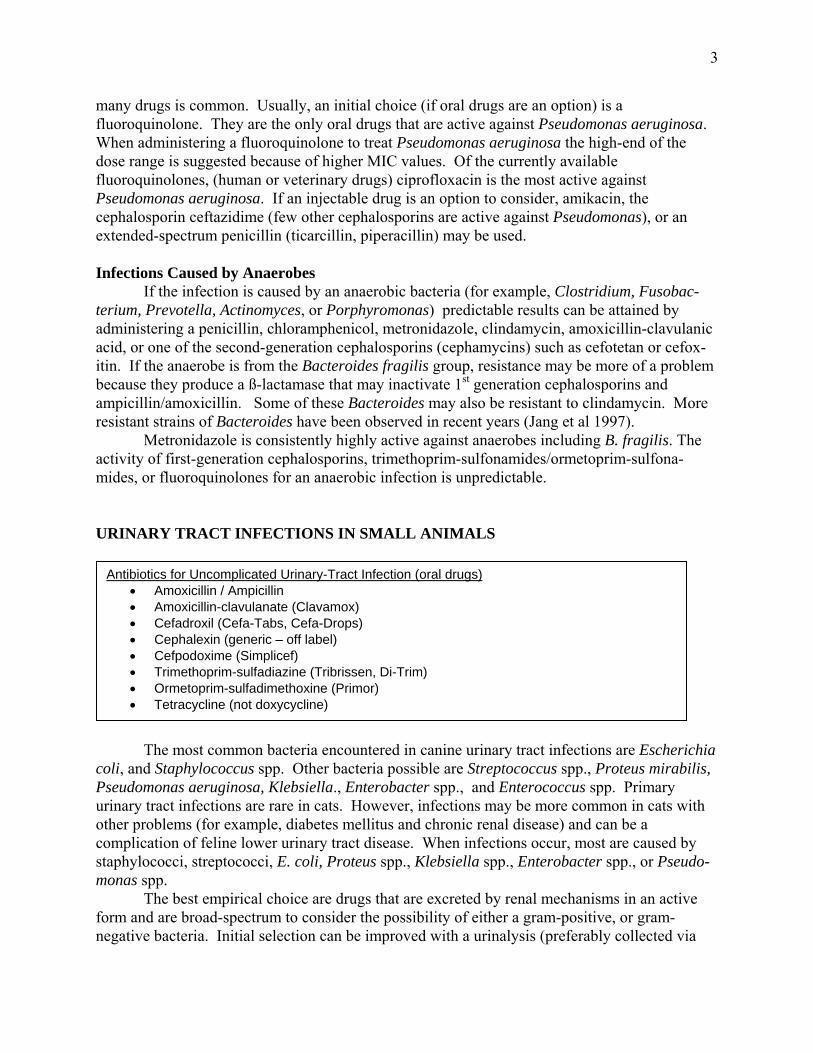
The most common bacteria encountered in canine urinary tract infections are Escherichia coli, and Staphylococcus spp. Other bacteria possible are Streptococcus spp., Proteus mirabilis, Pseudomonas aeruginosa, Klebsiella., Enterobacter spp., and Enterococcus spp. Primary urinary tract infections are rare in cats. However, infections may be more common in cats with other problems (for example, diabetes mellitus and chronic renal disease) and can be a complication of feline lower urinary tract disease. When infections occur, most are caused by staphylococci, streptococci, E. coli, Proteus spp., Klebsiella spp., Enterobacter spp., or Pseudo-monas spp. The best empirical choice are drugs that are excreted by renal mechanisms in an active form and are broad-spectrum to consider the possibility of either a gram-positive, or gram-negative bacteria. Initial selection can be improved with a urinalysis (preferably collected via
Antibiotics for Uncomplicated Urinary-Tract Infection (oral drugs) • Amoxicillin / Ampicillin • Amoxicillin-clavulanate (Clavamox) • Cefadroxil (Cefa-Tabs, Cefa-Drops) • Cephalexin (generic – off label) • Cefpodoxime (Simplicef) • Trimethoprim-sulfadiazine (Tribrissen, Di-Trim) • Ormetoprim-sulfadimethoxine (Primor) • Tetracycline (not doxycycline)
4
cystocentesis), examination of urine sediment, a culture, and quantification of the bacteria in the urine. Prior to culture results, empirical selection can be made with the following list of drugs (not necessarily in order of priority): amoxicillin, amoxicillin-clavulanate (Clavamox), first-generation cephalosporin, and trimethoprim-sulfonamide. If a gram-negative bacilli is suspected as the cause of the infection, and resistance to other drugs is a possibility (Oluoch et al, 2001; Cooke et al, 2002), a fluoroquinolone or cefpodoxime proxetil can also be considered. If treatment has been refractory, some are resistant to fluoroquinolones. In urinary tract infections (Torres et al, 2005) half of the E. coli were resistant to cephalexin, and only 22% were sensitive to enrofloxacin. When treating urinary tract infections, rule out complicating factors such as cystic calculi, metabolic disorders such as diabetes mellitus or hyperadrenocorticism, renal or prostate involvement. If the patient is an intact male dog, the prostate may be involved. Selection of a drug that will penetrate the prostate must be a factor in drug selection in that case. Appropriate drugs are trimethoprim-sulfonamides, or a fluoroquinolone.
High antibiotic concentrations achieved in renal tubules and the urine after routine therapy with modest doses of antibiotics is often sufficient to cure lower urinary tract infections, even those that are caused by organisms identified on a susceptibility test as “intermediate” in sensitivity (CLSI, 2008). Urine concentrations of antibiotics in animals with good renal function are at least 100 x the corresponding plasma concentrations because of the tubular concentration. When the infection is confined to the lower urinary tract, these high concentrations are an advantage (Stamey, et al. 1974). Cures of urinary tract infections are possible, even when the antibiotic levels do not attain concentrations high enough for a systemic infection. However, clinicians should be aware that if the concentrating ability of the kidneys is compromised, antibiotic concentrations in the urine may be low. Patients may have dilute urine because of renal disease, or treatment with corticosteroids, fluid therapy, or diuretics.
When the renal tissue is involved, (for example with a chronic infection) high urine drug concentrations offer little advantage. Drug concentrations in renal tissue – which are equivalent to the renal lymph concentrations – are correlated to plasma drug concentrations, not the drug concentrations in the urine. Therefore, consideration must be given to drugs that attain high concentrations in the renal tissue and that can be administered at doses and intervals that are optimum to achieve the pharmacokinetic-pharmacodynamic relationships for a clinical cure. RESPIRATORY TRACT INFECTIONS IN SMALL ANIMALS
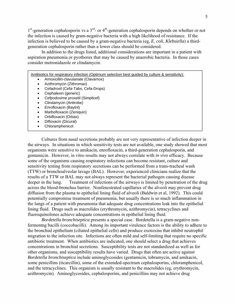
Upper and lower respiratory tract infections are common indications for empirical antibiotic therapy in animals. Upper respiratory infections are often self-limiting and will resolve without antibiotics However many upper and lower respiratory infections are secondary, and the result of a more serious disease (eg, megaesophagus), immunosuppression (eg, cancer), or foreign body (nasal cavity infection). Bacteria cultured from animals with pulmonary infections include Bordetella bronchi-septica, Streptococcus zooepidemicus, Escherichia coli, Pseudomonas aeruginosa, Klebsiella pneumoniae, Staphylococcus spp, alpha- and beta- streptococci, and Pasteurella multocida. Mycoplasma may play a role in some infections, but its importance has been controversial. Initial therapy for the bacteria listed above can be considered from the following list of drugs: amoxicillin-clavulanate (Clavamox), cephalosporins, clindamycin (Antirobe), fluoroquinolones (enrofloxacin, marbofloxacin, orbifloxacin, or difloxacin), and chloramphenicol. For infections known to be caused by gram-positive bacteria, azithromycin can be considered. The choice of a
5
1st-generation cephalosporin vs a 3rd- or 4th-generation cephalosporin depends on whether or not the infection is caused by gram-negative bacteria with a high likelihood of resistance. If the infection is believed to be caused by a gram-negative bacteria (eg, E. coli, Klebsiella) a third-generation cephalosporin rather than a lower class should be considered. In addition to the drugs listed, additional considerations are important in a patient with aspiration pneumonia or pyothorax that may be caused by anaerobic bacteria. In those cases consider metronidazole or clindamycin.
Cultures from nasal secretions probably are not very representative of infection deeper in the airways. In situations in which sensitivity tests are not available, one study showed that most organisms were sensitive to amikacin, enrofloxacin, a third-generation cephalosporin, and gentamicin. However, in vitro results may not always correlate with in vivo efficacy. Because some of the organisms causing respiratory infections can become resistant, culture and sensitivity testing from respiratory secretions can be performed from a trans-tracheal wash (TTW) or bronchoalveolar lavage (BAL). However, experienced clinicians realize that the results of a TTW or BAL may not always represent the bacterial pathogen causing disease deeper in the lung. Treatment of infections of the airways is limited by penetration of the drug across the blood-bronchus barrier. Nonfenestrated capillaries of the alveoli may prevent drug diffusion from the plasma to epithelial lining fluid of alveoli (Baldwin et al, 1992). This could potentially compromise treatment of pneumonia, but usually there is so much inflammation in the lungs of a patient with pneumonia that adequate drug concentrations leak into the epithelial lining fluid. Drugs such as macrolides (erythromycin, azithromycin), tetracyclines and fluoroquinolones achieve adequate concentrations in epithelial lining fluid. Bordetella bronchiseptica presents a special case. Bordetella is a gram-negative non-fermentng bacilli (coccobacilli). Among its important virulence factors is the ability to adhere to the bronchial epithelium (ciliated epithelial cells) and produce exotoxins that inhibit neutrophil migration to the infection site. Infections are often mild and self-limiting that require no specific antibiotic treatment. When antibiotics are indicated, one should select a drug that achieves concentrations in bronchial secretions. Susceptibility tests are not standardized as well as for other organisms, and susceptibility results have varied. Drugs that often are active against Bordetella bronchiseptica include aminoglycosides (gentamicin, tobramycin, and amikacin, some penicillins (ticarcillin), some of the extended-spectrum cephalosporins, chloramphenicol, and the tetracyclines. This organism is usually resistant to the macrolides (eg, erythromycin, azithromycin). Aminoglycosides, cephalosporins, and penicillins may not achieve drug
Antibiotics for respiratory infection (Optimum selection best guided by culture & sensitivity): • Amoxicillin-clavulanate (Clavamox) • Azithromycin (Zithromax) • Cefadroxil (Cefa-Tabs, Cefa-Drops) • Cephalexin (generic) • Cefpodoxime proxetil (Simplicef) • Clindamycin (Antirobe) • Enrofloxacin (Baytril) • Marbofloxacin (Zeniquin) • Orbifloxacin (Orbax) • Difloxacin (Dicural) • Chloramphenicol

6
concentrations at the infection site and fluoroquinolones are not consistently active against this organism. Another treatment route that is considered is aerosolization of antibiotics (eg, tobramycin). EMPIRICAL TREATMENT FOR INTRACELLULAR PATHOGENS Since most bacterial infections are located extracellularly, it is sufficient for a cure to achieve adequate drug concentrations in the extracellular (interstitial) space rather than intracellular space. However, intracellular infections present another problem. Only lipid-soluble drugs are able to reach high concentrations in cells. Intracellular organisms such as Brucella, Rhodococcus equi, Chlamydophilia, Ehrlichia, Rickettsia, Bartonella and Mycobacteria are examples of intracellular pathogens that may not be susceptible in vivo to drugs that cannot penetrate cells. Staphylococci may, in some cases, become refractory to treatment because of intracellular survival.
The concentration of drugs in cells often is expressed as the cellular to extracellular concentration ratio (C:E ratio). Examples of drugs that enter leukocytes, and other cells (That is, they have C:E ratios of one or greater than one.) are fluoroquinolones (enrofloxacin, ciproflox-acin, difloxacin, marbofloxacin, and orbifloxacin), tetracyclines, lincosamides (clindamycin, lincomycin), macrolides (erythromycin, clarithromycin), and the azalides (azithromycin) (Pasqual, 1995). β-lactam antibiotics and aminoglycosides do not reach effective concentrations within cells and are not useful for these infections. Tetracyclines (eg, doxycycline) and fluoroquinolones are often used to treat Chlamydia and Rickettsia infections because of their ability to kill intracellular organisms. There is good evidence for efficacy of doxycycline or fluoroquinolones (enrofloxacin is the only one tested) for treating Rickettsia, but only doxycycline should be considered for its efficacy for treating canine ehrlichiosis. The best treatment for Bartonella infections in dogs and cats has not been determined. However, azithromycin, with or without combinations of other drugs (eg, a tetracycline or fluoroquinolone) has been used. EMPIRICAL ANTIBIOTIC TREATMENT OF SEPSIS AND FEVER
Often the only sign of a potential infection is fever. If there also is evidence that the patient is immunosuppressed, antibiotic therapy is justified. Evidence of immunosuppression may include documented neutropenia, corticosteroid administration, hyperadrenocorticism (Cushing’s Disease), or anticancer treatment. In these instances, it is not unusual to fail to identify a bacterial cause. Blood cultures are often recommended, but may be unrewarding, or the result of a blood culture may not be available for 2 to 3 days or longer. In these cases, one should select a drug protocol that gives maximum coverage with minimal risk of adverse effect. Oral Drugs: For patients that can be treated with oral drugs, a combination of a fluoroquinolone (enrofloxacin, difloxacin, marbofloxacin, orbifloxacin, or ciprofloxacin) plus a potentiated amoxicillin (Clavamox) or an oral cephalosporin (cephalexin, cefadroxil, or cefpodoxime proxetil) is a rational choice. This combination is safe, and may be as efficacious as injectable drugs. However, if a patient is severely ill, do not rely on oral drug absorption alone and treatment should be initiated with an injectable regimen (see below). Also, in animals that have severe illness from sepsis, the adverse effects from oral drugs (gastrointestinal system effects such as nausea, vomiting, diarrhea), may complicate recovery. Injectable Drugs: If the patient is more critically ill, or if the infection becomes more life-threatening, injectable drugs should be considered. In these cases, injectable enrofloxacin
7
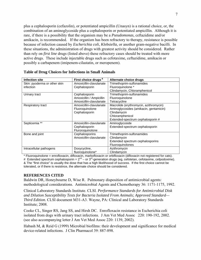
plus a cephalosporin (cefazolin), or potentiated ampicillin (Unasyn) is a rational choice, or, the combination of an aminoglycoside plus a cephalosporin or potentiated ampicillin. Although it is rare, if there is a possibility that the organism may be a Pseudomonas, ceftazidime and/or amikacin, is recommended. If the organism has been refractory to therapy, resistance is possible because of infection caused by Escherichia coli, Klebsiella, or another gram-negative bacilli. In these situations, the administration of drugs with greatest activity should be considered. Rather than rely on first line drugs (listed above) these refractory cases should be treated with more active drugs. These include injectable drugs such as cefotaxime, ceftazidime, amikacin or possibly a carbapenem (imipenem-cilastatin, or meropenem). Table of Drug Choices for Infections in Small Animals Infection site First choice drugs & Alternate choice drugs Skin: pyoderma or other skin infection
Amoxicillin-clavulanate Cephalosporin
Trimethoprim-sulfonamides Fluoroquinolone * Clindamycin, Chloramphenicol
Urinary tract Cephalosporin Amoxicillin / Ampicillin Amoxicillin-clavulanate
Trimethoprim-sulfonamides Fluoroquinolone Tetracycline
Respiratory tract Amoxicillin-clavulanate Fluoroquinolone Cephalosporin
Macrolide (erythromycin, azithromycin) Aminoglycosides (amikacin, gentamicin) Clindamycin Chloramphenicol Extended-spectrum cephalosporin #
Septicemia ** Amoxicillin-clavulanate Cephalosporin Fluroroquinolone
Aminoglycoside Extended-spectrum cephalosporin
Bone and joint Cephalosporins Amoxicillin-clavulanate
Trimethoprim-sulfonamides Clindamycin Extended spectrum cephalosporins Fluoroquinolones
Intracellular pathogens Doxycycline, fluoroquinolone*
Azithromycin Clindamycin
* Fluoroquinolone = enrofloxacin, difloxacin, marbofloxacin or orbifloxacin (difloxacin not registered for cats). # Extended spectrum cephalosporin = 2nd – or 3rd-generation drugs (eg, cefotetan, cefotaxime, cefpodoxime). & The “first choice” is usually the dose that has a high likelihoood of success. If the first choice cannot be tolerated, or if there is resistnce, the alternate choice should be considered. REFERENCES CITED Baldwin DR, Honeybourne D, Wise R. Pulmonary disposition of antimicrobial agents: methodological considerations. Antimicrobial Agents and Chemotherapy 36: 1171-1175, 1992. Clinical Laboratory Standards Institute. CLSI. Performance Standards for Antimicrobial Disk and Dilution Susceptibility Tests for Bacteria Isolated From Animals; Approved Standard—Third Edition. CLSI document M31-A3. Wayne, PA: Clinical and Laboratory Standards Institute; 2008. Cooke CL, Singer RS, Jang SS, and Hirsh DC. Enrofloxacin resistance in Escherichia coli isolated from dogs with urinary tract infections. J Am Vet Med Assoc 220: 190-192, 2002. (see also accompanying letter J Am Vet Med Assoc 220: 1139, 2002). Habash M, & Reid G (1999) Microbial biofilms: their development and significance for medical device-related infections. J Clin Pharmacol 39: 887-898.
8Jang SS, Breher JE, Dabaco LA, & Hirsh DC (1997) Organisms isolated from dogs and cats with anaerobic infections and susceptibility to selected antimicrobial agents. J Am Vet Med Assoc 210: 1610-1614. Lees GE, Rogers KS: Treatment of urinary tract infections in dogs and cats. J Am Vet Med Assoc 189: 648-652, 1986. Lloyd DH, Lamport AI, Feeney C: Sensitivity to antibiotics amongst cutaneous and mucosal isolates of canine pathogenic staphylococci in the UK, 1980-1996. Vet Derm 7: 171-175, 1996. Lorian V: Antibiotics in Laboratory Medicine 4th Ed. Williams & Wilkins, 1996. NCCLS (2002): Performance standards for antimicrobial disk and dilution susceptibility tests for bacteria isolated from animals; approved standards – second edition. M31-A2, 2002; 940 West Valley Road, Suite 1400, Wayne, Pennsylvania 19087. Nix DE, Goodwin SD, Peloquin CA, et al. Antibiotic tissue penetration and its relevance: Impact of tissue penetration on infection response. Antimicrob Agents Chemother 35: 1953-1959, 1991. Oluoch AO, Kim C-H, Weisiger RM, et al. Nonenteric Escherichia coli isolates from dogs: 674 cases (1990-1998). J Am Vet Med Assoc 218: 381-384, 2001. Pascual A: Uptake and intracellular activity of antimicrobial agents in phagocytic cells. Rev Med Microbiol 6: 228-235, 1995. Petersen AD, Walker RD, Bowman MM, Schott HC, Rosser EJ. Frequency of isolation and antimicrobial susceptibility patterns of Staphylococcus intermedius and Pseudomonas aeruginosa isolates from canine skin and ear samples over a 6 year period (1992-1997). J Am Anim Hosp Assoc 38: 407-413, 2002. Pinchbeck LR, Cole LK, Hillier A, Kowalski JJ, Rajala-Schultz PJ, Bannerman TL & York S. Pulsed-field gel electrophoresis patterns and antimicrobial susceptibility phenotypes for coagulase-positive staphylococcal isolates from pustules and carriage sites in dogs with superficial bacterial folliculitis. Am J Vet Res 68: 535-542, 2007. Stamey TA, Fair WR, Timothy MM. Et al. Serum versus urinary antimicrobial concentrations in cure of urinary-tract infections. New Engl J Med 291: 1159-1163, 1974. Torres SM, Diaz SF, Nogueira SA, Jessen C, Polzin DJ, Gilbert SM, and Horne LK. Frequency of urinary tract infection among dogs with pruritic disorders receiving long-term glucocorticoid treatment. J Am Vet Med Assoc 227: 239-243, 2005. Weese JS. Methicillin-resistant Staphylococcus aureus: an emerging pathogen in small animals. J Am Anim Hosp Assoc 41: 150-157, 2005.
9
STRATEGIES TO MANAGE ANTIBIOTIC-RESISTANT INFECTIONS Mark G. Papich
College of Veterinary Medicine, North Carolina State University Raleigh, North Carolina, USA
Treatment guidelines are established in textbooks and consensus documents available for treating routine infections in small animals. Dosage regimens have been established and drug manufacturers have produced several important drugs to treat the most common infections encountered in small animals. However, the drugs and approaches to therapy are more limited when the infection is more refractory, resistant, or is associated with another complicating factor. Susceptibility of the most common isolates has been documented well enough to make sound judgments and empirical antimicrobial drug choices. However, when the patient has a refractory and/or resistant infection, or is seriously ill with an infection, other strategies and drugs may be necessary. As with many new treatments, there are few veterinary clinical studies to support a recommended use and dose and many of these details have been extrapolated from human medicine. Bacterial Susceptibility Issues
Most bacteria that cause infections come from the following list: Staphylococcus pseudintermedius (and occasionally other staphylococci) Escherichia coli, Klebsiella pneumoniae, Pasteurella multocida, beta-hemolytic streptococci, Pseudomonas aeruginosa, Proteus mirabilis (and occasionally indole-positive Proteus), Enterobacter spp and Enterococcus spp. If the bacteria are accurately identified, antibiotic selection is simplified because the susceptibility pattern of many organisms is predictable. For example, if the bacteria is likely to be Pasteurella, Streptococcus, or Actinomyces, susceptibility is expected to penicillin or an aminopenicillin such as ampicillin, amoxicillin, or amoxicillin-clavulanic acid (Clavamox). Staphylococcus
Staphylococcus isolated from small animals is most likely to be S. pseudintermedius rather than S. aureus. (Note that previously identified Staph intermedius are now referred to as S. pseudintermedius by many laboratories. This species of Staphylococcus will usually have a predictable susceptibility to β-lactamase resistant β-lactam antibiotics such as amoxicillin combined with a β-lactamase inhibitor (Clavamox), or first-generation cephalosporin such as cephalexin or cefadroxil, or the third-generation cephalosporins, cefovecin (Convenia) and cefpodoxime (Simplicef). Staphylococcus also is susceptible to oxacillin and dicloxacillin but these are not used as commonly in small animal medicine. Reports of studies on S. pseudintermedius have shown that, despite frequent use of the above mentioned drugs in small animals, the incidence of resistance has not increased (Lloyd, et al, 1996; Pinchbeck et al, 2007). Most staphylococci are also sensitive to fluoroquinolones. The majority of staphylococci are sensitive to lincosamides (clindamycin, lincomycin), trimethoprim-sulfonamides, or erythromycin, but resistance can occur in as high as 25% of the cases. Methicillin-Resistant Staphylococcus
Thes favorable rates of susceptibility listed above do not diminish the importance of emergence of methicillin-resistant Staphylococcus in companion animals (Weese 2005). The mecA gene and methicillin resistance appears to be increasing in veterinary medicine based on

10
the number of reports in the last several years. Methicillin-resistant Staphylococcus aureus (MRSA) in human hospitals, and in the community has reached alarming rates.
Staphylococcal resistance can be caused by altered penicillin-binding proteins (PBP-2a), carried by the gene mecA. These are known as methicillin-resistant staphylococci – MRS (Gortel et al, 1999; Deresinski 2005; Jones et al, 2007; Bemis et al, 2006). If it is S. aureus the term methicillin-resistant S. aureus (MRSA) can be applied, but S. aureus is an infrequent pathogen in dogs, and only occasionally in cats. Bacteria previously identified as Staphylococcus intermedius are most likely Staph. pseudintermedius and any future studies and papers will likely use the new terminology (Sasaki et al, 2007; Devriese et al, 2005). Other Staphylococcus species also have been identified among veterinary isolates, such as coagulase-negative Staphylococcus.
Oxacillin is now used more commonly than methicillin as the marker for this type of resistance, and resistance to oxacillin is equivalent to methicillin-resistance. If staphylococci are resistant to oxacillin or methicillin, they should be considered resistant to all other β-lactams, including cephalosporins and amoxicillin-clavulanate (eg, Clavamox), regardless of the susceptibility test result. Adding a β-lactamase inhibitor will not overcome methicillin resistance. Unfortunately, these bacteria often carry co-resistance to many other non- β-lactam drugs, including clindamycin, fluroquinolones, macrolides, tetracyclines, and trimethoprim-sulfonamides. In the report by Bemis et al (2009), more than 90% of the methicillin-resistant isolates of S. pseudintermedius also were resistant to > 4 other drugs. The cause of the increased frequency of resistance has not been identified. Use of fluroquinolones and cephalosporins has been linked to emergence of resistance of methicillin-resistant staphylococci (Dancer, 2008; Harbarth & Samore, 2008).
Because susceptibility to non-β-lactam antibiotics is unpredictable, a susceptibility test is needed to identify which drug to administer for these infections. Clindamycin, chloramphenicol, tetracyclines, rifampin, and trimethoprim-sulfonamides are drugs to consider for these infections if a susceptibility test can confirm activity. However, in some instances the only drug that is active for treatment will be a glycopeptide such as vancomycin (Vancocin) or the oxazolidinone, linezolid (Zyvox). Vancomycin can only be administered by intravenous infusion. Linezolid is the first in the class of oxazolidinones to be used in medicine and it is used in people to treat resistant gram-positive infections caused by MRSA, enterococci and streptococci. It can be administered IV or orally and has excellent absorption, but is extremely expensive. Nevertheless, veterinary patients have been treated with this medication with a low incidence of adverse effects, good success, and no evidence of inducing further resistance.
Susceptibility testing issues for Staphylococcus:
The current standards published by the Clinical Laboratory Standards Institute (CLSI 2008; formerly NCCLS) do not differentiate the interpretive criteria of Staphylococcus aureus from that if Staphylococcus pseudintermedius or S. intermedius. The CLSI document states that the S. aureus interpretive criteria should be used for all other veterinary isolates of non-CNS (coagulase negative staphylococci). This interpretation lists a MIC value of ≥ 4.0 µg/mL as resistant. However, there is now evidence that other coagulase-positive Staphylococcus spp. should be considered resistant when the MIC is ≥ 0.5 µg/mL, which was the criteria in the older standard (Bemis et al, 2009). If the criteria of of ≥ 4.0 µg/mL is used, resistant staphylococci from animals may be misidentified. In the next published supplement of the CLSI standards, this recommendation will change to reflect this new evidence. Until then, diagnostic laboratories
11
should adopt the recommendation that if any non-aureus coagulase-positive Staphylococcus isolated from animals has a MIC value ≥ 0.5 µg/mL (corresponding to a zone diameter of ≤ 17 mm), it should be considered methicillin-resistant, mec-A positive, and resistant to all β-lactam antibiotics Resistant Enterococcus Enterococci are gram-positive cocci that have emerged as important causes of infections, especially those that are nosocomial. The most common species identified are Enterococcus faecalis and E. faecium. Enterococcus faecalis is more common, but E. faecium is usually the more resistant. Wild-strain enterococci may still be sensitive to penicillin G and ampicillin, or amoxicillin. However, the enterococci have an inherent resistance to cephalosporins and fluoroquinolones. These strains also are usually resistant to trimethoprim-sulfonamide combinations, clindamycin, and erythromycin. Susceptibility test results for cephalosporins, β-lactamase resistant penicillins (eg, oxacillin), trimethoprim-sulfonamide combinations, and clindamycin can give misleading results (CLSI, 2008). Even if isolates are shown to be susceptible to a fluoroquinolone, this class of drugs may not be a good alternative for treatment.
In human medicine frequent use of fluoroquinolones and cephalosporins (both of which have poor activity against enterococci), has been attributed to emergence of a higher rate of enterococcal infections. Evidence to document this trend is limited in veterinary medicine, but one study from a veterinary teaching hospital indicated increased rate of enterococcal urinary tract infections (Prescott, et al, 2002). Treatment of Enterococcus is frustrating because there are so few drug choices. If the Enterococcus isolated is sensitive to penicillins, one should administer amoxicillin or ampicillin at the high-end of the dose range. When possible, combine an aminoglycoside with a β-lactam antibiotic for treating serious infections. Occasionally, one of the carbapenems (imipenem-cilastatin) or an extended-spectrum penicillin (eg, piperacillin) can be considered for treatment of E. faecalis (but not E. faecium). When enterococci are present in wound infections, lower urinary tract, peritoneal infections, and body cavity infections (eg, peritonitis), the organism may exist with other bacteria such as gram-negative bacilli, or anaerobic bacteria. In these cases, there is evidence that treatment should be aimed at the anaerobe, and/or gram-negative bacilli and not directed at the enterococcus. Treatment cures are possible if the other organisms are eliminated without specific therapy for enterococcus (Bartlett et al 1978). As mentioned above for resistant Staphylococcus, sometimes the only active drug will be a glycopeptide or the oxazolidinone linezolid. Disadvantages of these drugs were discussed above. Problem with Gram-Negative Resistant Bacteria
If the organism is Pseudomonas aeruginosa, Enterobacter, Klebsiella, Escherichia coli, or Proteus, resistance against many common antibiotics is possible and a susceptibility test is ad-vised. For example, a report showed that among nonenteric E. coli, only 23% were sensitive to a 1st generation cephalosporin and less than half were sensitive to ampicillin. In the same study, 13%, and 23% were intermediate or resistant to enrofloxacin, and orbifloxacin, respectively (Oluoch, et al 2001). In urinary tract infections (Torres et al, 2005) half of the E. coli were resistant to cephalexin, and only 22% were sensitive to enrofloxacin. Based on these data as well as other studies, for initial therapy we usually expect the gram-negative enteric bacteria to be susceptible to fluoroquinolones and aminoglycosides. An extended-spectrum cephalosporin (second- or third-generation cephalosporin) usually is active against enteric-gram negative
12
bacteria, but will not be active against Pseudomonas aeruginosa. Pseudomonas aeruginosa
Infections caused by Pseudomonas aeruginosa present a special problem because so few drugs are active against this organism. Of the β-lactam antibiotics, a few are designated as anti-Pseudomonas antibiotics. Those with activity against this organism include the ureidopenicillins (mezlocillin, azlocillin, piperacillin) and the carboxylic derivatives of penicillin (carbencillin, ticarcillin). These derivatives are available as sodium salts for injection; there are no orally-effective formulations in this class, except indanyl carbenicillin (Geocillin, Geopen) which is poorly absorbed and not useful for systemic infections. These drugs are more expensive than the more-commonly used penicillins, and must be administered frequently (eg, at least 4 times daily) to be effective. Ticarcillin is available in combination with the β-lactamase inhibitor clavulanic acid (Timentin). Because these drugs degrade quickly after reconstitution, observe the storage recommendations on the package insert to preserve the drug’s potency.
In one published study, the in vivo activity was examined in 23 strains of Pseudomonas: 19 Ps. aeruginosa, 3 Ps. fluorescens and one Pseudomonas spp. The most effective antibiotics were tobramycin (100% susceptible), marbofloxacin (91.3%) and ceftazidime (91.3%). Ticarcillin and gentamicin, showed good activity (86 and 65.2% respectively). Lower susceptibility was found with enrofloxacin (52.1%) (Martin Barrasa et al, 2000). Isolates of Pseudomonas aeruginosa from otitis media showed that 97% were susceptible to ceftazidime, and 81% to carbenicillin (Colombini et al 2000). Fewer were susceptible to enrofloxacin (51%) and gentamicin (68%). In a study that isolated Pseudomonas aeruginosa from the skin and ears of dogs, the pattern of resistance is similar (Petersen et al, 2002). There were no trends identified, and most isolates were susceptible to ciprofloxacin, piperacillin, ticarcillin, amikacin, and gentamicin (enrofloxacin was not tested). However, isolates from the ears tended to be more resistant than isolates from the skin, with lower susceptibility to topical drugs such as gentamicin.
When administering a fluoroquinolone to treat Pseudomonas aeruginosa the high-end of the dose range is suggested because even among wild-type strains the MIC values are higher than other gram-negative bacteria. Of the currently available fluoroquinolones, (human or veterinary drugs) ciprofloxacin is the most active against Pseudomonas aeruginosa, followed by marbofloxacin, enrofloxacin, difloxacin, and orbifloxacin (Rubin et al, 2008; Riddle et al, 2000).
Drug Choices for Resistant Gram-Negative Infections
After a susceptibility report is available, one may find that the only drugs to which some gram-negative bacilli are sensitive, including Pseudomonas aeruginosa, are extended-spectrum cephalosporins, penems (carbapenems), or amikacin.
Cefpodoxime is more active than many other third-generation cephalosporins against Staphylococcus, and pharmacokinetic properties allow for once-daily dosing (Papich et al, 2007). However, it is not active against Pseudomonas aeruginosa, Enterococcus, or methicillin-resistant Staphylococcus. One should be aware that the break-point for susceptibility is lower than for other third-generations cephalosporins. Therefore, it is possible for a bacterial isolate to be sensitive to cefotaxime or ceftazidime (breakpoint 8 µg/mL) but resistant to cefpodoxime (breakpoint 2 µg/mL) (CLSI 2008). Specific disks are suggested for testing bacterial isolates, rather than relying on the results from other cephalosporins.
In the spring of 2008 cefovecin (Convenia) was registered by the FDA-CVM for use in

13
dogs and cats for treatment of skin infections. In December of 2006 cefovecin (Convenia) was introduced to small animal medicine in Europe and in Canada in October 2007. There have also been pharmacokinetic studies (Stegemann et al 2006ab) published for dogs and cats, pharmacodynamic studies published (Stegemann et al, 2006c), and clinical efficacy studies in dogs and cats (Stegemann et al, 2007ab; Passmore et al, 2007; Six et al, 2008). In the clinical studies, cefovecin was compared to another active antimicrobial (cefadroxil, cephalexin, or amoxicillin-clavulanate) and non-inferior to these other drugs.
In dogs and cats, cefovecin is registered in Europe and Canada for treatment of skin infections. In dogs it is also registered for urinary tract infections. In Europe, but not Canada, it is also registered for urinary tract infections in cats. The approved label dose in these countries is 8 mg/kg SC, once every 14 days. The studies published show efficacy with a 14 day interval for administration. The injection may be repeated for infections that require longer than 14 days for a cure (eg, canine pyoderma). The registration in the United States lists treatment of skin infections in dogs and cats and therapeutic concentrations are maintained for an interval of 7 days. Drug concentrations persist long enough for a 14 day interval for some indications.
There are currently not any CLSI approved standards for susceptibility testing established for cefovecin (CLSI 2008). Based on the distribution of organisms reported (Stegemann et al. 2006c) ≤ 2.0 µg/mL should be considered. It has equal or greater activity against Staphylococcus spp. isolates and gram-negative bacteria of the Enterobacteriaceae (eg, E. coli, Klebsiella). However, activity against Pseudomonas aeruginosa is poor and it will not be effective against methicillin-resistant staphylococci.
Cefovecin is a third-generation cephalosporin and is more active with lower MIC values than first generation cephalospsorins. This was demonstrated for pathogens from Europe and the United States (Stegemann et al, 2006c, Six et al, 2008). Cefovecin MIC90 values were 0.25 µg/mL for Staphylococcus intermedius compared to 2 µg/mL for cephalexin and cefadroxil. As a 3rd-generation cephalosporin, it is expected to have even greater activity against gram-negative bacteria as was demonstrated by the MIC90 values of 1 µg/mL compared to 16 µg/mL for cephalexin and cefadroxil (Six et al, 2008). Other MIC comparisons are provided in the tables in the paper by Stegemann et al (2006c).
When other injectable cephalospsorins are considered for small animals, the most often used are cefotaxime and ceftazidime, although individual veterinary hospitals have utilized others in this group. These drugs are expensive, injectable, and must be administered frequently. Of the cephalosporins, only the 3rd-generation cephalosporins, ceftazidime (Fortaz, Tazidime), cefoperazone (Cefobid), or cefepime (Maxipime), a 4th-generation cephalosporin, have predictable activity against Pseudomonas aeruginosa. Ceftazidime has greater activity than cefoperazone and is the one used most often in veterinary medicine. These drugs must all be injected, and are usually given IV, although SC, and IM routes have been used. As with the penicillins, frequent administration is necessary.
The β-lactam antibiotics with greatest activity against Pseudomonas aeruginosa are the carbapenems. The carbapenems are β-lactam antibiotics that include imipenem-cilastatin sodium (Primaxin), meropenem (Merrem), and most recently, ertapenem (Invanz). All three have activity against the enteric gram-negative bacilli. Ertapenem is a new addition to the class of carbapenems but it does not have anti-Pseudomonas activity. Resistance (carbapenemases) among veterinary isolates has been very rare. Imipenem is administered with cilastatin to decrease renal tubular metabolism. Imipenem has become a valuable antibiotic because it has a broad spectrum that includes many bacteria resistant to other drugs. Imipenem is not active
14
against methicillin-resistant staphylococci or resistant strains of Enterococcus faecium. The high activity of imipenem is attributed to its stability against most of the β-lactamases (including ESBL) and ability to penetrate porin channels that usually exclude other drugs (Livermore 2001). The carbapenems are more rapidly bactericidal than the cephalosporins and less likely to induce release of endotoxin in an animal from gram-negative sepsis.
Some disadvantages of imipenem are the inconvenience of administration, short shelf-life after reconstitution, and high cost. It must be diluted in fluids prior to administration. Meropenem, one of the newest of the carbapenem class of drugs (some experts consider it a 2nd –generation penem) and has antibacterial activity greater than imipenem against some isolates. One important advantage over imipenem is that it is more soluble and can be administered in less fluid volume and more rapidly. For example, small volumes can be administered subcutaneously with almost complete absorption. There also is a lower incidence of adverse effects to the central nervous system, such as seizures. Based on pharmacokinetic experiments in our laboratory (Bidgood & Papich, 2002), the recommended dose for Enterobactericeae and other sensitive organisms is 8.5 mg/kg SC every 12hr, or 24 mg/kg IV every 12 hr. For infections caused by Pseudomonas aeruginosa, or other similar organisms that may have MIC values as high as 1.0 mcg/mL: 12 mg/kg q8h, SC, or 25 mg/kg q8h, IV. For sensitive organisms in the urinary tract, 8 mg/kg, SC, every 12 hours can be used. In our experience, these doses have been well-tolerated except for slight hair loss over some of the SC dosing sites.
Aminoglycosides are active against most wild-type strains of Pseudomonas aeruginosa. Against resistant isolates, amikacin and tobramycin are more active than gentamicin, and resistance is less likely to these drugs (Petersen et al, 2002). Aminoglycosides are valuable for treating gram-negative bacilli that are resistant to other drugs. They are rapidly bactericidal, less expensive than injectable drugs listed above, and can be administered once-daily. Among these, amikacin is the most active. Therefore, it is often the first choice in small animal medicine. It has been administered once-daily IV, IM, or SC. There are two important disadvantages to systemic use of aminoglycosides: (1) Treatment usually must extend for at least two weeks or longer. Risk of nephrotoxicosis is greater with longer duration of treatment. (2) Activity of aminoglycosides is diminished in the presence of pus and cellular debris (Konig et al 1998). This may decrease their usefulness for the treatment of wound and ear infections caused by Pseudomonas aeruginosa. To decrease the risk of drug-induced nephrotoxicosis, therapeutic drug monitoring and careful evaluation of renal function during its use is recommended. STRATEGIES TO REDUCE RESISTANCE Restrict the Patients
When resistant bacteria are isolated from patients, a veterinary hospital should have measures in place to restrict these animals to prevent the spread of bacteria from one animal to another, or to decrease the possibility of a person handling the animal to act as a carrier. Patients with resistant infections should be identified clearly with a prominent identification marker, and a notation made in the patient’s record so the staff is aware of the patient’s status upon future visits.
Veterinarians and their staff handling patients with resistant infections should wear gloves and encouraged to wash their hands frequently with appropriate antibacterial cleansers. Animals should not be transferred from one cage to another. Transport to other areas of the clinic should be controlled and restricted as much as possible. If transported on a gurney or cart, the surface should be disinfected after use. All surfaces in contact with the patient, and
15
instruments used should be sterilized or disinfected after use. Cages or stalls should be disinfected when the animal leaves the hospital. A culture swab of the cage can be used to confirm proper and effective disinfecting practices. It is desirable to treat these patients, whenever possible, as out-patients to minimize their stay in the hospital. Restrict the Antibiotics?
Antibiotic administration – if not active enough to eliminate resistant isolates – can select for these resistant strains, which can multiply and flourish. There are examples of some drugs that can induce resistance mechanisms, but these are rare compared to selection of drug resistant strains. Many of the principles of bacterial resistance were covered thoroughly in a recent book (Guardabassi, et al, 2008). Inadequate antibiotic treatment consisting of doses too low, infrequent administrations, or selection of a poorly active drug, is probably the most common reason for emergence of drug resistance. As it was stated in one of the chapters of the book cited above, “It is exposure, and especially exposure to sub-optimal drug concentrations that is the most important single factor in resistance emergence and its subsequent spread.” (Lees et al, 2008). Because inadequate treatment may produce resistance, a strategy has been advocated that employs the use of highly active drugs, administered using appropriate regimens to attain pharmacokinetic-pharmacodynamic (PK-PD) targets, for as short of a duration as possible (Amyes et al, 2007). This suggests that in companion animals, veterinarians should not be reluctant to administer highly active drugs, provided appropriate regimens are used for only as long of a duration as necessary. When possible, therapy then can be de-escalated to less active, more narrow spectrum drugs, based on culture and susceptibility results and clinical response. This type of two-tiered approach to therapy has been successful in human medicine. If administered rationally, and at correct dosages, the administration of an antibiotic should eradicate the infection and actually decrease resistance. However, what we have observed over the 60 years (approximately) of antibiotic use is that resistant strains have been selected. Many of these resistant strains are now common and contributing to the difficulty in selecting effective antibiotics for some patients. As discussed previously, in small animals the most common bacteria producing resistant infections are Escherichia coli, Pseudomonas aeruginosa, Staphylococcus species, and Enterococcus species. These resistant strains have most likely been selected because of antibiotic pressure and inadequate exposure. The resistant strains emerge because the competition from more susceptible bacteria is reduced or eliminated following antibiotic administration. However, the extent to which prescribing practices can influence this trend is not straight-forward. The notion that antibiotic treatment causes resistance is too simplistic and does not reflect the complexity of the issue. Although antibiotic administration is certainly associated with resistance, it is not always clear. A recent review stated “... Thus, the emergence of resistance to antibiotics is associated with their use, although the precise correlation can be highly variable.” (Hawkey, 2008). For some bacteria, infection control may be more important than restrictions on antibiotic use (Harbarth & Samore, 2008). Resistance emerges both through horizontal spread – through transfer of genetic elements carrying genes for resistance – as well as mutations arising during treatment through selection of resistant strains. Reports have shown that resistant strains are more likely when the animal has previously been treated with antibiotics. However, consistent evidence is elusive for antibiotic use in companion animals. In a report from Europe, resistance was low to the antibiotics that were most frequently prescribed (Pedersen et al, 2007). Although resistance to enrofloxacin has been associated with prescribing practices (Cooke et al, 2002), resistance to fluoroquinolones in
16
other studies has not been associated with use (Meunier et al, 2002). Patterns of in vitro susceptibility of Staphylococcus have been reported for over 20 years (Lloyd, et al, 1996; Medleau et al, 1986, Noble & Kent, 1992, Petersen et al, 2002; Prescott et al, 2002; Pinchbeck et al, 2007). These published surveys have confirmed that a high proportion of organisms have retained susceptibility to cephalosporins, including cephalexin and cefadroxil (in addition to other drugs such as amoxicillin-clavulanate, aminoglycosides, and fluoroquinolones). In most papers published, the incidence of resistance to these drugs had not increased, despite frequent use of amoxicillin-clavulanate, cephalosporins (primarily cephalexin), and fluoroquinolones (Ganiere et al 2001; Normand et al, 2000; Petersen et al, 2002). In one contrasting report, and accompanying commentary (Prescott et al, 2002), there was a trend of increased resistance of Staphylococcus aureus and S. intermedius to enrofloxacin and cephalothin (1st generation cephalosporin equivalent to cephalexin) that the authors attributed to patterns of antibiotic use. However, the analysis of cephalothin resistance was not statistically significant the correlation weak (r2 = 0.25). It is not certain to what degree antibiotic use has influenced the emergence of methicillin-resistant staphylococci in animals. As discussed previously, methicillin-resistant Staphylococcus (MRS) have been identified in companion animals – primarily horses and dogs. These have been reported more frequently in recent years (Soulsby 2008). As reviewed by Harbarth & Samore (2008), in people there is a relationship between antibiotic use and MRSA rates. The drugs most often cited for driving MRSA acquisition and transmission is the use of cephalosporins and fluoroquinolones (Harbarth & Samore, 2008; Dancer, 2008), both of which are frequently prescribed in small animals (Guardabassi et al. 2008). The risk of increased bacterial resistance in pets as a result of veterinary prescribing practices should be taken seriously. In addition to increased difficulty treating resistant infections in pets, there may be a public health consequence. Transfer between animals and people of resistant bacteria, or genetic elements coding for resistance is possible (Guardabassi et al, 2004). The best documented resistant bacteria that may be transferred from pets to people are Salmonella, Campylobacter, and methicillin-resistant Staphylococcus (Jensen et al, 2008).
REFERENCES CITED
Amyes SGB, Walsh FM, Bradley JS. Best in class: a good principle for antibiotic usage to limit resistance development? Journal of Antimicrobial Chemotherapy 59: 825-826, 2007.
Bemis DA, Jones RD, Frank LA, Kania SA. Evaluation of susceptibility test breakpoints used to predict mecA-mediated resistance in Staphylococcus pseudintermedius isolated from dogs. J Vet Diagn Invest. 2009 Jan;21(1):53-8.
Bemis DA, Jones RD, Hiatt LE, Ofori ED, Rohrbach BW, Frank LA, Kania SA. Comparison of tests to detect oxacillin resistance in Staphylococcus intermedius, Staphylococcus schleiferi, and Staphylococcus aureus isolates from canine hosts. J Clin Microbiol. 2006 Sep;44(9):3374-6.
Bidgood T, Papich MG. Plasma pharmacokinetics and tissue fluid concentrations of meropenem after intravenous and subcutaneous administration in dogs. American Journal of Veterinary Research . 2002;63(12):1622-1628.
17
Clinical Laboratory Standards Institute. CLSI. Performance Standards for Antimicrobial Disk and Dilution Susceptibility Tests for Bacteria Isolated From Animals; Approved Standard—Third Edition. CLSI document M31-A3. Wayne, PA: Clinical and Laboratory Standards Institute; 2008.
Colombini S, Merchant RS, and Hosgood G. Microbial flora and antimicrobial susceptibility patterns from dogs with otitis media. Veterinary Dermatology 11: 235-239, 2000.
Cooke CL, Singer RS, Jang SS, & Hirsh DC. Enrofloxacin resistance in Escherichia coli isolated from dogs with urinary tract infections. J American Veterinary Medical Association 220: 190-192, 2002.
Dancer SJ. The effect of antibiotics on methicillin-resistant Staphylococcus aureus. Journal of Antimicrobial Chemotherapy 61: 246-253, 2008.
Deresinski S. Methicillin-resistant Staphylococcus aureus: an evoluationary, epidemiologic, and therapeutic odyssey. Clin Infect Dis 40: 562-573, 2005.
Devriese LA, Vancanneyt M, Baele M, Vaneechoutte M, De Graef E, Snauwaert CCleenwerck I, Dawyndt P, Swings J, Decostere A, & Haesebrouck F. Staphylocossus pseudintermedius sp. nov., a coagulase-positive species from animals. Int. Journal of Systematic and Evoluationary Microbiology 55: 1569-1573, 2005.
Ganiere JP, Medaille C, Limet A, et al. Antimicrobial activity of enrofloxacin against Staphylococcus intermedius strains isolated from canine pyodermas. Vet Dermatol 2001; 12:171-5.
Gortel K, Campbell KL, Kakoma I, Whittem T, Schaeffer DJ, Weisiger RM. Methicillin resistance among staphylococci isolated from dogs. Am J Vet Res 60: 1526-1530, 1999.
Guardabassi L, Houser GA, Frank LA & Papich MG. Guidelines for antimicrobial use in dogs and cats. (Chapter 11). In Guardabassi L, Jensen LB & Kruse H (editors). Guide to Antimicrobial Use in Animals. Blackwell Publishing, pages 183-206, 2008.
Guardabassi L, Schwarz S, Lloyd DH. Pet animals as reservoirs of antimicrobial-resistant bacteria. J Antimicrob Chemother 54: 321-332, 2004.
Harbarth S, & Samore MH. Interventions to control MRSA: high time for time-series analysis? Journal of Antimicrobial Chemotherapy 62: 431-433, 2008.
Hawkey PM. The growing burden of antimicrobial resistance. Journal of Antimicrobial Chemotherapy 62 (Suppl 1): i1-i9, 2008.
Jensen LB, Angulo FJ, Mølbak K, Wegener HC. Human health risks associated with antimicrobial use in animals. (Chapter 2). In Guardabassi L, Jensen LB & Kruse H (editors). Guide to Antimicrobial Use in Animals. Blackwell Publishing, pages, 13-26, 2008
Jones RD, Kania SA, Rohrbach BW, Frank LA, Bemis DA. Prevalence of oxacillin- and
18
multidrug-resistant staphylococci in clinical samples from dogs: 1,772 samples (2001-2005). J Am Vet Med Assoc. 2007 Jan 15;230(2):221-7.
Konig C, Simmen HP, Blaser J. Bacterial concentrations in pus and infected peritoneal fluid – implication of bactericidal activity of antibiotics. J Antimicrob Chemother 42: 227-232, 1998.
Lees P, Svendsen O, & Wiuff C. Strategies to minimize the impact of antimicrobial treatment on the selection of resistant bacteria. (Chapter 6). Guardabassi L, Jensen LB & Kruse H (editors). Guide to Antimicrobial Use in Animals. Blackwell Publishing, pages, 77-101, 2008.
Livermore DM. Of Pseudomonas, porins, pumps, and carbapenems. J Antimicrob Chemother 47: 247-250, 2001.
Lloyd DH, Lamport AI, Feeney C: Sensitivity to antibiotics amongst cutaneous and mucosal isolates of canine pathogenic staphylococci in the UK, 1980-1996. Vet Derm 7: 171-175, 1996.
Martin Barrasa JL, Lupiola Gomez P, Gonzalez Lama Z, et al. Antibacterial susceptibility patterns of Pseudomonas strains isolated from chronic canine otitis externa. J Vet Med B Infect Dis Vet Public Health 2000;47:191-6.
Medleau L, Long RE, Brown J, Miller WH. Frequency and antimicrobial susceptibility of Staphylococcus species isolated from canine pyoderma. Am J Vet Res 47: 229-231, 1986.
Meunier D, Acar J-F, Martel J-L, Kroemer S, & Valle M. A severn-year survey of susceptibility to marbofloxacin of pathogenic strains isolated from pets. International Journal of Antimicrobial Agents 24: 592-598, 2004.
Noble WC, Kent LE. Antibiotic resistance in Staphylococcus intermedius isolated from cases of pyoderma in the dog. Veterinary Dermatology 3: 71-74, 1992.
Normand EH, Gibson NR, Taylor DJ, et al. Trends of antimicrobial resistance in bacterial isolates from a small animal referral hospital. Vet Record 146: 151-155, 2000.
Oluoch AO, Kim C-H, Weisiger RM, et al. Nonenteric Escherichia coli isolates from dogs: 674 cases (1990-1998). J Am Vet Med Assoc 218: 381-384, 2001.
Papich MG, Davis JL, Floerchinger AM. Cefpodoxime and cephalexin plasma pharmacokinetics, protein binding, and tissue distribution after oral administration to dogs. [abstract # 161] American College of Veterinary Internal Medicine Annual Forum, Seattle, Washington, 2007.
Passmore CA, Sherington J, Stegemann MR. Efficacy and safety of cefovecin (Convenia) for the treatment of urinary tract infections in dogs. J Small Anim Pract. 2007; 48(3):139-44. PMID: 17355604.
Pedersen K, Pedersen K, Jensen H, Finster K, Jensen VF, & Heuer OE. Occurrence of antimicrobial resistance from diagnostic samples from dogs. Journal of Antimicrobial Chemotherapy 60: 775-781, 2007.
19
Petersen AD, Walker RD, Bowman MM, Schott HC, Rosser EJ. Frequency of isolation and antimicrobial susceptibility patterns of Staphylococcus intermedius and Pseudomonas aeruginosa isolates from canine skin and ear samples over a 6 year period (1992-1997). J Am Anim Hosp Assoc 38: 407-413, 2002.
Pinchbeck LR, Cole LK, Hillier A, Kowalski JJ, Rajala-Schultz PJ, Bannerman TL, York S. Pulsed-field gel electrophoresis patterns and antimicrobial susceptibility phenotypes for coagulase-positive staphylococcal isolates from pustules and carriage sites in dogs with superficial bacterial folliculitis.Am J Vet Res. 2007 May;68(5):535-42.
Prescott JF, Hanna WJB, Reid-Smith R, and Drost K. Antimicrobial drug use and resistance in dogs. Canadian Veterinary Journal 43: 107-116, 2002.
Riddle, C., Lemons, C., Papich, M.G., and Altier, C. Evaluation of ciprofloxacin as a representative of veterinary fluoroquinolones in susceptibility testing. Journal of Clinical Microbiology. 2000; 38: 1636-1637.
Rubin J, Walker RD, Blickenstaff K, Bodeis-Jones S, Zhao S. Antimicrobial resistance and genetic characterization of fluoroquinolone resistance of Pseudomonas aeruginosa isolated from canine infections.Vet Microbiol. 2008;131(1-2):164-172.
Sasaki T, Kikuchi K, Tanaka Y, Takahashi N, Kamata S, Hiramatsu K.Reclassification of phenotypically identified staphylococcus intermedius strains.J Clin Microbiol. 2007 Sep;45(9):2770-8.
Six R, Cherni J, Chesebrough R, Cleaver D, Lindeman CJ, Papp G, Skogerboe TL, Weigel DJ, Boucher JF, Stegemann MR. Efficacy and safety of cefovecin in treating bacterial folliculitis, abscesses, or infected wounds in dogs. Journal of the American Veterinary Medical Association 233(3):433-439, 2008.
Soulsby L. The 2008 Garrod Lecture: Antimicrobial resistance—animals and the environment. Journal of Antimicrobial Chemotherapy 62: 229-233, 2008.
Stegemann MR, Coati N, Passmore CA, Sherington J. Clinical efficacy and safety of cefovecin in the treatment of canine pyoderma and wound infections. J Small Anim Pract. 2007a; 48(7):378-86. PMID: 17559523.
Stegemann MR, Passmore CA, Sherington J, Lindeman CJ, Papp G, Weigel DJ, Skogerboe TL. Antimicrobial activity and spectrum of cefovecin, a new extended- spectrum cephalosporin, against pathogens collected from dogs and cats in Europe and North America. Antimicrob Agents Chemother. 2006c;50(7):2286-92. PMID: 16801403.
Stegemann MR, Sherington J, Blanchflower S. Pharmacokinetics and pharmacodynamics of cefovecin in dogs.J Vet Pharmacol Ther. 2006a; 29(6):501-11. PMID: 17083454.
Stegemann MR, Sherington J, Coati N, Brown SA, Blanchflower S. Pharmacokinetics of cefovecin in cats. J Vet Pharmacol Ther. 2006b; 29(6):513-24. PMID: 17083455.

20
Stegemann MR, Sherington J, Passmore C. The efficacy and safety of cefovecin in the treatment of feline abscesses and infected wounds. J Small Anim Pract. 2007b; 48(12):683-9. PMID: 17725587
Torres SM, Diaz SF, Nogueira SA, et al. Frequency of urinary tract infection among dogs with pruritic disorders receiving long-term glucocorticoid treatment. J Am Vet Med Assoc. 2005;227:239-243.
Weese JS.Methicillin-resistant Staphylococcus aureus: an emerging pathogen in small animals.J Am Anim Hosp Assoc. 2005 May-Jun;41(3):150-7.

21
ANALGESIC DRUGS FOR SMALL ANIMALS FROM NSAIDs to OPIATES
Mark G. Papich, DVM, MS, Diplomate ACVCP North Carolina State University, Raleigh, North Carolina, USA
NONSTEROIDAL ANTI-INFLAMMATORY DRUGS (NSAID): There are many NSAIDs available that have been used to treat osteoarthritis and pain. Many are registered for use in people (but we use them in animals also) and several are approved for use specifically for animals, particularly dogs. Current state of understanding of NSAID pharmacology: It has been accepted since Dr. Vane's work of the early 1970's that the most important mechanism of action of NSAID is inhibition of the cyclo-oxygenase enzyme (Abramson, et al, 1988, 1985), and the inhibition of prostaglandin synthesis. Tepoxalin (Zubrin) is only veterinary NSAID for which there is evidence that it also inhibits the lipoxygenase cascade (inhibition of leukotriene synthesis). Other drugs that are inhibitors of the lipo-oxygenase enzyme, or that block leukotrienes, are only used to treat asthma in people. The non-steroidal anti-inflammatory drugs (NSAIDs) have been among the most rapidly expanding group of drugs for dogs. Older approved drugs include carprofen (Rimadyl) and etodolac (EtoGesic). We now have several new additions such as deracoxib (Deramaxx), firocoxib (Previcox, Equioxx), tepoxalin (Zubrin), and meloxicam (Metacam). In other countries, additional drugs are available, such as tolfenamic acid (Tolfedine), nimesulide, and ketoprofen (Anafen). The pharmacologic action of the nonsteroidal anti-inflammatory drugs (NSAID) has been reviewed (Vane and Botting, 1995; Papich, 2000; Papich 2008). These drugs act to inhibit the isoenzymes of cyclo-oxygenase (COX). Cyclo-oxygenase 1 (COX 1) is a constitutive enzyme expressed in tissues (Meade et al 1994). Prostaglandins, prostacyclin, and thromboxane synthesized by this enzyme are responsible for normal physiological functions. Cyclo-oxygenase 2 (COX-2), on the other hand, is inducible and synthesized by macrophages and inflammatory cells after stimulation by cytokines and other mediators of inflammation. In some tissues, COX-2 may be constitutive, or may be induced to maintain favorable conditions in healthy tissue. The target of recently-developed NSAID has been COX-2, with the goal of producing analgesia and suppressing inflammation without inhibiting physiologically important prostanoids (Laneuville et al, 1994; Bergh & Budsberg, 2005). Whether or not selective inhibition of COX-2 is the safest
References: Visit the FDA website for current information on FDA-registered NSAIDs for animals. This website may be viewed at: http://www.fda.gov/cvm/nsaids.htm Reviews of these drugs are available that provide good summaries of their pharmacology and use. Many of the popular journals contain review papers on the use of these drugs (eg, Lascelles 2005). A review of clinical trials for treating osteoarthritis may be fund in the paper by Aragon et al (2007). Comprehensive references on this topic were provided in the Veterinary Clinics of North America (July 2000, Vol. 30(4); and November 2008 Vol. 38, both edited by K.A. Mathews. These issues contain several articles on drugs used for pain relief and control of inflammation. A discussion of the physiologic characteristics of cyclooxygenase products can be found in the reference by Jones & Budsberg (2000). For information specific to cats, there are recent reviews, Lascelles et al, 2007; Carroll and Simonson, (2005); Taylor & Robertson (Part 1, 2004) and Robertson & Taylor (Part 2 2004). For a comparison on how osteoarthritis is treated in people, consult the review by Steinmeyer & Konttinen (2006).

22
and most effective approach for animal treatment has yet to be established. Studies in Veterinary Medicine When one examines the other drugs registered for veterinary medicine, the effect of COX-2 inhibition is inconsistent. For example, deracoxib is considered a highly selective COX-2 inhibitor based on an assay performed with purified enzymes (Gierse et al 2002). In this study, the COX-1:COX-2 ratio was 1275; much higher than other drugs tested. But when tested in canine whole blood and compared to other NSAIDs, deracoxib had a ratio of only 12 (carprofen had a ratio of 6-7) (McCann et al, 2004). Drugs that had wide differences for in vitro COX-1/COX-2 inhibition ratios did not show the same degree of differences in vivo when samples were assayed from the stomach, inflamed joint, and blood (Sessions et al, 2005). Some of the confusion regarding understanding the action of the veterinary NSAID is that in vitro studies to examine their relative effects on COX-1 vs COX-2 have varied in their techniques and the cell system used. For example, in a study using canine enzyme systems, carprofen had a COX-1:COX-2 ratio of 129 (Ricketts et al 1998). In another study, using cell lines of another species (sheep and rodent) the ratio was 1.0 (Vane and Botting, 1995), and in a study using canine macrophages, the ratio was 1.75 (Kay-Mugford et al 2000). Yet another study on carprofen showed a ratio of 5.3 and that it was 1,000 times less potent in whole blood than in cell culture (Wilson et al, 2004). This emphasizes the effect of protein binding on in vitro assays. There has also been conflicting results when other drugs have been examined. The ratios for etodolac, another NSAID approved for dogs, has a COX-1:COX-2 ratio of 8.1 in humans, but 0.52-0.53 in dogs. Another study with etodolac showed that the selectivity for COX-2 was 10 times greater in people than dogs (Gierse et al 2002; Glaser 1995). Dr. Vane, a preeminent expert on COX inhibition, concluded that, Athe inhibitory activity of a drug for COX-1 to its inhibitory activity for COX-2 can vary according to whether tests are done on pure enzymes, cell homogenates, intact cells, or with the types of cells used (Vane and Botting, 1995). According to Dr. Lees, one of the leading investigators of NSAIDs in veterinary medicine, there are several unexplored questions to be answered for veterinary drugs (Lees, 2003). In some instances, the mechanism of action may not be entirely known. For example, carprofen appears to be a COX-1 sparing drug, (Ricketts et al 1998) but there is not agreement among investigators on whether or not it also inhibits COX-2 in vivo. Although there is evidence for inhibitory effects on cyclooxygenase in some models, carprofen did not show an in vivo anti-prostaglandin effect in dogs (McKellar et al 1994), which may explain the low rate of gastrointestinal adverse effects at approved doses. In another study, the investigators were unable to show that carprofen inhibited either COX-1 or COX-2 (Bryant et al, 2003). Some NSAIDs, including salicylates have been suggested to also inhibit nuclear factor kappa-B (NF-κB). NF-κB is an important promoter for inflammatory mediators. Veterinary drugs, such as carprofen and others also may act through inhibition of the activation of NF-κB (Bryant et al, 2003). Are the Selective COX-2 Inhibitors Better? The evidence for superior efficacy for selective COX-2 inhibitors is lacking. They are not necessarily more effective than older drugs, but they may be safer for the gastrointestinal tract (Peterson and Cryer 1999). However, the studies demonstrating safety in people have been criticized (Malhotra et al, 2004). Some skeptics have proposed that selective COX-2 inhibitors may not be appropriate for all patients because COX-2 enzyme products may be involved in actions other than inflammation. For example, COX-2 products may be biologically important
23
for angiogenesis, renal function, regulation of bone resorption, reproductive function, and healing of gastroduodenal ulcers (Wolfe et al 1999). There are high endogenous levels of COX-1 in the stomach, which is subject to high acid levels and shear forces. Inhibition of COX-1 in the stomach increases the risk of gastric ulceration. On the other hand, in the duodenum, COX-2 may be induced as a result of other treatments or injury to the duodenal mucosa. Inhibition of COX-2 in the duodenum may produce serious ulcers when this risk is high. COX-2 selective drugs also may cause a higher risk of cardiovascular problems in people because it preserves COX-1 which may promote platelet aggregation and vasoconstriction (Mukherjee et al, 2001). High COX-2 selectivity may increase risk of cardiovascular events (Topol 2004). This is the reason that popular drugs rofecoxib (Vioxx) and valdecoxib (Bextra) were discontinued in 2004. There has been serious concerns expressed about the events that preceeded this withdrawal. Dual Inhibitors There have been older drugs promoted to be "dual inhibitors" of arachidonic acid metabolites, but none were commercially successful. Dual inhibitor drugs effectively inhibit both cyclo-oxygenase (COX) and lipoxygenase (LOX). Therefore, they inhibit synthesis of both inflammatory prostaglandins (PG) and leukotrienes (LT). Interest in a dual inhibitor has focused on the potential benefits in inhibiting LOX, which may include higher GI safety, and greater analgesic efficacy. Lipoxygenase metabolites are involved in hyperalgesia, and inflammatory responses (Bertolini et al, 2001). Traditional NSAIDs that only block COX enzymes may increase leukotriene synthesis (Peters-Golden & Henderson, 2007). Older drugs thought to have dual inhibitor capability were benoxaprofen and ketoprofen. Benoxaprofen was taken off the market, and the evidence for ketoprofen acting as a dual inhibitor is weak. The only currently available drugs that acts as a dual inhibit of both LOX and COX is tepoxalin (Zubrin). The metabolite is active, but only acts as a COX inhibitor. Tepoxalin is more specific for COX-1 than COX-2, although this was not a canine-specific assay (data from Schering-Plough). Nevertheless, tepoxalin has a good gastrointestinal safety profile that matches other more selective COX-2 inhibitors. Tepoxalin has been shown to be effective in dogs with osteoarthritis and showed GI safety at several times the label dose. The only question remaining about tepoxalin is the duration of the LOX inhibitory effect. As shown in the accompanying table, the half-life for the LOX inhibitor parent drug is much shorter than the metabolite, which has little LOX inhibition. The other question remaining to be answered for tepoxalin is the contribution of anti-LOX action on the overall therapeutic effect. Studies in osteoarthritis in dogs (the registered indication for tepoxalin) have not revealed whether or not it is the COX or the LOX inhibition (or possibly some other mechanism) that is responsible for a favorable clinical effect. Whether or not the dual inhibition action of tepoxalin will be effective for other inflammatory diseases (for example, respiratory disease, dermatitis) has not been reported.
24
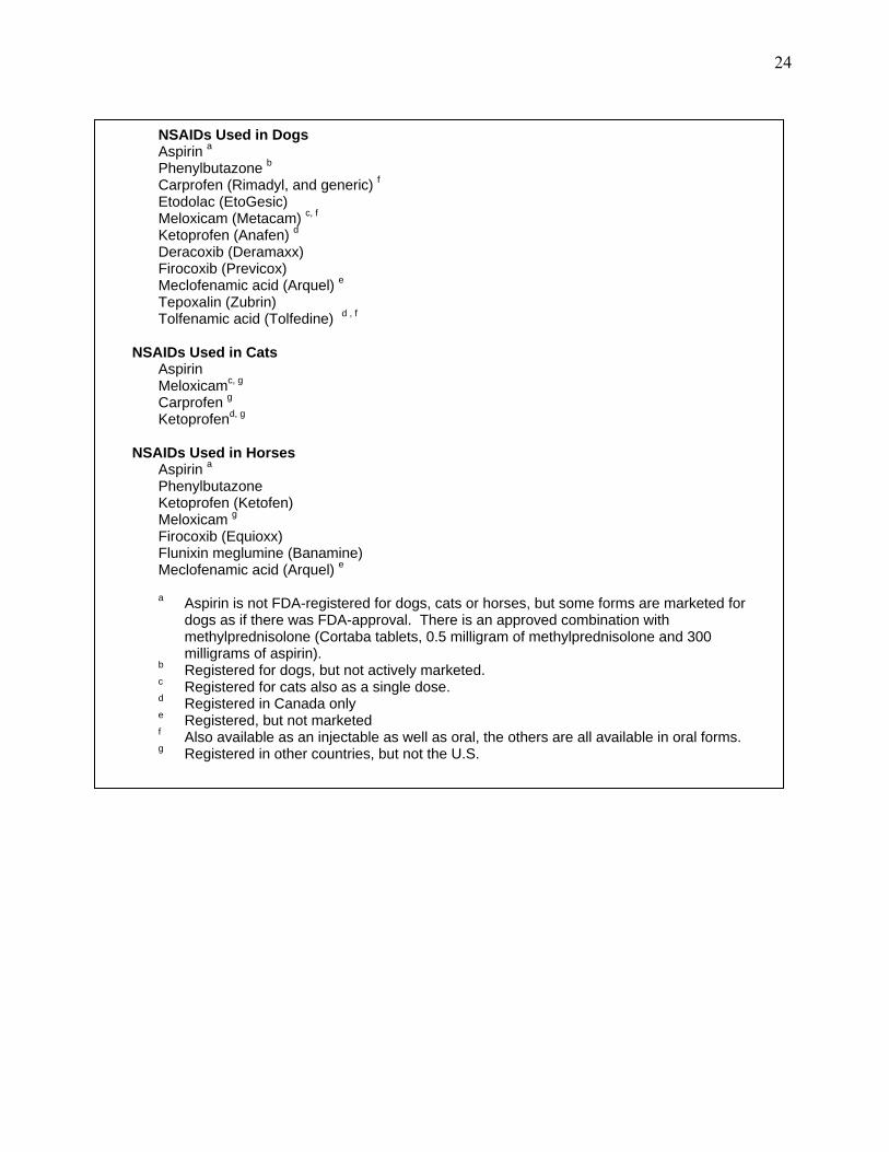
NSAIDs Used in Dogs Aspirin a Phenylbutazone b Carprofen (Rimadyl, and generic) f Etodolac (EtoGesic) Meloxicam (Metacam) c, f
Ketoprofen (Anafen) d
Deracoxib (Deramaxx) Firocoxib (Previcox) Meclofenamic acid (Arquel) e
Tepoxalin (Zubrin) Tolfenamic acid (Tolfedine) d , f
NSAIDs Used in Cats Aspirin Meloxicamc, g Carprofen g
Ketoprofend, g
NSAIDs Used in Horses Aspirin a
Phenylbutazone Ketoprofen (Ketofen) Meloxicam g Firocoxib (Equioxx) Flunixin meglumine (Banamine) Meclofenamic acid (Arquel) e
a Aspirin is not FDA-registered for dogs, cats or horses, but some forms are marketed for dogs as if there was FDA-approval. There is an approved combination with methylprednisolone (Cortaba tablets, 0.5 milligram of methylprednisolone and 300 milligrams of aspirin).
b Registered for dogs, but not actively marketed. c Registered for cats also as a single dose. d Registered in Canada only e Registered, but not marketed f Also available as an injectable as well as oral, the others are all available in oral forms. g Registered in other countries, but not the U.S.

25
COX 1/ COX 2 Inhibitor Ratios in Published Studies Drug
Streppa, et al 2002*
Ricketts, et al 1998*
Kay-Mugford et al. 2000*
Cryer et al 1998 **
Brideau et al. 2001 **
Wilson et al 2004**
Gierse et al 2002***
Ketoprofen
0.17
0.23 0.36 0.125 0.6
0.5
-
Aspirin
0.39
<0.3 - 0.32 -
0.37
-
Etodolac
0.53
0.52 - 7.92 -
6.3
3.4
Ibuprofen
0.74
- - 0.6 -
-
-
Piroxicam
2
- - 1.27 -
1.75
-
Meloxicam
2.72
2.9 12.3 - 10
-
-
Meclofenamic acid
5
15.4 - 12.1 -
5
-
Phenylbuta-zone
9.7
> 2.6 - - 0.6
-
Carprofen
16.8
129 1.75 - 6.5
5.3
65
Deracoxib
-
1275
* Assay with canine cell lines.** Assay with human cell lines; . *** assay with purified enzymes.
Adverse Effects of NSAIDs: • Gastrointestinal (GI) toxicity (Wolfe et al 1999): There are two forms GI injury that can
occur as a result of NSAID administration. (a) NSAID can directly injure the lining of the stomach because of direct irritation of an oral medication. This is common from aspirin. It usually is not serious, but can cause stomach discomfort, dyspepsia, and nausea. (b) A more serious form of GI injury occurs because prostaglandins are responsible for a healthy GI tract. When these prostaglandins are inhibited by NSAID, gastritis, GI ulcers, GI bleeding, perforations, diarrhea, and protein losing enteropathy have all been described in animals. Gastrointestinal toxicity is exacerbated by co-administration with corticosteroids in dogs.
• Renal toxicity: COX-1 and COX-2 products play an important role in renal vascular tone
(perfusion) and tubular function (natriuretic effect). Both COX-1 and COX-2 enzymes synthesize prostaglandins important for renal tubular function and blood perfusion during hypovolemia, hypotension, and salt depletion (Jones & Budsberg, 2000). Prostaglandins can have vasodilatory effects, and in some organs, are responsible for increasing blood perfusion during times of stress. In animals with decreased renal perfusion, NSAIDs may cause ischemic nephropathy.
• Injury to cartilage: NSAIDs have been shown to accelerate articular cartilage lesions in
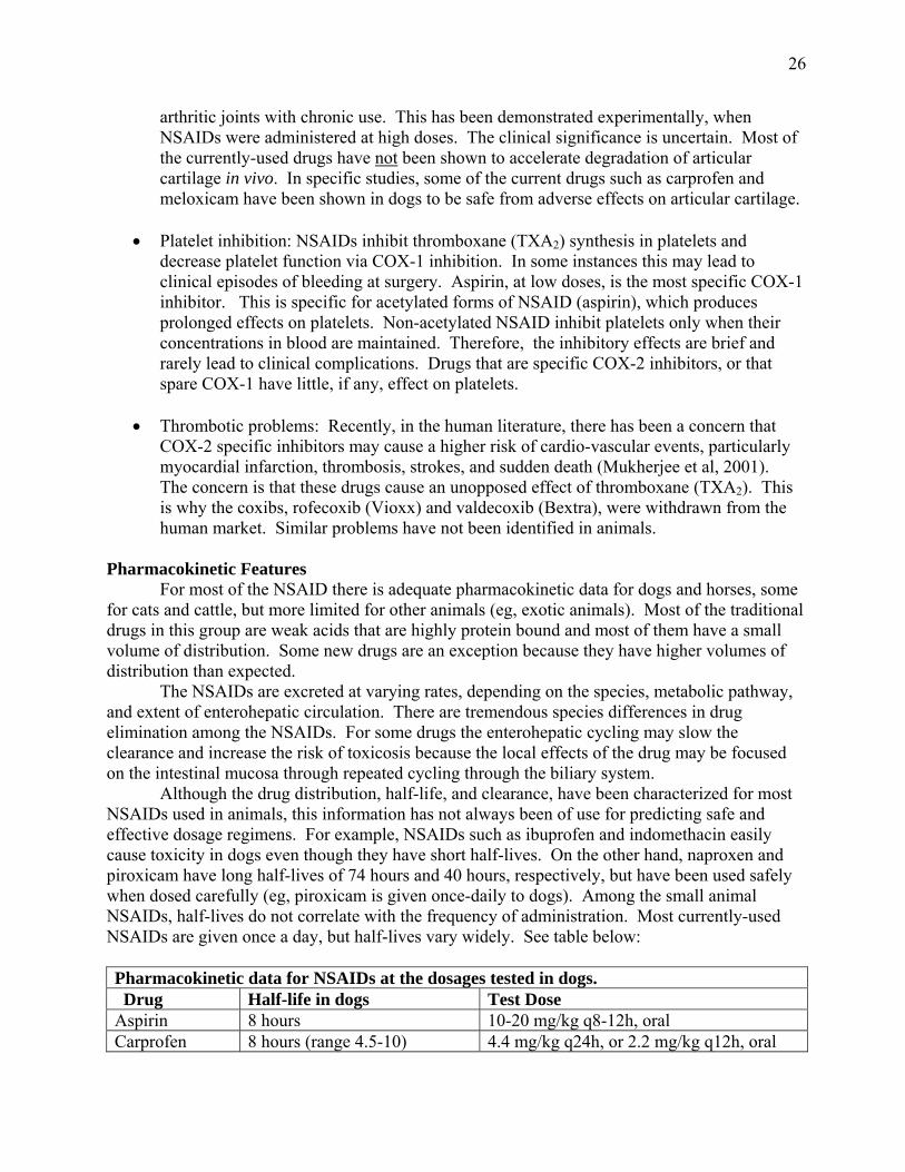
26
arthritic joints with chronic use. This has been demonstrated experimentally, when NSAIDs were administered at high doses. The clinical significance is uncertain. Most of the currently-used drugs have not been shown to accelerate degradation of articular cartilage in vivo. In specific studies, some of the current drugs such as carprofen and meloxicam have been shown in dogs to be safe from adverse effects on articular cartilage.
• Platelet inhibition: NSAIDs inhibit thromboxane (TXA2) synthesis in platelets and
decrease platelet function via COX-1 inhibition. In some instances this may lead to clinical episodes of bleeding at surgery. Aspirin, at low doses, is the most specific COX-1 inhibitor. This is specific for acetylated forms of NSAID (aspirin), which produces prolonged effects on platelets. Non-acetylated NSAID inhibit platelets only when their concentrations in blood are maintained. Therefore, the inhibitory effects are brief and rarely lead to clinical complications. Drugs that are specific COX-2 inhibitors, or that spare COX-1 have little, if any, effect on platelets.
• Thrombotic problems: Recently, in the human literature, there has been a concern that
COX-2 specific inhibitors may cause a higher risk of cardio-vascular events, particularly myocardial infarction, thrombosis, strokes, and sudden death (Mukherjee et al, 2001). The concern is that these drugs cause an unopposed effect of thromboxane (TXA2). This is why the coxibs, rofecoxib (Vioxx) and valdecoxib (Bextra), were withdrawn from the human market. Similar problems have not been identified in animals.
Pharmacokinetic Features For most of the NSAID there is adequate pharmacokinetic data for dogs and horses, some for cats and cattle, but more limited for other animals (eg, exotic animals). Most of the traditional drugs in this group are weak acids that are highly protein bound and most of them have a small volume of distribution. Some new drugs are an exception because they have higher volumes of distribution than expected. The NSAIDs are excreted at varying rates, depending on the species, metabolic pathway, and extent of enterohepatic circulation. There are tremendous species differences in drug elimination among the NSAIDs. For some drugs the enterohepatic cycling may slow the clearance and increase the risk of toxicosis because the local effects of the drug may be focused on the intestinal mucosa through repeated cycling through the biliary system. Although the drug distribution, half-life, and clearance, have been characterized for most NSAIDs used in animals, this information has not always been of use for predicting safe and effective dosage regimens. For example, NSAIDs such as ibuprofen and indomethacin easily cause toxicity in dogs even though they have short half-lives. On the other hand, naproxen and piroxicam have long half-lives of 74 hours and 40 hours, respectively, but have been used safely when dosed carefully (eg, piroxicam is given once-daily to dogs). Among the small animal NSAIDs, half-lives do not correlate with the frequency of administration. Most currently-used NSAIDs are given once a day, but half-lives vary widely. See table below: Pharmacokinetic data for NSAIDs at the dosages tested in dogs. Drug Half-life in dogs Test Dose Aspirin 8 hours 10-20 mg/kg q8-12h, oral Carprofen 8 hours (range 4.5-10) 4.4 mg/kg q24h, or 2.2 mg/kg q12h, oral
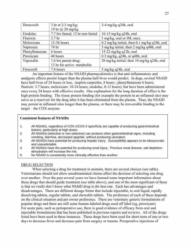
27
Deracoxib 3 hr at 2-3 mg/kg; 19 hr @ 20 mg/kg
3-4 mg/kg q24h, oral
Etodolac 7.7 hrs fasted; 12 hr non fasted 10-15 mg/kg q24h, oral Flunixin 3.7 hr 1 mg/kg, oral or IM, once. Meloxicam 12-36 hours 0.2 mg/kg initial, then 0.1 mg/kg q24h, oral Naproxen 74 hr 5 mg/kg initial, then 2 mg/kg q48h, oral Phenylbutazone 6 hours 15-22 mg/kg q12h, oral Piroxicam 40 hours 0.3 mg/kg, q24h, or q48h, oral Tepoxalin 1.6 hrs parent drug;
13 hr for active metabolite 20 mg/kg initial; then 10 mg/kg q24h, oral
Firocoxib 7.8 hours 5 mg/kg q24h, oral An important feature of the NSAID pharmacokinetics is that anti-inflammatory and analgesic effects persist longer than the plasma half-lives would predict. In dogs, several NSAID have half-lives of 24 hours or less, (aspirin carprofen, 8 hours ; phenylbutazone 6 hours; flunixin: 3.7 hours; meloxicam: 10-24 hours; etodolac, 8-12 hours), but have been administered once every 24 hours with effective results. One explanation for the long duration of effect is the high protein binding. The tissue protein binding (for example the protein in an inflamed site) may serve as a reservoir for the drug after it has been eliminated from the plasma. Thus, the NSAID may persist in inflamed sites longer than the plasma, or there may be irreversible binding to the target – the COX enzyme.
DRUG SELECTION When selecting a drug for treatment in animals, there are several choices (see table). Veterinarians should not allow unsubstantiated claims affect the decision of selecting one drug over another. Over the past several years we have learned some important information about these drugs that should guide treatment (see table above), and one of the most significant of these is that we really don’t know what NSAID drug is the best one. Each has advantages and disadvantages. There are different dosage forms that include injectable, to oral liquid, rapidly dissolving tablets, regular tablets, and chewable tablets. The preference of each of these depends on the clinical situation and pet owner preference. There are veterinary generic formulations of popular drugs and there are still some human-labeled drugs used off label (eg, piroxicam). For acute pain, such as perioperative use, there is good evidence of efficacy from oral and injectable formulations that has been published in previous reports and reviews. All of the drugs listed have been used in these instances. These drugs have been used for short-term of one or two days to decrease fever and decrease pain from surgery or trauma. Preoperative injections of
Consistent features of NSAIDs 1. All NSAIDs, regardless of COX-1/COX-2 specificity are capable of producing gastrointestinal
lesions, particularly at high doses. 2. All NSAIDs (selective or non-selective) can produce other gastrointestinal signs, including
vomiting, diarrhea, decreased appetite, without producing ulceration. 3. All NSAIDs have potential for producing hepatic injury. Susceptibility appears to be idiosyncratic
and unpredictable. 4. All NSAIDs have the potential for producing renal injury. Previous renal disease, salt depletion,
dehydration will increase the risk. 5. No NSAID is consistently more clinically effective than another.

28
carprofen to dogs were shown to be beneficial to decrease post-operative pain in dogs after ovariohysterectomy (Lascelles et al 1998). Meloxicam has been evaluated in two published studies for perioperative use, and was shown to be superior to butorphanol in some of the pain assessments that were measured. Oral NSAIDs also may be used for acute treatment of myositis, arthritis, and post-operative pain, or they may be administered chronically for osteoarthritis. Drugs that have been administered in the U.S. to small animals were listed above, and some veterinarians also have used human-label drugs such as aspirin, piroxicam, and naproxen. If these human-label drugs are considered, consult appropriate references for accurate dosing because it may differ from the human dose schedule. The most recently approved drugs are carprofen, etodolac, meloxicam, firocoxib, tepoxalin, and deracoxib. Doses for dogs were provided previously, and for other species the doses are widely available in formularies and textbooks. For long-term use there are no controlled studies that compare which is the most effective. When drugs are compared to one another, it is difficult, using subjective measurements, to demonstrate differences between these drugs for reducing pain in animals. Without a very large number of patients, the statistical power to detect differences among drugs in clinical veterinary studies is difficult. It is a rational approach to consider a rotating schedule of two or more drugs to identify which drug is better tolerated, effective, and easier to administer in each patient. NSAIDs for Cats: For a review of NSAID drug selection for cats, consult the references cited earlier (Lascelles et al, 2007; Carroll and Simonson, (2005); Taylor & Robertson (Part 1, 2004) and Robertson & Taylor (Part 2 2004). Meloxicam is commonly used in cats because it can be injected initially, and follow-up is possible with oral treatment. The oral solution has been palatable for cats, but the dose should be reduced compared to the canine dose. In addition, ketoprofen, aspirin (at an extended interval), and occasionally other drugs listed for dogs have been used off-label. For short-term use, carprofen and deracoxib, and flunixin have been used. REFERENCES AND ADDITIONAL READING Abramson SB, Korchak H, Ludewig R, et al: Modes of action of aspirin-like drugs. Proc Nat Acad Sci 82: 7227-7231, 1985.
Abramson SB, Weissmann G: The mechanisms of action of nonsteroidal anti-inflammatory drugs. Arth Rheum 32: 1-9, 1988.
Aragon CL, Hofmeister EH, Budsberg SC. Systematic review of clinical trials of treatments for osteoarthritis in dogs. J Am Vet Med Assoc. 2007 Feb 15;230(4):514-21.
Bergh MS & Budsgerg SC. The Coxib NSAIDs: Potential clinical and pharmacologic importance in veterinary medicine. J Vet Int Medicine 19: 633-643, 2005.
Bertolini A, Ottani A, Sandrini M. Dual acting anti-inflammatory drugs: a reappraisal. Pharmacol Res 2001; 44: 437-50.
Bryant CE, Farnfield BA, Janicke HJ. Evaluation of the ability of carprofen and flunixin meglumine to inhibit activation of nuclear factor kappa B. Am J Vet Res 2003; 64: 211-15.
Carroll GL, Simonson SM. Recent developments in nonsteroidal antiinflammatory drugs in cats. J Am Anim Hosp Assoc. 2005 Nov-Dec;41(6):347-54.
Day RO, Graham GG, Williams KM, Brooks PM: Variability in response to NSAIDs: Fact or Fiction? DRUGS 36: 643-651, 1988.
FitzGerald GA, Patrono C. The Coxibs, selective inhibitors of cyclooxygenase-2. N Engl J Med 2001; 345: 433-42.
Gierse JK, Staten NR, Casperson GF, et al. Cloning, expression, and selective inhibition of canine cyclooxygenase-1 and cyclooxygenase-2. Veterinary Therapeutics 3: 270-280, 2002.

29
Glaser KB. Cyclooxygenase selectivity and NSAIDs: cyclooxygenase-2 selectivity of etodolac (Lodine). Inflammopharmacology 1995; 3: 335-45.
Johnston SA, Budsberg SC. Nonsteroidal anti-inflammatory drugs and corticosteroids for the management of canine osteoarthritis. Vet Clin North Amer (Small Animal Practice) 27(4): 841-861, 1997.
Johnston SA, Fox SM: Mechanisms of action of anti-inflammatory medications used for the treatment of osteoarthritis. J Am Vet Med Assoc 210: 1486-1492, 1997.
Jones CJ, Streppa HK, Harmon BG, Budsberg SC. In vivo effects of meloxicam and aspirin on blood, gastric mucosal, and synovial fluid prostanoid synthesis in dogs. Am J Vet Res 63: 1527-1531, 2003.
Jones CJ, Budsberg SC. Physiologic characteristics and clinical importance of the cyclooxygenase isoforms in dogs and cats. J Am Vet Med Assoc. 2000 Sep 1;217(5):721-9. Review.
Kay-Mugford P, Benn SJ, LaMarre J, Conlon P. In vitro effects of non-steroidal anti-inflammatory drugs on cyclooxygenase activity in dogs. Am J Vet Res 2000; 61: 802-10.
Laneuville O, Breuer DK, Dewitt DL, Hla T, Funk CD, Smith WL. Differential inhibition of human prostaglandin endoperoxide synthases-1 and -2 by non-steroidal anti-inflammatory drugs. J Pharmacol Exp Ther 1994; 271(2): 927-34.
Lascelles BDX, Butterworth SJ, Waterman AE: Postoperative analgesic and sedative effects of carprofen and pethidine in dogs. Vet Rec 134: 187-191, 1994.
Lascelles BDX, Cripps PJ, Jones A, Waterman-Pearson AE. Efficacy and kinetics of carprofen, administered preoperatively or postoperatively, for the prevention of pain in dogs undergoing ovariohysterectomy. Vet Surg 27: 568-582, 1998.
Lascelles BDX, McFarland JM, Swann H. Guidelines for safe and effective use of NSAIDs in dogs. Veterinary Therapeutics 6 (3): 237-250, 2005.
Lascelles BDX, Blikslager AT, Fox SM, & Reece D. Gastrointestinal tract perforation in dogs treated with a selective cyclooxygenase-2 inhibitor: 29 cases (2002-2003). J Am Vet Med Assoc 227: 1112-1117, 2005.
Lascelles BDX, Court MH, Hardie EM, Robertson SA. Nonsteroidal anti-inflammatory drugs in cats : a review. Veterinary Anaesthesia and Analgesia 34 : 228-250, 2007.
Lascelles BDX, Cripps PJ, Jones A, and Waterman-Pearson AE. Efficacy and kinetics of carprofen, administered preoperatively or postoperatively, for the prevention of pain in dogs undergoing ovariohysterectomy. Veterinary Surgery 27: 568-582, 1998.
Lees P. Pharmacology of drugs used to treat osteoarthritis in veterinary practice. Inflammopharmacology 2003; 11: 385-99.
Lobetti RG, Joubert KE. Effect of administration of non-steroidal anti-inflammatory drugs before surgery on renal function in clinically normal dogs. Am J Vet Res 2000; 61(12): 1501-7.
MacPhail CM, Lappin MR, Meyer DJ, Smith SG, Webster CR, Armstrong PJ. Hepatocellular toxicosis associated with administration of carprofen in 21 dogs. J Am Vet Med Assoc 1998; 212(12): 1895-901.
Malhotra S, Shafiq N, Pandhi P. COX-2 inhibitors: a CLASS act, or just VIGORously promoted. MedGenMed 2004; 6(1); 6.
Mathews KA. Non-steroidal anti-inflammatory analgesics in pain management in dogs and cats. Can Vet J 1996; 37: 539-45.
Mathews KA: (Editor) Management of Pain. Veterinary Clinics of North America (Small Animal Practice) 2000; 30(4).
Mathews KA: Nonsteroidal anti-inflammatory analgesics: indications and contraindications for pain management in dogs and cats. Veterinary Clinics of North America (Small Animal Practice) 2000; 30(4) 783-804.
McCann ME, Andersen DR, Zhang D, Brideau C, Black WC, Hanson PD, Hickey GJ. In vitro effects and in vivo efficacy of a novel cyclo-oxygenase-2 inhibitor in dogs with experimentally induced synovitis. Am J Vet Res 2004; 65: 503-12.
McKellar QA, Delatour P, Lees P: Stereospecific pharmacodynamics and pharmacokinetics of carprofen in the dog. J Vet Pharmacol Therap 17: 447-454, 1994.
McNamara PS, Johnston SA, Todhunter RJ. Slow-acting, disease-modifying osteoarthritis agents. Vet Clinics of North America (Small Animal Practice) 27: 863-881, 1997.
Meade EA, Smith WL, DeWitt DL. Pharmacology of prostaglandin endoperoxide synthase isozymes-1 and -2. Ann N Y Acad Sci 1994; 714: 136-42.
Moreau M, Dupuis J, Bonneau H, Desnoyers M. Clinical evaluation of a nutriceutical, carprofen and meloxicam for the treatment of dogs with osteoarthritis. Veterinary Record 152: 323-329, 2003.
Mukherjee D, Nissen SE, Topol EJ. Risk of cardiovascular events associated with selective COX-2 inhibitors. JAMA 2001; 286: 954-9.

30
Papich MG. A update on nonsteroidal anti-inflammatory drugs (nsaid) in small animals. Veterinary Clinics of North America (Small Animal) 2008; 38: 1243-1266.
Papich MG: Principles of analgesic drug therapy. Seminars in Vet Med and Surg (Small Animal) 12(2): 80-93, 1997.
Papich MG: Pharmacologic considerations for opiate analgesic and nonsteroidal anti-inflammatory drugs. Veterinary Clinics of North America (Small Animal Practice) 2000; 30(4) 815-837.
Peters-Golden M, Henderson WR. Leukotrienes. New Engl J Medicine 357: 1841-1854, 2007.
Peterson WL, Cryer B. COX-1-sparing NSAIDs: is the enthusiasm justified? J Am Med Assoc 1999; 282: 1961-3
Ricketts AP, Lundy KM, Seibel SB. Evaluation of selective inhibition of canine cyclooxygenase 1 and 2 by carprofen and other nonsteroidal anti-inflammatory drugs. Am J Vet Res 59: 1441-1446, 1998.
Robertson SA, Taylor PM. Pain management in cats--past, present and future. Part 2. Treatment of pain--clinical pharmacology. J Feline Med Surg. 2004 Oct;6(5):321-33. Review.
Sessions JK, Reynolds LR, Budsberg SC. In vivo effects of carprofen, deracoxib, and etodolac on prostanoid production in blood, gastric mucosa, and synovial fluid in dogs with chronic osteoarthritis. Am J Vet Res 66: 812-817, 2005.
Steinmeyer J & Konttinen YT. Oral treatment options for degenerative joint disease – presence and future. Advanced Drug Delivery Reviews 58: 168-211, 2006.
Taylor PM, Robertson SA. Pain management in cats – past, present and future. Part 1: the Cat is unique. And Robertson SA, Taylor PM. Part 1: Treatment of pain – clinical pharmacology. Journal of Feline Medicine & Surgery 6: 313-320, 2004 And 6: 321-333, 2004.
Topol EJ. Failing the public health-rofecoxib, Merck and the FDA. N Engl J Med 2004; 351: 1707-9.
Vane JR, Botting RM. New insights into the mode of action of anti-inflammatory drugs. Inflamm Res 1995; 44: 1-10.
Vane JR: Inhibition of prostaglandin synthesis as a mechanism of action for aspirin-like drugs. Nature New Biology 234: 231-238, 1971.
Wilson JE, Chandrasekharan NV, Westover KD, Eager KB, Simmons DL. Determination of expression of cyclooxygenase-1 and -2 isozymes in canine tissues and their differential sensitivity to non-steroidal anti-inflammatory drugs. Am J Vet Res 2004; 65: 810-18.
Wolfe MM, Lichtenstein DR, Singh G: Gastrointestinal toxicity of nonsteroidal anti-inflammatory drugs. New Engl Journal of Medicine 340: 1888-1899, 1999.
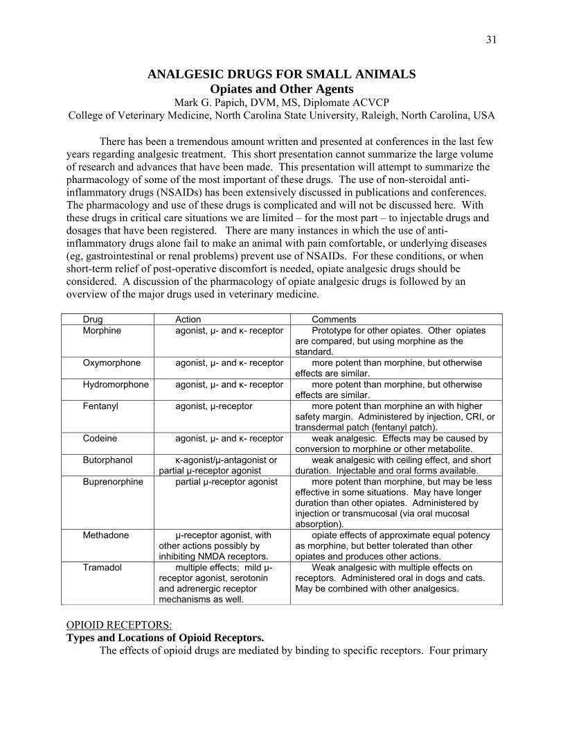
31
ANALGESIC DRUGS FOR SMALL ANIMALS Opiates and Other Agents
Mark G. Papich, DVM, MS, Diplomate ACVCP College of Veterinary Medicine, North Carolina State University, Raleigh, North Carolina, USA
There has been a tremendous amount written and presented at conferences in the last few
years regarding analgesic treatment. This short presentation cannot summarize the large volume of research and advances that have been made. This presentation will attempt to summarize the pharmacology of some of the most important of these drugs. The use of non-steroidal anti-inflammatory drugs (NSAIDs) has been extensively discussed in publications and conferences. The pharmacology and use of these drugs is complicated and will not be discussed here. With these drugs in critical care situations we are limited – for the most part – to injectable drugs and dosages that have been registered. There are many instances in which the use of anti-inflammatory drugs alone fail to make an animal with pain comfortable, or underlying diseases (eg, gastrointestinal or renal problems) prevent use of NSAIDs. For these conditions, or when short-term relief of post-operative discomfort is needed, opiate analgesic drugs should be considered. A discussion of the pharmacology of opiate analgesic drugs is followed by an overview of the major drugs used in veterinary medicine.
Drug Action Comments Morphine agonist, µ- and κ- receptor Prototype for other opiates. Other opiates
are compared, but using morphine as the standard.
Oxymorphone agonist, µ- and κ- receptor more potent than morphine, but otherwise effects are similar.
Hydromorphone agonist, µ- and κ- receptor more potent than morphine, but otherwise effects are similar.
Fentanyl agonist, µ-receptor more potent than morphine an with higher safety margin. Administered by injection, CRI, or transdermal patch (fentanyl patch).
Codeine agonist, µ- and κ- receptor weak analgesic. Effects may be caused by conversion to morphine or other metabolite.
Butorphanol κ-agonist/µ-antagonist or partial µ-receptor agonist
weak analgesic with ceiling effect, and short duration. Injectable and oral forms available.
Buprenorphine partial µ-receptor agonist more potent than morphine, but may be less effective in some situations. May have longer duration than other opiates. Administered by injection or transmucosal (via oral mucosal absorption).
Methadone µ-receptor agonist, with other actions possibly by inhibiting NMDA receptors.
opiate effects of approximate equal potency as morphine, but better tolerated than other opiates and produces other actions.
Tramadol multiple effects; mild µ-receptor agonist, serotonin and adrenergic receptor mechanisms as well.
Weak analgesic with multiple effects on receptors. Administered oral in dogs and cats. May be combined with other analgesics.
OPIOID RECEPTORS: Types and Locations of Opioid Receptors.
The effects of opioid drugs are mediated by binding to specific receptors. Four primary
32
types of opioid receptors in the body have been described. Opiate receptors also are found outside the central nervous system (CNS) such as in the gastrointestinal tract (GIT). Some aspects of opiate pharmacology can be found summarized in a special issue of Veterinary Clinics of North America (30(4): 2000, (K. Mathews editor). Opiate receptors produce the following effects:
• mu (μ) receptor (supraspinal): euphoria, sedation, analgesia, respiratory depression, and addiction.
• kappa (κ) receptor (spinal): analgesia at spinal level, and sedation. • delta (δ) receptor: spinal and supraspinal analgesia. • sigma (σ) receptor: unknown significance, not recognized by some investigators.
SYSTEMIC EFFECTS OF OPIOID AGONISTS:
In the central nervous system, opioids produce analgesia, euphoria, sedation, and excitement (depending on the species). Usually, sedation and analgesia is seen with low doses, anesthesia at high doses, and unconsciousness at the highest doses. When used for anesthesia or sedation, opiates are often used with other compounds (eg, phenothiazine tranquilizers) to poten-tiate the sedative effects. ADVERSE EFFECTS / SIDE EFFECTS:
Opiates have a good safety record. However, one should be aware of the important side effects and adverse effects associated with therapy. Serious adverse effects can reversed with an opiate antagonist, such as naloxone (Narcan) at 0.01 to 0.04 mg/kg IV, SC, IM, as needed. Naloxone administration should be done only as a last resort to an animal in pain because it can elicit a severe reaction. Some adverse effects can be alleviated with the administration of a partial antagonist such as buprenorphine or agonist/antagonist, such as butorphanol. Adverse effects of opioid therapy can include: • Sedation (dose related) • Excitement and dysphoria: Excitement can be seen in cats, cattle and horses, but may not be
a clinical problem if other sedative drugs are added, or if recommended dose rates are followed. Very high doses may cause convulsions, but this is rare. The cause of the excitement has varied, depending on the study. Some experts believe that the reaction is dopaminergic, adrenergic, or caused by decreased inhibitory activity of GABA. Release of acetylcholine or histamine release also may cause excitement from opiates.
• GI Effects: constipation, nausea (Some clinicians recommend stool softeners with chronic therapy because of the constipation.), and vomiting.
• Vasodilation (related to histamine release from morphine) • Cardiac depression (bradycardia). • Panting (seen primarily in dogs) because of lowering of temperature regulation in
hypothalamus by approximately 2Ε. • Respiratory depression. This is the most serious life-threatening effect with high doses. • Decreased urination: Inhibition of voiding reflex, increased tone of urinary sphincters, and
diuretic effect.
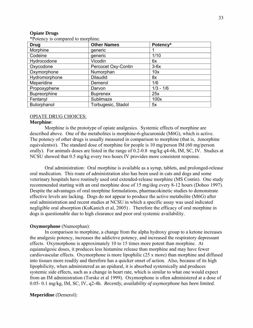
33
Opiate Drugs *Potency is compared to morphine. Drug Other Names Potency* Morphine generic 1 Codeine generic 1/10 Hydrocodone Vicodin 6x Oxycodone Percocet Oxy-Contin 3-6x Oxymorphone Numorphan 10x Hydromorphone Dilaudid 8x Meperidine Demerol 1/6 Propoxyphene Darvon 1/3 - 1/6 Bupreorphine Buprenex 25x Fentanyl Sublimaze 100x Butorphanol Torbugesic, Stadol 5x OPIATE DRUG CHOICES: Morphine:
Morphine is the prototype of opiate analgesics. Systemic effects of morphine are described above. One of the metabolites is morphine-6-glucuronide (M6G), which is active. The potency of other drugs is usually measured in comparison to morphine (that is, Amorphine equivalents@). The standard dose of morphine for people is 10 mg/person IM (60 mg/person orally). For animals doses are listed in the range of 0.2-0.8 mg/kg q4-6h, IM, SC, IV. Studies at NCSU showed that 0.5 mg/kg every two hours IV provides more consistent response.
Oral administration: Oral morphine is available as a syrup, tablets, and prolonged-release oral medication. This route of administration also has been used in cats and dogs and some veterinary hospitals have routinely used oral extended-release morphine (MS Contin). One study recommended starting with an oral morphine dose of 15 mg/dog every 8-12 hours (Dohoo 1997). Despite the advantages of oral morphine formulations, pharmacokinetic studies to demonstrate effective levels are lacking. Dogs do not appear to produce the active metabolite (M6G) after oral administration and recent studies at NCSU in which a specific assay was used indicated negligible oral absorption (KuKanich et al, 2005) . Therefore the efficacy of oral morphine in dogs is questionable due to high clearance and poor oral systemic availability.
Oxymorphone (Numorphan): In comparison to morphine, a change from the alpha hydroxy group to a ketone increases the analgesic potency, increases the addictive potency, and increased the respiratory depressant effects. Oxymorphone is approximately 10 to 15 times more potent than morphine. At equianalgesic doses, it produces less histamine release than morphine and may have fewer cardiovascular effects. Oxymorphone is more lipophilic (25 x more) than morphine and diffused into tissues more readily and therefore has a quicker onset of action. Also, because of its high lipophilicity, when administered as an epidural, it is absorbed systemically and produces systemic side effects, such as a change in heart rate, which is similar to what one would expect from an IM administration (Torske et al 1999). Oxymorphone is often administered at a dose of 0.05- 0.1 mg/kg, IM, SC, IV, q2-4h. Recently, availability of oxymorphone has been limited. Meperidine (Demerol):
34
Meperidine has 1/3 to 1/6 the potency of morphine, and is shorter acting (at 3 to 5 mg/kg its duration of action is less than 1 hour). With repeated use it may have fewer effects on the GI tract as compared to other opioids. The usual dose is 5-10 mg/kg for dogs and 3-5 mg/kg for cats, IV, IM. Because of its short duration of action, it is not used commonly in animals. Fentanyl: Fentanyl has been combined with droperidol in the preparation Innovar (no longer available) or used alone in Sublimaze: Although it is 80 to 100 times more potent than morphine, fentanyl can be used safely when administered with care. The usually dose is 0.02-0.04 mg/kg IM, SC, IV or as intravenous infusion. It is most often used as a transdermal preparation (see below). Codeine: Codeine is relatively safe and inexpensive, but is comparably weak as an analgesic. In humans, approximately 10% of a dose of codeine is converted to morphine. In dogs, there is only limited evidence of oral absorption. It is available as a 15, 30 and 60 mg tablet (codeine sulfate, codeine phosphate) and a 5 mg/ml syrup. Some formulations are combined with acetaminophen and/or caffeine (For example, Tylenol-2 or -3). Dextromethorphan: Dextromethorphan is not a true opiate, because it does not bind μ- or κ-opiate receptors, but it still has antitussive effects. It is the d-isomer of levorphan (the l-isomer, levorphan is an opiate with addictive properties, but the d-isomer does not). Dextromethorphan produces mild analgesia and modulate pain via its abilitiy to act as an NMDA (n-methyl D-aspartate) antagonist, but this is unrelated to the antitussive action.
Dextromethorphan has been administered to dogs and cats, but recent pharmacokinetic studies in dogs indicated that dextromethorphan does not attain effective concentrations after oral administration (KuKanich & Papich, 2004). After injections IV it produced adverse effects in dogs (vomiting after oral doses, and CNS reactions after IV administration). Even after IV administration, concentrations of the parent drug and active metabolite persisted for only a short time after dosing. Therefore, routine use in dogs is not recommended until more data is available to establish safe and effective doses. OPIATE AGONISTS/ANTAGONISTS AND PARTIAL AGONISTS: Opiate agonists/antagonists have effects that may differ qualitatively from those of pure opioid agonists such as morphine. Such differences may include less respiratory depression, fewer psychotic effects, fewer hemodynamic effects, and less physical dependency. A ceiling on the analgesic effects (that is, a limit to the analgesic efficacy of these drugs), distinguishes them from pure opioid agonists. Butorphanol (Stadol, Torbugesic, Torbutrol): Butorphanol is a weak analgesic with a short duration. Butorphanol has mixed effects because it is a μ-antagonist, or a partial μ-agonist, but a κ-agonist. It has opiate agonistic activity that is considered 5 times that of morphine. Its antagonistic effects, on the other hand are weak and only 1/40, or less than that of naloxone (some references suggest that it has no activity on the μ-receptor). Therefore, its agonist actions predominate. Butorphanol has been
35
used as an antitussive and analgesic. It may have fewer gastrointestinal effects compared to pure opioid agonists.
Butorphanol is used in dogs, horses, cats, and some zoo and exotic animals. In dogs it has been used frequently for pre- and post-operative analgesia by injection at 0.1 to 0.5 mg/kg. Efficacy is limited to mild pain. In some studies its efficacy is low and not as great as NSAIDs. For cats, for post-operative pain, (example, after declaw surgery) it has been administered with good results at a dose of 0.4 mg/kg, IM, q4h (Carroll et al 1998), but in other studies has not been as effective as NSAIDs (eg, meloxicam). In horses it is commonly administered for relief of acute pain. In horses, it has been administered as an IV infusion for post-operative pain. Butorphanol has been administered at a starting oral dosage of 1 to 4 mg/kg every 1-4 hrs. It is available as 1, 5 and 10 mg tablets (Torbutrol). Buprenorphine: Buprenorphine (Buprenex) is a partial μ-receptor agonist, with little effects on the κ-receptor. It is 25-50 times more potent than morphine. It is available as a 300 μg/ml injection. In animals it is reported that the duration of analgesia is longer (for example 6-8 hours) compared to the duration of action of morphine, perhaps because it dissociates slower from receptors. Because of the higher affinity for the μ-receptor, it will take higher doses of naloxone to reverse buprenorphine.
Buprenorphine has been used in dogs and cats because there is an impression that the duration of action is longer. In cats, buprenorphine was administered at a dose of 0.01 mg/kg and had a duration of action between 4 and 12 hours after injection (Robertson, et al. 2003). It also has been administered orally to cats (sublingually) at a dose of 0.066 mL per kg (Robertson et al. 2003).
Other Opiates: Drugs used occasionally, but not discussed in this section include oxycodone (Percodan), hydromorphan (Dilaudid), hydrocodone (Hycodan), and hydrocodone + acetaminophen (Vicodin). Tramadol (Ultram):
Although not a true opioid, tramadol will be discussed in this section. Tramadol is a racemic mixture (R & S) that has a complicated mechanism of action. It has some mu-opioid receptor action, but this effect is 10 times lower than codeine and 6,000 times lower than morphine. Tramadol also inhibits the reuptake of norepinephrine (NE) and serotonin (5 HT) and produces secondary effects on alpha-2 adrenergic receptors in pain pathways. One isomer has greater effect on 5 HT reuptake and greater affinity for mu-opiate receptors. The other isomer is more potent for NE reuptake and less active for inhibiting 5 HT reuptake. Taken together, the effects of tramadol may be explained through inhibition of 5 HT reuptake, action on α2 receptors, and mild activity on opiate mu-receptors. Tramadol has as many as 11 metabolites. One metabolite (o-desmethyl tramadol, also called M1) may have greater opiate effects than the parent drug (for example, 200-300 times greater opiate effect than tramadol), but still lower than morphine. In animals that produce this metabolite in sufficient amounts, some analgesic action may be attributed to opiate-mediated effects from the active metabolite. The other metabolites have not been shown to have active analgesic activity. The pharmacokinetics of tramadol and metabolites have been studied extensively in dogs, horses, and cats. The pharmacokinetics are

36
inconsistent with variation in clearance, oral absorption, and metaboism to the active metaboite among studies, within and between species. Some studies have shown that the active metabolite (M1) in dogs as a minor metabolite (10-16 % of tramadol concentrations), but in other studies the levels of this metabolie were either too low to quantify, or non-existent and may not contribute significantly to the analgesic effects. Tramadol half-life in dogs is approximately 1-2.7 hours with variable oral absorption, but oral absorption that may be as high as 65%. In cats the half-life is 3-4 hours and they produce higher and more sustained concentrations of the active metabolite (M1) than other animals – presumably because of differences in enzyme activity. Active metabolite concentration in cats parallel the concentrations of tramadol. A drug with similar structure and activity as tramadol is tapentadol (Nucynta). Tapentadol is used for moderate pain in people (50-200 mg q4-6h), but its use has not been reported in animals.
Tramadol has been used as an analgesic in people, dogs, cats, horses, and minor species (eg, rabbits, goats). It is an alternative to pure opiate analgesics and nonsteroidal antiinflammatory drugs (NSAIDs) in patients that require treatment for mild to moderate pain. In people it is regarded as a mild analgesic, but the pharmacokinetics of tramadol are different in people compared to animals. Clinical effects in humans cannot necessarily be extrapolated to animals. In animal studies – both clinical and experimental – the results have been inconsistent to demonstrate its effectiveness as an analgesic. However, studies have varied in the dose, route, and pain stimulus evaluated. There has been some evidence of analgesia after administration for treating pain associated with elective surgery, but a lack of evidence when experimental models of pain have been used. It may be more effective when used with an NSAID. Tramadol has also been administered by the epidural route (diluted in saline) in horses, dogs, and cats (1 – 2 mg/kg). Although some analgesic was documented in these studies, the effects and pharmacokinetics were similar as other parenteral routes of administration and it is assumed that tramadol is rapidly distributed systemically after epidural administration. OTHER ROUTES OF ADMINISTRATION FOR OPIATES: Epidural: Epidural administration of morphine has been an effective tool for decreasing pain from surgery or trauma. Following surgery, epidural analgesia may produce acceptable levels of analgesia for as long as 12-18 hrs. Some protocols for epidural use combine a local anesthetic (for example bupivacaine or mepivacaine) with an opiate. Transdermal drug delivery:
Transdermal delivery of potent opiates has been examined in several veterinary species. One such delivery device consists of a patch containing fentanyl (Duragesic) which is absorbed through the skin after it is applied. Transdermal administration of fentanyl at a delivery rate of 100 μg/hr is therapeutically equivalent to intramuscular administration of 60 mg of morphine. Use in Dogs: A study at NCSU showed that the analgesia from a patch to dogs controlled postoperative pain as well as injections of oxymorphone (Kyles et al 1998). One patch (eg, Duragesic-50) applied to the skin of dogs may provide analgesia for at least 72 hours. The dose delivered depends on the patch's surface area. Patches are available that deliver 25, 50, 75, and 100 μg/hr. Duragesic-50 patches are appropriate for dogs from 10-20 kg and perhaps larger dogs. The dose delivered may be variable among patients. For example, rate of release of fentanyl has varied from 27% to 98% (mean 71%) of the theoretical value (Kyles et al 1996). Because of this variability, in some animals clinical analgesic effects may not be observed.
37
Use in Cats: Research at NCSU showed that a Duragesic-25 patch appears to be appropriate for average size cats (Lee, et al 2000). Cats absorbed the fentanyl at an average rate of approximately a that of the theoretical delivery rate, but one patch will maintain consistent concentrations of fentanyl in the plasma for at least 118 hours. Fentanyl delivered via this route has been well-tolerated in cats. Fentanyl patches (25 μg/hr) applied to cats were effective and safe to relieve pain from onychectomy surgery (Franks et al 2000). Cats that have received fentanyl patches have had improvement in temperament, attitude, and appetite. GUIDELINES FOR USING OPIOID DRUGS FOR TREATING SEVERE PAIN: Mild Pain:
Mild to moderate pain can often be controlled with NSAIDs, but when these drugs do not control pain, or when risk of adverse effects are great (gastrointestinal toxicosis), opioids should be considered. Moderate Pain:
Therapy can be initiated with opioids such as codeine (or codeine + NSAIDs), butorphanol, pentazocine (rarely available), and buprenorphine. Propoxyphene (Darvon) has not been used because of its low efficacy. Butorphanol has been a popular opioid because it was not controlled by the DEA, but that status changed in 1997. One disadvantages of butorphanol is that it has produced a high degree of sedation at doses required for maximum pain relief. Because butorphanol, pentazocine are mixed opioid agonists/antagonists they have low maximal efficacy. (That is, they show a "ceiling effect" whereby there is no advantage to increasingly higher doses.) Antagonists and partial agonists, can potentially reverse analgesia produced by full opiate agonists, but the degree to which this occurs clinically has been debated. Buprenor-phine is a partial agonist with longer-lasting effects compared to other opiates, but it has low maximal efficacy and it may produce toxicity without additional efficacy at high doses. These opioids may be acceptable for moderate pain, but for severe pain one may have to escalate to opioids with higher efficacy, or combine an opiate with an NSAID. NSAIDs can be administered for moderate to severe pain, but their onset of action is delayed compared to opiates. Although there is not much clinical information on the use of tramadol in dogs, it may be considered for moderate pain. Experience with tramadol in other species is lacking. Severe Pain: For severe pain, when NSAIDs or opiates with low efficacy are ineffective, potent opiates with maximal efficacy should be used. Opiates can be combined with NSAID and other adjunctive treatments for control of severe pain. The opiate drugs that have been used include morphine, oxymorphone, hydromorphone, meperidine, and fentanyl. Others include oxycodone and methadone, but there is less experience with these drugs in animals. Meperidine usually is not a popular choice because of its short duration of action (<2 hrs), however compared to other opiates, it has fewer GI side effects. When using oxymorphone, morphine, hydromorphone, or fentanyl injections, administer them on a regular basis (eg, every 4 hours). If maximal efficacy is not achieved, increase the dose (by as much as 50% increase with each dose) at each 4 hr. dosing interval. When a 4 hr dose interval cannot be used, consider epidural administration (if
38
convenient in a hospitalized patient), or a transdermal fentanyl patch. OTHER DRUGS:
Other drugs have been used for pain that has not responded to traditional treatments. Included in this list is amantadine, dextromethorphan, tramadol, and gabapentin (Neurontin). Dextromethorphan is not well absorbed in dogs and injectable administration produced severe side effects. None of the other drugs have been studied well enough to provide information.
Gabapentin, Pregabalin
Gabapentin (Neurontin) is ordinarily used as an anticonvulsant but may have analgesic properties as well. Gabapentin is a structural analogue of gamma aminobutyric acid. It does not interfere with sodium-dependent channels or exhibit affinity for other neurotransmitter receptors, such as those affected by benzodiazepines (i.e., glutamate, dopamine, NMDA). The mechanism of action appears to be via blocking calcium-dependent channels. Gabapentin inhibits the alpha2-delta subunit of the N-type voltage-dependent calcium channel on neurons. After binding to this subunit, it reduces the calcium influx needed for release of neurotransmitters--specifically excitatory amino acids--from presynaptic neurons. This channel becomes up-regulated when nerves are stimulated, such as in epileptic or neuropathologic conditions. Blocking the channels has little effect on normal neurons, but appears to suppress stimulated neurons; therefore, gabapentin is associated with few adverse effects. Gabapentin and a related drug, pregabalin (Lyrica), have been used in humans to treat many pain states, including fibromyalgia, inflammatory pain, diabetic neuropathy, malignant pain, central pain, complex regional pain syndrome, and trigeminal neuralgia. In small animals there is mostly anecdotal experience and little guidance on the use for pain. One study (Wagner et al 2010) did not show a benefit over placebo for treating pain associated with limb amputation in dogs. However, the type of pain and method of assessment may have been inappropriate for this medication. Gabapentin is available in 100-, 300-, and 400-mg capsules; 100-, 300-, 400-, 600-, and 800-mg scored tablets; and a 50-mg/ml oral solution. The oral solution contains xylitol, which is toxic to dogs if administered at high concentrations. Gabapentin is primarily excreted via renal mechanisms and thus has no hepatic interactions. For rescue analgesia (when other drugs have not been effective), gabapentin has been used in both dogs and cats; however, the dose range is very wide. Patients are started at the lower end, and the dose is increased gradually. Sedation is possible if high doses are given initially, but this is rare if the doses are increased gradually. Patients need to be weaned from gabapentin over 2 to 3 weeks to prevent seizures (reported in humans) and a rebound pain phenomenon.
Other Analgesics, (with only limited evaluation in small animals) Amantadine (potentially effective) Gabapentin (Neurontin) Potentially effective Pregabalin (lyrica) Potentially effective Tricyclic Antidepressants (eg, clomipramine, amitriptyline) Serotonin-Reuptake Inhibitors (SSRI) (eg, fluoxetine, paroxetine) Dextromethorphan (probably not effective) Bisphosphonates (eg, pamidronate) potentially effective for bone pain

39
NMDA Antagonists NMDA-receptor antagonists prevent and/or attenuate central sensitization (wind-up) at the dorsal horn of the spinal cord and thus may also prevent neuroma or phantom limb pain following amputations. NMDA antagonists can also potentiate the effects of opioids and may prevent or delay tolerance that develops during repeated administration of opiates. Few NMDA antagonists are used clinically. One of the drugs for which NMDA antagonism is possible is the antitussive dextromethorphan (a common ingredient in over-the-counter cough medications). However, dextromethorphan has been shown to not be absorbed orally in dogs, and we do not recommend its use in veterinary medicine. Other clinically useful drugs with this activity are ketamine, amantadine, and methadone. Amantadine Amantadine is an antiviral drug that has also been used to treat pain. The proposed mechanism of action for pain in animals is via NMDA-receptor antagonism, which produces central sensitization and pain in animals. Oral absorption of amantadine is good, but the precise duration of action and dosing regimens have not been fully investigated in animals. In a study from NCSU, it was used in combination with NSAIDs for treatment of pain in dogs. At a dose of 3 to 5 mg/kg PO administered once daily with meloxicam, dogs responded better than if meloxicam were given alone (Lascelles et al, 2008). Other doses that have been cited for pain are 2 to 10 mg/kg PO q 8 to 12 hr in dogs and 2 mg/kg orally q 24 hr in cats. Amantadine is available in a 100-mg capsule or in a foul-tasting 10-mg/ml liquid. For rescue analgesia, amantadine is often given with other drugs and may take days to weeks to reach full effect. Ketamine Ketamine (injectable) also is an NMDA-receptor antagonist. It is a commonly used dissociative injectable anesthetic. For rescue analgesia, ketamine is generally administered as CRI for at least 12 hours and usually 24 hours. The dose administered for analgesia is lower than the anesthetic dose and is not expected to produce behavioral changes. Methadone Methadone has traditionally been used in humans to treat opiate addiction but has also been used for pain. It is a mu-receptor opiate agonist with high intrinsic activity and may have analgesic effects beyond the opiate action. Methadone also has some activity as an antagonist at NMDA receptors and inhibits reuptake of norepinephrine and serotonin, all of which contribute variably to its analgesic activity. As a result, pain that is poorly controlled with other opioids (for example, morphine, hydromorphone, and fentanyl), such as chronic and neuropathic pain, may be controlled more effectively with methadone. Activity on the NMDA receptor inhibits development of resistance to the opiate effects. In addition, methadone has demonstrated synergistic analgesic effects when coadministered with morphine and additive effects when given in conjunction with oxymorphone, oxycodone, fentanyl, alfentanil, or meperidine. Methadone is available as an oral cherry-flavored concentrate (10 mg/ml) (Methadose) and an oral tablet (40 mg). It is also available as an injectable solution. It is a 50:50 racemic mixture, with the l-chiral isomer (l-enantiomer) primarily responsible for the opioid pharmacologic effect and both isomers, l- and d-methadone, capable of binding to the NMDA receptor. Levomethadone contains only the active enantiomer; therefore if levomethadone is administered,

40
the dose should be one half the racemic formulation. Although oral formulations of methadone are inexpensive and readily available, oral bioavailability is low, and detectable plasma concentrations are not achieved in dogs dosed orally.
Although it is not absorbed sufficiently from oral administration, methadone appears to be well tolerated following IV administration, except for minor sedation. Minimal cardiovascular effects have been observed in healthy dogs after clinically relevant methadone dosages, but increased systemic vascular resistance and coronary vascular resistance have been documented. Therefore, methadone should be used cautiously in animals prone to congestive heart failure, with underlying cardiac disease, or with hypertension. The recommended dose of methadone for dogs is 0.5 mg/kg IV q 6 hr or 0.5 to 1 mg/kg q 6 to 8 hr IV, IM, SQ.
In cats, methadone (0.6 mg/kg IM) was well tolerated when administered as a preanesthetic and was effective for postoperative pain at a dose of 0.5 to 0.6 mg/kg. Although repeated doses were not evaluated, a suggested dose interval based on pharmacokinetics and clinical observations is q 6 to 8 hr in cats. At a dose of 0.2 mg/kg SQ to healthy cats, no adverse effects were observed, euphoria was exhibited, and methadone was as effective against nociceptive stimuli as morphine. Tricyclic Antidepressants Tricyclic antidepressants (TCAs) are ordinarily associated with treating anxiety and behavior problems in animals; however, they have also been used to treat pain syndromes. There is evidence to indicate that TCAs are effective in humans for treating pain, especially neuropathic pain, and they may be used in combination with other drugs. The analgesic mechanism is not entirely understood, although it is probably a different mechanism from that affecting anxiety and depression, because analgesic effects have been documented to occur at lower doses and the onset of pain relief occurs faster than relief from depression and anxiety. The proposed mechanism is increased availability of norepinephrine and serotonin at the synapse, but other neurotransmitters could also be involved. In addition to the human drugs in this class--amitriptyline (Elavil), doxepin (Sinequan), and imipramine (Tofranil)--there also is a veterinary drug, clomipramine (Clomicalm). Amitriptyline seems to be the gold standard for analgesic uses, and other antidepressants, such as selective serotonin reuptake inhibitors, do not appear to be as effective. Bisphosphonates Drugs in the bisphosphonate class include pamidronate, alendronate, etidronate, and pyrophosphate. They are characterized by a germinal bisphosphonate bond and slow formation and dissolution of hydroxyapatite crystals. Their clinical utility resides in the ability to inhibit bone resorption, which occurs by inhibiting the mevalonate pathway. These drugs decrease bone turnover by inhibiting osteoclast activity (inducing osteoclast apoptosis), retarding bone resorption, and decreasing the rate of osteoporosis. Bisphosphonates have a good safety margin (they were approved for use in humans 20 years ago); however, side effects can include hypocalcemia and renal toxicity. Oral administration in humans can cause esophageal lesions. Bisphosphonates can reduce the pain associated with bone cancer or cancer that has metastasized to bone. The drug most often used for this purpose is IV pamidronate; zoledronate (Zometa) and ibandronate have also been studied. Pamidronate (at 1 to 2 mg/kg IV over 2 hr q 21 to 28 d) has been shown to provide pain relief in about 50% of dogs with skeletal neoplasia. Another clinical study reported on two dogs
41
treated with alendronate sodium (Fosamax) at a dose of 10 to 20 mg. In a more recent study, zoledronate was administered to dogs with malignant osteolysis caused by bone tumors. A zoledronate dose of 0.25 mg/kg was administered IV during a 15-minute infusion. In dogs with osteosarcoma, zoledronate suppressed pathologic bone resorption and subjective alleviation of pain. Anecdotal success has been reported in dogs with ibandronate sodium (Boniva). Tiludronate has been used to reduce bone pain in horses. REFERENCES: Carroll GL, Howe LB, Slater MR, et al: Evaluation of analgesia provided by postoperative administration of butorphanol to cats undergoing onychectomy. J Am Vet Med Assoc 213: 246-250, 1998.
Dohoo SE: Steady-state pharmacokinetics of oral sustained-release morphine sulphate in dogs. J Vet Pharmacol Therap 20: 129-133, 1997.
Franks JN, Boothe HW, Taylor L, Geller S, Carroll GL, Cracas V, Boothe DM: Evaluation of transdermal fentanyl patches for analgesia in cats undergoing onychectomy. J Am Vet Med Assoc 217: 1013-1018, 2000.
KuKanich B, Papich MG. Pharmacokinetics of tramadol and the metabolite O-desmethyltramadol in dogs. J Vet Pharmacol Ther. 2004 Aug;27(4):239-46.
Kukanich B, Papich MG. Plasma profile and pharmacokinetics of dextromethorphan after intravenous and oral administration in healthy dogs. J Vet Pharmacol Ther. 2004 Oct;27(5):337-41.
KuKanich B, Lascelles BD, & Papich MG. Pharmacokinetics of morphine and plasma concentrations of morphine-6-glucuronide following morphine administration to dogs. Journal of Veterinary Pharmacology and Therapeutics. Aug;28(4):371-376, 2005.
Kyles, A. E., Hardie, E.M., Hansen, B.D., and Papich, M.G. Comparison of transdermal fentanyl and intramuscular oxymorphone on post-operative behaviour after ovariohysterectomy in dogs. Research in Veterinary Science 1998; 65: 245-251.
Kyles, A.E. M.G. Papich, and E.M. Hardie. 1996. Disposition of transdermally administered fentanyl in dogs. American Journal of Veterinary Research 57: 715-719.
Lascelles BD, Gaynor JS, Smith ES, Roe SC, Marcellin-Little DJ, Davidson G, Boland E, Carr J Amantadine in a multimodal analgesic regimen for alleviation of refractory osteoarthritis pain in dogs. J Vet Intern Med. 2008 Jan-Feb;22(1):53-9.
Lee, D.D., Papich, M.G., Hardie, E.M. Comparison of pharmacokinetics of fentanyl after intravenous and transdermal administration in cats. American Journal of Veterinary Research 2000; 61: 672-677.
Mathews KA: (Editor) Management of Pain. Veterinary Clinics of North America (Small Animal Practice) 2000; 30(4).
Maxwell LK, Thomasy SM, Slovis N, Kollias-Baker C. Pharmacokinetics of fentanyl following intravenous and transdermal administration in horses. Equine Vet J. 2003 Jul;35(5):484-90.
Papich MG. Pharmacologic considerations for opiate analgesic and non-steroidal anti-inflammatory drugs. Vet Clin North Am Small Anim Pract 2000; 30(4): 815-37.
Papich MG: Principles of analgesic drug therapy. Seminars in Vet Med and Surg (Small Animal) 12(2): 80-93, 1997.
Papich MG: Pharmacologic considerations for opiate analgesic and nonsteroidal anti-inflammatory drugs. Veterinary Clinics of North America (Small Animal Practice) 2000; 30(4) 815-837.

42
Robertson SA, Taylor PM, and Sear JW. Systemic uptake of buprenorphine by cats after oral mucosal administration. Vet Record 152: 675-678, 2003.
Robertson SA, Taylor PM, Lascelles BDX, Dixon MJ. Changes in thermal threshold response in eight cats after buprenorphine, butorphanol, and morphine. Vet Record 153: 462-465, 2003.
Torske KE, Dyson DH, Conlon PD: Cardiovascular effects of epidurally administered oxymorphone and an oxymorphone-bupivacaine combination in halothane-anesthetized dogs. Am J Vet Res 60: 194-200, 1999.
Wagner AE, Mich PM, Uhrig SR, & Hellyer PW. Clinical evaluation of perioperative administration of gabapentin as an adjunct for postoperative analgesia in dogs undergoing amputation of a forelimb. J Am Vet Med Assoc 236: 751-756, 2010.


