Clinical Outcome of 14 Obese, Laminitic Horses … form of rubber hoof boots and/or hoof casts (see...
9
Case Report Clinical Outcome of 14 Obese, Laminitic Horses Managed with the Same Rehabilitation Protocol Debra Taylor DVM, MS a, * , Alex Sperandeo b , John Schumacher DVM, MS a , Thomas Passler DVM, PhD a , Anne Wooldridge DVM, PhD a , Rhodes Bell DVM c , Adam Cooner DVM a , Leah Guidry BS a , Hannah Matz-Creel DVM a , Ivy Ramey d , Pete Ramey d a Department of Clinical Sciences, College of Veterinary Medicine, JohnThomas Vaughan Veterinary Teaching Hospital, Auburn University, AL b Sperandeo Hoof Care, Cornelia, GA c Department of Veterinary Medicine and Surgery, University of Missouri Veterinary Medical Teaching Hospital, Columbia, MO d Hoof Rehabilitation Publishing LLC, Lakemont, GA article info Article history: Received 5 July 2013 Received in revised form 13 September 2013 Accepted 15 October 2013 Available online 18 October 2013 Keywords: Laminitis Equine metabolic syndrome Founder Hoof abstract A specific method of rehabilitation was used to manage obese horses with laminitis, and clinical outcome was evaluated after 5 to 20 months. Clinical data from 14 similar laminitis cases were statistically analyzed to evaluate response to rehabilitation. Data were analyzed using repeated measures or logistic regression methodologies. Each horse presented as obese and laminitic with no history of a systemic inflammatory disease. The rehabilitation method emphasized a mineral-balanced, low nonstructural carbohydrate diet; daily exercise; hoof trimming that minimized hoof wall loading; and sole protection in the form of rubber hoof boots and/or hoof casts. Distal phalanx alignment within the hoof capsule was significantly improved, and hoof wall thickness was significantly decreased (P < .0001) following treatment. Solar depth was significantly increased (P < .0015). Reduction of palmar angle measurements was detected in acutely and chronically affected horses. This treatment effect was statistically greater for horses with chronic laminitis than for horses with acute laminitis (P interaction < .0001). Horses were 5.5 times more likely to be sound post-treatment than before treatment. Daily exercise, dietary modification, and removal of ground reaction force from the hoof wall were foci of the rehabilitation program. Hoof care and husbandry as applied to these horses may be an effective method of rehabilitation of horses from obesity-associated laminitis. Ó 2014 The Authors. Published by Elsevier Inc. 1. Introduction It is generally accepted that prognosis for laminitic horses with significant palmar rotation of the distal phalanx is guarded to poor [1]. Other indicators predicting outcome of laminitic horses include severity of lameness [2], white blood cell count [3], weight of the horse [4], number of feet involved [5], and the magnitude of the corrected (for magnification) distance between the proximal aspect of the extensor process of the third phalanx and the most proximal extent of the proximodorsal wall measured on a later- omedial radiograph [6]. The inciting cause of laminitis may also be a factor in outcome. Recent studies indicate that the basement membrane of epidermal laminae of ponies with insulin-induced laminitis remains intact [7] and minimal upregulation of matrix metalloproteases occurs in the laminae when laminitis is insulin induced [8]. Preservation of the basement membrane in these laminitic ponies suggests that some horses with endocrinopathic laminitis * Corresponding author at: Debra Taylor, DVM, MS, Auburn University, 1500 Wire Rd, AL 36879. E-mail address: ruffi[email protected] (D. Taylor). Contents lists available at ScienceDirect Journal of Equine Veterinary Science journal homepage: www.j-evs.com 0737-0806 Ó 2014 The Authors . Published by Elsevier Inc. http://dx.doi.org/10.1016/j.jevs.2013.10.003 Journal of Equine Veterinary Science 34 (2014) 556–564 Open access under CC BY-NC-SA license. Open access under CC BY-NC-SA license.
Transcript of Clinical Outcome of 14 Obese, Laminitic Horses … form of rubber hoof boots and/or hoof casts (see...

ilable at ScienceDirect
Journal of Equine Veterinary Science 34 (2014) 556–564
Contents lists ava
Journal of Equine Veterinary Science
journal homepage: www.j -evs.com
Case Report
Clinical Outcome of 14 Obese, Laminitic Horses Managedwith the Same Rehabilitation Protocol
Debra Taylor DVM, MS a,*, Alex Sperandeo b, John Schumacher DVM, MS a,Thomas Passler DVM, PhD a, Anne Wooldridge DVM, PhD a, Rhodes Bell DVM c,Adam Cooner DVMa, Leah Guidry BS a, Hannah Matz-Creel DVMa, Ivy Ramey d, Pete Ramey d
aDepartment of Clinical Sciences, College of Veterinary Medicine, John Thomas Vaughan Veterinary Teaching Hospital, Auburn University, ALb Sperandeo Hoof Care, Cornelia, GAcDepartment of Veterinary Medicine and Surgery, University of Missouri Veterinary Medical Teaching Hospital, Columbia, MOdHoof Rehabilitation Publishing LLC, Lakemont, GA
a r t i c l e i n f o
Article history:Received 5 July 2013Received in revised form 13 September 2013Accepted 15 October 2013Available online 18 October 2013
Keywords:LaminitisEquine metabolic syndromeFounderHoof
* Corresponding author at: Debra Taylor, DVM, M1500 Wire Rd, AL 36879.
E-mail address: [email protected] (D. Taylor).
0737-0806 � 2014 The Authors Published by Elsevhttp://dx.doi.org/10.1016/j.jevs.2013.10.003
a b s t r a c t
A specific method of rehabilitation was used to manage obese horses with laminitis, andclinical outcome was evaluated after 5 to 20 months. Clinical data from 14 similar laminitiscases were statistically analyzed to evaluate response to rehabilitation. Data were analyzedusing repeated measures or logistic regression methodologies. Each horse presented asobese and laminitic with no history of a systemic inflammatory disease. The rehabilitationmethod emphasized a mineral-balanced, low nonstructural carbohydrate diet; dailyexercise; hoof trimming that minimized hoof wall loading; and sole protection in the formof rubber hoof boots and/or hoof casts. Distal phalanx alignment within the hoof capsulewas significantly improved, and hoof wall thickness was significantly decreased (P < .0001)following treatment. Solar depth was significantly increased (P < .0015). Reduction ofpalmar angle measurements was detected in acutely and chronically affected horses. Thistreatment effect was statistically greater for horses with chronic laminitis than for horseswith acute laminitis (P interaction < .0001). Horses were 5.5 times more likely to be soundpost-treatment than before treatment. Daily exercise, dietary modification, and removal ofground reaction force from the hoof wall were foci of the rehabilitation program. Hoof careand husbandry as applied to these horses may be an effective method of rehabilitation ofhorses from obesity-associated laminitis.
� 2014 The Authors. Published by Elsevier Inc. Open access under CC BY-NC-SA license.
1. Introduction
It is generally accepted that prognosis for laminitichorseswith significant palmar rotation of the distal phalanxis guarded to poor [1]. Other indicators predicting outcomeof laminitic horses include severity of lameness [2], whiteblood cell count [3], weight of the horse [4], number of feet
S, Auburn University,
.ier Inc. Open access under CC B
involved [5], and the magnitude of the corrected (formagnification) distance between the proximal aspect of theextensorprocess of the thirdphalanx and themost proximalextent of the proximodorsal wall measured on a later-omedial radiograph [6]. The inciting cause of laminitis mayalso be a factor in outcome. Recent studies indicate that thebasement membrane of epidermal laminae of ponies withinsulin-induced laminitis remains intact [7] and minimalupregulation of matrix metalloproteases occurs in thelaminae when laminitis is insulin induced [8]. Preservationof the basement membrane in these laminitic poniessuggests that some horses with endocrinopathic laminitis
Y-NC-SA license.

Fig. 1. (A) Method used to measure the degree of rotation (yellow angle) andthe palmar angle (black angle), which were evaluated before and aftertreatment on both front feet of each horse. (B) The red line shows themeasurement of the horizontal distance from the coronary band to theextensor process (CE); the green line shows sole depth measurement (SD),and the blue lines show measurement locations of the thickness of theproximal and distal aspects of the dorsal hoof wall (H:L zones). Each of thesemeasurements was evaluated before and after treatment on both front feetof each horse.
D. Taylor et al. / Journal of Equine Veterinary Science 34 (2014) 556–564 557
mayhave completeor at least improvedhoof repair once theongoing insulin-induced laminar insult is eliminated.
Results of studies indicate that exercise and controlledfeed intake decrease insulin resistance in ponies [9-11].However, exercising laminitic horses is controversialwhen movement causes pain and/or may further damageinflamed lamina.
The goal of this study was to evaluate the outcomeof laminitic horses subjected to a specific managementprogram that emphasized a mineral-balanced, lownonstructural carbohydrate diet; daily exercise; hooftrimming that minimized hoof wall loading and re-established a lower palmar angle; and sole protection inthe form of rubber hoof boots and/or hoof casts (seesections 2.3 to 2.7 below) when needed for horse comfortand safety. This program was expected to improve footmorphology, radiographic parameters, and gait. Themedical records of 14 obese, laminitic horses that partici-pated in the management program were examined, andobjective parameters were statistically analyzed.
2. Materials and Methods
2.1. Case Selection
The medical records of 14, obese (body condition sco-re >6), laminitic horses that had acutely or insidiouslydeveloped bilateral forelimb (13 horses) or bilateral fore-limb and hindlimb (1 horse) lameness with clinical andradiographic signs of laminitis were evaluated. A horse’smedical record was included for evaluation if the horsehad a history of each of the following: development oflaminitis while on grass pasture, a presenting Obel lame-ness score �2 [12] (subjectively determined by D.R.T.),radiographic evidence of palmar rotation of the distalphalanx or proximal rotation of the hoof capsule of bothforelimbs, a presenting body condition score >6, andmanagement with the protocol described below. Horsesthat had either divergent growth rings of the hoof capsuleor remodelling of the distal phalanx, or both, were classi-fied as having “chronic” laminitis, and those who lackeddivergent growth rings of the hoof capsule and had noevidence of remodelling of the distal phalanx at the time ofpresentation were considered to have acute laminitis.“Lipping” of the distal phalanx on the lateral radiographwas considered evidence of remodelling. Growth rings thatwere wider in the heel region than in the toe region wereconsidered divergent. Statistical analysis was performedusing clinical data at 2 time points: initial presentation andwhen the client requested the final radiographic re-evaluation and the horse had returned to its previouslevel of soundness (5-12 months postinitial presentation).
2.2. Radiographs and Measurements
All lateromedial radiographs of the front feet wereacquired by the same clinician (D.R.T.) [13], using criteriadescribed by Redden [14,15], and included the followingrecommended standards: a true lateral projection with theprimary beam striking the foot in a horizontal plane 1 cmabove the bearing surface; a zero subject-to-film distance
by ensuring that the medial aspect of the hoof was incontact with the radiographic cassette and maintaininga consistent distance between the radiograph machine andthe cassette; detailing the face of the hoof wall withbarium; and having the horse standing on 2 wire-embedded positioning blocks of equal height, with thelimbs in a vertical position. The proximal aspect of the hoofcapsule and dorsal margin of the hoof capsule in all caseshad been marked with barium paste to bisect the foot fromthe roots of the most distal hairs of the coronary band tothe tip of the hoof capsule at the bearing surface. Thesagittal plane of the sole of the hoof was marked withbarium on some horses.
Five radiographic parameters were measured andrecorded for statistical analysis (Fig. 1A, B): (1) The thick-nesses of the dorsal horn and lamellar tissues weremeasured proximally at the level of the base of the extensorprocess and distally at the level of the tip of the distal
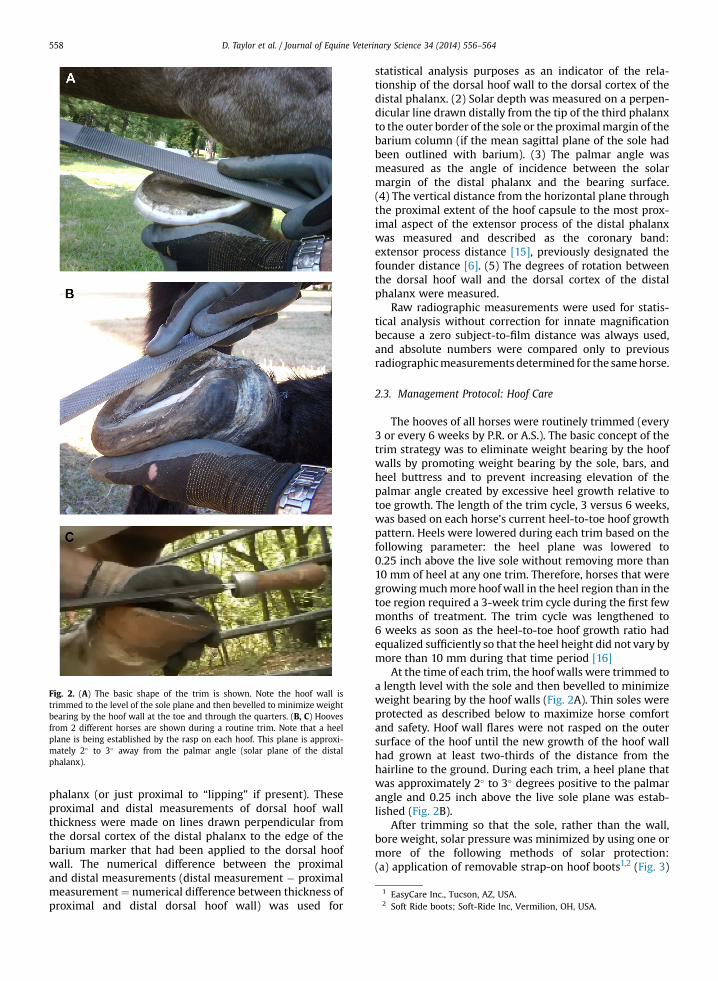
Fig. 2. (A) The basic shape of the trim is shown. Note the hoof wall istrimmed to the level of the sole plane and then bevelled to minimize weightbearing by the hoof wall at the toe and through the quarters. (B, C) Hoovesfrom 2 different horses are shown during a routine trim. Note that a heelplane is being established by the rasp on each hoof. This plane is approxi-mately 2� to 3� away from the palmar angle (solar plane of the distalphalanx).
1 EasyCare Inc., Tucson, AZ, USA.2 Soft Ride boots; Soft-Ride Inc, Vermilion, OH, USA.
D. Taylor et al. / Journal of Equine Veterinary Science 34 (2014) 556–564558
phalanx (or just proximal to “lipping” if present). Theseproximal and distal measurements of dorsal hoof wallthickness were made on lines drawn perpendicular fromthe dorsal cortex of the distal phalanx to the edge of thebarium marker that had been applied to the dorsal hoofwall. The numerical difference between the proximaland distal measurements (distal measurement � proximalmeasurement¼ numerical difference between thickness ofproximal and distal dorsal hoof wall) was used for
statistical analysis purposes as an indicator of the rela-tionship of the dorsal hoof wall to the dorsal cortex of thedistal phalanx. (2) Solar depth was measured on a perpen-dicular line drawn distally from the tip of the third phalanxto the outer border of the sole or the proximalmargin of thebarium column (if the mean sagittal plane of the sole hadbeen outlined with barium). (3) The palmar angle wasmeasured as the angle of incidence between the solarmargin of the distal phalanx and the bearing surface.(4) The vertical distance from the horizontal plane throughthe proximal extent of the hoof capsule to the most prox-imal aspect of the extensor process of the distal phalanxwas measured and described as the coronary band:extensor process distance [15], previously designated thefounder distance [6]. (5) The degrees of rotation betweenthe dorsal hoof wall and the dorsal cortex of the distalphalanx were measured.
Raw radiographic measurements were used for statis-tical analysis without correction for innate magnificationbecause a zero subject-to-film distance was always used,and absolute numbers were compared only to previousradiographicmeasurements determined for the samehorse.
2.3. Management Protocol: Hoof Care
The hooves of all horses were routinely trimmed (every3 or every 6 weeks by P.R. or A.S.). The basic concept of thetrim strategy was to eliminate weight bearing by the hoofwalls by promoting weight bearing by the sole, bars, andheel buttress and to prevent increasing elevation of thepalmar angle created by excessive heel growth relative totoe growth. The length of the trim cycle, 3 versus 6 weeks,was based on each horse’s current heel-to-toe hoof growthpattern. Heels were lowered during each trim based on thefollowing parameter: the heel plane was lowered to0.25 inch above the live sole without removing more than10 mm of heel at any one trim. Therefore, horses that weregrowingmuchmore hoof wall in the heel region than in thetoe region required a 3-week trim cycle during the first fewmonths of treatment. The trim cycle was lengthened to6 weeks as soon as the heel-to-toe hoof growth ratio hadequalized sufficiently so that the heel height did not vary bymore than 10 mm during that time period [16]
At the time of each trim, the hoof walls were trimmed toa length level with the sole and then bevelled to minimizeweight bearing by the hoof walls (Fig. 2A). Thin soles wereprotected as described below to maximize horse comfortand safety. Hoof wall flares were not rasped on the outersurface of the hoof until the new growth of the hoof wallhad grown at least two-thirds of the distance from thehairline to the ground. During each trim, a heel plane thatwas approximately 2� to 3� degrees positive to the palmarangle and 0.25 inch above the live sole plane was estab-lished (Fig. 2B).
After trimming so that the sole, rather than the wall,bore weight, solar pressure was minimized by using one ormore of the following methods of solar protection:(a) application of removable strap-on hoof boots1,2 (Fig. 3)

Fig. 3. Two styles of protective hoof boots1,2 used to protect and hold soft protective pads1,3 in place on the feet of the horses of this report. Human athletic socks(at left) enhanced hoof hygiene with either boot design when horses were wearing hoof boots 24 hours a day, 7 days a week.
D. Taylor et al. / Journal of Equine Veterinary Science 34 (2014) 556–564 559
with foam rubber pads1,3 and/or dental impression mate-rial4,5; (b) application of hoof casts6 to cover pads1,3 and/ordental impression material4,5 that had been applied to fillthe solar concavity and collateral sulci; (c) application ofglue-on hoof boots1 with dental impression material4,5
filling the solar concavity and collateral sulci; or(d) allowing the horse to go barefoot on yielding terrainincluding soft ground free of rocks or loose beds of peagravel (5- to 8-mm-diameter stones) 10 cm deep.
Removable, strap-on hoof boots1,2 with pads1,3 werealways the first choice as an initial method of solarprotection and were used as needed for horse comfort andsafety on the available terrain. Horses wore hoof bootsnearly 24 hours per day until their use no longer improvedthe level of horse comfort. Horse comfort was subjectivelyjudged based on the Obel lameness scale and the horse’sdaily habits of self- initiated movement and recumbency.Owners were instructed to remove the strap-on hoof bootswith pads from the hooves briefly each day to allowcleaning and drying of the hoof and the boot. During thelater stages of treatment (when the horse had 2-3 cm ofuniform new hoof growth from the coronary band),removable hoof boots (used as a method of solar protec-tion) were, for some horses, replaced by hoof casts or glue-on hoof boots (for periods of 6-12 weeks). The decision touse hoof casts or glue-on hoof boots (in lieu of removablestrap-on boots) was based on one of following factors: theowner’s inability to maintain the removable strap-on hoof
3 Wrestling mat pads; Rood and Riddle Equine Podiatry Center, Lex-ington, KY, USA.
4 Advanced Cushion Support; Nanric Inc, Lawrenceburg, KY, USA.5 EDSS sole support impression material; Hope for Soundness Inc.,
Penrose, CO, USA.6 Equicast, Equicast Inc, Aberdeen, NC, USA.
boots or the need for establishment of a shallow (5- to6-mm) air space under a very thin sole (ie, <7 mm) ora solar perforation or defect. Hoof casts were always chosenover glue-on boots when the need to establish and securepads with cutout air spaces under jeopardized sole regionsarose. The use of deep pea gravel footing for horses withsole depths of 8 to 10 mm or more was encouraged. It wasnoted that rock diameter needed to be matched to soledepth in order to observe signs of increased comfort. Use ofpea gravel footing was recommended only for periods ofrehabilitation process when the current sole depth and peagravel diameter combination allowed horses to exhibitsigns of increased comfort while on the gravel. Signs ofincreased comfort included (1) the horse choosing to standin the gravel rather than other terrain, (2) the horse turningmore easily in the gravel, (3) the horse spending more timestanding while on the gravel (as opposed to other availableterrain), and (4) the horse assuming a more normal stancewhen standing on the gravel. Barefoot turnout was notallowed until a horse had sole depth �12 mm and was ableto move comfortably on the current terrain without hoofprotection.
2.4. Management Protocol: Diet
The weight loss goal was to decrease each horse’s bodycondition score to a five [18]. Dietary restrictions recom-mended for each laminitic horse included elimination ofgrains and/or processed feeds, elimination of fruits, vege-tables, and other sweet or starchy treats, partial orcomplete restriction from pasture grazing (this restrictionvaried according body condition of the horse and size orcondition of available pasture). Horses were fed Bermudagrass hay ad libitum. During the initial weight loss period,
D. Taylor et al. / Journal of Equine Veterinary Science 34 (2014) 556–564560
owners were advised to have their hay tested for nutrientcontent. Advice was also offered at the onset of treatmentthat any untested hay be soaked inwater prior to feeding ineffort to minimize the chance of feeding high nonstructuralcarbohydrate hay. Retrospectively, none of the owners re-ported having tested the types of hay fed to these 14 horses.Owners reported soaking their hay in water for varioustime periods ranging from 30 minutes to 12 hours prior tofeeding. Mineral supplementation was provided empiri-cally to balance suspected nutritional content of local hay/grass to meet recommendations of the National ResearchCouncil [17].
2.5. Management Protocol: Exercise
Turnout in a grass-free paddock or daily in-hand exer-cise was encouraged after the following (1) hooves hadbeen trimmed tominimizeweight bearing by the hoofwall;the desired heel plane and palmar angle (� 10 degrees)had been established by trimming; and when the hooveshad been protected by soft protective hoof boots with pads(as described in Section 2.3) that provided enough comfortfor the horse to have a heel-first hoof landing. In-handexercise was increased daily by adding 5- to 10-minuteincrements to each exercise session until horses werewalking 30 to 45 minutes 2 or 3 times daily. Owners wereinstructed to observe for the intended heel-first impact ofthe hoof boot and to walk the horse only while the hoofboots were securely in place. They were instructed not towalk the horse if the hoof impact appeared to be toe-firstand to discontinue daily walking (and call the veterinary/hoof care provider team) if the horse seemed to haveincreasing pain after walking.
2.6. Management Protocol: Medication
Phenylbutazone (4.4 mg/kg once every 24 hours or2.2 mg/kg once every 12 hours orally) was used forameliorating pain when needed to maintain an Obel gradescore �2. Acepromazine (20 mg every 8 hours intramus-cular) was administered to 6 horses as a peripheral vaso-dilator during the initial 2 to 4 weeks of therapy. The solesof horses with solar necrosis and prolapsed solar corium(5 horses) were treated with topical tetracycline and/ormetronidazole. One horse with solar necrosis receivedintravenous oxytetracycline (7 mg/kg every 12 hoursintravenously for 14 days). During periods of active abscessformation and/or drainage of exudates from the sole,owners were advised to soak the feet 3 to 5 times weekly ineither magnesium sulfate solution or 50% acetic acidsolution.
7 SAS 9.2.1, SAS Institute Inc., Cary, NC, USA.
2.7. Body Condition Scoring
The body condition scores of horses were determined byD.R.T., using the system described by Henneke [18] beforetreatment and at the endpoint radiographic examinationafter the owners reported a return to the prelaminitis levelof soundness.
2.8. Statistical Analysis
Response variables, including coronary band-to-extensor process distance, palmar angle, degree of rota-tion, sole depth, and difference of the dorsal hoof wallthickness at the proximal and distal aspects, weremeasured or scored before and after treatment, hence thedata had the character of repeated measures, ie, multipleobservations on the same experimental unit. Data wereanalyzed using repeated measures methodology as imple-mented in SAS� PROC GLIMMIX.7 To account for thepossibility of an interaction, data were analyzed usingrepeated measures methodology using a linear model inwhich the main effects condition (condition refers tochronicity (acute or chronic) of laminitis) and treatment aswell as their interaction were included. Inspection of stu-dentized residuals (¼ standardized residuals distributed asa t random variable) indicated that the normally assump-tion was warranted for all response variables, except OBEL.The interaction was significant only for palmar angle (P ¼0.002). Because all other response variables had a non-significant interaction term (P � 0.63) it was dropped fromthe model resulting in a main effects model. Significance ofpairwise differences was calculated using the PDIFF orSLICEDIFF option of the LSMEANS statement in the abovenamed PROC. The dichotomous OBEL before and aftertreatment scores were analyzed using logistic regression asimplemented in in SAS� PROC LOGISTIC. Means and 95%confidence intervals on the logit scale were back-trans-formed to frequencies.
2.9. Long-term Follow-up Owner Survey
Owners of 12 horses were contacted 23 to 73 monthsafter initial onset and asked the following questions: “Hasthe horse had any foot pain due to laminitis relapses or hoofabscesses since the follow-up radiographic exam?” and “Isyour horse currently considered rideable?”
3. Results
Six different breeds were represented within this groupof horses (7 Tennessee Walking Horses, 2 Appaloosa,2 Quarter Horses, 1 grade horse, 1 Rocky Mountain SaddleHorse, and 1 Peruvian Paso; 10 geldings and 4 mares,ranging in age from 4-22 years; mean age, 13 years).Descriptive information for the measurements taken atpre- and post-treatment examinations is given in Table 1. Atinitial examinations, themedian Obel lameness score for allpretreatment laminitic horses was 3.5 (range: 0.5-4). Incontrast, at post-treatment evaluations, 12 horses appearedsubjectively to be sound at a trot or equivalent gait, and themedian Obel lameness score for all horses was 0 (range: 0-1). All owners considered the quality of their horse’s gait tobe equivalent to their prelaminitic gait. One of the authors(D.R.T.) evaluated the gaits of all horses at the time ofendpoint radiographic examination and subjectively scored12 horses (0 of 10) and 2 horses appeared mildly lame ata trot (2 of 10). Statistically, horses were 5.5 times more

Table 1Radiographic measurements before and after treatment
Measurement Examination Minimum Maximum Mean SEM
Coronary band: extensor process (mm) Pre-treatment 6.00 19.00 12.64 0.918Post-treatment 5.00 18.00 11.58 0.889
Palmar angle Pre-treatment 3.00 31.00 11.75 0.965Post-treatment 0.00 15.00 5.46 0.680
Degrees of rotation Pre-treatment 5.00 29.00 12.20 1.061Post-treatment 0.00 20.00 6.09 1.085
Sole depth (mm) Pre-treatment 4.00 13.00 8.38 0.553Post-treatment 8.00 16.00 11.14 0.531
DDHWT (mm) Pre-treatment 2.00 19.00 7.87 0.824Post-treatment 0 12.00 5.06 0.739
DDHWT, difference in dorsal hoof wall thickness- proximal vs. distal; SEM, standard error of the mean.
D. Taylor et al. / Journal of Equine Veterinary Science 34 (2014) 556–564 561
likely to be sound at post-treatment examination than atpre-treatment examinations. A statistically significantdecrease of the third phalanx rotation was detected post-treatment (P < .0001, 95% CI for differences: 3.8�-6.5�).
Table 2Long-term follow-up owner survey
Case Signalment(breed, age,sex)
Date ofPresentation(mo, yr)
Months fromPresentation toEndpointRadiographicExamination
Acute 1 Peruvian Paso, 10 yr,mare
September 2006 7
2 Tennessee WalkingHorse, 4 yr, mare
May 2007 5
3 Appaloosa, 8 yr, mare May 2007 114 Tennessee Walking
Horse 9 y.o., geldingApril 2008 10
5 Quarter Horse, 19 yr,gelding
August. 2008 5
6 Rocky MountainHorse, 10 yr, gelding
April 2009 20
Chronic 7 Tennessee WalkingHorse, 13 yr, gelding
May 2007 9
8 Grade, 15 yr, gelding June 2007 79 Appaloosa, 22 yr,
geldingJanuary 2009 6.5
10 Tennessee WalkingHorse, 15 yr, gelding
April 2009 13.5
11 Tennessee WalkingHorse, 6 yr, mare
April 2010 7
12 Tennessee WalkingHorse, 15 yr, gelding
April 2010 9
13 Tennessee WalkingHorse, 10 yr, gelding
May 2010 6
14 Quarter Horse 13 yr,gelding
May 2010 9
DSLD, degenerative suspensory ligament disease; NA, not applicable.* See Comments for explanation of statement.
The differences of the dorsal hoof wall thicknesses betweenthe proximal and distal aspects of the distal phalanx wassignificantly reduced at post-treatment evaluation (P <
.0001, 95% CI for differences: 2.0-3.5 mm). The reduction of
Months fromPresentationto FinalFollow-Up
Follow-Up Owner Survey Comments
Has the HorseHad Any FootPain: Relapses/Abscesses?
Is the HorseCurrentlyConsideredRideable?
45 No NA* *Euthanized June 2010due to DSLD; wassound April 2007until onset of DSDLin Fall 2009
73 No Yes
73 No Yes62 No Yes
58 No No* *Not rideable due topreexisting proximallimb arthritis
50 Two minorrelapses*
Yes *Both relapses wereassociated with anincrease in bodycondition score anda lack of timely hoofcare
73 NA* NA* *Horse lost to follow-up
NA* NA* NA* *Horse lost to follow-up53 No No Was rideable prior to
becomingneurologic in 2011
50 No Yes
38 No Yes
23 Chronic footabscessesin left front
NA* *Euthanized March2012 due to colic;sound betweenabscesses episodes14 months post-rehabilitation
37 No Yes
37 No Yes

Fig. 4. Oblique radiographic view of the left front foot of one of the subjecthorses. Note that the red line, which represents the hoof wall, is not parallelto the yellow line, which represents the cortical surface of the distal phalanx.This radiograph provides evidence that obesity-associated laminitis mayproduce lamellar pathology resulting in wall flares in the quarters as well asin the toe regions [16].
D. Taylor et al. / Journal of Equine Veterinary Science 34 (2014) 556–564562
the coronary band-to-extensor process distance was notstatistically significant (P¼ .10, 95% CI for differences:�0.2-2.3 mm). Solar depth was significantly increased at post-treatment evaluation (P ¼ .0015, 95% CI for differences:3.1-5.4 mm). An overall reduction of palmar anglemeasurements was detected in acutely and chronicallyaffected horses. This treatment effect was statisticallygreater for horses with chronic laminitis than for horseswith acute laminitis (P interaction < .0001).
A decrease of the body condition scores of each horsewas observed during the treatment period, and while themedian body condition score was 8.5 at initial evaluation,a median body condition score of 5 was observed at post-treatment. One horse became obese again subsequent topasture turn-out in the spring–summer season post-treatment, but, according to the owner, primary careveterinarian (D.R.T.) and hoof care professional (A.S.),remained sound while obese.
Results of long-term follow-up (2-6 years after initialonset of laminitis) of 12 horses indicated that 8 of 12owners considered their horse to be rideable (2 horseswere lost to follow-up and 2 horses were euthanized due tononlaminitis-related diseases; 1 acquired chronic neuro-logic deficits, and 1 has pre-existing arthritis of the prox-imal pelvic limbs). Only 1 owner reported transient footpain due to apparent laminitis relapse after allowing thehorse unlimited access to pasture, resulting in bodycondition score of 8 in conjunction with a lapse in hoofcare. One other owner reported recurrent abscess episodesin one foot (Table 2).
4. Discussion
It is likely that all horses in this study suffered fromendocrinopathic (grass-induced) laminitis because eachwas obese and there was no history or clinical evidence ofan acute systemic inflammatory disease or clinical signs ofpituitary disease. It is possible that some of these horsessuffered from pituitary pars intermedia dysfunction, butthe horses had no clinical signs of this syndrome before orafter the study. Because of the retrospective nature of thisstudy, insulin concentrations were determined only fora few horses, and other measures of glucose homeostasiswere not performed in any of the horses; therefore, thedata were not analyzed.
The prognosis for return to former athletic function forhorses with >11.5� of distal phalangeal rotation is consid-ered poor, and the prognosis for horses with 5.5� to 11.5� ofrotation is guarded [1]. Even though all horses in this studyhad>5.5� rotation of the distal phalanx and 6 had>11.5� ofrotation, all returned to their prelaminitis level of sound-ness as evaluated by owners. While it would have beenideal for a blinded unbiased clinician to have performedlameness evaluation on each of these horses, they weremanaged as field cases by a single veterinarian (D.R.T.), andthere was no clinical indication for objective evaluation ofgait at the time of initial and subsequent evaluations.
After laminar failure, the primary mechanical forceworking to separate the laminae is the weight of the horseopposed by the hoof wall [19]. When the hoof wall bearsweight, the lamellae are forced to suspend the horse’s
weight and bear foot impact forces. Even though tension ofthe deep digital flexor muscle and tendon exerts a rota-tional force on the distal phalanx, we suspect that tensionof the deep digital flexor tendon did not result in addedstress to laminae in these horses because the hoof wall inthe toe region was trimmed so it remained out of contactwith the bearing surface.
Because a significant overall reduction of the palmarangle of these horses was detected (from a pretreatmentmean of 11.75� to a post-treatment mean of 5.46�), thismethod of laminitis management should be furtherinvestigated as an alternative nonsurgical method forrestoring alignment of the distal phalanx [20] in laminitichorses.
The treatment effect on palmar angle reduction wasstatistically larger for chronic cases than for acute cases.
The authors believe that the amount of lamellar sepa-ration at the quarter walls (or anywhere around theperimeter of the foot) may be as significant as the amountof lamellar separation in the region of the toe [16] (Fig. 4).Eliminating or at least reducing weight-bearing by theentire perimeter of the hoof wall (with the exception of theheel buttress) may be important in stopping and possiblyreversing distal decent of the third phalanx in horses witheither acute or chronic laminitis. Reduction or eliminationof weight-bearing by the hoof wall theoretically decreasesstrain on laminar attachments by removing mechanicalforces, thus minimizing or preventing additional rotationor sinking [21]. Elimination of weight-bearing by the hoofwall may also reverse distal descent of the distal phalanx insome horses as demonstrated in lateromedial radiographsof the right front foot of horse 11 (Fig. 5) obtained at initialevaluation and 2 years 7 months later.
This hoof care method used the sole to support thedistal phalanx. While using the sole to support the distal

Fig. 5. The radiographs8 show that the coronary band to extensor process distance (CE) may become smaller over time in this hoof care system. The radiograph atright (CE ¼ 13 mm) is a follow-up examination of the right front foot 2 years and 7 months after the initial evaluation in the radiograph on the left (CE ¼ 17 mm).
D. Taylor et al. / Journal of Equine Veterinary Science 34 (2014) 556–564 563
phalanx, it is important that the sole be protected duringweight-bearing yet not be subjected to pressure duringhoof flight. When sole thickness is <12 mm and/or whenweight-bearing by the hoof wall is eliminated, excesspressure on the solar corium can result. When the soleunder the margin of the distal phalanx was less than 7mm,bulging, or cracked, an air space had been established in thehoof pad under the solar margin. We suspect that lack ofsolar pressure during hoof flight is critical to maintainingsolar blood flow to prevent solar corium injury. Eachmethod of solar protection used in these cases was inten-ded to release pressure on the solar corium during hoofflight. Efforts were made to protect the laminar corium byallowing the hoof wall to bear little or no weight by bev-eling the hoof wall away from the ground and not allowingapplication of a shoe to the hoof wall (Fig. 2).
When the horse’s foot is bearing weight, arterial bloodflow to the foot is occluded, in part, by force of the deepdigital flexor tendon on the medial and lateral digitalarteries. This is a normal physiological event, and it is the
Fig. 6. Radiographs8 show change in the dimension of the soft tissues of the heel an2, 2009; right image, October 20, 2011). Note that the weight-bearing surface of therealignment of the bones of the distal limb and the lowering of the palmar angle (
likely mechanism preventing backflow of arterial bloodduring the loading phase of the stride [22]. When the footdoes not bear weight, arterial blood flow to the foot isrestored. We suspect that exercise of these horses causedan intermittent lack of pressure to the sole and preventedischemia of the laminar and solar corium that occurs whenthe horse’s foot bears weight continuously. It is importantto understand that these horses were exercised with thehoof wall bevelled to minimize weight-bearing by the hoofwall, the sole well protected by soft pads held on the hoofby hoof boots or occasionally casts and a near normalpalmar angle of less than 10�. If horses did not land heel-first in their protective hoof wear during exercise, exer-cise was discontinued until this desired hoof landing wasachieved by altering the hoof mechanics and padding [16].The exercise regimen seemed to alleviate pain in thesehorses as shown by increased comfort (decreased pain –
modified Obel pain scale) [22] during and after exercise.Exercise is recommended for horses suffering from insulinsensitivity [9,11]. This laminitis management method may
d shortening of the digital breakover over 2 years 6 months (left image, Aprilheel region has increased 6 mm in length (from 9.3 to 9.9 cm). Also note thepre: 22�; post: 12�).

D. Taylor et al. / Journal of Equine Veterinary Science 34 (2014) 556–564564
provide a means of exercising insulin resistant laminitichorses without jeopardizing their hooves. These horseswere fed a diet that was intended to minimize the intake ofnonstructural carbohydrates, provided free-choice accessto hay and also provide a mineral-balanced diet.
Because none of the types of hay fed to the 14 horses ofthis study were tested for nutritional content and ownerssoaked the hay for various amounts of time, the actual die-tary intake of these horses could not be determined retro-spectively. The hay “as fed” combined with the exerciseprogram “as applied” did cause a statistically significantdecrease in body condition score, which indicates that thediet and exercise regime was clinically appropriate.However, close attention to diet to include nutrient testingof hay fed to obese laminitic horses would obviously besuperior to the empirical feeding programs of these horses.
This method of laminitis management apparently alle-viated pain and returned this group of horses to theirformer level of athletic ability despite suspected minor tomoderate nutritional imbalance, distal phalanx remodel-ling in some horses, and incomplete resolution of hoof wallrotation in some horses (Fig. 5). It is hypothesized thatincreased heel volume (Fig. 6) may in some way compen-sate for distal phalanx remodelling and laminar damage asaltered heel dimension similar to the improved heel anglesdescribed by Clayton et al [23] was also noted in thesehorses.
As none of these horses was hospitalized during thelaminitis treatment period, owner compliance with dailycare recommendations likely played a role in the favorableoutcome in these horses. Successful management of obese,laminitic horses requires a concentrated long-term teameffort among the veterinarian, the owner, and the hoof careprofessional to manage the horse’s diet, exercise, hoof care,and medical needs. In this series of cases, the radiographicparameters of the hoof improved, and lameness decreasedduring the period of dietary restriction, weight loss, andexercise.
Using the described management protocol, 14 of 14laminitic horses with rotation�5� and a guarded prognosisreturned to their prelaminitis level of soundness by thetime of the endpoint radiographic evaluation. Long-termfollow-up survey indicated that most of these horsesmaintained the same level of soundness without incidenceof laminitis recurrence or hoof abscess formation. Thismethod of laminitis management is effective and warrantsfurther evaluation.
Acknowledgments
We would like to acknowledge the dedicated ownersand care takers of the horses described in the manuscript.
8 SoundEklin, Carlsbad, CA, USA.
References
[1] Stick JA, Jann HW, Scott EA, Robinson NE. Pedal bone rotation asa prognostic sign in laminitis of horses. J Am Vet Med Assoc 1982;180:251–3.
[2] Hunt RJ. A retrospective evaluation of laminitis in horses. Equine VetJ 1993;25:61–4.
[3] Coffman JR, Hammond LS, Garner HE, Thawley DG, Selby LA. Hae-matology as an aid to prognosis of chronic laminitis. Equine Vet J1980;12:30–1.
[4] Coffman JR, Johnson JH, Finocchio EJ, Guffy MM. Biomechanics ofpedal rotation in equine laminitis. J Vet Med Assoc 1970;156:219–21.
[5] Colles CM, Jeffcott LB. Laminitis in the horse. Vet Rec 1977;100:262–4.
[6] Cripps PJ, Eustace RA. Factors involved in the prognosis of equinelaminitis in the UK. Equine Vet J 1999;31:433–42.
[7] Asplin KE, Patterson-Kane JC, Sillence MN, Pollitt CC, McGowan CM.Histopathology of insulin-induced laminitis in ponies. Equine Vet J2010;42:700–6.
[8] de Laat MA, Kyaw-Tanner MT, Nourian AR, McGowan CM,Sillence MN, Pollitt CC. The developmental and acute phases ofinsulin-induced laminitis involve minimal metalloproteinaseactivity. Vet Immunol Immunopathol 2011;120:275–81.
[9] Freestone JF, Beadle R, Shoemaker K, Bessin RT, Wolfsheimer KJ,Church C. Improved insulin sensitivity in hyperinsulinaemic poniesthrough physical conditioning and controlled feed intake. EquineVet J 1992;24:187–90.
[10] Stewart-Hunt L, Geor RJ, McCutcheon LJ. Effects of short-termtraining on insulin sensitivity and skeletal muscle glucosemetabolism in Standardbred horses. Equine Vet J Suppl 2006;36:226–32.
[11] Frank N, Geor RJ, Baily SR. Equine metabolic syndrome. ACVIMconsensus. J Vet Intern Med 2010;24:467–75.
[12] Obel N. Studies of the Histopathology of Acute Laminitis [disserta-tion]. Uppsala, Sweden: Almqvist and Wilksells Boktryckeri AB;1948.
[13] Taylor DR. Radiographic imaging of the laminitis patient. In:Ramey P, editor. Care and rehabilitation of the equine foot. Lake-mont, GA: Hoof Rehabilitation Publishing LLC; 2011. p. 234–63.
[14] Redden RF. Shoeing the laminitic horse. In: Norwood G, editor.Proceedings of the Forty-third Annual Convention of the AmericanAssociation of Equine Practitioners. Phoenix, Arizona, USA: AAEPPublishing; 1997. p. 356–9.
[15] Redden RF. Clinical and radiographic examination of the equinefoot. In: Bramlage L, editor. Proceedings of the Forty-ninth AnnualConvention of the American Association of Equine Practitioners.New Orleans, Lousianna, USA: AAEP Publishing; 2003. p. 169–85.
[16] Ramey P, editor. Care and rehabilitation of the equine foot. Lake-mont, GA: Hoof Rehabilitation Publishing LLC; 2011. p. 284–352.
[17] National Research Council nutrient requirements of horses, 6th rev.ed. Washington, DC: National Academies Press; 2007.
[18] Henneke DR. A condition score system for horses. Equine Pract1985;8:13–5.
[19] Steven D. Functional anatomy of the horse’s foot. Pract 1981;3:22–7.[20] O’Grady SE. How to restore alignment of P3 in horses with
chronic laminitis. In: Bramlage L, editor. Proceedings of the Forty-ninth Annual Convention of the American Association of EquinePractitioners. New Orleans, Lousianna, USA: AAEP Publishing; 2003.p. 328–36.
[21] Ramey P. Care and rehabilitation of the hoof walls and lamellarattachment. In: Ramey P, editor. Care and rehabilitation of theequine foot. Lakemont, GA: Hoof Rehabilitation Publishing LLC;2011. p. 345.
[22] Van Eps A, Collins SN, Pollitt CC. Supporting limb laminitis.Advances in laminitis. Part II. Equine Pract 2010;26:287–302.
[23] Clayton HM, Gray S, Kaiser LJ, Bowker RM. Effect of barefoot trim-ming on hoof morphology. Aust Vet J 2011;89:305–11.