Clinical Interpretation Standards and Quality Assurance ...
7
Clinical Interpretation Standards and Quality Assurance for the Multicenter PET/CT Trial Rubidium-ARMI Jennifer M. Renaud 1 , Ilias Mylonas 1 , Brian McArdle 1 , Taylor Dowsley 1 , Kathy Yip 2 , Eric Turcotte 3 , Jean Guimond 4 , Mikael Trottier 4 , Philipe Pibarot 4 , Conor Maguire 5 , Lucille Lalonde 5 , Karen Gulenchyn 6 , Gerald Wisenberg 7 , R. Glenn Wells 1 , Terrence Ruddy 1 , Benjamin Chow 1 , Rob S.B. Beanlands 1 , and Robert A. deKemp 1 1 University of Ottawa Heart Institute, Ottawa, Ontario, Canada; 2 KMH Cardiology and Diagnostic Centres, Mississauga, Ontario, Canada; 3 Centre Hospitalier Universitaire de Sherbrooke, Quebec, Canada; 4 Institut Universitaire de Cardiologie et de Pneumologie de Québec, Quebec, Canada; 5 University of Alberta Hospital, Edmonton, Alberta, Canada; 6 St. Joseph’s Healthcare, Hamilton, Ontario, Canada; and 7 St. Joseph’s Health Care, London, Ontario, Canada Rubidium-ARMI ( 82 Rb as an Alternative Radiopharmaceutical for Myocardial Imaging) is a multicenter trial to evaluate the accuracy, outcomes, and cost-effectiveness of low-dose 82 Rb perfusion im- aging using 3-dimensional (3D) PET/CT technology. Standardized imaging protocols are essential to ensure consistent interpretation. Methods: Cardiac phantom qualifying scans were obtained at 7 recruiting centers. Low-dose (10 MBq/kg) rest and pharmacologic stress 82 Rb PET scans were obtained in 25 patients at each site. Summed stress scores, summed rest scores, and summed differ- ence scores (SSS, SRS, and SDS [respectively] 5 SSS–SRS) were evaluated using 17-segment visual interpretation with a discretized color map. All scans were coread at the core lab (University of Ottawa Heart Institute) to assess agreement of scoring, clinical di- agnosis, and image quality. Scoring differences greater than 3 un- derwent a third review to improve consensus. Scoring agreement was evaluated with intraclass correlation coefficient (ICC-r), concor- dance of clinical interpretation, and image quality using k coefficient and percentage agreement. Patient 99m Tc and 201 Tl SPECT scans (n 5 25) from 2 centers were analyzed similarly for comparison to 82 Rb. Results: Qualifying scores of SSS 5 2, SDS 5 2, were achieved uniformly at all imaging sites on 9 different 3D PET/CT scanners. Patient scores showed good agreement between core and recruiting sites: ICC-r 5 0.92, 0.77 for SSS, SDS. Eighty-five and eighty-seven percent of SSS and SDS scores, respectively, had site–core differences of 3 or less. After consensus review, scoring agreement improved to ICC-r 5 0.97, 0.96 for SSS, SDS (P , 0.05). The agreement of normal versus abnormal (SSS $ 4) and nonische- mic versus ischemic (SDS $ 2) studies was excellent: ICC-r 5 0.90 and 0.88. Overall interpretation showed excellent agreement, with a k 5 0.94. Image quality was perceived differently by the site versus core reviewers (90% vs. 76% good or better; P , 0.05). By comparison, scoring agreement of the SPECT scans was ICC-r 5 0.82, 0.72 for SSS, SDS. Seventy-six and eighty-eight per- cent of SSS and SDS scores, respectively, had site–core differen- ces of 3 or less. Consensus review again improved scoring agree- ment to ICC-r 5 0.97, 0.90 for SSS, SDS (P , 0.05). Conclusion: 82 Rb myocardial perfusion imaging protocols were implemented with highly repeatable interpretation in centers using 3D PET/CT technology, through an effective standardization and quality assurance program. Site scoring of 82 Rb PET myocardial perfusion imaging scans was found to be in good agreement with core lab standards, suggesting that the data from these centers may be combined for analysis of the rubidium-ARMI endpoints. Key Words: positron emission tomography; myocardial perfusion imaging; 82 Rb J Nucl Med 2014; 55:58–64 DOI: 10.2967/jnumed.112.117515 Myocardial perfusion imaging (MPI) using 201 Tl- and 99m Tc-labeled tracers is an accepted indication for detection of obstructive coronary artery disease and stratification of patients at risk for adverse cardiovascular events (1,2). 82 Rb is an alternative isotope with the lowest radiation dose among perfusion imaging tracers (0.7–1.3 mSv/GBq) (3,4) and is considered to have superior accuracy and incremental prognostic value (5–10). Rubidium-ARMI ( 82 Rb as an Alternative Radiopharmaceutical for Myocardial Imaging) is a multicenter trial to evaluate the ac- curacy, clinical outcomes, and cost-effectiveness of low-dose 82 Rb MPI using 3D PET/CT, compared with conventional 99m Tc and 201 Tl SPECT imaging (10). Standardization of acquisition and in- terpretation methods is essential to allow consistent analysis of pooled data from multiple participating centers. A single core site (University of Ottawa Heart Institute) had previous experience per- forming 82 Rb PET in Canada because the tracer was not yet ap- proved for sale. Therefore, an initial process of knowledge transfer was proposed to establish the training and standardized procedures for high-quality 82 Rb perfusion imaging and interpretation at nu- clear imaging centers across Canada. We hypothesized that a com- prehensive quality assurance (QA) program would achieve low in- terobserver variability between the new 82 Rb imaging sites and the QA core site. MATERIALS AND METHODS Qualifying Scans Image Acquisition. Standards were established using the Discovery 690 PET/CT system (GE Healthcare) at the core site (University of Ottawa Heart Institute) (8,9). Qualifying scans simulating normal rest and abnormal stress 82 Rb MPI were then obtained at all sites using an anthropomorphic torso phantom (Data Spectrum) to standardize Received Dec. 10, 2012; revision accepted Jul. 31, 2013. For correspondence or reprints contact: Robert deKemp, University of Ottawa Heart Institute, 40 Ruskin St., Ottawa, ON, Canada K1Y 4W7. E-mail: [email protected] Published online Nov. 18, 2013. COPYRIGHT ª 2014 by the Society of Nuclear Medicine and Molecular Imaging, Inc. 58 THE JOURNAL OF NUCLEAR MEDICINE • Vol. 55 • No. 1 • January 2014
Transcript of Clinical Interpretation Standards and Quality Assurance ...
Clinical Interpretation Standards and Quality Assurance forthe Multicenter PET/CT Trial Rubidium-ARMI
Jennifer M. Renaud1, Ilias Mylonas1, Brian McArdle1, Taylor Dowsley1, Kathy Yip2, Eric Turcotte3, Jean Guimond4,Mikael Trottier4, Philipe Pibarot4, Conor Maguire5, Lucille Lalonde5, Karen Gulenchyn6, Gerald Wisenberg7,R. Glenn Wells1, Terrence Ruddy1, Benjamin Chow1, Rob S.B. Beanlands1, and Robert A. deKemp1
1University of Ottawa Heart Institute, Ottawa, Ontario, Canada; 2KMH Cardiology and Diagnostic Centres, Mississauga, Ontario,Canada; 3Centre Hospitalier Universitaire de Sherbrooke, Quebec, Canada; 4Institut Universitaire de Cardiologie et de Pneumologiede Québec, Quebec, Canada; 5University of Alberta Hospital, Edmonton, Alberta, Canada; 6St. Joseph’s Healthcare, Hamilton,Ontario, Canada; and 7St. Joseph’s Health Care, London, Ontario, Canada
Rubidium-ARMI (82Rb as an Alternative Radiopharmaceutical for
Myocardial Imaging) is a multicenter trial to evaluate the accuracy,outcomes, and cost-effectiveness of low-dose 82Rb perfusion im-
aging using 3-dimensional (3D) PET/CT technology. Standardized
imaging protocols are essential to ensure consistent interpretation.
Methods: Cardiac phantom qualifying scans were obtained at 7recruiting centers. Low-dose (10 MBq/kg) rest and pharmacologic
stress 82Rb PET scans were obtained in 25 patients at each site.
Summed stress scores, summed rest scores, and summed differ-
ence scores (SSS, SRS, and SDS [respectively] 5 SSS–SRS) wereevaluated using 17-segment visual interpretation with a discretized
color map. All scans were coread at the core lab (University of
Ottawa Heart Institute) to assess agreement of scoring, clinical di-agnosis, and image quality. Scoring differences greater than 3 un-
derwent a third review to improve consensus. Scoring agreement
was evaluated with intraclass correlation coefficient (ICC-r), concor-
dance of clinical interpretation, and image quality using k coefficientand percentage agreement. Patient 99mTc and 201Tl SPECT scans
(n 5 25) from 2 centers were analyzed similarly for comparison
to 82Rb. Results: Qualifying scores of SSS 5 2, SDS 5 2, were
achieved uniformly at all imaging sites on 9 different 3D PET/CTscanners. Patient scores showed good agreement between core
and recruiting sites: ICC-r 5 0.92, 0.77 for SSS, SDS. Eighty-five
and eighty-seven percent of SSS and SDS scores, respectively, had
site–core differences of 3 or less. After consensus review, scoringagreement improved to ICC-r 5 0.97, 0.96 for SSS, SDS (P , 0.05).
The agreement of normal versus abnormal (SSS $ 4) and nonische-
mic versus ischemic (SDS $ 2) studies was excellent: ICC-r 5 0.90and 0.88. Overall interpretation showed excellent agreement, with
a k 5 0.94. Image quality was perceived differently by the site
versus core reviewers (90% vs. 76% good or better; P , 0.05).
By comparison, scoring agreement of the SPECT scans wasICC-r 5 0.82, 0.72 for SSS, SDS. Seventy-six and eighty-eight per-
cent of SSS and SDS scores, respectively, had site–core differen-
ces of 3 or less. Consensus review again improved scoring agree-
ment to ICC-r 5 0.97, 0.90 for SSS, SDS (P , 0.05). Conclusion:82Rb myocardial perfusion imaging protocols were implemented
with highly repeatable interpretation in centers using 3D PET/CT
technology, through an effective standardization and quality
assurance program. Site scoring of 82Rb PET myocardial perfusionimaging scans was found to be in good agreement with core lab
standards, suggesting that the data from these centers may be
combined for analysis of the rubidium-ARMI endpoints.
Key Words: positron emission tomography; myocardial perfusion
imaging; 82Rb
J Nucl Med 2014; 55:58–64DOI: 10.2967/jnumed.112.117515
Myocardial perfusion imaging (MPI) using 201Tl- and99mTc-labeled tracers is an accepted indication for detection of
obstructive coronary artery disease and stratification of patients at
risk for adverse cardiovascular events (1,2). 82Rb is an alternativeisotope with the lowest radiation dose among perfusion imaging
tracers (0.7–1.3 mSv/GBq) (3,4) and is considered to have superior
accuracy and incremental prognostic value (5–10).Rubidium-ARMI (82Rb as an Alternative Radiopharmaceutical
for Myocardial Imaging) is a multicenter trial to evaluate the ac-curacy, clinical outcomes, and cost-effectiveness of low-dose 82Rb
MPI using 3D PET/CT, compared with conventional 99mTc and201Tl SPECT imaging (10). Standardization of acquisition and in-
terpretation methods is essential to allow consistent analysis ofpooled data from multiple participating centers. A single core site
(University of Ottawa Heart Institute) had previous experience per-
forming 82Rb PET in Canada because the tracer was not yet ap-proved for sale. Therefore, an initial process of knowledge transfer
was proposed to establish the training and standardized procedures
for high-quality 82Rb perfusion imaging and interpretation at nu-
clear imaging centers across Canada. We hypothesized that a com-prehensive quality assurance (QA) program would achieve low in-
terobserver variability between the new 82Rb imaging sites and the
QA core site.
MATERIALS AND METHODS
Qualifying Scans
Image Acquisition. Standards were established using the Discovery690 PET/CT system (GE Healthcare) at the core site (University of
Ottawa Heart Institute) (8,9). Qualifying scans simulating normal rest
and abnormal stress 82Rb MPI were then obtained at all sites using
an anthropomorphic torso phantom (Data Spectrum) to standardize
Received Dec. 10, 2012; revision accepted Jul. 31, 2013.For correspondence or reprints contact: Robert deKemp, University of
Ottawa Heart Institute, 40 Ruskin St., Ottawa, ON, Canada K1Y 4W7.E-mail: [email protected] online Nov. 18, 2013.COPYRIGHT ª 2014 by the Society of Nuclear Medicine and Molecular
Imaging, Inc.
58 THE JOURNAL OF NUCLEAR MEDICINE • Vol. 55 • No. 1 • January 2014

reconstructed image resolution, perfusion defect contrast, and CT at-
tenuation correction (CTAC) image alignment. The phantom contained
heart and liver inserts allowing scatter from abdominal organs to be
simulated (Fig. 1A). With the phantom placed in a prone position,
a CT scout scan was obtained to center the heart in the field of view.
The phantom was removed from the bed, and 1,000–1,500 MBq of82Rb activity were infused rapidly into the water-filled liver cavity.
After vigorous mixing, 60 mL were withdrawn from the liver and
injected directly into the heart wall chamber and the remaining volume
filled with water. The resulting 2:1 activity concentration in liver:myo-
cardium simulated image contrast observed at rest, when the liver,
spleen, pancreas, or stomach wall is often visualized. The phantom
was repositioned on the scanner bed, and 2 min after infusion an 8-min
scan was acquired, followed by a CTAC scan. To simulate stress-
induced ischemia, a 1-cm3 transmural plastic defect was placed in
the inferior wall of the myocardium chamber. The rubidium imaging
procedure was repeated, except 120 mL were withdrawn from the
liver and injected into the heart wall, resulting in liver:myocardium
stress contrast of 1:1.
Image Reconstruction. Tracer uptake images were reconstructedusing the vendor default iterative reconstruction settings with 12-mm
postfiltering and all corrections enabled. Images were corrected
explicitly for the 776.5-keV cascade/prompt g emissions from 82Rb
decay (;15% abundance) on some PET systems (Table 1). Prompt g
emissions recorded in coincidence with annihilation photons produce
a background signal distinct from scatter and randoms, reported to
produce septal artifacts on some 3D PET systems (11). On other
systems without explicit cor-
rection, a 50-cm CTAC field of
view was used to minimize the
potential prompt g effects. Vendor-
specific fusion display of the
PET and CTAC images was used
to correct or verify alignment
of the CT images for proper
attenuation correction. Sites were
instructed to verify that the 50%
PET activity contour fell within
the CT soft-tissue contour on the
fusion display (Figs. 1B and 1C).
Semiquantitative Analysis.The uptake defect summed rest
score (SRS), summed stress
score (SSS), and summed dif-
ference score (SDS) was com-
puted automatically with Corri-
dor4DM (INVIA), comparing
scans against a simulated data-
base with a uniform mean of
75% and SD of 15%. Defect
scores were assigned on a 0–4
scale (0, normal tracer uptake;
4, absent tracer) of defect se-
verity in 17 myocardial seg-
ments according to the thresh-
olds shown in Figure 2.
Patient Scans
Population. 82Rb PET im-ages acquired consecutively
from the trial start date at each
site (May 2010 to February 2012)
were evaluated from 25 pa-
tients enrolled at 6 of the sites
and 24 patients from the seventh site (n 5 174); 1 patient could not
complete the stress scan because of claustrophobia. The 99mTc
SPECT images of 25 patients acquired at a single recruiting site
from October to November 2011 and 201Tl SPECT images of 25
patients acquired at another site during the 99mTc shortage (June to
September 2009) were assessed. All patients were referred for clin-
ically indicated myocardial perfusion scans for diagnosis or risk
stratification of coronary artery disease. Summary demographic data
are presented for PET and SPECT patients in Table 2 and by site in
Supplemental Table 1 (supplemental materials are available at http://jnm.snmjournals.org). The study was approved by the research ethics
boards at all participating centers. All patients signed a written in-
formed consent form before enrollment.Patients were instructed to fast overnight and abstain from caffeine
and theophylline-containing medications for 12 h before the test as perguidelines of the American Society of Nuclear Cardiology (12). Anti-
anginal medications (b-blockers, calcium antagonists, and nitrates)were withheld on the morning of the study.
PET Imaging. Patients underwent low-dose rest and dipyridamolestress 82Rb PET MPI; 10 MBq/kg of body weight were infused
over 30 s using a custom infusion system (13). A 6- to 8-min staticscan was started 2 min after injection, when the randoms rate was
less than two thirds the total coincidence counting rate. The acqui-sition sequence was rest CTAC, rest PET, dipyridamole (0.140 mg/
kg/min · 4–5 min), stress PET, aminophylline (optional), and stressCTAC (Fig. 3). The CTAC scans were fast helical (,5 s), low-dose
scans acquired after breath-hold or normal end expiration. Staticimages were reconstructed using the same methods as described
above.SPECT Imaging. Patients imaged with 201Tl SPECT underwent
a 1-d stress-redistribution study with a single injection of 130 MBqat peak dipyridamole (0.140 mg/kg/min · 5 min) stress. Stress scans
were acquired on an Infinia Hawkeye 4 dual-head SPECT/CT camera(GE Healthcare) at 10–15 min after injection and after 4 h of redis-
tribution. Thirty projections were obtained at 45 s/projection over180� rotation. Static images were reconstructed using filtered back
projection with a 10th-order Butterworth filter, 0.35 cycles/cm cutoff
frequency, and no attention correction as per standard clinical practice
at the site.99mTc-sestamibi SPECT patients underwent a 2-d stress–rest pro-
tocol. Eight minutes after dipyridamole injection (0.140 mg/kg/min ·5 min), patients received 555–1,110 MBq of 99mTc-sestamibi, and
4 min later 100–200 mg of aminophylline were administered. Stress
imaging was performed 45–90 min after dipyridamole using a Vertex
or Forte dual-head SPECT camera (ADAC Laboratories). Counts
were collected over 180� rotation with 64 projections of 28 s each.
Static images were reconstructed using ordered-subset expectation
maximization (10 subsets, 2 iterations), a fifth-order Butterworth
filter, 0.52 cycles/cm cutoff frequency, and no attenuation correction
as per standard clinical practice at the site. Rest imaging was
performed the day before or after the stress study using the same
protocol.
Semiquantitative Analysis. PET and SPECT scans were assessedvisually for image quality as good, fair, or poor. Semiquantitative
segmental scoring of SSS, SRS, and SDS was performed with
Corridor4DM as described above. Default scores were set automat-
ically using the simulated database with a uniform 75% normal cutoff
to establish a consistent starting point for clinical interpretation, as
scanner-specific normal databases were not available for 3D PET/CT.
A discretized, 10-step color map was used for visualization, with the
scores corresponding to the colors and database percentages shown inFigure 2. Default scores were modified by the interpreting physician
according to their expert visual assessment and the following guide-
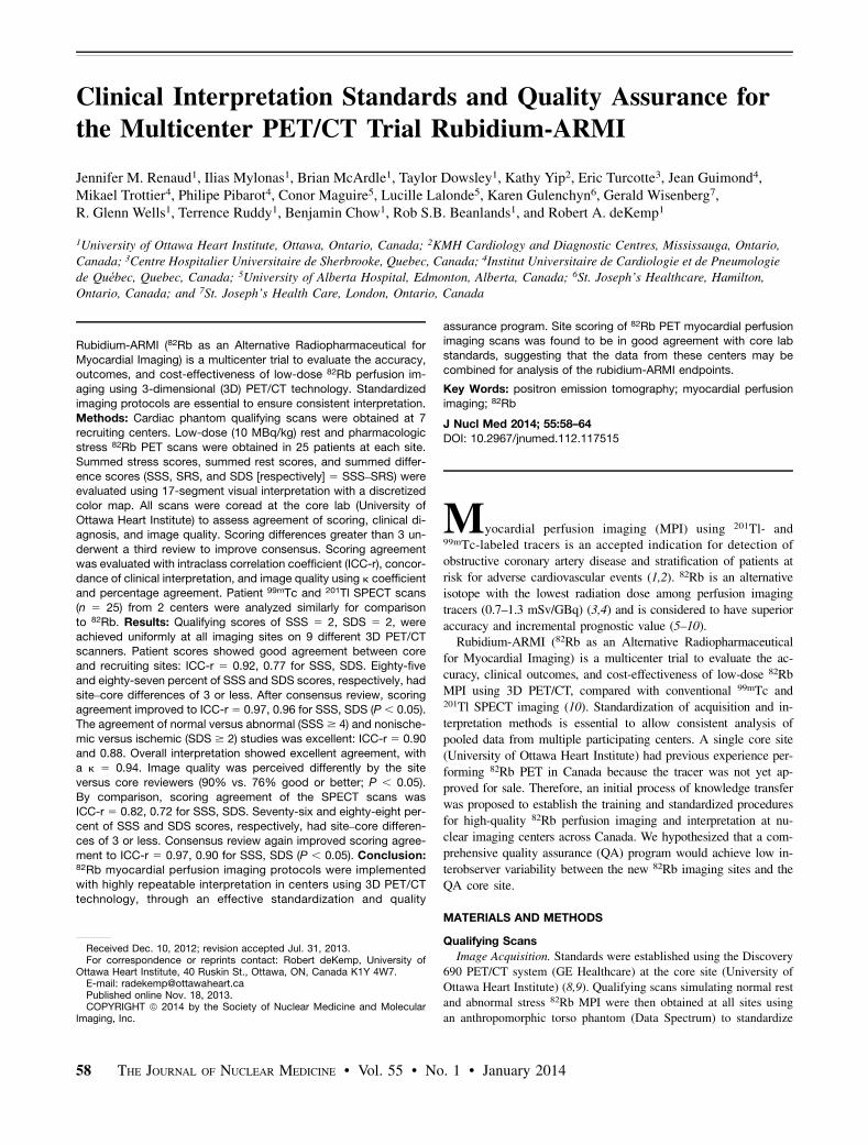
FIGURE 1. (A) Anthropomorphic
torso phantom with heart and liver
inserts, used to simulate rest and
stress perfusion scans. Corri-
dor4DM display of CTAC (B) and
fused PET CTAC images (C) used
to verify alignment for proper atten-
uation correction. Fifty percent PET
activity (green-yellow) should fall
within CT soft-tissue region for
proper alignment.
INTERPRETATION STANDARDS FOR RUBIDIUM-ARMI • Renaud et al. 59

lines: stress defect scores represent infarct plus ischemia, rest defectscores represent infarct only, and stress–rest difference scores reflect
ischemia. Physicians reviewed 5 example cases before the startof the trial to familiarize themselves with these guidelines. The
example scans were selected by the core lab reviewer and included2 normal and 3 abnormal cases, representing a combination of straight-
forward versus challenging interpretations. The site reviewers scoredthese cases independently based on the guidelines above and then
compared their scores with those assigned by the core lab reviewer;any discrepancies were discussed with the core lab to increase inter-
pretation consistency and experience.Site Versus Core Interpretation. Scans were analyzed by a single
physician per scan, per site, with several sites having two or morereporting physicians. The 25 cases from the recruiting sites were
then coread by a single physician at the core lab to assess the var-iability in image quality, SSS, SDS, and clinical diagnosis between
the sites and core. All physicians were senior nuclear medicine re-
porting staff experienced with SPECT perfusion imaging. With theexception of the core lab reviewer, all were new to the reporting of
PET perfusion scans because 82Rb was not yet approved for sale inCanada. 82Rb PET MPI was initiated at the recruiting sites for the
rubidium-ARMI study, hence the need for the common interpreta-tion software and specific interpretation guidelines above. Scores
with differences greater than 3 underwent a third review via discus-sion between the core lab reader and the recruiting site physician to
improve scoring consensus. A difference greater than 3 was chosenas consensus review cutoff because it is equivalent to the range of
SSS diagnostic categories (e.g., 0–3, 4–7, and 8–11) used commonlyin prognosis studies of MPI (9).
After the quantitative perfusion scoring, clinical diagnosis was classifiedas normal, abnormal, equivocal, or uninterpretable. The core reviewer
diagnosis was based only on the perfusion scores when electrocardio-gram-gated and coronary calcium results were not available at the core
lab. Cases in which the image quality was not indicated by one or bothof the interpreters (n 5 21) were excluded from the analysis of that
metric. The core lab reader was masked to clinical history.Statistical Analysis. PET and SPECT demographics were compared
via nonparametric Wilcoxon rank-sum tests for continuous variablesand Fisher exact tests for categoric variables. The interclass correla-
tion coefficient (ICC-r), determined with a 2-way random model withsingle measures, was used to assess agreement of 82Rb and combined201Tl and 99mTc SSS and SDS values, before and after consensusreview. ICC-r was also used to assess the scoring agreement of normal
versus abnormal (SSS $ 4) and ischemic (SDS $ 2) versus nonische-mic cases. Bland–Altman analysis was used to evaluate the mean 6SD and reproducibility coefficient (RPC) between the site and corescores. Means were compared via the Student t test. The Fisher exact
test was used to determine the significance of the change in correlationbefore versus after consensus review and between PET versus SPECT.
The F test was used to compare the RPC values before versus afterconsensus review and between PET versus SPECT. P values of less
than 0.05 were considered significant. Agreement between imagequality was assessed with percentage agreement and the Fisher exact
test. The agreement of clinical interpretation was assessed with the k
coefficient. Site-by-site variability of image quality assessment was
evaluated using ANOVA.
RESULTS
Qualifying Scans
Phantom scans consistently resulted in polar map scores ofSSS 5 2, SRS 5 0, and SDS 5 2 at all imaging sites using 9
TABLE 1PET/CT Systems Used in Rubidium-ARMI Study
Scanner Reconstruction algorithm Filter Cutoff Prompt g correction SSS SRS SDS
D690 PET/VCT-64 VUE Point HD (4 iterations, 24 subsets) Hann 12 mm Yes 2 0 2D600 PET/CT-16 VUE Point HD (4 iterations, 32 subsets) Hann 12 mm Yes 2 0 2
DRX PET/CT-16 3D FORE OSEM (5 iterations, 21 subsets) Hann 12 mm No 2 0 2
TF PET/CT-16 3D RAMLA NA Normal No 2 0 2
GXL PET/CT-16 3D RAMLA NA Normal No 2 0 2DSTE/VCT-16 3D FORE OSEM (8 iterations, 20 subsets) Hann 12 mm No 2 0 2
TF PET/CT-16 3D RAMLA (2 iterations, 4 subsets) NA Normal No 2 0 2
Biograph PET/CT-16 3D FORE OSEM (2 iterations, 24 subsets) Gauss 12 mm Yes 2 0 2DSTE/VCT-16 3D FORE OSEM (8 iterations, 20 subsets) Hann 12 mm No 2 0 2
FORE 5 Fourier rebinning; OSEM 5 ordered-subset expectation maximization; RAMLA 5 row action maximum likelihood algorithm.
FIGURE 2. Qualifying phantom scan results showing normal perfusion
at rest and inferior wall defect at stress, resulting in SRS 5 0, SSS 5 SDS
5 2. Vertical-long-axis (VLA) views of myocardium at stress and rest are
shown. Normalized stress, rest, and reversibility 17-segment polar maps
show assigned database percentages and spectrum 10-step colors. SSS,
SRS, and SDS polar maps show expected defect in inferior wall.
60 THE JOURNAL OF NUCLEAR MEDICINE • Vol. 55 • No. 1 • January 2014

different 3D PET/CT scanners (Table 1). Normal resting scanswith an SRS 5 0 were expected by design, using the phantomwith uniform myocardial activity. Similarly, all simulated stressscans with the defect in the inferior wall resulted in an SSS 5 2,with an SDS 5 2 accurately reflecting the pattern of reversibleischemia under these idealized conditions with a small 1-cm3 de-fect centered in the midinferior segment (Fig. 2).
Patient Scans
PET and SPECT patients had a mean age of 65 y (34–93 and39–89 y, respectively). None of the demographics were signifi-cantly different between the groups (Table 2).
82Rb PET Site Versus Core Interpretation
Recruiting site SSS and SDS scores were 5.6 6 7.9 and 3.3 65.2, respectively. Comparison between core and site scoresresulted in good overall agreement: ICC-r 5 0.92 for SSS and0.77 for SDS. Eighty-five percent (148/174) of SSS and 87% (151/174) of SDS scores had absolute differences |site – core| D # 3.The difference was 0 in 40% of the cases for SSS and 51% ofcases for SDS, with D # 6 in 95% of the cases for SSS (range, –15to 115) and 98% of cases for SDS (range, –15 to 113) (Figs. 4Aand 4B). Mean differences were SSS D 5 20.52 6 3.3 and SDSD 5 0.35 6 3.5 (P 5 not significant vs. zero) (Figs. 4C and 4D).The reproducibility coefficient for SSS and SDS was 6.4 and 5.4,respectively. Scoring agreement was most highly correlated in thenormal to moderate range (SSS and SDS , 15); this is importantbecause small scoring differences in this range can shift a patientfrom one diagnostic category to another.After consensus review, overall agreement was significantly
improved to ICC-r 5 0.97 for SSS and 0.96 for SDS (P , 0.05for both). Considering the sites individually, 5 of 7 had significantimprovement in SSS or SDS agreement after consensus review. Incases for which a nonsignificant improvement was observed, the
initial agreement rate was already high (ICC-r . 0.8) so there wasless room for improvement. The number of cases with an overallscoring D of 0 increased to 44% for SSS and 55% for SDS. Therange of SSS and SDS differences was also reduced (Figs. 4A and4B). Mean differences were SSS D 5 20.36 6 1.8 and SDS D 50.26 6 1.4 (P 5 not significant vs. zero) (Figs. 4C and 4D). RPCfor SSS and SDS improved significantly to 3.5 and 2.8, respec-tively (P , 0.05). A smaller range of scoring differences wasobserved after consensus review for both SSS and SDS. Therewas little change in the low-score range; however, the distributionwas narrower in the highly abnormal cases (i.e., high SSS andSDS). Ninety-three percent of the SSS data and 95% of the SDSwere within a difference of 3 after consensus review. The largestdiscrepancies occurred in cases with large defects spanning mul-tiple segments, as in the example in Figure 5. Despite the scoringdifferences, these cases were all correctly identified as abnormal atboth the recruiting site and the core lab. The scoring agreement ofnormal versus abnormal (SSS $ 4) scans and ischemic (SDS $ 2)versus nonischemic was found to be excellent, with an ICC-r 50.90 and 0.88, respectively.For overall diagnostic interpretation, before consensus review
the site and core interpretations were in 86% agreement (k 50.74); after review agreement improved to 94% (k 5 0.89). Thesame 82 of 82 cases were interpreted as abnormal by both coreand site reviewers, and 81 of 85 were also considered normalby both core and site. In the other 4 of 85 cases interpreted asnormal by the core, 3 were considered abnormal and 1 was equiv-ocal by the site. In 5 cases, the site reported the scans as unin-terpretable because of technical difficulties including CTAC mis-registration (n 5 4) or truncation of the heart within the field ofview (n 5 1), whereas the core reported these cases as abnormal.Lastly, in 2 cases considered equivocal by the core, the sitereported 1 as normal and 1 as abnormal.Good diagnostic image quality was obtained for most scans ac-
quired (Supplemental Fig. 1). Recruiting sites ranked 90% of theircombined images as good quality, whereas the core reviewer in-dicated that 76% of the same images fell into that category. Siteversus core percentages in the other categories were 7% versus 16%fair and 3% versus 9% poor. These represent a significant differencein the perception of image quality between the core and site re-viewers (P , 0.05). ANOVA of site rankings of image qualityshowed no significant difference between sites, but each site con-sistently ranked its own image quality higher than the core re-viewer’s ranking (P , 0.001).
201Tl and 99mTc SPECT Site Versus Core Interpretation
SPECT scans were merged into a single cohort for analysis. SiteSSS and SDS scores were 5.7 6 8.3 and 3.3 6 5.7, respectively.Moderate to good agreement between site and core scores was ob-served: ICC-r 5 0.82 for SSS and 0.72 for SDS. Seventy-six percent(38/50) of SSS scores and 88% (44/50) of SDS scores had differences|site – core| D # 3. Thirty-six percent of the cases for SSS and 54%
for SDS had D 5 0; the scoring differencewas# 6 in 86% of the cases for SSS (range,218 to 14) and 98% for SDS (range, –6 to113) (Figs. 6A and 6B). There was a mod-erate correlation between site and corescores: r 5 0.81 for SSS and r 5 0.77 forSDS, with a mean SSS D5 –0.406 4.8 andSDS D5 0.546 2.7 (P5 not significant vs.zero) (Figs. 6C and 6D). RPC values for SSS
TABLE 2Patient Demographics
Description PET SPECT
Male sex 56% 58%Age (y) 65 6 11 65 6 11
Body mass index (kg/m2) 30 6 7 29 6 5
Diabetes 36% 30%
Smoker* 63% 66%Hypertension 68% 68%
Dyslipidemia 79% 76%
Family history 46% 50%Typical angina 21% 12%
Previous myocardial infarction 28% 26%
Previous percutaneous
coronary intervention
26% 28%
*Past or current.
FIGURE 3. 82Rb PET rest and stress MPI protocol.
INTERPRETATION STANDARDS FOR RUBIDIUM-ARMI • Renaud et al. 61

and SDS were 9.4 and 5.3, respectively. Overall diagnostic interpre-tation between the core and sites showed 84% agreement (k 5 0.74).After consensus review, scoring agreement improved signifi-
cantly to ICC-r 5 0.97 for SSS and ICC-r 5 0.90 for SDS (P ,0.05 for both). SDS correlation was significantly lower versusPET (P , 0.001), whereas SSS correlation was not significantly
different. Cases with an overall SSS differ-ence of 0 increased to 48%, with no changeobserved for SDS. The consensus rangeswere reduced accordingly (Figs. 6A and6B). Mean differences were SSS D 50.16 6 1.9 and SDS D 5 20.14 6 1.4(P 5 not significant vs. zero and vs.PET) (Figs. 6C and 6D). RPC for SSSand SDS improved to 3.8 and 2.8, respec-tively (P , 0.05) but were not significantlydifferent from PET. Scoring agreement ofnormal versus abnormal (SSS $ 4) scansand ischemic (SDS $ 2) versus nonischemicwas good: ICC-r 5 0.84 and 0.73, respec-tively. The latter is significantly worse thanthe PET scoring agreement for ischemic(SDS $ 2) versus nonischemic cases, forwhich ICC-r 5 0.90 and 0.88, respectively.Overall agreement in diagnostic classificationbetween the core and sites improved to 98%(k 5 0.96), similar to the PET results. Thesame 24 of 24 cases were interpreted as nor-mal by both core and site reviewers, and 22of 23 were considered abnormal by both coreand site. In 1 of 23 cases interpreted as nor-mal by the core, the site considered the caseequivocal because of a perceived artifact.
Image quality was rated as fair in most cases (54%) by the corereviewer, whereas the sites ranked the majority as good (54%). Overallrankings of the site versus core were 54% versus 46% good, 44%versus 54% fair, and 2% versus 0% poor, representing lower overallimage quality as compared with PET (P , 0.05). This reflects a 64%overall agreement in SPECT image quality rating, with no significant
difference in the perception of quality be-tween the site and core reviewers (P 5 notsignificant).
DISCUSSION
This study successfully standardized 82Rb
imaging protocols at several centers using
3D PET/CT scanners. After initial qualify-
ing phantom scans, clinical 82Rb scans were
coread to assess the agreement of perfusion
scores (SSS, SDS), image quality, and over-
all interpretation between the recruiting site
and core lab reviewers. Polar map scoring
consensus improved after cases with differ-
ences of more than 3 were reviewed. Com-
parison with the combined results of stan-
dard 201Tl- and 99mTc-based SPECT
imaging was performed. SPECT data were
merged into a single cohort because separate
analysis of the 201Tl- and 99mTc-based datadid not show a significant difference be-tween the 2 tracers.All 3D PET/CT scanners in this study
achieved consistent qualifying phantomscan results despite differences in technol-ogy, such as scintillation detectors, numberof CT slices, and prompt g correction. This
FIGURE 4. (A and B) Difference between core lab and recruiting site PET SSS (A) and SDS (B)
before and after consensus review. (C and D) Bland–Altman analysis of site vs. core PET SSS (C)
and SDS (D) before and after consensus review.
FIGURE 5. 82Rb perfusion images and 17-segment consensus scores for patient where
core–site scoring difference was greater than 3 (core: SSS, SDS 5 28; site: SSS, SDS 5 13).
Discrepancy is from large defect spanning multiple segments. Regardless of scoring differences,
the case was correctly identified as definitely abnormal by both interpreters. S 5 stress; R 5 rest.
62 THE JOURNAL OF NUCLEAR MEDICINE • Vol. 55 • No. 1 • January 2014

latter effect is highly dependent on the particular vendor imple-mentation of scatter correction, which has not been systematicallyinvestigated on the GE Healthcare and Philips systems. The phan-tom studies suggest that any potential bias is small but should beconfirmed in future studies comparing perfusion results against anaccepted gold standard such as invasive coronary angiography.The standardized imaging and scoring methods allowed ex-
cellent agreement of SSS and SDS between site and core labinterpretations. The improvement in scoring agreement after con-sensus review demonstrates the added value of this process inimproving consistency and training experience. Significant im-provement in half of the sites demonstrates that they required theconsensus review process to improve their technique and experi-ence to accurately score more difficult cases, whereas the othersites already had adequate understanding of the scoring methodsafter initial training. Greater differences were observed in the site–core scoring in cases with large SSS and SDS. These cases werestill recognized as highly abnormal by both readers; thus, thesescoring differences were not clinically significant. However, in thecases of mild-to-moderate disease (SSS, SDS , 15), for whicha small scoring difference can change the disease classification,a high correlation between the site and core scores was observed.The equivocal range of 60%–75% used in the present study isconsistent with the established abnormal threshold of PET perfu-sion, less than 60% of maximum, as reported previously (14).Without an independent gold standard, the present study resultsmay rely on the core lab expertise and experience relative to thenewly recruited centers. Consensus reviewing was shown to im-prove agreement but did not introduce a bias toward the core labinterpretation scores because an equal proportion was shifted ei-ther toward the site scores or toward the core scores (31% each),and the remainder shifted to the average of the 2 scores (39%).Overall agreement might be further improved using scanner-specific 82Rb normal databases to assign default segmental scores,
reducing the need for user modificationand decreasing interoperator variability.We recently reported the accuracy of low-dose 82Rb MPI using a 3D PET/CT normaldatabase (8), showing results similar tostandard-dose imaging (15) and traditional2D PET methods. Consistent scan interpre-tation between recruiting centers is essen-tial to permit pooling of the data for furtherevaluation of the rubidium-ARMI end-points of clinical outcomes and cost-effec-tiveness. Agreement between site and corescoring was assessed only after the initialtraining period. It is not known whetherthese results may diverge or converge overthe course of the study, but it seems likelythat the agreement should be maintainedover time, assuming that the site reviewerscontinue to follow the simple interpretationmethods as described.The 94% agreement of PET interpreta-
tion after consensus review is similar to, orbetter than, previous studies comparingSPECT perfusion scores between readers(16). In those studies, agreement was in therange of 68%–97% (k 5 0.56–0.89). In thepresent study, SPECT interpretation agree-
ment was 98% (k 5 0.96), similar to PET. The PET interpretationagreement might have been even further improved if the core re-viewer had access to the CTAC images for all studies. This is a lim-itation of this study and highlights the importance of reviewing theCTAC and PET alignment in all cases. Attenuation-corrected SPECTimages were not evaluated in the present study. While this may beviewed as a limitation, the procedure is not widely used in clinicalpractice.Good PET image quality was observed in most cases when the
site and core reviewers used a 3-point scale of good, fair, or poor.The higher percentage of site rankings of good PET image qualitymay reflect the reviewers’ relative inexperience at the start of thetrial. The core interpreter had observed a larger number of casesacross the sites and thus had a wider frame of reference than thesite reviewers, who reviewed only images from their own partic-ular site. Image quality may vary from one site to another becauseof the specific equipment and patient populations—for example,one site had a significant population of bariatric surgery patientswith high body mass index, resulting in lower-quality images.Comparison of image quality in patients scanned on both PETand SPECT systems may further elucidate the relative quality of82Rb PET versus SPECT images.
82Rb is produced from a 82Sr/Rb generator, allowing wide-spread distribution. The short 76-s half-life allows rapid scanningtimes and lower radiation exposure to the patient. The short scantime permits rapid rest and stress imaging to be completed in 30–45 min, which is convenient for the patient and permits high-throughput imaging and efficient use of the technology. Thelow-dose (10 MBq/kg) protocol may also allow simultaneous quan-tification of absolute flow on 3D PET systems with adequate dy-namic range to permit accurate measurement of the bolus first-passactivity (9,17,18) and measurement of ventricular function closeto peak hyperemia with pharmacologic stress. Ischemia-inducedperfusion and wall-motion changes with 82Rb PET may enhance
FIGURE 6. (A and B) Difference between core lab and recruiting site SPECT SSS (A) and SDS
(B) before and after consensus review. (C and D) Bland–Altman analysis of site vs. core SPECT
SSS (C) and SDS (D) before and after consensus review.
INTERPRETATION STANDARDS FOR RUBIDIUM-ARMI • Renaud et al. 63

sensitivity for detecting clinically significant disease (19). The re-ported sensitivity and specificity for 82Rb PET imaging was ap-proximately 90%–93% and 81%–88%, respectively, in systematicreviews by our group and others (2,7,20). These reviews also dem-onstrated that 99mTc-based SPECT sensitivity and specificity were80%–85% and 76%–85%, respectively (7,20). 82Rb PETwas shownto be superior to electrocardiogram-gated 99mTc-SPECT MPI interms of overall accuracy, normalcy rates, and improved imagequality, with increased confidence in interpretation (fewer equiv-ocal studies) (6). In comparison, mean accuracies were reported tobe 85% versus 79% for PET and 201Tl SPECT (5).Our results show the benefit of a QA and standardization pro-
gram for the dissemination and use of a new PET tracer in theclinical routine. The present study supports the ability to combinedata from several centers for evaluation of the full multicenter trialresults. This model may also be helpful in the investigation andimplementation of other novel tracers, such as 18F-labeled perfu-sion agents currently in phase III development (21).
CONCLUSION
Reproducible imaging standards are essential for clinical trialresults to be pooled across participating centers. With effectivetraining and standardization of 82Rb PET MPI, good interpretationagreement was found between the recruiting sites and the core lab.Agreement was further improved with consensus reading of themost discrepant cases, suggesting that improved scoring consis-tency is achieved with increased training and experience. Theresults indicate that repeatable interpretation is achievable acrossmultiple imaging centers using different 3D PET/CT scanners,following these imaging standards and quality assurance methods.
DISCLOSURE
The costs of publication of this article were defrayed in partby the payment of page charges. Therefore, and solely to indicatethis fact, this article is hereby marked “advertisement” in accor-dance with 18 USC section 1734. This study was funded byresearch grants from CIHR MIS-100935 (Rubidium-ARMI) andML1-112246 (MITNEC), HSFO PRG-6242 (Molecular Functionand Imaging program), and ORF RE-02038 (ICT) in partnershipwith Jubilant DraxImage (JDI) and INVIA in-kind. Rob S.B.Beanlands was supported in part by a career investigator awardfrom HSFO and Tier 1 Research Chair at the University ofOttawa. Ilias Mylonas, Brian McArdle, and Taylor Dowsley weresupported in part by the HSFO program grant, the Whit & HeatherTucker Endowed Fellowship, the Vered-Beanlands Endowed Re-search Fellowship in Cardiology, and the UOHI Associates in Car-diology. Robert A. deKemp receives royalties from rubidium PETtechnology licenses. Rob S.B. Beanlands and Robert A. deKempare consultants with JDI. No other potential conflict of interestrelevant to this article was reported.
ACKNOWLEDGMENTS
We thank the research coordinators, technologists, and nurses atthe participating sites for their efforts acquiring the PET and SPECTscans. We acknowledge Jubilant DraxImage for providing the 82Rb
generators, elution system, training, and support and INVIA Med-ical Solutions for providing Corridor4DM software and support.
REFERENCES
1. Hendel RC, Berman DS, Di Carli MF, et al. ACCF/ASNC/ACR/AHA/ASE/
SCCT/SCMR/SNM 2009 appropriate use criteria for cardiac radionuclide imag-
ing. J Am Coll Cardiol. 2009;53:2201–2229.
2. Beanlands RS, Chow BJ, Dick A, et al. CCS/CAR/CANM/CNCS/CanSCMR
joint position statement on advanced noninvasive cardiac imaging using positron
emission tomography, magnetic resonance imaging and multidetector computed
tomographic angiography in the diagnosis and evaluation of ischemic heart
disease: executive summary. Can J Cardiol. 2007;23:107–119.
3. Ruby-FillTM product monograph, rubidium Rb 82 generator [package insert].
Kirkland, Quebec, Canada: Jubilant DraxImage; 2011.
4. Senthamizhchelvan S, Bravo PE, Lodge MA, et al. Radiation dosimetry of 82Rb
in humans under pharmacologic stress. J Nucl Med. 2011;52:485–491.
5. Stewart RE, Schwaiger M, Molina M, et al. Comparison of rubidium-82 positron
emission tomography and thallium-201 SPECT imaging for detection of coro-
nary artery disease. Am J Cardiol. 1991;67:1303–1310.
6. Bateman TM, Heller GV, McGhie A, et al. Diagnostic accuracy of rest/stress
ECG-gated 82Rb myocardial perfusion PET: comparison with ECG-gated
Tc-99m sestamibi SPECT. J Nucl Cardiol. 2006;13:24–33.
7. Mc Ardle BA, Dowsley TF, Wells G, et al. Systematic review of the accuracy of82Rb PET for the diagnosis of CAD in comparison to SPECT. J Am Coll Cardiol.
2012;60:1828–1837.
8. Kaster T, Mylonas I, Renaud JM, et al. Accuracy of low-dose rubidium-82
myocardial perfusion imaging for detection of coronary artery disease using
3D PET and normal database interpretation. J Nucl Cardiol. 2012;19:1135–1145.
9. Yoshinaga K, Katoh C, Manabe O, et al. Incremental diagnostic value of regional
myocardial blood flow quantification over relative perfusion imaging with gen-
erator-produced rubidium-82 PET. Circ J. 2011;75:2628–2634.
10. Rubidium-82: an alternative radiopharmaceutical for myocardial imaging (ARMI).
Clinicaltrials.gov. http://clinicaltrials.gov/ct2/show/NCT01128023. Accessed Oc-
tober 21, 2013.
11. Esteves FP, Nye JA, Khan A, et al. Prompt-gamma compensation in 82Rb myo-
cardial perfusion 3D PET/CT. J Nucl Cardiol. 2010;17:247–253.
12. Dilsizian V, Bacharach SL, Beanlands R, et al. ASNC imaging guidelines for
nuclear cardiology procedures: PET myocardial perfusion and metabolism
imaging. J Nucl Cardiol [serial online]. http://www.asnc.org/imageuploads/
ImagingGuidelinesPETJuly2009.pdf. Accessed November 8, 2013.
13. Klein R, Adler A, Beanlands RS, deKemp RA. Precision-controlled elution of
a 82Sr/82Rb generator for cardiac perfusion imaging with positron emission to-
mography. Phys Med Biol. 2007;52:659–673.
14. Sdringola S, Nakagawa K, Nakagawa Y, et al. Combined intense lifestyle and
pharmacologic lipid treatment further reduce coronary events and myocardial
perfusion abnormalities compared with usual-care cholesterol-lowering drugs in
coronary artery disease. J Am Coll Cardiol. 2003;41:263–272.
15. Nakazato R, Berman DS, Dey D, et al. Automated quantitative 82Rb 3D PET/CT
myocardial perfusion imaging: normal limits and correlation with invasive cor-
onary angiography. J Nucl Cardiol. 2012;19:265–276.
16. Johansen A, Høilund-Carlsen PF, Christensen HW, et al. Observer variability in
the evaluation of dual-isotope Tl-201/Tc-99m sestamibi rest/stress myocardial
perfusion SPECT in men and women with known or suspected stable angina
pectoris. J Nucl Cardiol. 2004;11:710–718.
17. Klein R, Renaud JM, Ziadi MC, et al. Intra- and inter-operator repeatability of
myocardial blood flow and myocardial flow reserve measurements using rubidium-82
pet and a highly automated analysis program. J Nucl Cardiol. 2010;17:600–616.
18. Lortie M, Beanlands RS, Yoshinaga K, et al. Quantification of myocardial blood flow
with 82Rb dynamic PET imaging. Eur J Nucl Med Mol Imaging. 2007;34:1765–1774.
19. Dorbala S, Vangala D, Sampson U, et al. Value of vasodilator left ventricular
ejection fraction reserve in evaluating the magnitude of myocardium at risk and
the extent of angiographic coronary artery disease: a 82Rb PET/CT study. J Nucl
Med. 2007;48:349–358.
20. Parker MW, Iskandar A, Limone B, et al. Diagnostic accuracy of cardiac positron emis-
sion tomography versus single photon emission computed tomography for coronary
artery disease: a bivariate meta-analysis. Circ Cardiovasc Imaging. 2012;5:700–707.
21. Rischpler C, Park MJ, Fung GS, Javadi M, Tsui BM, Higuchi T. Advances in PET
myocardial perfusion imaging: F-18 labeled tracers. Ann Nucl Med. 2012;26:1–6.
64 THE JOURNAL OF NUCLEAR MEDICINE • Vol. 55 • No. 1 • January 2014