1 Renal Physiology PART ONE Renal Physiology overview PART TWO Renal Physiology details.
Clinical Implications of Renal Physiology
September 2021
DR. SURJ IT TARAFDAR
NEPHROLOGIST
BLACKTOWN HOSPITAL , SYDNEY
CONJOINT SENIOR LECTURER DEPARTMENT OF MEDICINE WESTERN SYDNEY UNIVERSITY

The Kidney

Glomerulus§1.3 to 1.8 million nephrons in each kidney
§Glomerulus is formed by the invagination of a tuft of capillaries into the dilated proximal end of the nephron- Bowman’s capsule
§Glomerular capillary supplied by afferent and drained by efferent arteriole
§Glomerular basement membrane (GBM) is the skeleton of the glomerular tuft with podocytes on outer side and capillaries/endothelial cells and mesangium on inner side
§Blood inside capillary is separated from the urinary space by endothelium, GBM and podocyte
§Glomerular pathologies- nephrotic and nephritic syndrome

Filtration Barrier

Filtration Barrier§Endothelium: 50 to 100nm pores; luminal surface highly electronegative
§Visceral epithelium (podocytes): Big cell body floating in the urinary space with long primary processes which affix on capillaries by foot processes with filtration slits sized 30-40nm…….slits are bridged by the slit diaphragm which are themselves perforated by smaller pores sized about 8nm….
§GBM: With podocytes externally and endothelium/mesangium internally main constituents -collagen IV, laminin and heparin (all negative charged)
§So….for NEGATIVELY CHARGED PROTEINS like albumin to leak through this barrier…..either slit diaphragm pores should get bigger and/or the barriers should lose its negative charges
§Q- What is the size of albumin molecule?

Question•What are the criterion to diagnose nephrotic syndrome?
•When to call it nephritic syndrome?
• Do you need proteinuria to diagnose nephritis?
•What is the commonest glomerular disease worldwide?
From previous slide- diameter of albumin is 7.2 nm and smallest pores in filtration slits are 8 nm

Answer• Nephrotic syndrome- Proteinuria>3.5g/24 hours, hypoalbuminemia, generalised oedema and hyperlipidaemia
• Nephritic syndrome- Haematuria with RBC casts and dysmorphic RBCs, often some degree of oliguria with hypertension and progressive reduction in GFR
• Proteinuria is not strictly needed to diagnose nephritis
• Commonest glomerular disease worldwide is IgA nephropathy

Some diseases of GBM§Type IV collagen molecules are composed of three alpha chains that form triple-helical rope like structure
§GBM and alveolar capillary basement membrane collagen consist of alpha3, 4 and 5 chains unlike other basement membranes with alpha1, alpha1 and alpha2 chains
§Goodpasteur disease characterised by antibody against alpha3 chain of type IV collagen (anti-GBM antibody )
§Alport syndrome results from mutations in genes encoding the alpha-3, 4, and 5 chains of type IV collagen (80% due to alpha-5 mutation)
§Q. Time: Almost all patients with Goodpasteur have GN but only half have hemoptysis. Why?
What does the kidney do??§GFR of 125ml/minute…….180 L fluid filtered from the glomeruli daily
§>98-99% of above reabsorbed in the tubules
§>90% of the water reabsorbed is a passive process…water follows Na
§Only in the collecting duct is water absorbed actively and independent of Na by Aquaporin 2 channel stimulated by ADH

Tubular Reabsorption

Absorption of various ions and water across different segments of the renal tubules
Ion or substance PCT Loop of Henle DCT CD
Na 65 to 70% 30 to 35% 5% 1-2%
Water 65 to 70% 25% 5% 5 to 10 %
K 45 to 50% 40 to 45% Is secreted here
Ca 70% 20% 10 to 15%
Mg 15 to 25% 60 to 70% 5 to 10%
Ph 70 to 80% Insignificant Insignificant Insignificant
HCO3 85 to 90% 5-10% by beta intercalated cells

Why is Na reabsorbed by the tubular cell so easily?§In tubular epithelial cells, the basolateral Na K-ATPase extrudes 3 Na and pulls in 2 K ions from blood
§Result: intracellular Na deficiency and K excess in tubular cells
§Na deficient tubular epithelial cells reclaim Na from tubular lumen
§Water follows the Na from the tubule into the tubular cells
§In the collecting duct aldosterone accounts for an extra Na reabsorption (1-2% of total Na reabsorbed)

QuestionWhat is the unifying mechanistic principle of action of all diuretics?
a. Prevent water reabsorption
b. Prevent Na secretion
c. Prevent Na reabsorption
d. Cause K secretion

Diuretics action§All diuretics act by inhibiting tubular Na reabsorption
§Diuretics cause secondary hyperaldosteronism by causing intra vascular volume contraction
§Raised aldosterone due to loop and thiazide diuretics leads to tubular K and H loss and resultant hypokalemia and metabolic alkalosis
§K sparing diuretics antagonize the actions of aldosterone leading to K and H retention and the resultant hyperkalemia and acidosis

Proximal convoluted tubule (PCT)
§Reabsorbs >65% of Na by apical Na-H exchange protein
§Linked to regeneration of HCO3 [Carbonic anhydrase involved]
§Acetozolamide will interfere with above….Na and water lost in urine….HCO3 not regenerated……chances of metabolic acidosis
§Na reabsorption is coupled to reabsorption of glucose (SGLUT), amino acids, phosphate and uric acid
§Defect in above leads to inefficient regeneration of HCO3 and hence proximal renal tubular acidosis…….Fanconi syndrome if reabsorption of glucose, amino acids, phosphate and uric acid effected
§SGLUT 2 inhibitors for the treatment of type 2 diabetes mellitus: a mild diuretic effect as reabsorption of both glucose and Na prevented

PCT- Role of Carbonic Anhydrase (CA)

Thick Ascending Limb of Loop of Henle§Na-K-2Cl cotransporter reabsorbs 1 Na and K each along with 2 chloride (Cl) ions
§High intracellular K concentration causes the reabsorbed K to be promptly secreted back into the lumen via ROMK
§ Cl is absorbed into blood by the basolateral Cl channel (ClC-Kb) which has barttin as a crucial accessory protein
§Movement of 1 positive ion(K+) into the lumen and net of one negative ion into peritubular capillary(Na+ and 2Cl-) causes luminal positivity…….
§Which in turn leads to paracellular reabsorption of Ca+ and Mg+

Na reabsorption in TAL- nil water reabsorbed
Loop diuretics and Bartter syndrome§Frusemide inhibits the Na-K-2Cl cotransporter [NKCC2]
§ Bartter syndrome is due to defect in Na reabsorption here (so same as being on Frusemide)§ 5 types depending on the mutation in the protein involved-
§ Na-K-2Cl - type 1, ROMK- type 2, ClC-kb type 3, barttin- type 4, upregulation of CaSR-type 5§ Metabolic alkalosis and hypokalemia with low to normal BP
Q. What do Frusemide and thiazide diuretics do to K and acid base? ( check slide no. 14)

Question Frusemide action on calcium ?
Thiazide action on calcium?

Answer§Frusemide by inhibiting the Na+K+2Cl- co-transporter in TAL inhibits all the events
downstream including paracellular reabsorption of Ca and Mg
§Blocking the DTC with thiazides causes up regulation of other portions of the renal
tubules….upregulation of Na+K+2Cl- co-transporter in TAL causes increased Ca
reabsorption

Increased medullary osmolality-counter-current mechanism§Loop of henle reabsorb upto 40% of total Na but only 25% of water
§The descending limb is highly permeable to water but the thick ascending limb is impermeable to water……basis for countercurrent mechanism
§Helps to maintain osmotic gradient from 290mOsm/kg at cortex to 1200 mOsm/kg at tip of medulla
NOTE: ADH controlled reabsorption of water in the cortical duct is dependent on the above osmotic gradient

Counter-current Mechanism

QuestionCan Frusemide lead to hyponatremia?
Hint- let us go back to the diagram in previous slide

Answer§Frusemide by interrupting the Na-K-2Cl co-transporter in the ascending limb diminishes the osmotic gradient
§Reabsorption of water in the collecting duct under the action of ADH is dependent on this gradient
§Make sense? If not let us discuss this again………..
Calcium Sensing Receptor (CaSR)§Apart from parathyroid gland also found in basolateral membrane on the cells of the thick ascending limb of the loop of Henle
§Calcium binding to CaSR inhibits the K channel in the luminal membrane and the Na-K ATPase pump in the basolateral membrane
§ALSO….Ca stimulation of CaSR also leads to upregulation of inhibitory claudrin 14 that causes decrease in claudrin 16 and 19 which are the carrier proteins for Ca movement through the paracellular area
§Net result of renal CaSR stimulation by plasma Ca - Decrease in paracellular Ca and Mg reabsorption (increased urinary Ca loss)


Question§Autosomal dominant condition with inactivating mutation of CaSR?
§How do we diagnose?
§Treatment?

Answer is……..FHH §Familial hypocalciuric hypercalcemia (FHH) is caused by inactivating mutations of CaSR
§Mild hypercalcemia, hypocalciuria, inappropriately normal to highish PTH and high-normal to frankly elevated serum magnesium levels
§ Urinary Ca/Cr ratio< 0.01 or low 24 hours urinary calcium
§Benign condition that does not require parathyroidectomy

Distal Convoluted Tubule (DCT) and Gitelman syndrome§Na is reabsorbed by the apical NaCl cotransporter (NCCT)
§Inhibition of NCCT by thiazide diuretic lead to volume contraction, leading to increased TAL reabsorption of Na which in turn leads to increased paracellular reabsorption of Ca+
§Gitelman syndrome characterised by impaired NCCT (so same as being on thiazide diuretics)
§Characterised by hypokalemia, metabolic alkalosis and hypomagnesemia with lowish BP….what is the urine Ca going to be??

QuestionWhich of the following does not lead to hypokalemia with metabolic alkalosis?
a. Bartter syndrome
b. Addison disease
c. Hyperaldosteronism
d. Chronic vomiting
e. Diuretic abuse

Answer ADDISON DISEASE characterised by inability to excrete K+ and H+
How do we differentiate between the rest?
§Hyperaldosteronism will be characterised by hypertension while Bartter and Gitelman will have low BP
§Patient who abuse diuretics will show variable urinary Cl levels depending on the timing of diuretic abuse and in cases of doubt, a urine diuretic screen may be helpful
§Chronic vomiting leads to hypovolemia and associated increased NaCl reabsorption and hence low urinary Cl
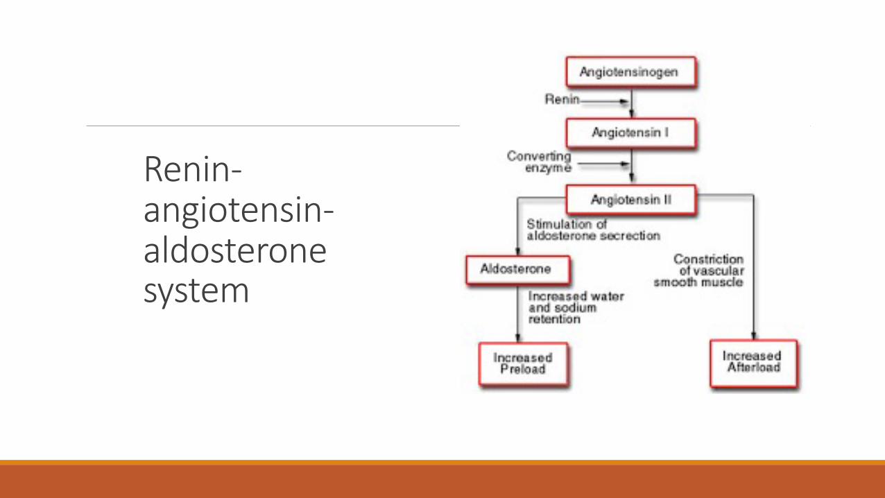
Renin-angiotensin-aldosterone system

Juxta Glomerular Apparatus (JGA)
§Macula Densa: specialised cells in DCT which sense urinary Na and Cl
§Granular Cells (JG Cells): modified smooth muscle cells in the terminal afferent arterioles that secrete renin when stimulated by the macula densa
§Renin splits the decapeptide Angiotensin I from the 32 amino-acid angiotensinogen produced in the liver
§Angiotensin Converting Enzyme in the lung vessels splits off two more aminoacids from angiotensin I to form angiotensin II
§Angiotensin II is a vasoconstrictor and increases aldosterone secretion
NOTE: Angiotensin II constricts efferent more than afferent arterioles• ACE-I and ARB decrease intraglomerular pressure and filtration thus diminishing
proteinuria

Collecting Duct (CD)..ADH§Principal cells contain aquaphorin 2 (AQP 2) which reabsorb water
§Vasopressin (ADH) via the V2 receptor moves AQP 2 from endosomes to the otherwise water impermeable luminal surface
§Remember ….the high interstitial osmolality due to countercurrent mechanism needed for water to move into the cells from CD lumen
§DI results when there is a vasopressin deficiency (central DI) or when the kidneys fail to respond to the hormone (nephrogenic DI)
§Lithium causes nephrogenic DI by causing decreased expression of AQP 2 genes
§Vasopressin V2 receptor antagonist Tolvaptan prevent renal enlargement by decreasing renal cAMP levels in ADPKD

Collecting Duct (CD)…….Aldosterone§Aldosterone secreted by the zona glomerulosa of adrenal cortex helps in Na reabsorption and K and H excretion
§Upregulates and activates the basolateral Na-K-ATPase causing a concentration gradient with low intracellular Na
§At the same time upregulates epithelial sodium channels- ENaCs, increasing apical membrane permeability for Na+
What about K and H?
§Intracellular movement of Na causes luminal negativity leading to K secretion
§The same luminal negativity leads to H secretion by the alpha- intercalated cells

Aldosterone action

Potassium and acid base balance-In metabolic acidosis excess H+ enters cells to be buffered and electroneutrality
maintained by intracellular K+ moving out
-Hyperkalaemia potentiates metabolic acidosis by three methods-o Excess K+ enters the cell and in exchange H+ comes out of cellso K+ competes with H+ for secretion by the collecting ducto Hyperkalaemia decreases renal ammonia production and so inhibits H+ excretion
(ammonia is the chief urinary buffer for H+)
-Additionally, hypokalaemia causes alkalosis by augmenting the H-K-ATPase pumps which secrete H+ and reabsorb K+ in the type A intercalated cells in CD

Renal Tubular Acidosis (RTA)§Disorders of the tubule characterized by a normal anion gap (hyperchloremic) metabolic acidosis despite a relatively well-preserved GFR with either hypokalaemia (types 1 and 2) or hyperkalaemia (type 4)
§Defect in distal tubule H+ leads to distal (type1) RTA while defect in HCO3 reabsorption in proximal tubule leads to proximal (type2)RTA
§Type 4 RTA (commonest RTA) is characterised by decreased production of aldosterone or diminished responsiveness of the cortical duct to aldosterone

RTA….Low K+ in both types 1 and 2DISTAL RTA
•Inability to secrete H+
•Urine pH >5.5 (no H+ in urine)
•Proximal tubules reabsorb all alkali including citrate which normally keeps Ca in urine soluble and so…. nephrololitiasis,nephrocalcinosis
•No Fanconi syndrome
•Sjogren’s syndrome, SLE, PBC, autoimmune hepatitis
•Treat with alkali and K+ replacement
PROXIMAL RTA
•Inability to reabsorb HCO3
•Urine pH < 5.5 (distal tubules secrete the excess H+ as in any acidosis)
•No renal stones
•Fanconi syndrome -glycosuria,phosphaturia, uricaciduria and aminoaciduria
•Myeloma,drugs- tenofovir, acetazolamide
•Same treatment…..needs bigger doses of alkali though…..

Type 4 RTA….hyper and NOT hypokalemia§Decreased production or diminished responsiveness to aldosterone
§Associated with DM (commonest), NSAIDs, ACE-I , calcineurin inhibitors (cyclosporine and tacrolimus), K sparing diuretics and high dose heparin
§In those not hypertensive or volume overloaded, synthetic mineralocorticoid such as fludrocortisone may help
§In patients with hypertension or fluid overload, a thiazide or loop diuretic may help by increasing distal delivery of Na and consequently increase urinary secretion of H+ and K+


Secondary hyperparathyroidism in CKDFollowing series of events cause hyperparathyroidism in CKD
● Phosphate retention (INITIAL TRIGGER)
● Decreased calcium concentration
● Decreased 1,25-dihydroxyvitamin D (calcitriol) concentration
● Increased fibroblast growth factor 23 (FGF23) concentration
● The reduced expression of calcium-sensing receptors ,FGF23 receptors and klotho (co-receptors for FGF23)

Thank you