Clinical Genetics Renata Gaillyová. Clinical genetics Dept. of medical genetics Genetic prevention...
184
Clinical Genetics Renata Gaillyová
-
Upload
polly-mcdonald -
Category
Documents
-
view
234 -
download
1
Transcript of Clinical Genetics Renata Gaillyová. Clinical genetics Dept. of medical genetics Genetic prevention...
Clinical Genetics
Renata Gaillyová
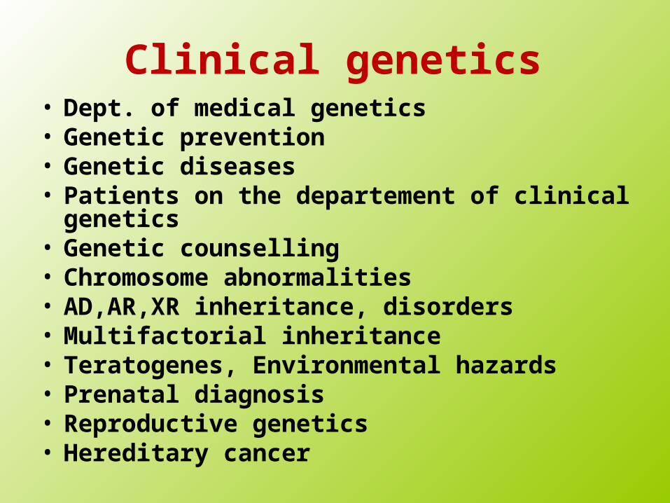
Clinical genetics• Dept. of medical genetics• Genetic prevention• Genetic diseases• Patients on the departement of clinical
genetics• Genetic counselling• Chromosome abnormalities• AD,AR,XR inheritance, disorders• Multifactorial inheritance• Teratogenes, Environmental hazards• Prenatal diagnosis• Reproductive genetics• Hereditary cancer
Dept. of Medical genetics
• Genetic ambulance genetic counselling• Laboratory part • Cytogenetic laboratoriesPrenatal cytogeneticsPostnatal cytogeneticsOncocytogeneticsMolecular – cytogenetics• Lab. for DNA and RNA analysis
(clinical genetics and oncogenetics)
Characteristic of Medical Genetics
• Preventive Medicine• Interdisciplinary cooperation• Information from genetics
(disease, testing, posibilities)• Voluntary choice for patients• Informed agreement
Primary prevention of genetic
• Before pregnancy• Folic acid (cca 0,8 mg/day, 3+3
months)• Vaccination (rubella)• Genetic counselling• Contraception, family can opt for
adoption or donor of gamets (oocytes, sperm)
• Pregnancy planning• Rediction of environmental hazards
(drugs, radiation, chemicals…)
Reproduction of the optimal age
• In women increases the risk of accidental congenital chromosomal aberrations in the offspring
• In men may increase the risk of de novo mutations in monogenic diseases (Neurofibromatosis, Achondroplasia..)
Prevention of spontaneous and induced mutations
• Healthy Lifestyle
• The restriction of harmful substances - drugs, environmental hazards
Vacctination, infection prevention
• Prevention of rubella embryopathie
Prevention of congenital toxoplasmosis
• Testing for infectious disease risk in mothers (CMV, varicella-zoster virus, ...)
Vitamin prevention of neural tube defects, anterior
abdominal wall defects, clefts• Folic acid at a dose of 0.8 mg daily (twice the
dose in non-pregnant) for 3-6 months prior to conception and till the end of 12. week of pregnancy
Pre-conception consultation with the doctor
• Family history
• Long term therapy
• Chronic diseases
Examination of acquired chromosomal aberrations
• Preventive examinations of persons exposed to environmetal risks at work or persons with risk of long-term therapy (immunosuppressants, cytostatics, ....)
• The possibility of vitamin therapy to improve repair of DNA (3-6 months)
Contraception, sterilization
• Contraception - temporarily prevents conception in the limited impact of risk (treatment)
• Sterilization - the long-term inhibition of pregnancy in a high risk of disease in the offspring (Hereditary disease)
Adopce• Alternative family care as an option
at high genetic risk families
Donor (oocytes, sperm)
• The possibility of sperm, oocytes and embryos donor
• reduction in high genetic risk
• reproductive problems
Secondary prevention of genetic
• Prenatal diagnosis• Prenatal screening • Prenatal tests• Genetic counselling• Termination of pregnancy (the law
in Czech Republic- end of 24. week of gestation)
• Postnatal screening• Newborn screening
Genetics diseases
• Chromosome abnormalities • about 0,6 - 0,7%
• Monogen diseases • about 0,36% (study in 1 000 000 newborns)• most then 90% of monogen diseases occur
in childhood
• Multifactorial (polygenic or complex) disorders
• Occur in about 80% in the population
Patients on genetic departements
•Dead person•Adults•Pregnant women•Fetuses•Children
Patients on genetic departements
• Positive family history (chromosome abnormality, congenital malformations, mental retardation, diseases…)
• Pregnant women with encrease risk for the fetus
• Infertility – sterility, repeated fetal loss
• Donors (gamets)• Patients with tumours
Children
•Congenital malformations
Children• Suspition of mongenic
hereditary diseases or inherited metabolic disorders and their families
Children
•Suspition on congenital chromosom aberations (children with congenital malformations, abnormal face, atipical visage, pre- or postnatal growth retardation, premature birth)
Children• Precocious or delayed puberty • Malformations of the external or
internal genitalia• Low or high figure
Children• Before adoption
Children or adults
• Mental retardation• Psychomotor retardation• Developmental delay
Children and adults
• Gender identity disorder
Children and adults
• people with long-term exposure to environmental pollutants
• (alcohol, cigarettes, drugs, radiation)
Children and adulds
• patients with suspected hereditary cancer
• patients with cancer (sporadic occurrence)
Adults
• Gamete donors(preventive tests)
Adults• Related partners (increased risk for hereditary disease
with AR inheritance)
adults• Infertility• Repeated spontaneous abortions
Pregnant women
• With unfavorable family history
Pregnant women• with adverse pregnancy history (chronic
diseases with established therapies, acute disease in early pregnancy - temperature, drugs, X-rays, CT, vaccinations, toxoplasmosis, rubella, ...)
Pregnant women
• Prenatal biochemical screening
(Pathology results)
Pregnant women• Ultrasound
prenatal screening – pathology results
• Congenital malformations
• Risk of chromosomal aberrations in the fetus
Pregnant women• ??? Age of parents ???relative indications
Genetic clinic
Genetic counselling
• Anamnesis• Family history• Pedigree analysis• Examining the patient• Laboratory analysis• Other examining - neurology,
psychology, hematology, CT, MRI …
Mother• Name, surname, date of birth,
maiden name• Place of birth• Place of birth of mothers parents• Relationship• Jobs - employment risks• Addictive substances alcohol, cigarettes, medication ..
Mother• Health problems from birth until
today• Long-term medication• Long-term monitoring of a
doctor• Gynecological anamnesis• The number of births, children,
pregnancy, birth weight children, the health status of the children
• The number of abortions, unsuccessful pregnancies
• Unsuccessful attempt to pregnancy
Mother
• In the case of health problems, if possible, to provide medical records from the attending physician
• Long-term used drugs, how long
Father• Name, surname, date of birth• Place of birth• Place of birth ot hte fathers
parents• Relationship• Jobs - employment risks• Addictive substances alcohol, cigarettes, drugs ..
Father• Health problems from birth
until today• Long-term medication• Long-term monitoring of a
doctor• Number of children from any
previous partners, their health status
• The number of abortions, failed pregnancy (if any previous partner)
• Unsuccessful attempt to become pregnant in previous partner
Father• In the case of health problems, if
possible, to provide medical records from the attending physician
• Long-term used drugs, how long
Child - Patient• Pregnancy• Swelling, nausea, protein in
urine, sugar in urine, high blood pressure
• Diseases in Pregnancy• Drugs in Pregnancy• Prenatal tests results
Ultrasound, blood tests
Child
• Birth - in time, early, after the deadline?
• Complications, neonatal icterus, birth weight and length, nutrition
• The mental and motor development
• Diseases• Monitoring of specialists• Drugs• Test results
Child
• Clinical genetic examination• Weight, height• Atypical visage• Malformations• Psychological state• Behavior
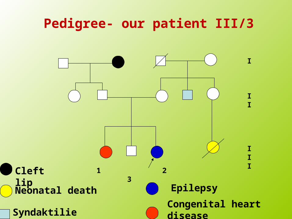
Pedigree- our patient III/3
Neonatal death
Syndaktilie
Epilepsy
Congenital heart disease
Cleft lip
I
II
III
1 2 3
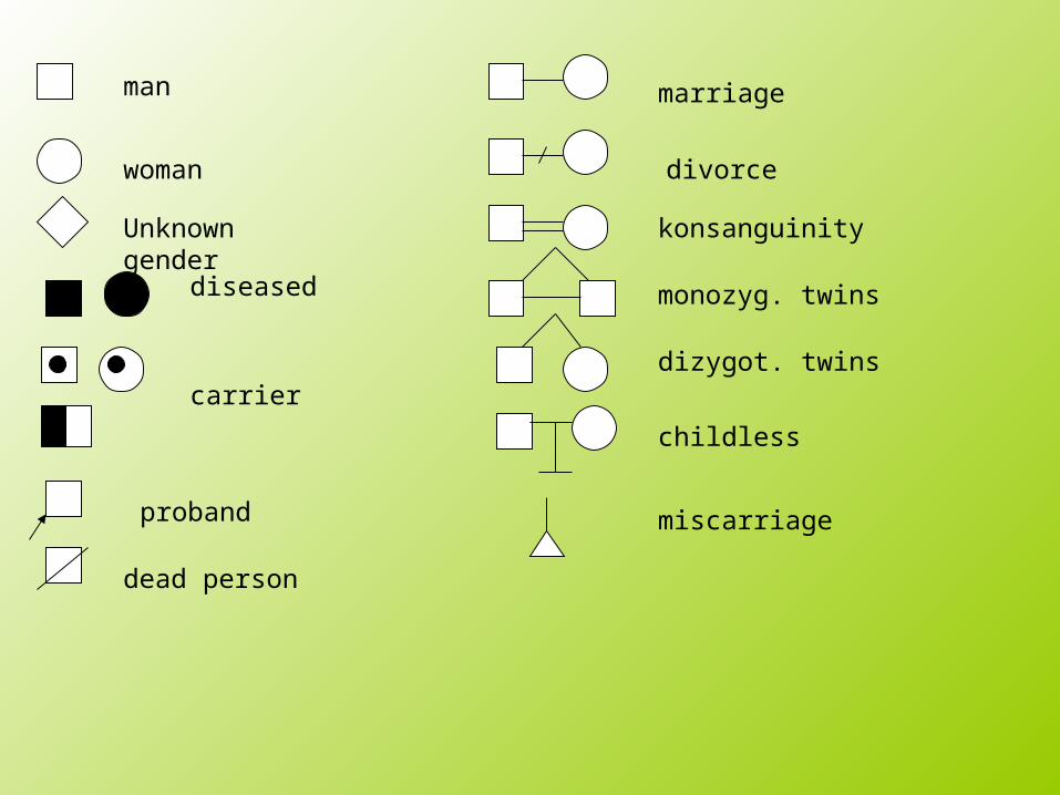
marriage
divorce
konsanguinity
monozyg. twins
dizygot. twins
childless
miscarriage
man
woman
diseased
Unknown gender
carrier
proband
dead person
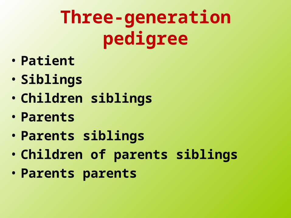
Three-generation pedigree
• Patient• Siblings• Children siblings• Parents• Parents siblings• Children of parents siblings• Parents parents
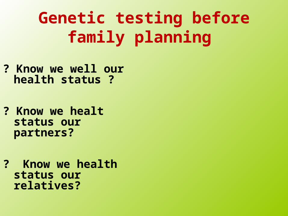
Genetic testing before family planning
? Know we well our health status ?
? Know we healt status our partners?
? Know we health status our relatives?
Next steps
• Recommend the laboratory genetic testing
• Recommend other specialists if needed
• Require medical records • Make photodocumentation
The result of genetic counselling
• Specify exact diagnosis (if possible)
• Determine genetic prognosis • Is the disease hereditary?• Type of inheritance• Genetic risks for other family
members• Posibilities of treatment, prenatal
analysis
Chromosome abnormalities
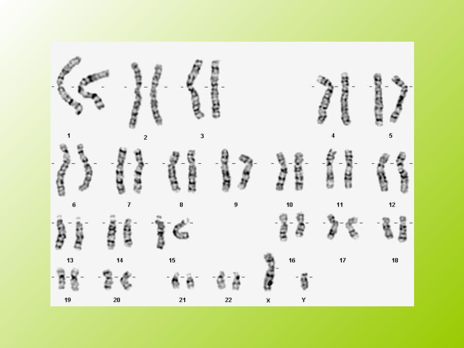
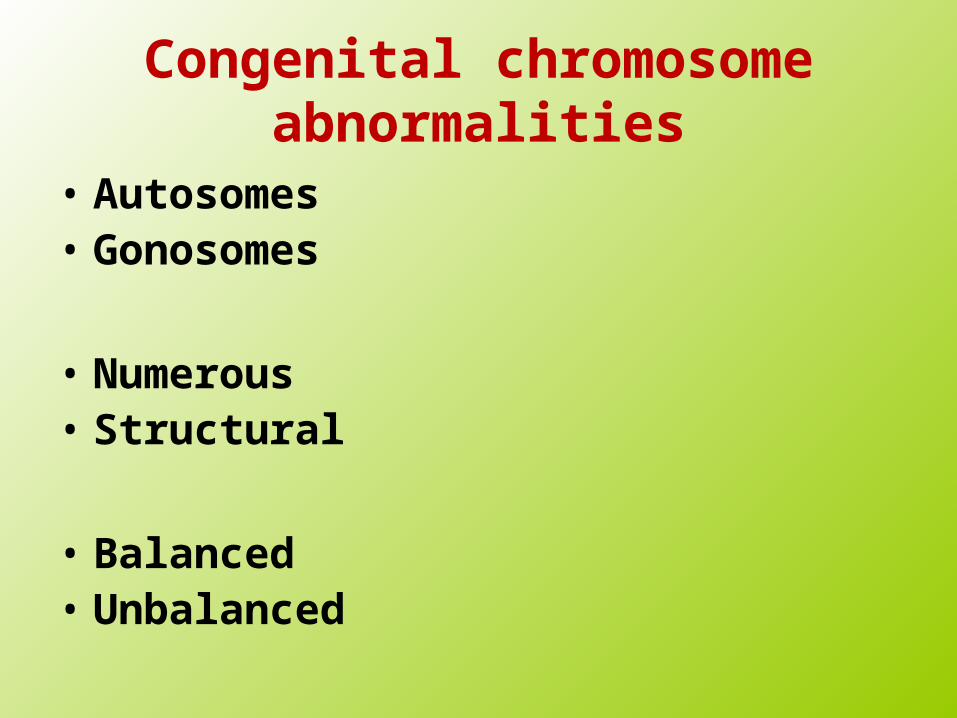
Congenital chromosome abnormalities
• Autosomes• Gonosomes
• Numerous• Structural
• Balanced• Unbalanced
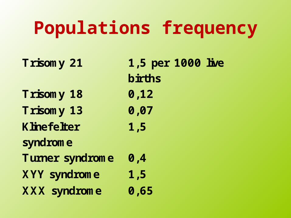
Populations frequency
Trisomy 21 1,5 per 1000 live births
Trisomy 18 0,12
Trisomy 13 0,07
Klinefelter syndrome
1,5
Turner syndrome 0,4
XYY syndrome 1,5
XXX syndrome 0,65
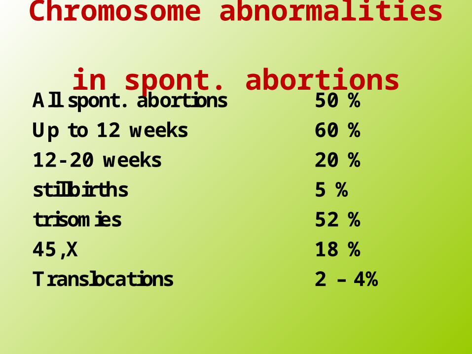
Chromosome abnormalities
in spont. abortionsAll spont. abortions 50 %
Up to 12 weeks 60 %
12- 20 weeks 20 %
stillbirths 5 %
trisomies 52 %
45,X 18 %
Translocations 2 – 4%
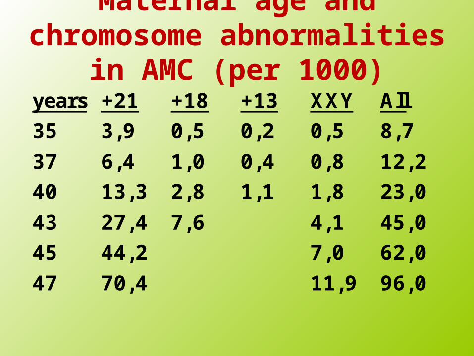
Maternal age and chromosome abnormalities
in AMC (per 1000)years +21 +18 +13 XXY All
35 3,9 0,5 0,2 0,5 8,7
37 6,4 1,0 0,4 0,8 12,2
40 13,3 2,8 1,1 1,8 23,0
43 27,4 7,6 4,1 45,0
45 44,2 7,0 62,0
47 70,4 11,9 96,0
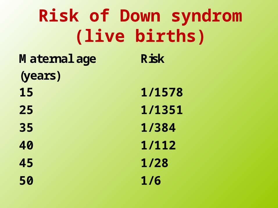
Risk of Down syndrom(live births)
Maternal age (years)
Risk
15 1/1578
25 1/1351
35 1/384
40 1/112
45 1/28
50 1/6
Down syndrome
Down syndrome• 47,XX,+21 or 47,XY,+21• About 1/800-1000 newborns, 1/75 SA• Hypotonia, joint laxicity, soft skin,
flat face, prominent intercanthal folds, slanted palpebral fissurs, Brushfield´s spots of the irides, small, down set ears, small nose, protruding tongue, simian crease in the hands (about 45%), short statue, mental retardation, congenital heart disease in about 50% of patients with DS, (atrioventricular canal)
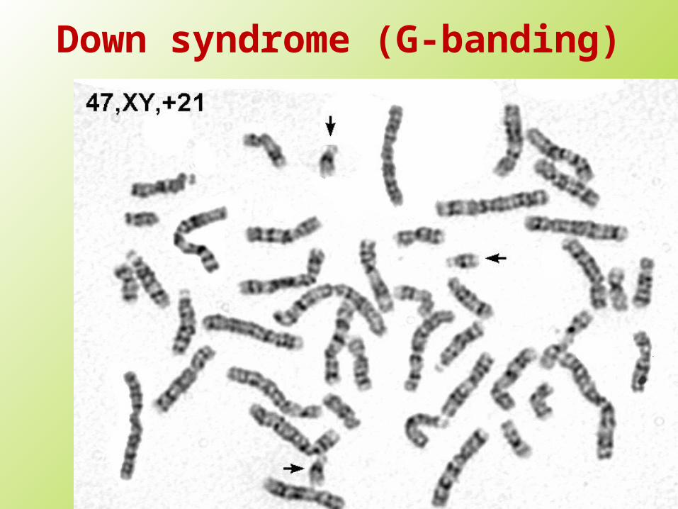
Down syndrome (G-banding)
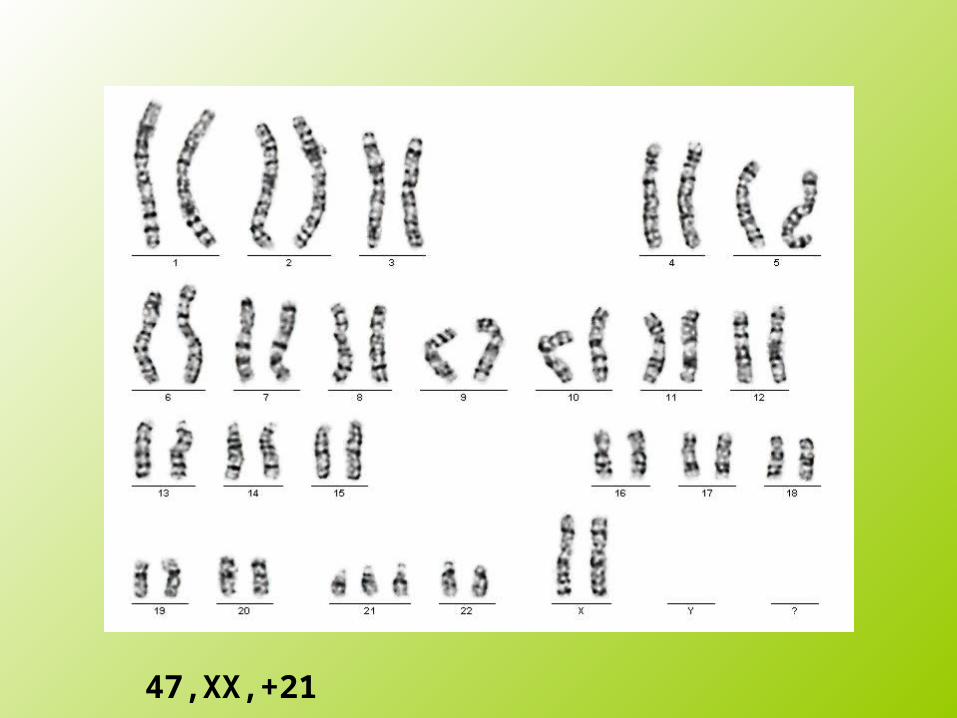
47,XX,+21
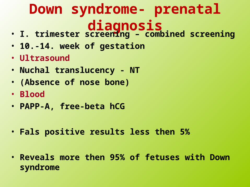
Down syndrome- prenatal diagnosis
• I. trimester screening – combined screening• 10.-14. week of gestation• Ultrasound• Nuchal translucency - NT • (Absence of nose bone)• Blood• PAPP-A, free-beta hCG
• Fals positive results less then 5%
• Reveals more then 95% of fetuses with Down syndrome
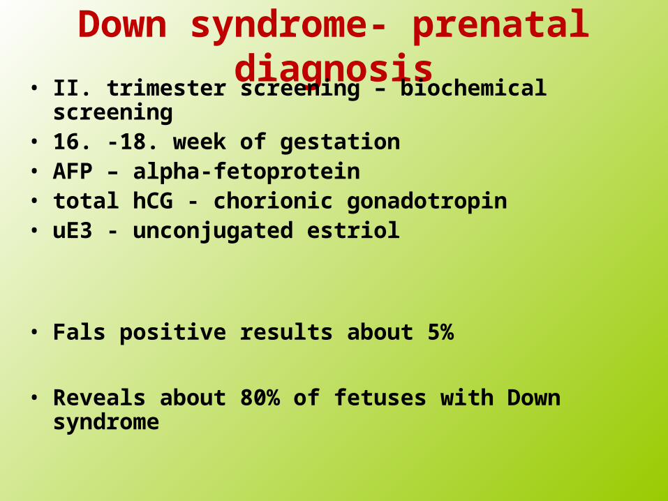
Down syndrome- prenatal diagnosis
• II. trimester screening – biochemical screening
• 16. -18. week of gestation• AFP – alpha-fetoprotein• total hCG - chorionic gonadotropin • uE3 - unconjugated estriol
• Fals positive results about 5%
• Reveals about 80% of fetuses with Down syndrome
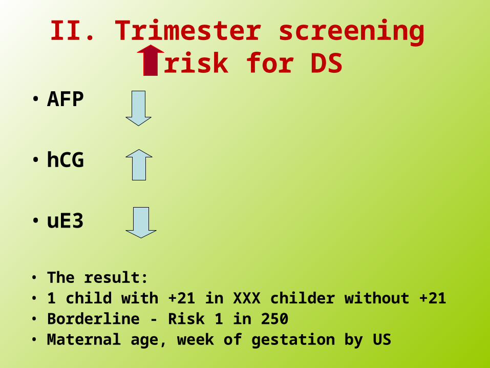
II. Trimester screening risk for DS
• AFP
• hCG
• uE3
• The result:• 1 child with +21 in XXX childer without +21• Borderline - Risk 1 in 250 • Maternal age, week of gestation by US
Down syndrome- prenatal diagnosis
• Ultrasound
• 10.-14. week• NT• NB
• 20. week • US- congenital heart disease and
other malformations
0
10
20
30
40
50
60
70
19
87
19
88
19
89
19
90
19
91
19
92
19
93
19
94
19
95
19
96
19
97
19
98
19
99
20
00
20
01
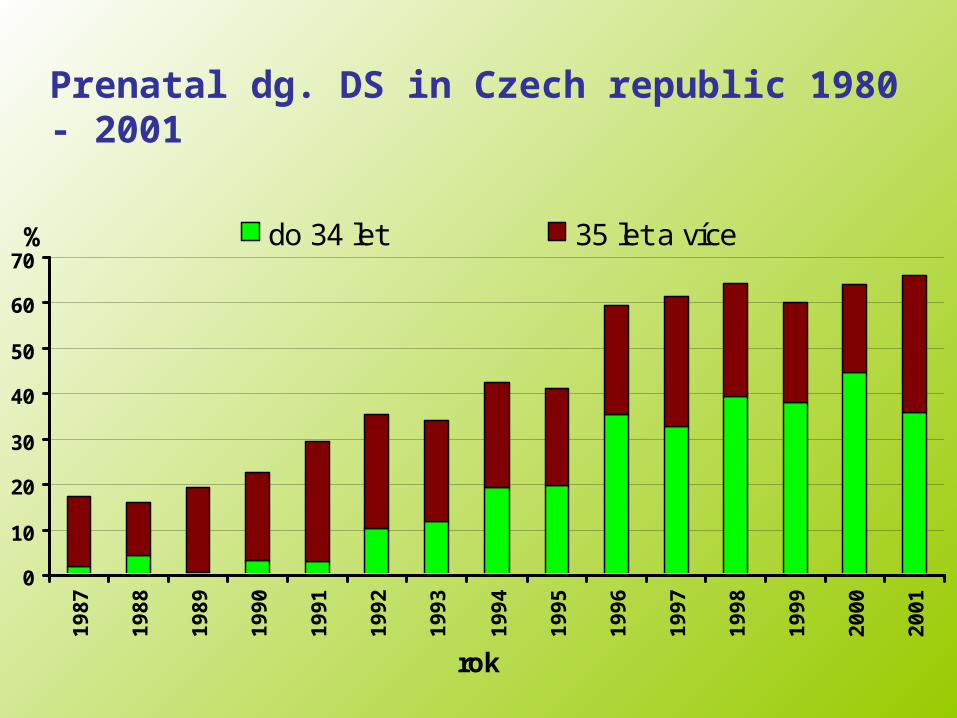
do 34 let 35 let a více%
rok
Prenatal dg. DS in Czech republic 1980 - 2001
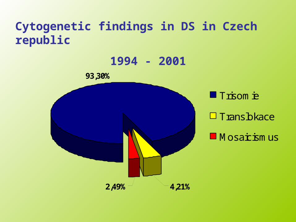
93,30%
2,49% 4,21%
Trisomie
Translokace
Mosaicismus
Cytogenetic findings in DS in Czech republic
1994 - 2001
Edwards syndrome
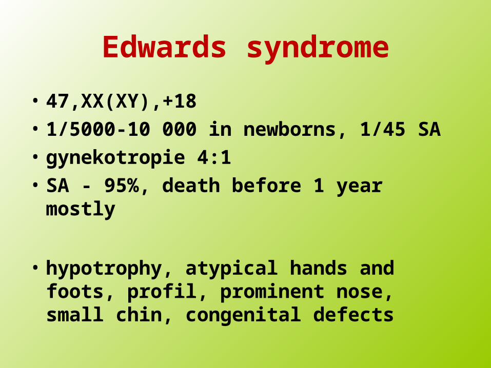
• 47,XX(XY),+18• 1/5000-10 000 in newborns, 1/45 SA• gynekotropie 4:1 • SA - 95%, death before 1 year
mostly
• hypotrophy, atypical hands and foots, profil, prominent nose, small chin, congenital defects
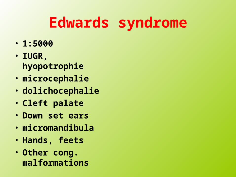
Edwards syndrome• 1:5000• IUGR,
hyopotrophie• microcephalie • dolichocephalie• Cleft palate• Down set ears• micromandibula• Hands, feets• Other cong.
malformations
Prenatal dg. +18 – II. trimester
• AFP, HCG, uE3
• Risk 1/250 - borderline• Ultrasonography
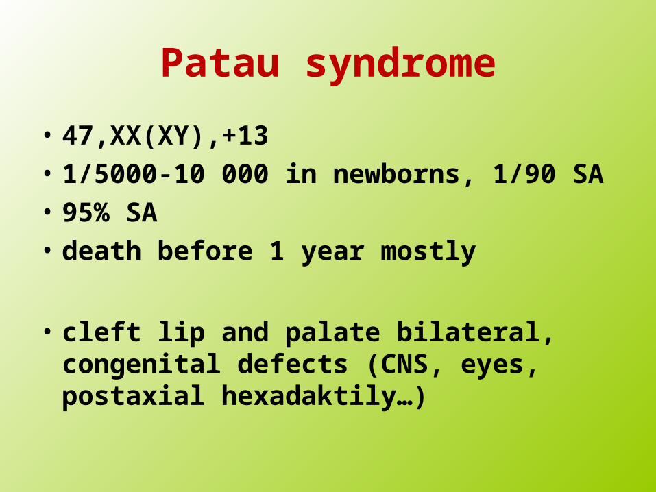
Patau syndrome
• 47,XX(XY),+13• 1/5000-10 000 in newborns, 1/90
SA• 95% SA• death before 1 year mostly
• cleft lip and palate bilateral, congenital defects (CNS, eyes, postaxial hexadaktily…)
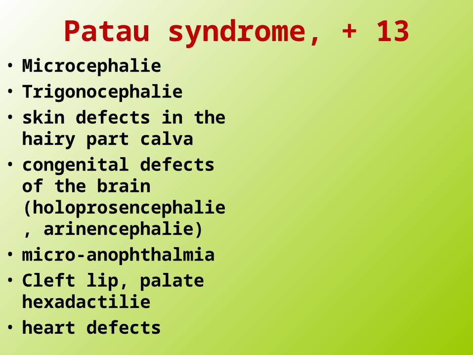
Patau syndrome, + 13• Microcephalie• Trigonocephalie• skin defects in the
hairy part calva• congenital defects
of the brain(holoprosencephalie, arinencephalie)
• micro-anophthalmia• Cleft lip, palate
hexadactilie• heart defects
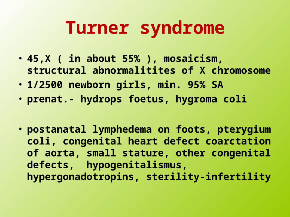
Turner syndrome
• 45,X ( in about 55% ), mosaicism, structural abnormalitites of X chromosome
• 1/2500 newborn girls, min. 95% SA• prenat.- hydrops foetus, hygroma coli
• postanatal lymphedema on foots, pterygium coli, congenital heart defect coarctation of aorta, small stature, other congenital defects, hypogenitalismus, hypergonadotropins, sterility-infertility
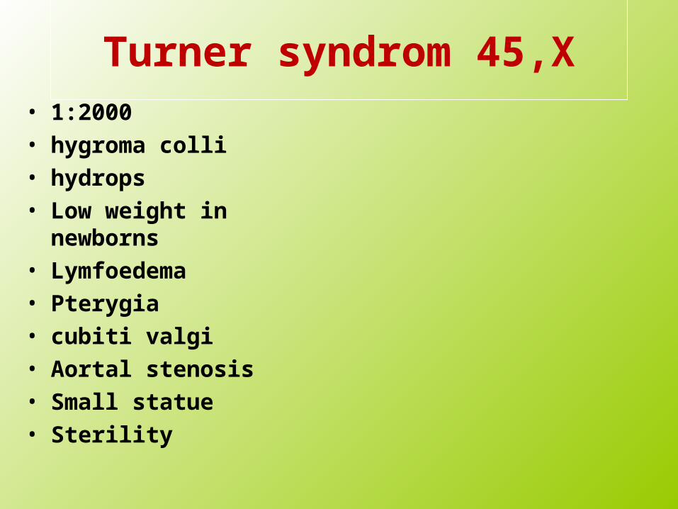
Turner syndrom 45,X• 1:2000• hygroma colli• hydrops• Low weight in
newborns • Lymfoedema• Pterygia• cubiti valgi• Aortal stenosis• Small statue• Sterility
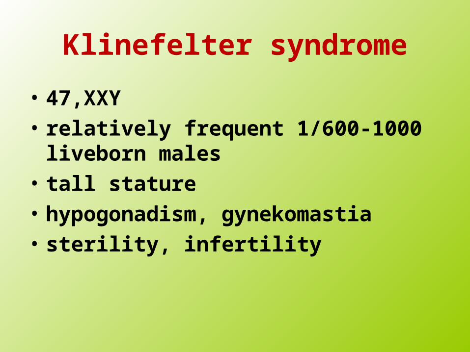
Klinefelter syndrome
• 47,XXY• relatively frequent 1/600-1000
liveborn males• tall stature• hypogonadism, gynekomastia• sterility, infertility
Others gonoseme abnormalities
• 47,XXX• 47,XYY• 48,XXXX• 48,XXYY….
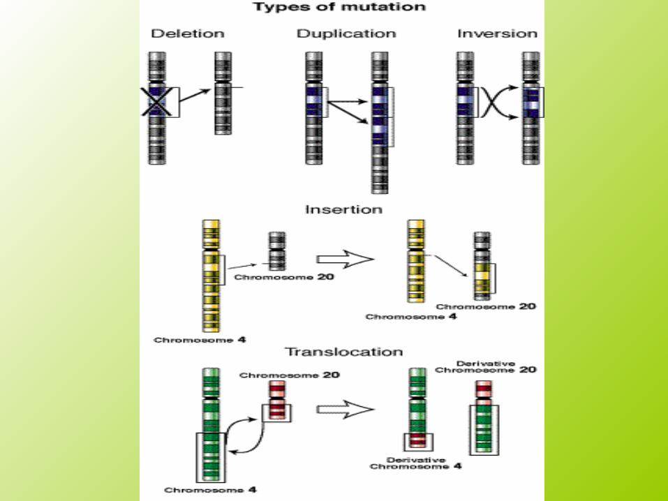
Structural chromosomal aberrations
• deletion or a duplication of the genetic material of any chromosome, atypical structure - side by side to get the genetic material, which there normally is not - the effect of positional
• partial-partial deletions• partial trisomy• inversions, insertions,
duplications ....
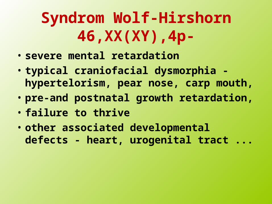
Syndrom Wolf-Hirshorn 46,XX(XY),4p-
• severe mental retardation• typical craniofacial dysmorphia -
hypertelorism, pear nose, carp mouth,
• pre-and postnatal growth retardation,
• failure to thrive• other associated developmental
defects - heart, urogenital tract ...
Wolf-Hirschhorn syndrom (46,XX,4p-)
Incidence?
IUGR
Hypotonia
Charakteristic face
Heart defects
Hypotonie
Hypotrophie
Severe mental retardation
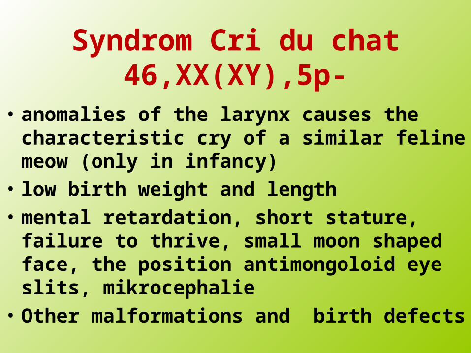
Syndrom Cri du chat 46,XX(XY),5p-
• anomalies of the larynx causes the characteristic cry of a similar feline meow (only in infancy)
• low birth weight and length• mental retardation, short stature,
failure to thrive, small moon shaped face, the position antimongoloid eye slits, mikrocephalie
• Other malformations and birth defects
Cri du chat 46,XX(XY),5p-
• 1:50 000• Typicaly cri in
newborns• laryngomalacie • antimongoloid • epicanthi• hypotonie• hypotrofie
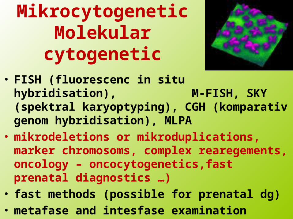
Mikrocytogenetic Molekular
cytogenetic• FISH (fluorescenc in situ hybridisation),
M-FISH, SKY (spektral karyoptyping), CGH (komparativ genom hybridisation), MLPA
• mikrodeletions or mikroduplications, marker chromosoms, complex rearegements, oncology – oncocytogenetics,fast prenatal diagnostics …)
• fast methods (possible for prenatal dg)• metafase and intesfase examination
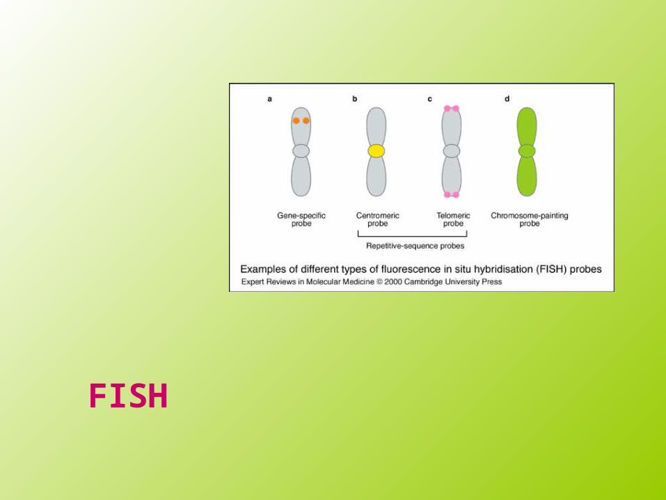
FISH
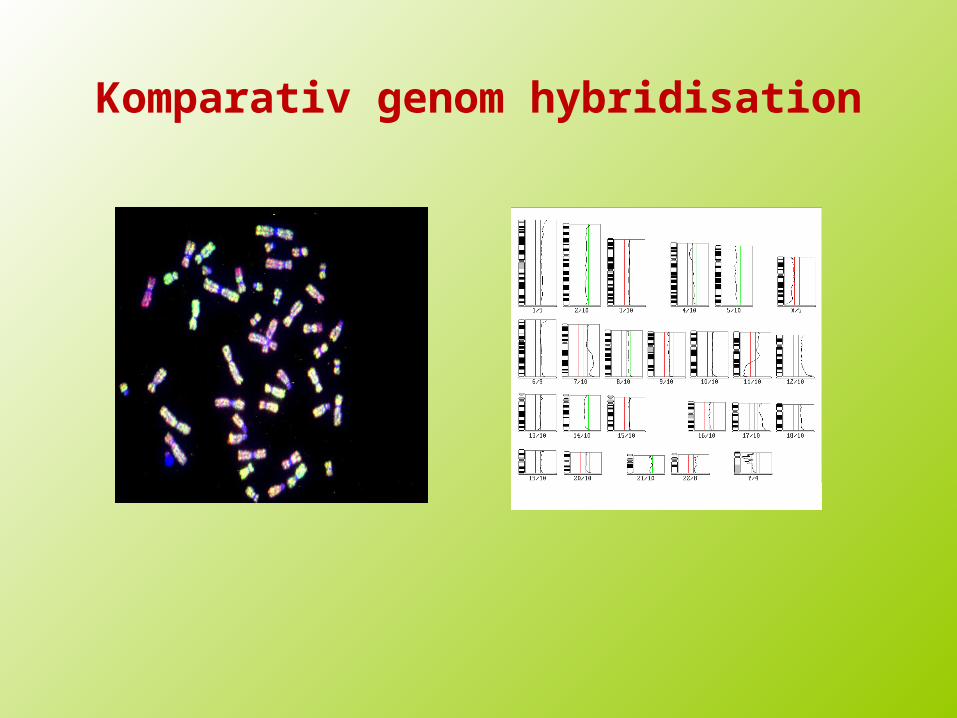
Komparativ genom hybridisation
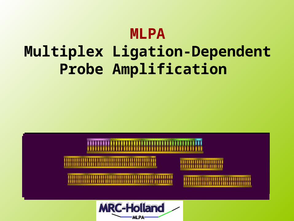
MLPAMultiplex Ligation-Dependent
Probe Amplification
Microdeletions
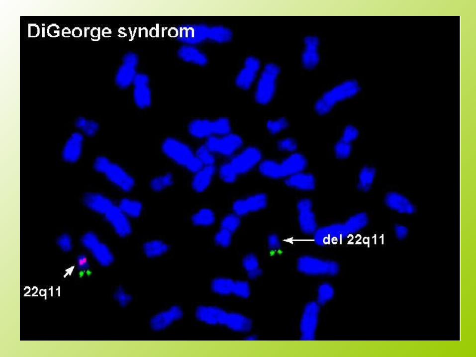
• Di George syndrome (del 22q11)
• Prader-Willi / Angelman syndrome (del15q11-13)
• Williams Beuren syndrome (del7q11.23)
Syndrom Di George
• Velo - Kardio- Facial syndrome• CATCH 22• Congenital heart desease -
conotruncal, craniofacial dysmorfism, thymus aplasie, imunodefitient¨cy, hypoparathyreoidismus
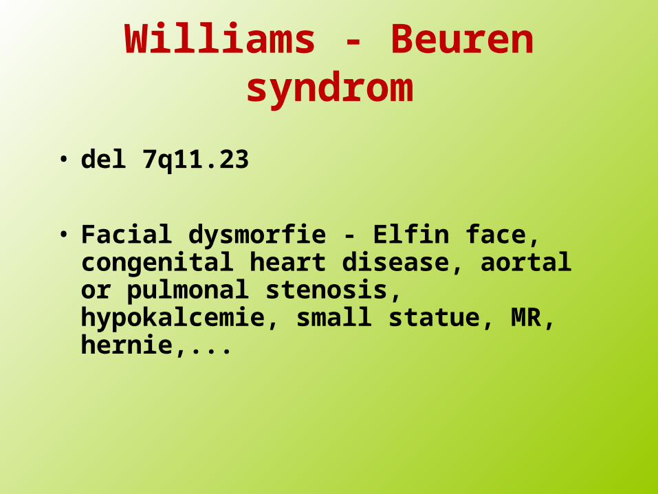
Williams - Beuren syndrom
• del 7q11.23
• Facial dysmorfie - Elfin face, congenital heart disease, aortal or pulmonal stenosis, hypokalcemie, small statue, MR, hernie,...
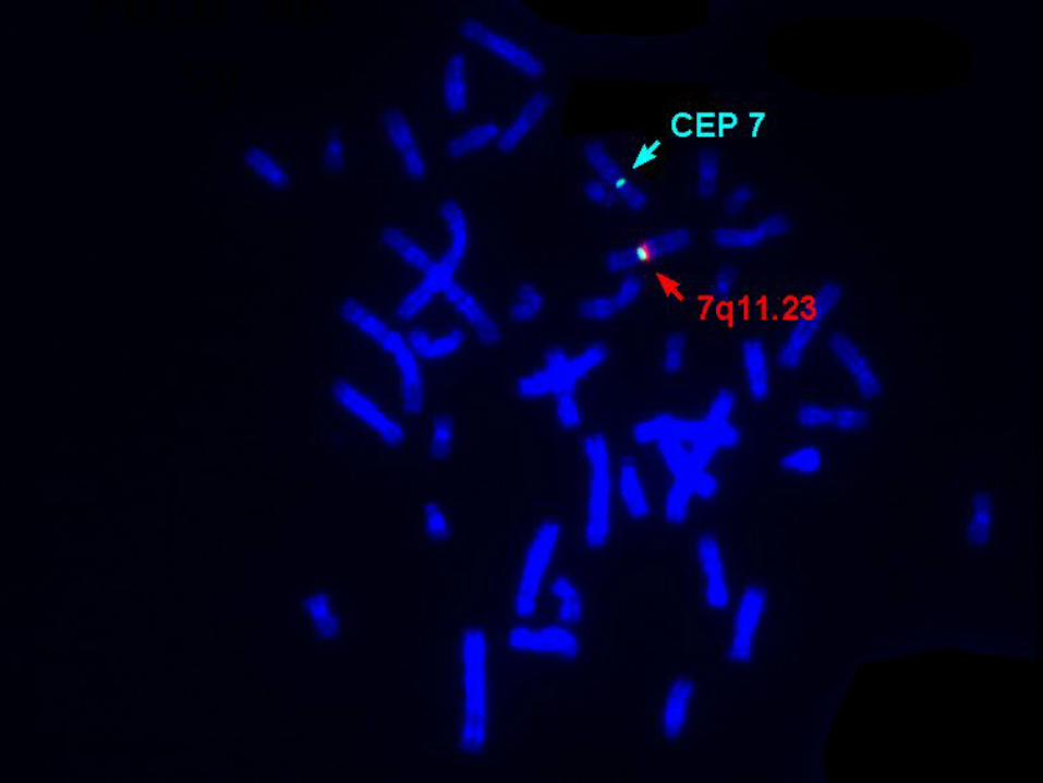
Foto WB sy
Prader-Willi syndrom
• Hypotonie, hypotrofie in small children
• PMR, small statue, obesity, hyperfagie, akromikrie, hypogonadismus
• mikrodeletion15q11-12 paternal
Angelman syndrom
• Severe mental retardation
• Epilepsie• Laughter• severely delayed
speech development
• mikrodeletion 15q11-12 mat
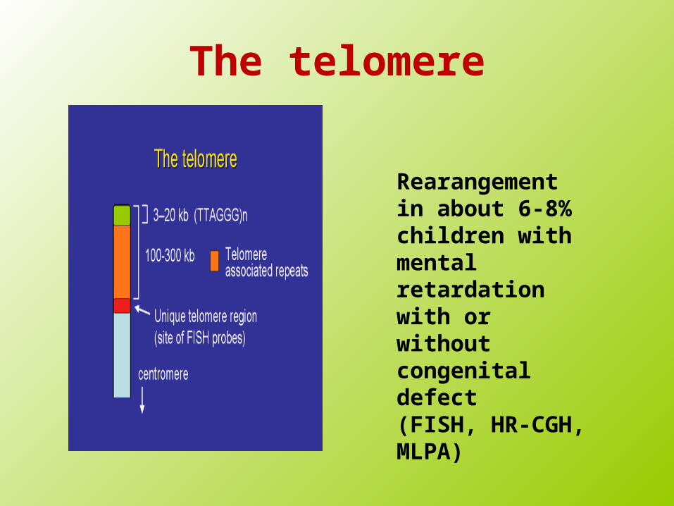
The telomere
Rearangement in about 6-8% children with mental retardation with or without congenital defect (FISH, HR-CGH, MLPA)

Mendelian inheritance
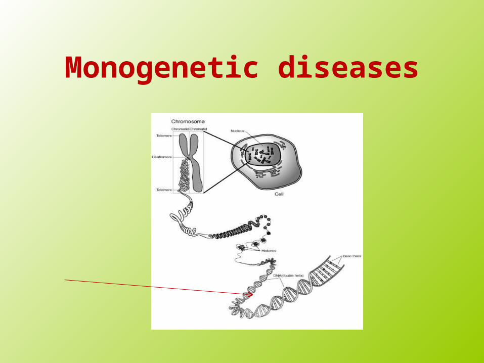
Monogenetic diseases
Autosomal Dominant
• The sexes are involved equaly• Heterozygotes are mostly
affected clinically• risk 50% for sibs and children• new mutations• penetrance, expresivity
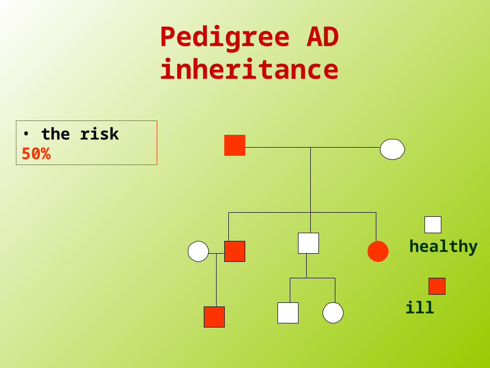
Pedigree AD inheritance
• the risk 50%
healthy
ill
AD - diseases
• Neurofibromatosis 1 and 2• Achondoplasia• Huntington disease• Marfan syndrome• Myotonic dystrophy
Neurofibtromatosis 1
• Neurofibromatosis type I is an autosomal dominant disorder
characterized by cafe-au-lait spots, Lisch nodules in the eye, and fibromatous tumors of the skin
• 50% of cases are caused by new mutations
• Caused by mutations in the neurofibromin gene (NF1)
Myotonic dystrophy
MD 1: 19q13.32 Caused by a trinucleotide repeat expansion (CTG)n in the dystrophia myotonica-protein kinase gene (DMPK), Prevalence of in 1 in 8,000 MD 2: 3q21.3Caused by a (CCTG)n repeat expansion in the zinc fingerprotein 9 gene (ZNF9)
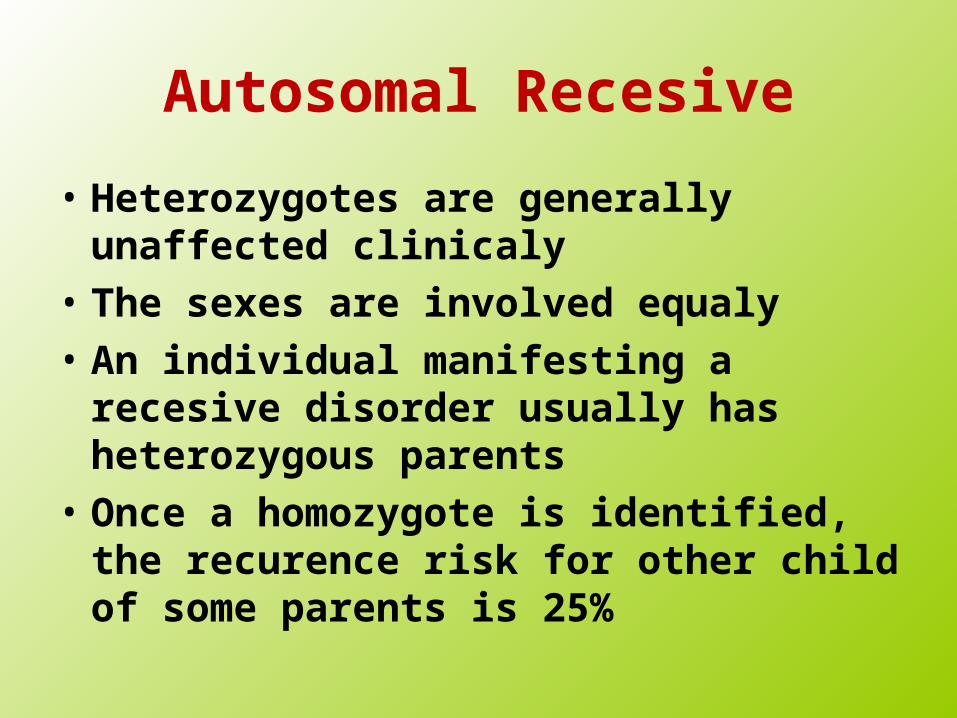
Autosomal Recesive
• Heterozygotes are generally unaffected clinicaly
• The sexes are involved equaly• An individual manifesting a
recesive disorder usually has heterozygous parents
• Once a homozygote is identified, the recurence risk for other child of some parents is 25%
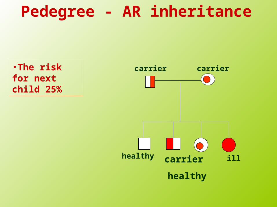
Pedegree - AR inheritance
•The risk for next child 25%
carriercarrier
healthy
illcarrier
healthy
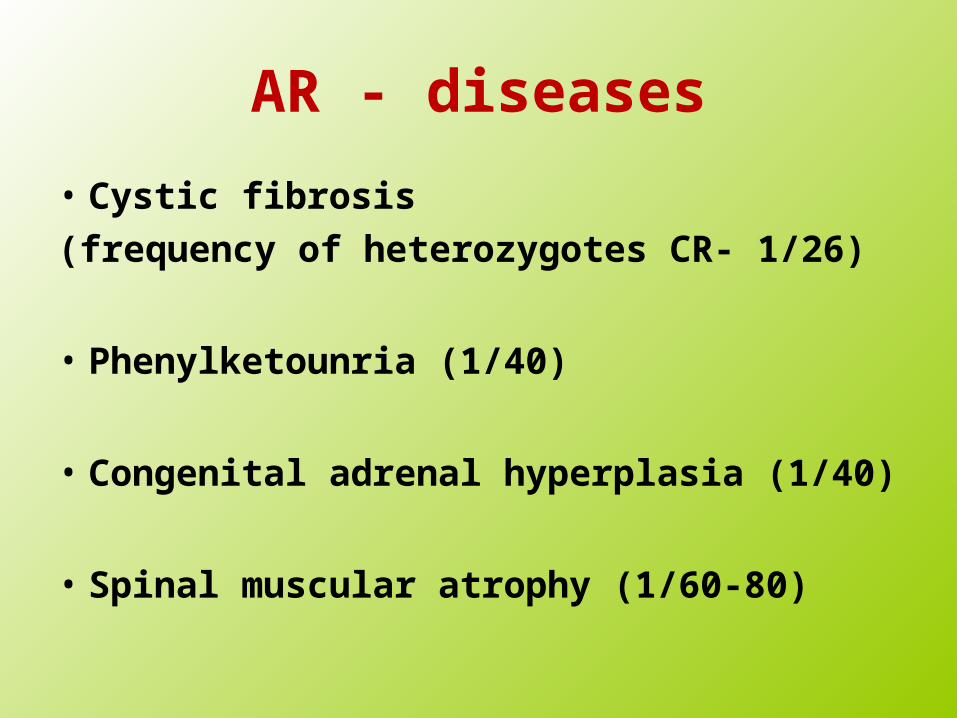
AR - diseases
• Cystic fibrosis (frequency of heterozygotes CR- 1/26)
• Phenylketounria (1/40)
• Congenital adrenal hyperplasia (1/40)
• Spinal muscular atrophy (1/60-80)
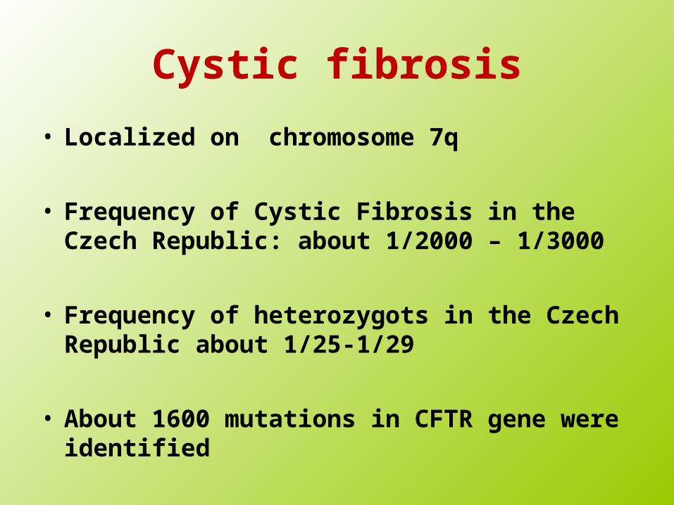
Cystic fibrosis
• Localized on chromosome 7q
• Frequency of Cystic Fibrosis in the Czech Republic: about 1/2000 – 1/3000
• Frequency of heterozygots in the Czech Republic about 1/25-1/29
• About 1600 mutations in CFTR gene were identified
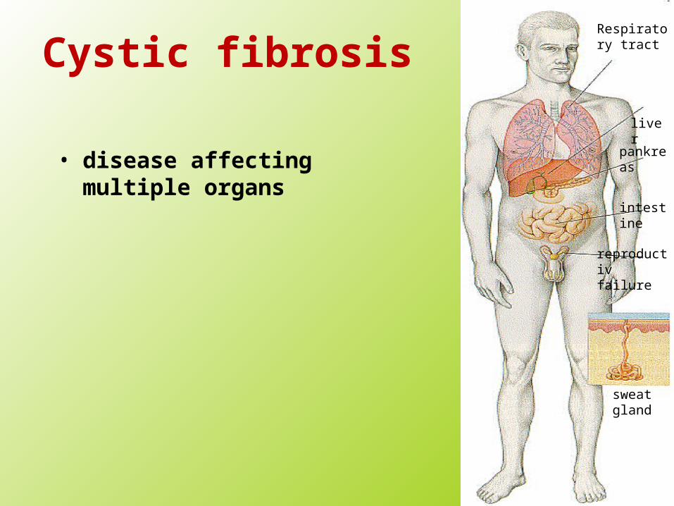
Cystic fibrosis
• disease affecting multiple organs
Respiratory tract
liver
pankreas
intestine
reproductiv failure
sweat gland
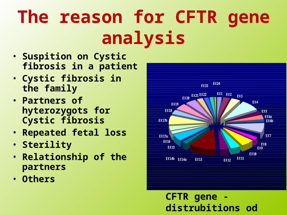
The reason for CFTR gene analysis
• Suspition on Cystic fibrosis in a patient
• Cystic fibrosis in the family
• Partners of hyterozygots for Cystic fibrosis
• Repeated fetal loss• Sterility• Relationship of the
partners• Others
EX1 EX2 EX3
EX4
EX5
EX6aEX6b
EX7
EX8EX9
EX10EX11EX12EX13
EX15
EX16EX17a
EX17b
EX18
EX19
EX20EX21 EX22
EX14aEX14b
EX23EX24
CFTR gene - distrubitions od mutations
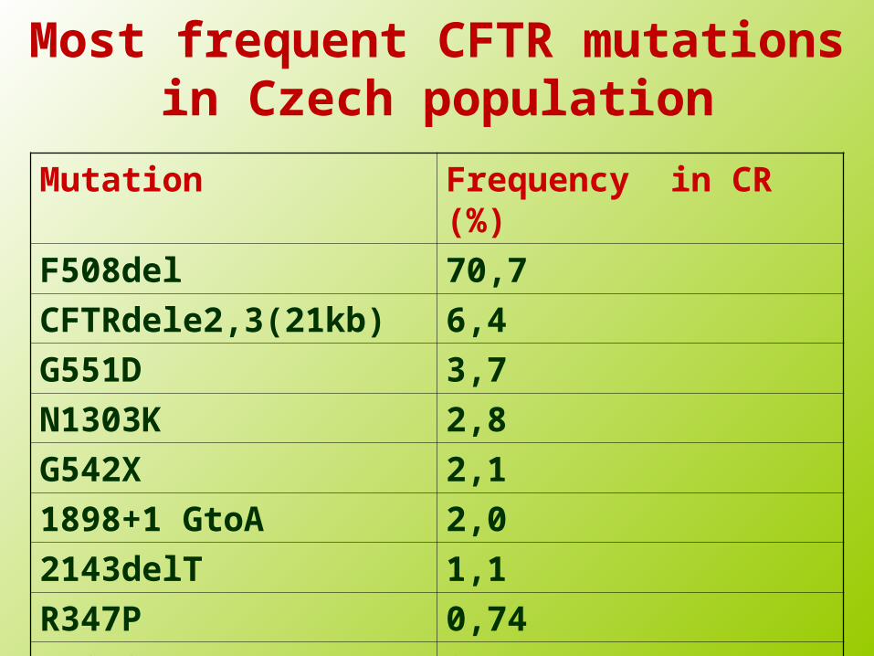
Most frequent CFTR mutations in Czech population
Mutation Frequency in CR (%)
F508del 70,7
CFTRdele2,3(21kb) 6,4
G551D 3,7
N1303K 2,8
G542X 2,1
1898+1 GtoA 2,0
2143delT 1,1
R347P 0,74
W1282X 0,6
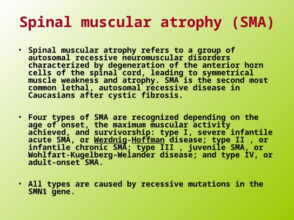
Spinal muscular atrophy (SMA)
• Spinal muscular atrophy refers to a group of autosomal recessive neuromuscular disorders characterized by degeneration of the anterior horn cells of the spinal cord, leading to symmetrical muscle weakness and atrophy. SMA is the second most common lethal, autosomal recessive disease in Caucasians after cystic fibrosis.
• Four types of SMA are recognized depending on the age of onset, the maximum muscular activity achieved, and survivorship: type I, severe infantile acute SMA, or Werdnig-Hoffman disease; type II , or infantile chronic SMA; type III , juvenile SMA, or Wohlfart-Kugelberg-Welander disease; and type IV, or adult-onset SMA.
• All types are caused by recessive mutations in the SMN1 gene.
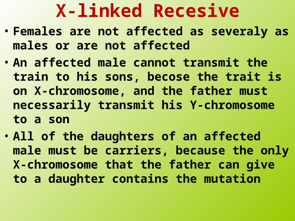
X-linked Recesive• Females are not affected as severaly as
males or are not affected• An affected male cannot transmit the
train to his sons, becose the trait is on X-chromosome, and the father must necessarily transmit his Y-chromosome to a son
• All of the daughters of an affected male must be carriers, because the only X-chromosome that the father can give to a daughter contains the mutation
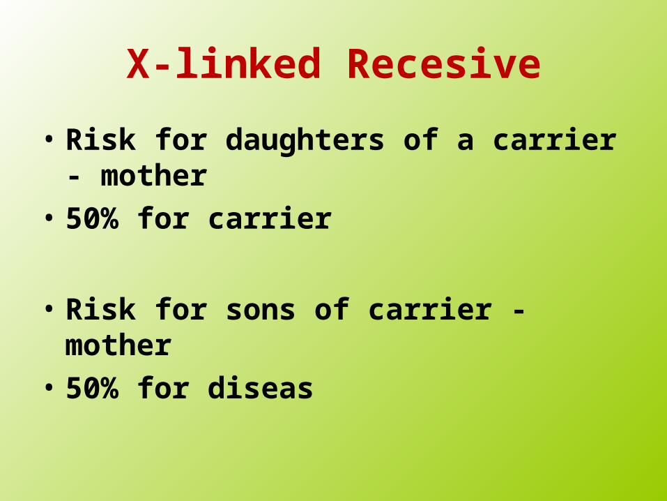
X-linked Recesive
• Risk for daughters of a carrier - mother
• 50% for carrier
• Risk for sons of carrier - mother• 50% for diseas
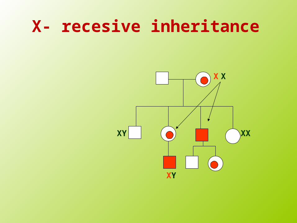
X- recesive inheritance
X
XY
XX
X
XY
XR - diseases
• Hemophilia A and B
• Duchenne and Becker muscular dystrophy
• Fragile X chromosome - X-linked disease
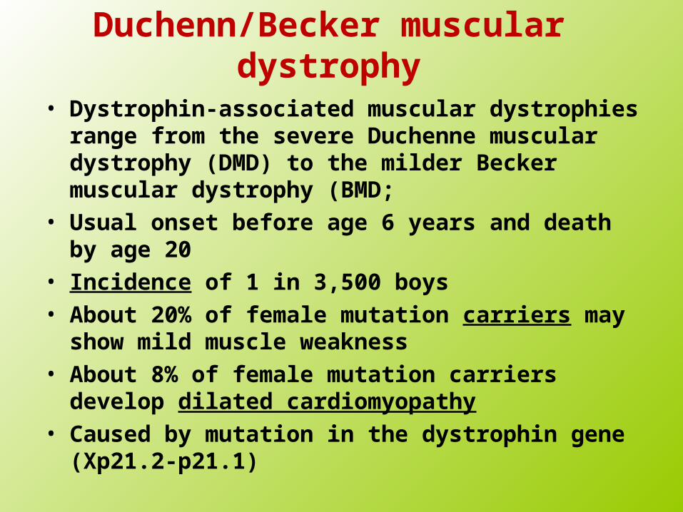
Duchenn/Becker muscular dystrophy
• Dystrophin-associated muscular dystrophies range from the severe Duchenne muscular dystrophy (DMD) to the milder Becker muscular dystrophy (BMD;
• Usual onset before age 6 years and death by age 20
• Incidence of 1 in 3,500 boys • About 20% of female mutation carriers may
show mild muscle weakness • About 8% of female mutation carriers
develop dilated cardiomyopathy• Caused by mutation in the dystrophin gene
(Xp21.2-p21.1)
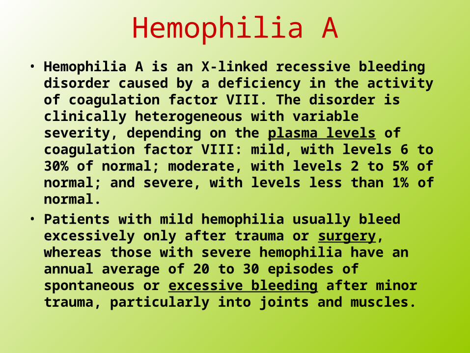
Hemophilia A• Hemophilia A is an X-linked recessive bleeding
disorder caused by a deficiency in the activity of coagulation factor VIII. The disorder is clinically heterogeneous with variable severity, depending on the plasma levels of coagulation factor VIII: mild, with levels 6 to 30% of normal; moderate, with levels 2 to 5% of normal; and severe, with levels less than 1% of normal.
• Patients with mild hemophilia usually bleed excessively only after trauma or surgery, whereas those with severe hemophilia have an annual average of 20 to 30 episodes of spontaneous or excessive bleeding after minor trauma, particularly into joints and muscles.
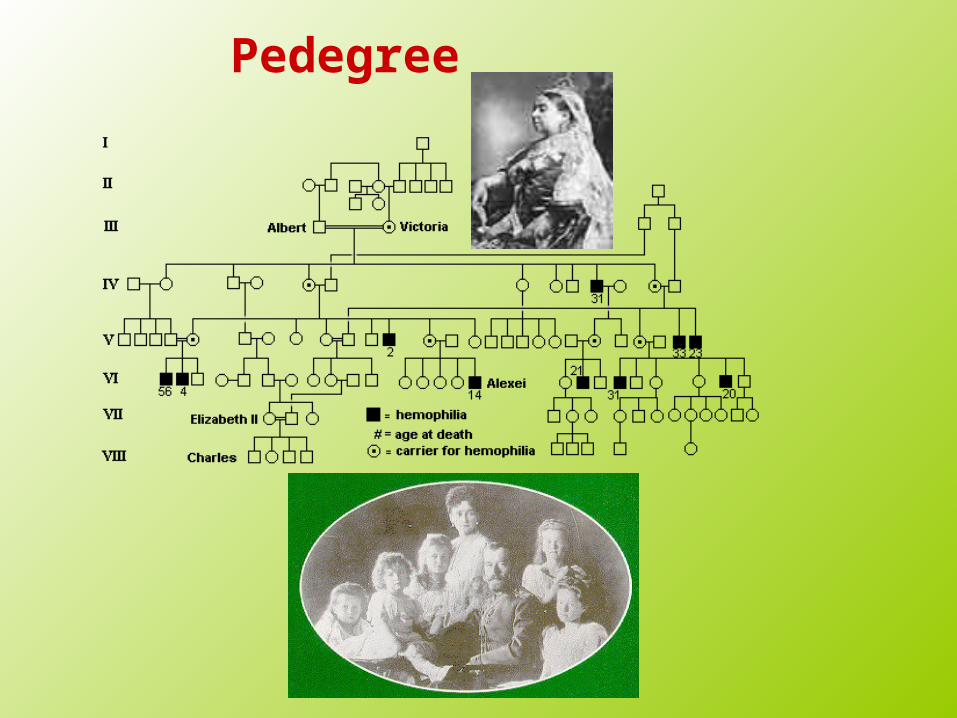
Pedegree
Multifaktorial –polygenic inheritance
Dieseases with complex heritability
Teratogens
Charakterization• disease with multifactorial
inheritance include not mendelian types of inheritance
• diseases exhibit familial aggregation, because the relatives of affected individuals more likely than unrelated people to carry diseases predisposing predisposition
Charakterization
• in the pathogenesis of the disease play a basic role non-genetic factors
• disease is more common among close relatives and in distant relatives is becoming less frequent
Examples• Congenitzal heart defects (VCC) 4-8/1000• Cleft lip and palate (CL/P) 1/1000• Neural tube defects (NTD, anencefalie,
spina bifida,..) 0,2-1/1000• Pylorostenosis• Congenital hip dislocation• Diabetes mellitus – most types• Ischemic heart desoease• Esential epilepsy
Common congenital defects
Congenital heart diseases
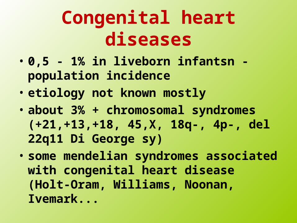
• 0,5 - 1% in liveborn infantsn - population incidence
• etiology not known mostly• about 3% + chromosomal syndromes
(+21,+13,+18, 45,X, 18q-, 4p-, del 22q11 Di George sy)
• some mendelian syndromes associated with congenital heart disease (Holt-Oram, Williams, Noonan, Ivemark...
Congenital heart diseases
prenatal diagnosis
• For most serious congenital heart diseases
• Ultrasonography in 21. week of gestation - by specialists for prenatal kardiology
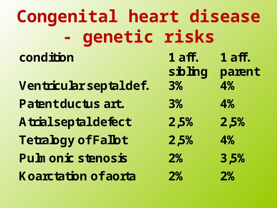
Congenital heart disease - genetic risks
condition 1 aff.sibling
1 aff.parent
Ventricular septal def. 3% 4%
Patent ductus art. 3% 4%
Atrial septal defect 2,5% 2,5%
Tetralogy of Fallot 2,5% 4%
Pulmonic stenosis 2% 3,5%
Koarctation of aorta 2% 2%
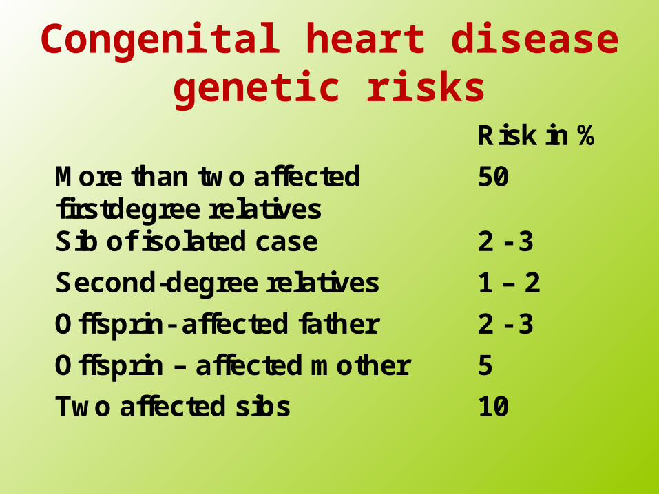
Congenital heart diseasegenetic risks
Risk in %
More than two affectedfirstdegree relatives
50
Sib of isolated case 2 - 3
Second-degree relatives 1 – 2
Offsprin- affected father 2 - 3
Offsprin – affected mother 5
Two affected sibs 10
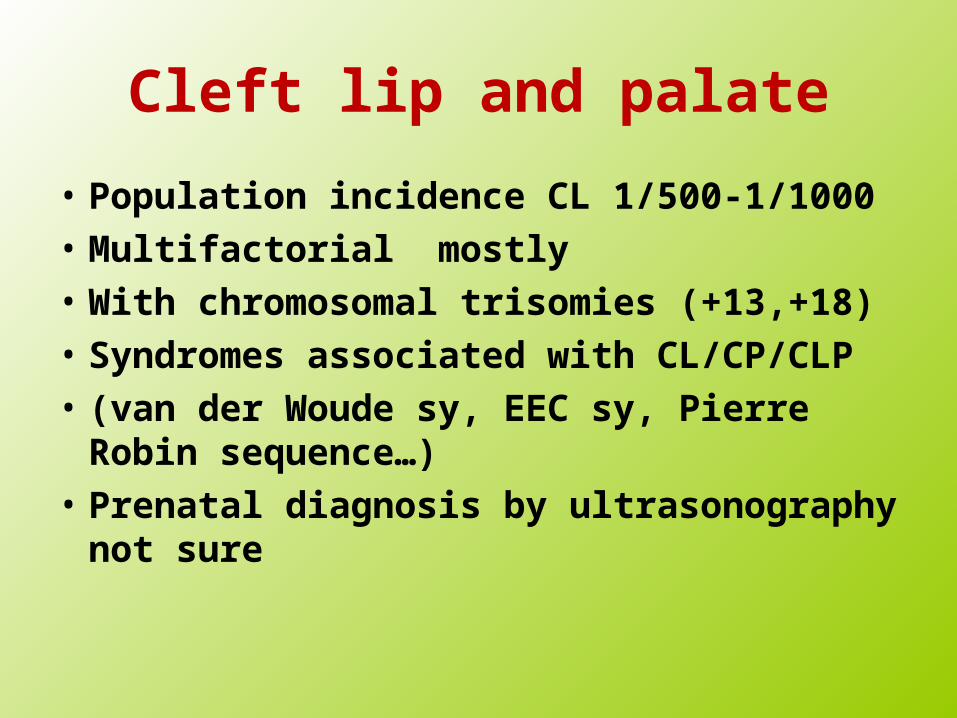
Cleft lip and palate
• Population incidence CL 1/500-1/1000• Multifactorial mostly• With chromosomal trisomies
(+13,+18)• Syndromes associated with CL/CP/CLP• (van der Woude sy, EEC sy, Pierre
Robin sequence…)• Prenatal diagnosis by
ultrasonography not sure
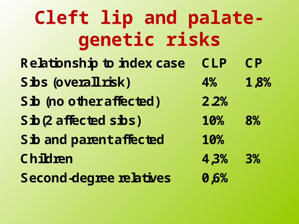
Cleft lip and palate- genetic risks
Relationship to index case CLP CP
Sibs (overall risk) 4% 1,8%
Sib (no other affected) 2.2%
Sib(2 affected sibs) 10% 8%
Sib and parent affected 10%
Children 4,3% 3%
Second-degree relatives 0,6%
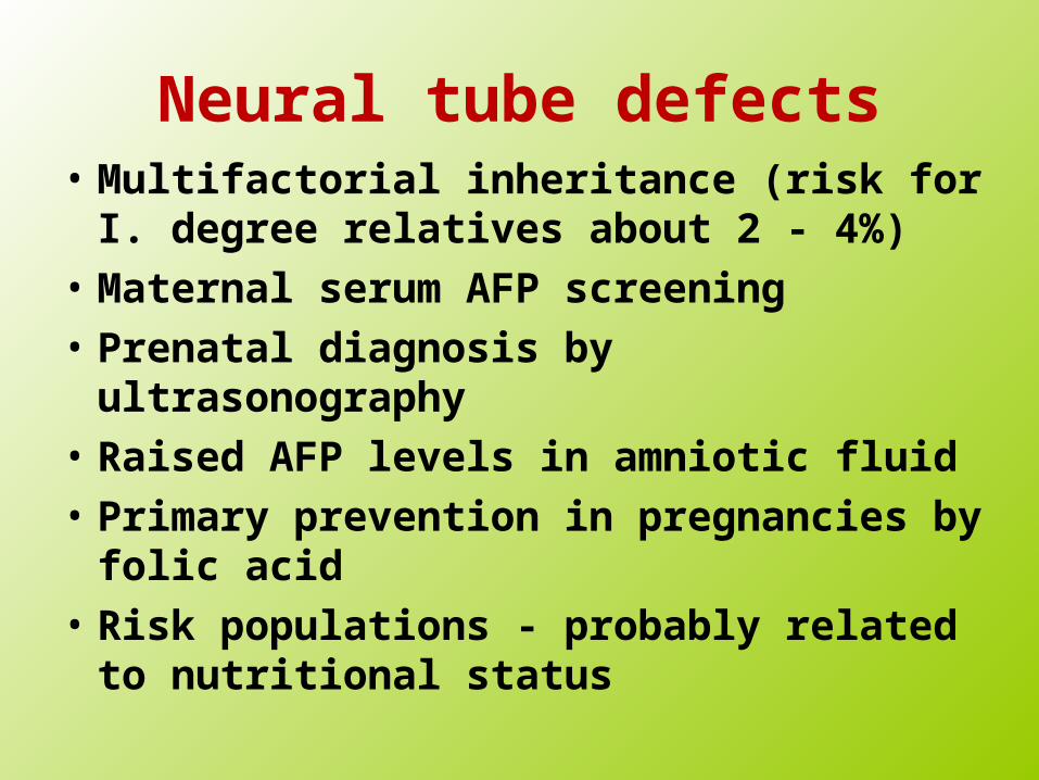
Neural tube defects• Multifactorial inheritance (risk for
I. degree relatives about 2 - 4%)• Maternal serum AFP screening• Prenatal diagnosis by
ultrasonography• Raised AFP levels in amniotic fluid• Primary prevention in pregnancies
by folic acid• Risk populations - probably
related to nutritional status
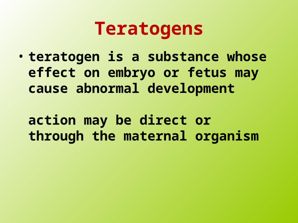
Teratogens• teratogen is a substance whose
effect on embryo or fetus may cause abnormal development
action may be direct or through the maternal organism

Human Teratogens
• Physical (radiation, heat (fever), mechanical impact)
• Chemical (chemicals, drugs)
• Biological (infection, fungus ...)
• Metabolic imbalance (disease mother)
The effect of teratogens depends on :
• dose
• length of the action
• contact time
• genetic equipment of the fetus and the mother
Critical period
• 14.-18. days after conception – the rule „all od nothing “
• 18.-90. day – organogenesis• The most sensitive period for the
emergence of developmental defects
Drugs
• Distribution of medicines practice into categories
• A• B• C• D• X • Food and Drug Administarion, 1980
A
• in controlled studies have shown no evidence of risk to the fetus in the first trimester of fetal development or influence in the next period of pregnancy
product appears to be safe
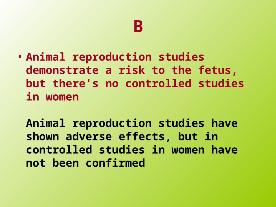
B
• Animal reproduction studies demonstrate a risk to the fetus, but there's no controlled studies in women
Animal reproduction studies have shown adverse effects, but in controlled studies in women have not been confirmed
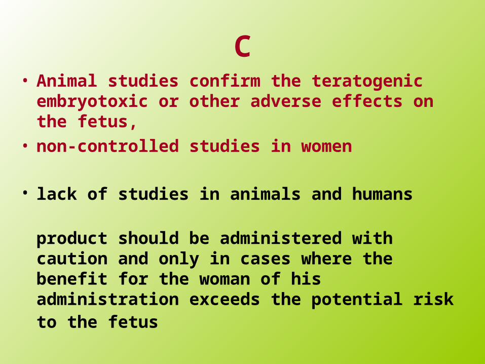
C• Animal studies confirm the teratogenic
embryotoxic or other adverse effects on the fetus,
• non-controlled studies in women
• lack of studies in animals and humans product should be administered with caution and only in cases where the benefit for the woman of his administration exceeds the potential risk to the fetus
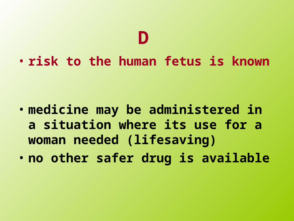
D• risk to the human fetus is known
• medicine may be administered in a situation where its use for a woman needed (lifesaving)
• no other safer drug is available
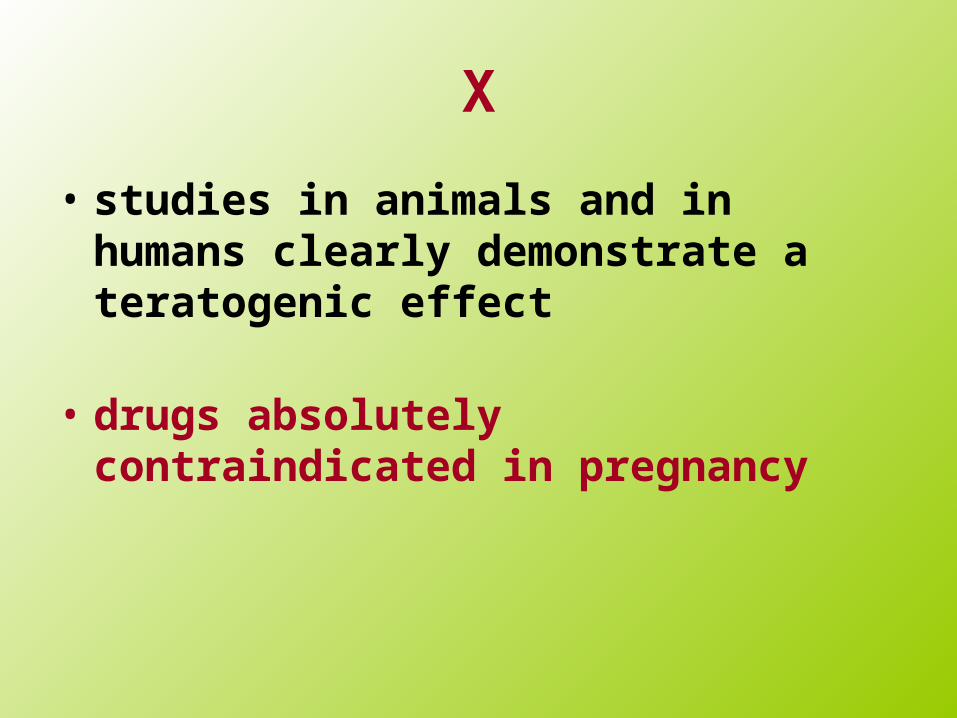
X
• studies in animals and in humans clearly demonstrate a teratogenic effect
• drugs absolutely contraindicated in pregnancy
Drugs with teratogenic effect
• Thalidomid• Hydantoin • Valproic acid• Anti coagulans - Warfarin• Trimetadion• Aminopterin • Methotrexat• Cyklophosphamid
Drugs with teratogenic effect
• Retinoids• Lithium• Thyxreostatic drugs• Androgens• Penicilamin• Enelapril, Captopril• Antituberkulotics-Streptomycin
Thalaidomid
• congenital heart defects• limb reduction anomalies• Other congenital defects
(gastrointestinal, urogenital tract orofacial – ears anomalies, CNS defects..)
Hydantoin
• Atypicaly face, growth retardation, mild mental retardation, behavioral problems, hypoplastic nails and fingers
Aminopterin a Methotrexat
• folic acid antagonist facial dysmorfism, cleft lip and/or palate, small mandible, ears anomalies, hydrocephaly, growth and mental retardation, miscarriage
Warfarin
• coumarin antikoagulans• facial dysmorfism – nasal
cartilage hypoplasia, CNS - defects
Retinoids
• Cleft lip and palate, mikrognatia, eyes anomalies, ears dysplasia
• Defects of CNS• Thymus hypoplasia• Limb defects
Infection
• Toxoplasmosis• Rubella• Cytomegalovirus• Herpesvirus• Others (parvovirus, antropozoonosy,
chlamydia..)
• TORCH
Toxoplasmosis
• chorioretinitis• hydrocephaly or microcephaly• intracranial calcification, mental
retardation• icterus, hepatosplenomegalia, carditis• prematurity
• positiv IgM in the mother – treatment with Rovamycin
• Prenatal dg.: serology, DNA-PCR)
Rubella
• hearing and vision impairment (cataract, glaucoma, mikroftalmia, blidness)
• mental retardation• Cong. heart defects• icterus, hepatosplenomegalia
• prevention- vaccination
Cytomegalovirus• Intrauterin growth retardation• mikrocephaly, cacification in the
brain, mental retardation, • hepatosplenomegaly
• Repeated maternal infection is possible
• Prenatal dg.: serology,DNA-PCR
Varicella zoster• Skin lesions and defects• Brain domage, mental retardation• Eye defects
• Prenatal dg. - serology, DNA-PCR

Metabolic dysbalance
• Fetal alcohol syndrom (FAS)• Maternal Phenylketonuria • Maternal Diabetes mellitus• Maternal Hypothyreosis
Fetal alcohol syndrom
• Hypotrophy, growth retardation, mental retardation
• facial dysmorphism• Congenital heart defects• Limb defekts
• Abuse of 60g pure alcohol / day (longterm)
• Combine with malnutrition, folic acid deficit...

Maternal Phenylketonuria
• Low birth weith• hypertonia• mikrocefaly, mental retardation• Cong. heart defects• hyperaktivity
• newborn screening • (frequency 1/10 000 newborns• inheritance - AR)• initiation of treatment within three weeks
to prevent mental retardation in the child
Reproductive Genetics Preconceptional testing
Genetic counselling and analysisin couples with reproductive disorders
Prenatal diagnosis Preimplantation genetic diagnosis
Examination of potential donor gametes
Secondary prevention of genetic
•The procedures in pregnancy - prenatal diagnosis and early postnatal diagnosis
Prenatal diagnosis
• Non invasive methods- screening
• Invasive methods • CVS – after the 10. week of gestation• AMC – 15.-18. week of gestation• Kordocentesis – after the 20. week of
gestation
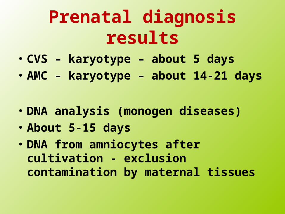
Prenatal diagnosis results
• CVS – karyotype – about 5 days• AMC – karyotype – about 14-21
days
• DNA analysis (monogen diseases) • About 5-15 days • DNA from amniocytes after
cultivation - exclusion contamination by maternal tissues
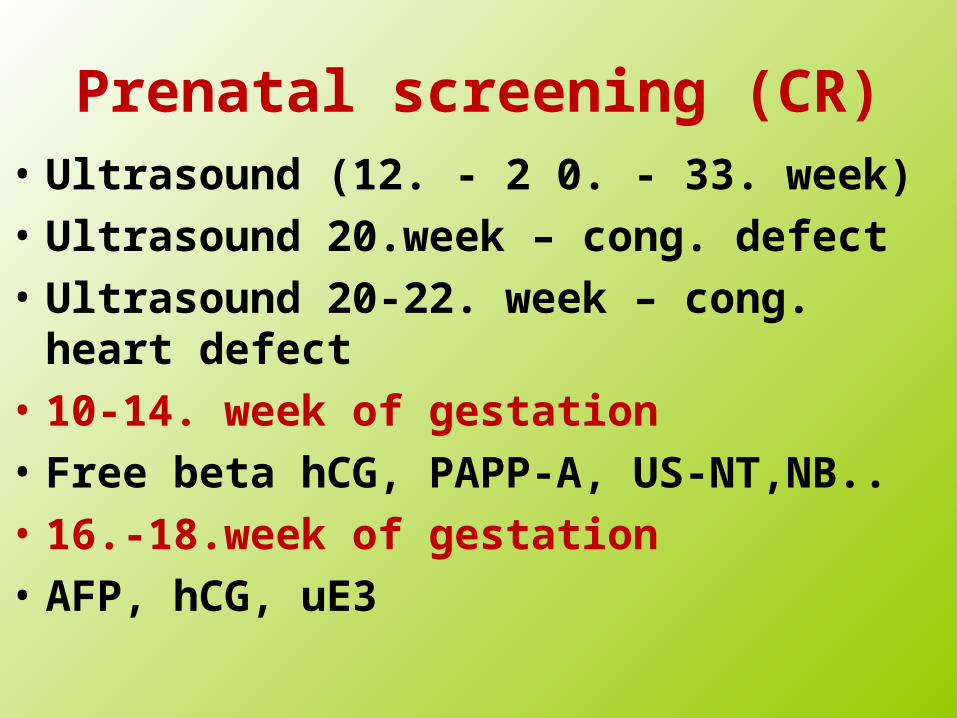
Prenatal screening (CR)• Ultrasound (12. - 2 0. - 33. week)• Ultrasound 20.week – cong. defect• Ultrasound 20-22. week – cong. heart
defect• 10-14. week of gestation• Free beta hCG, PAPP-A, US-NT,NB..• 16.-18.week of gestation• AFP, hCG, uE3

Indications for prenatal diagnosis / counselling
• Advanced maternal age (35-38 years)• Risk factors – US – congenital defects• Family history of known conditions for
which diagnosis is possible (DNA analysis)
• Known chromosomal abnormality (de novo finding in previous child, structural change in parents)
• Positive prenatal screening for chromosomal abnormalities
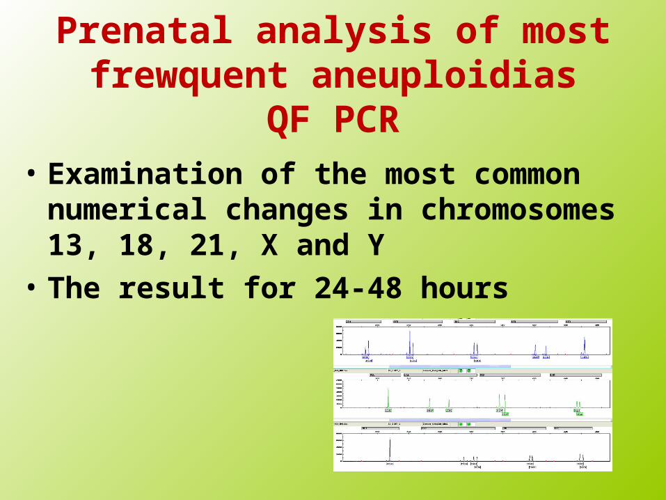
Prenatal analysis of most frewquent aneuploidias
QF PCR• Examination of the most common
numerical changes in chromosomes 13, 18, 21, X and Y
• The result for 24-48 hours
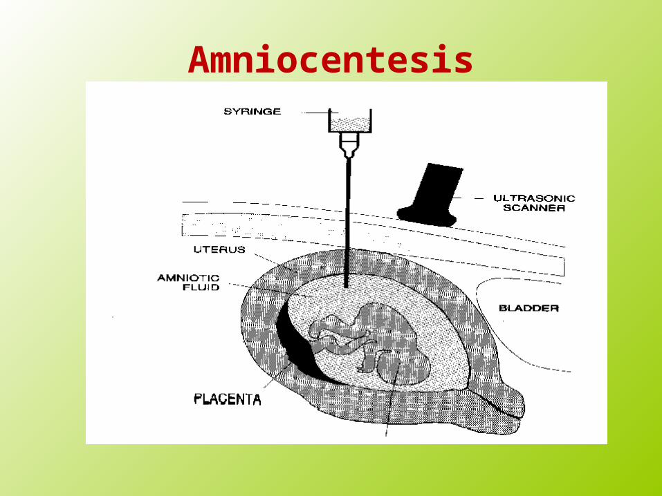
Amniocentesis
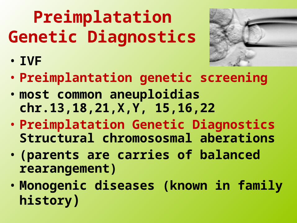
Preimplatation Genetic Diagnostics• IVF• Preimplantation genetic screening • most common aneuploidias
chr.13,18,21,X,Y, 15,16,22• Preimplatation Genetic Diagnostics
Structural chromososmal aberations• (parents are carries of balanced
rearangement)• Monogenic diseases (known in
family history)
PG DiagnosticX
PG Screening
• PGD high genetic risk
• PGS most common aneuploidies
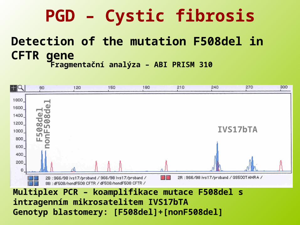
PGD – Cystic fibrosisDetection of the mutation F508del in CFTR gene
Fragmentační analýza – ABI PRISM 310
Multiplex PCR – koamplifikace mutace F508del s intragenním mikrosatelitem IVS17bTAGenotyp blastomery: [F508del]+[nonF508del]
F5
08d
eln
on
F5
08d
el
IVS17bTA
Genetic counselling in infertility
Infertility• Is the infertility one aspect of a
genetic disorder that might be transmitted?
• Will correction if infertility give an increased risk of malformations in the offspring?
• Genetic testing before use of metods of asisted reproduction.
Infertility• Patological examination of the abortus
where possible, this may identify major structural malformations.
• Cytogenetic study of parents, this is especialy important where a structural abnormality is present.
• In general the finding of a chromosome abnormality in the abortus but not in parent is not likely to be relevant or affect the genetic risks.
Infertility• A search for possible lethal
mendelian causes (consanguinity- risk for AR diseases, X-linked dominant disorders lethal in male, myotonic dystrophy which gives heavy fetal loss in the offspring of mildly affected women)
• Inherited trombophilias in women with recurrent abortions ( factor V Leiden, factor II - G20210A, hyperhomocystinaemia ? (MTHFR - C677T)
Factor V - Leiden• frequency in the white European
population of about 5 - 9%• AD inheritance• increased risk of thromboembolism
in homozygots for FVL 50-100x, in heterozygots 5-10x
• increased risk of fetal loss after the 10. week of gestation
Sterility in male
• AZF (azoospermia factor) deletions of the DAZ gene Yq (deleted in azoospermia)
• Infertile man – 4-5%• Men with azoospermia – about 15%
• CFTR mutations and polymorphisms
Genetic risk in cancer
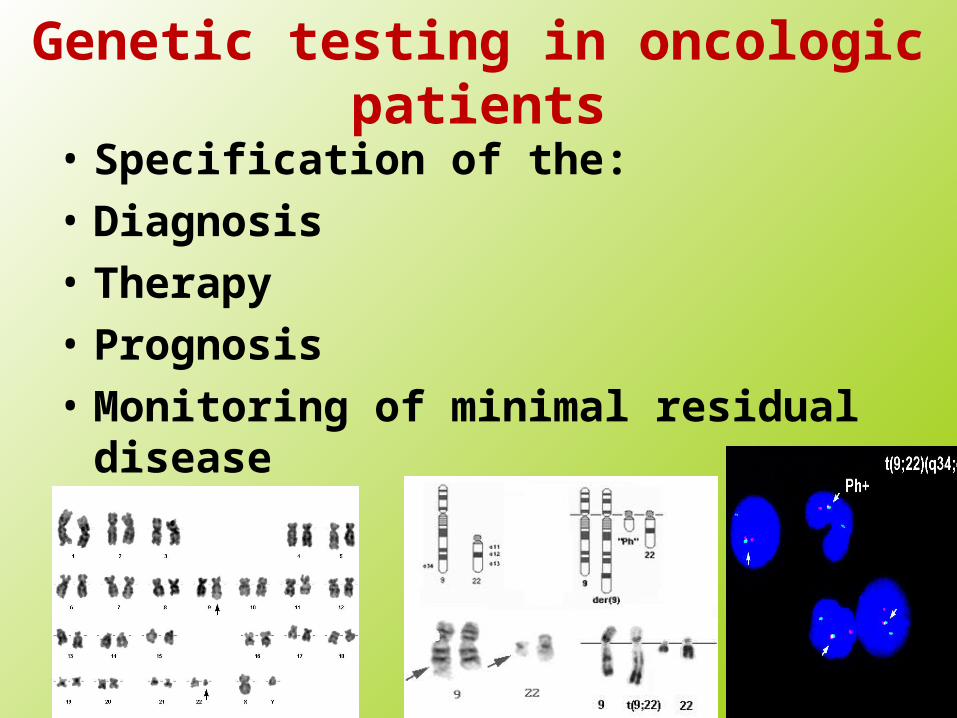
Genetic testing in oncologic patients
• Specification of the:• Diagnosis• Therapy• Prognosis• Monitoring of minimal residual
disease
Genetic risks in cancer
• Tumours following mendelian inheritance (most AD, about 5%)
• Genetic syndromes predisposing to malignancy
Hereditary cancer syndromes
• AD inheritance
• Preventive, pre-symptomatic testing
• Prevention
• Assotiated problems
Hereditary cancer syndromes following AD
inheritance• Brest cancer – BRCA 1 and BRCA 2
• Familial Adenomatous Polyposis coli – FAP
• Von Hippel – Lindau syndrome- VHL
• Retinoblastoma
• Neurofibromatosis- NF1, NF2
• Li-Fraumeni syndrome (p53 gene)
• Lynch syndrome – hereditary non polypous colon cancer - HNPCC
Genetic testing in Hereditary cancer
syndromes • Tests are voluntary• Mostly in adults only
• In children only when prevention in childhood is present and when the risk of tumours is in childhood
Postnatal care and neonatal screening
• Early diagnosis
Dispensary
Specialized Care
Prenatal and perinatal managment of prenagncies
with malformation or genetic disease in the fetus
• Consultation with experts, who will continue to take care of the pregnant woman - ultrasound specialist, gynecologist, obstetrician, psychological support ..
Consultions with specialists, who will care after the birth of newborns with disabilities
The planned delivery of specialized care workplace - kardiocentrum, pediatric surgery, cardiology…
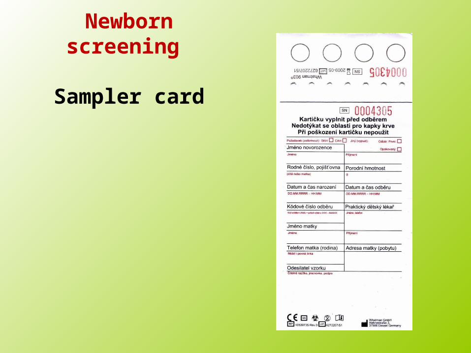
Newborn screening
Sampler card
Screened diseases in CR from 10/2009
• Kongenital hypothyreosis• Kongenital adrenal hyperplasia
– CAH
(cumulative risk 1/2900)
Screened diseases in CR from 10/2009
• Inborn errors of metabolism• Fenylketonuria (PKU, HPA)• Leucinosis• MCAD• LCHAD• VLCAD• Def.karnitinpalmitoyltransferasis I a II• Def.karnitinacylkarnitintranslocasis• Glutaric aciduria• Izovaleric acidurie
(cumulative risk 1/4000)
Screened diseases
• Cystic fibrosis
(1/4000)
• cumulative risk of all 13 screened diseases in CR - 1/1200