CLINICAL EVALUATION AND PATCH TESTING IN HAND ECZEMA
86
CLINICAL EVALUATION AND PATCH TESTING IN HAND ECZEMA Dissertation Submitted in fulfillment of the university regulations for MD DEGREE IN DERMATOLOGY, VENEREOLOGY AND LEPROSY (BRANCH XII A) THE TAMILNADU DR.M.G.R.MEDICAL UNIVERSITY, CHENNAI. MARCH 2010
Transcript of CLINICAL EVALUATION AND PATCH TESTING IN HAND ECZEMA

CLINICAL EVALUATION AND PATCH TESTING IN
HAND ECZEMA
Dissertation Submitted in fulfillment of the university regulations for
MD DEGREE IN
DERMATOLOGY, VENEREOLOGY AND LEPROSY
(BRANCH XII A)
THE TAMILNADU DR.M.G.R.MEDICAL UNIVERSITY, CHENNAI.
MARCH 2010
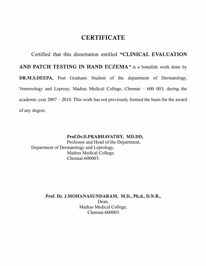
CERTIFICATE
Certified that this dissertation entitled “CLINICAL EVALUATION
AND PATCH TESTING IN HAND ECZEMA” is a bonafide work done by
DR.M.S.DEEPA, Post Graduate Student of the department of Dermatology,
Venereology and Leprosy, Madras Medical College, Chennai – 600 003, during the
academic year 2007 – 2010. This work has not previously formed the basis for the award
of any degree.
Prof.Dr.D.PRABHAVATHY, MD.DD,Professor and Head of the Department,
Department of Dermatology and Leprology,Madras Medical College,Chennai-600003.
Prof. Dr. J.MOHANASUNDARAM, M.D., Ph.d., D.N.B.,Dean,
Madras Medical College,Chennai-600003

SPECIAL ACKNOWLEDGEMENT
My sincere thanks to Prof. Dr.J.MOHANASUNDARAM, M.D., Ph.d.,
D.N.B., Dean, Madras Medical College for allowing me to do this dissertation
and utilize the institutional facilities.

ACKNOWLEDGEMENT
I am gratefully indebted to Prof. Dr. D.Prabhavathy MD., DD., Professor and
Head Department of Dermatology and Leprosy for her invaluable guidance, motivation
and help through out the study.
I express my gratefulness to Prof. Dr. V. Somasundaram MD., DD., Professor
and head Department of Occupational dermatology and Contact dermatitis for his
constant motivation and guidance.
I am very grateful to Prof. Dr. S.Jayakumar MD., DD., Additional Professor,
Department of Dermatology for his invaluable guidance and help.
I sincerely thank Prof. Dr.C.Janaki MD., DD., Additional Professor, Department
of Dermatology for her priceless support. I express my sincere gratitude to Prof.
Dr.R.Aruna Devi MD., DD., Professor of Leprosy, Department of Dermatology for her
support. I wish to thank Prof. Dr.N.kumar MD., DV., DMRD, Additional Professor
Institute of Venerology for his constant support.
I thank Dr.Manjula MD., DNB., Assistant Professor , Department of
Occupational Dermatology and contact dermatitis for her kind support and guidance.
I incline to thank Dr. G.K. Tharini MD., Dr.N.Hema MD., Dr.Samuel Jayaraj
Daniel MD., Dr.S.Anupama DDVL., Dr.S.Kumaravel MD., Assistant professors
Department of Dermatology for their kind support and encouragement.
I thank Dr.Hameedulla MD., DD., Dr.Afthab Jamila Wahab MD., DD.,
Dr.Thirunavukarasu MD., DD., Assistant professors, Department of Occupational
Dermatology and Contact dermatitis for their constant support.
My sincere thanks to Dr.V.Thrunavukarasu MD., DV., Dr.K.Venkateswaran
MD., DV.,Dr.P.Mohan MD., DV., Dr.S.Arun Kumar MD., DV., Dr.S.Kalaivani MD.,
DV., Dr.S.Prabakar MD(DVL)., Dr.Ahamed Sheriff MD (DVL)., Assistant Professors
Institute of Venerology for their help and suggestions.
A special mention of thanks to the patients for their cooperation without whom
this study would not have been possible.

CONTENTS
Sl.No. Title Page No.
1. INTRODUCTION 1
2. REVIEW OF LITERATURE 2
3. AIM OF THE STUDY 40
4. MATERIALS AND METHODS 41
5. OBSERVATION & RESULTS 46
6. DISCUSSION 57
7. SUMMARY 65
8. CONCLUSION 67
ANNEXURES
BIBLIOGRAPHY
PROFORMA
MASTER CHART

INTRODUCTION
Hand eczema is a descriptive diagnosis for dermatitis largely confined to the
hands, and it does not make any presumption about the etiology. 1 It may be endogenous
or exogenous (allergic or irritants) in origin. 1 Most of the cases of hand eczema have a
multifactorial etiology, 1 wherein the eczema is caused and perpetuated by exogenous
factors in individuals who are susceptible to such processes due to endogenous factors.
1,2 Identification and avoidance of the external contactants is of paramount importance
in appropriate management of hand eczema. As clinical differentiation between chronic
allergic and irritant hand eczemas is often difficult, patch testing becomes an important
diagnostic tool for identification of the allergen/allergens responsible for the eczema. 3
Patch testing is a well established method of diagnosing allergic contact dermatitis.
Patients with a history and clinical picture compatible with contact dermatitis are re-
exposed to suspected allergens under controlled conditions to verify the diagnosis.
Properly applied and correctly interpreted patch tests are, at present, the only
scientific proof of allergic contact dermatitis. This study was conducted to identify the
allergens showing positive reactions in patch test in patients with hand eczema.

REVIEW OF LITERATURE
Hand eczema
Historical back ground
It may be considered curious to single out eczema of the hands as being worthy
of special study. In his long treatise of eczema, Hebra 4 devoted less than a page to the
eczema of hands and feet, and this is in morphological terms. Fox5 stated that eczema in
these sites “is chiefly remarkable for the peculiar tenacity and persistence of the
vesicles” and mentioned grocer’s and baker’s itch, but little else. Radcliffe – Crocker6
emphasized the role of external irritants.
The recognition of hand eczema as a region of peculiar interest has come about
gradually during this century and increasingly so in the last 50 years. There are several
reasons for it. The most important was the rapid growth of industrialization in the west,
especially enormous growth and development in dye and chemical industries. This led to
an increase in realization of the importance of irritant and allergic dermatitis. In the
increasingly complex environment of 20 th century the house wife too, encountered new
causes of hand dermatitis.
Finally with increasing affluence, personal adornment flourished and the social,
professional, and psychological effect of disfigurement on a visible area, such as the
hands, undoubtedly prompted the increased use of potentially sensitizing hand creams

and a greater degree of medical attention.
Hand eczema is one of the commonest occupational health problems encountered
in dermatology. It affects 1 % of adults7, with a male: female ratio of 2:1.8 The lifetime
prevalence varies between 5-7 % and 16.7% for women and between 5.2% and 9.5% for
men.9
The incidence and causative factors vary from region to region, from rural to
urban areas and from non industrialized areas to industrialized areas. Climate and
occupation play an important part in determining the incidence. Women of child bearing
age have a high incidence due to greater contact with soaps, detergents, vegetables,
spices, etc. The incidence is high among certain occupational groups engaged in wet
work such as hairdressers, cooks, domestics, nurses and print workers.10
Pathogenesis
Hand eczema is multifactorial in etiology. A unifying feature in most cases is an
underlying disruption in the stratum corneum, altering its barrier function.
Transepidermal water loss increases with barrier disruption and is exacerbated by
additional exposure to water.11
Understanding the Characteristics of the Hands and the Barrier Function ofthe
Stratum Corneum 12,13,14,15
1. Characteristics of the hands:
The palms have a peculiar skin structure; it is related to the fact that the hands are
the body part most frequently exposed to external stimuli. The stratum corneum of the
palms consists of approximately 50 layers, and is much thicker than the skin on other
parts of the hands (about 15–20 layers). Also unlike the facial skin, the stratum corneum
has no hair follicles and sebaceous glands. In areas where hair follicles are present, the
super surface lipid membrane overlying the stratum corneum is predominantly produced
by the sebaceous glands associated with hair follicles, while in the palms and soles the
membrane is composed exclusively of lipid produced by metabolism of epidermal cells.
The super surface lipid membrane is well developed in the face, whereas the membrane
on the palm is thinner, which is compensated for by a thick stratum corneum.
Additionally the back of the fingers, distal phalanx has no hair follicles and that the nail
margins and fingertip have the same properties as the palms and finger-pulps.
2. Barrier function of the stratum corneum
It has been shown that homeostasis of barrier function of the stratum corneum is
maintained primarily by three factors;
1) Surfacelipid
2) Intrinsic hydrophobic lipid of the stratum corneum such as ceramide.
3) Natural moisture-retentive factors.

Among these factors, ceramide has recently emerged as an important contributor
to the moisture retention. Ceramide is the hydrophobic lipid that bridges the gap
between the horny cells and forms the barrier that keeps water from passing through.
The substance is supplied by a structure of the epidermal cell called a lamellar granule
(or Odland body), and the process of its metabolism and production is under
investigation.
The so-called natural moisturizing factors are thought to bind with water within
the horny cells and play a role in enhancing the flexibility of the keratin. This factor
originates from the keratohyaline granules of the epidermal cells, which are the soluble
amino acids produced by degradation of fillagrin. In the cosmetic industry, great
importance is placed on this factor. When the super surface lipid membrane and lipids
such as ceramide between the horny cells are removed by artificial cause, internal water
is lost from the stratum corneum (trans-epidermal water loss [TEWL]) and the way is
left open for chemical stimuli or external substances including allergens and
microorganisms to invade the body, leading to susceptibility to inflammation and
allergic sensitization. Itchscratching also occurs (itch-scratch cycle) promoting entry and
inflammation. The body part most vulnerable to the influences that permit this
sequential process is the hand.
The most common clinical presentations of hand eczema are atopic hand
dermatitis, pompholyx, and contact dermatitis (irritant contact dermatitis [ICD], allergic
contact dermatitis [ACD]).12 The diagnosis of hand dermatitis is determined by a review
of the patient's medical history, a physical examination including other body sites as well
as the hands, and a thorough overview of the patient's daily activities with emphasis on
occupation and hobbies. Irritant contact dermatitis usually is diagnosed by the absence
of a positive patch test result; however, patch testing is essential in confirming a clinical
diagnosis of ACD by identifying the allergens to which the patient has been sensitized.
Classification
1. Based on etiological factors:
Endogenous:
Atopic dermatitis
Discoid eczema
Pompholyx
Hyperkeratotic eczema
Exogenous:
Irritant contact dermatitis
Allergic contact dermatitis
Systemic allergens – drugs, metals
Dissemination from a focus
Infective dermatitis involving hands and feet.
2. Based on Morphologic pattern:
Pompholyx

Recurrent focal palmar peeling
Hyperkeratotic palmar eczema
Finger tip eczema
Ring eczema
Housewives eczema (Wear and tear dermatitis)
Apron eczema
Discoid eczema
Chronic acral dermatitis
Gut eczema
Other patterns ( e.g patchy vesiculosquamous)
3. Based on duration of symptoms:
Acute
Chronic
Acute on chronic
Atopic dermatitis:
The most common site of atopic dermatitis is the hand in adults. Atopic
adolescents and young adults develop hand eczema when exposed to school work,
hobbies or occupational contacts. Meding et al reported that 22% of hand eczema
patients were atopic.16 Predisposing factors for developing hand eczema in atopics are,
dry itchy skin, persistent diffuse atopic dermatitis, widespread dermatitis in childhood
and atopic dermatitis of hand in childhood.17 The characteristic distribution is on the

dorsal aspect of hand and fingers where it is seen as patchy vesicular rash that is itchy
and irritable.18 Wet work is the most important factor causing or aggravating hand
eczema in atopics. Although atopic eczema resolves by puberty, it may recur as hand
eczema in adults. Diagnosis is usually ascertained through history of atopy, distribution
of lesion and occasionally raised IgE levels.
Irritant contact dermatitis
Contact irritants are the commonest exogenous cause of hand eczema.19 Acute
irritant contact dermatitis results from contact with usually a strong reactive acidic or
alkaline chemical, presents with erythema, edema, vesiculation and exudation. Chronic
irritant dermatitis is caused by either the repetitive or cumulative effect of a variety of
minor damaging factors. Chapping is predominantly seen on the back of the hands,
while fissuring is seen on the palmar aspect.
Allergic contact dermatitis:
Allergic contact dermatitis is an exogenous cause for hand eczema. Contact
allergens produce hand dermatitis in individuals who are already sensitized to these
antigens through two types of immunological responses. One is through the delayed type
of hypersensitivity reaction (Type IV), as seen with chromium, nickel and rubber
allergies. The second one is the immediate type of hypersensitivity reaction (Type I),
that occurs as sudden itchy eruption of the hands following ingestion of sea food in
sensitized individuals. Suman 20 et al reported 67 % of hand eczema was due to allergic
contact dermatitis in their study.1 Hand eczema is aggravated in sensitized individuals

due to oral ingestion of nickel or chromium. The commonest allergens are nickel,
chromium, vegetables and natural rubber latex
Hyperkeratotic palmar eczema:
Prolonged and repeated contact with certain agents can induce a reaction pattern
on palmar skin manifesting as thickening, scaling and fissuring particularly involving
tips, palmar surface of fingers and palms of one or both the hands associated with
itching and pain. Such lesions termed as hyperkeratotic palmar eczema can occur either
due to physical factors like dryness and friction, irritant reaction or allergic reaction. 21
Patterns of distribution of the lesions as a result of allergic contact dermatitis depend
largely upon the causative agents pertaining to the habits, activities and occupation of an
individual providing valuable clues for establishing the cause by patch testing. 22 This
condition needs to be differentiated from psoriasis.
Finger tip eczema:
It is a recurrent recalcitrant eczema seen on the palmar surface of the finger tips. It
involves the palmar surface of the tips of some or all the fingers. The skin is dry, cracked
and sometimes breaks into painful fissures. Usually it remains localized. It may
occasionally extend down the palmar surfaces of the fingers and merge with palmar
eczema. It involves most or all of the fingers, more predominantly those of the master
hand and particularly the thumb and forefinger. It may start as a moist lesion, but
eventually it becomes dry, cracked and scaly. Beneath the peeling skin a raw, red,
cracked, painful surface is seen. It may represent cumulative irritant reaction or a

allergic contact dermatitis. Patch testing is useful in identifying the etiology.
Ring eczema:
This form of hand eczema starts under a ring but frequently spreads to adjacent
side of third finger or palm. It is more common in women, often starting after marriage
or the arrival of child, but it may affect men. The onset is usully in third decade, but can
be earlier in women wearing metal rings. This form of eczema is considered to be an
irritant reaction to the concentration of soap and detergents under the rings. Patch test
usually gives a low yield, except for nickel, but this is common in women of this age
and it is usually irrelevant unless associated with metal use.
Dyshidrotic Eczema ( Pompholyx):
Dyshidrotic eczema is a recurrent or chronic relapsing form of vesicular
palmoplantar dermatitis of unknown etiology. Dyshidrotic eczema is also termed
pompholyx, which derives from cheiropompholyx, which means "hand and bubble" in
Greek. The etiology is multifactorial, it is considered a reaction pattern caused by
various endogenous conditions and exogenous factors. Dyshidrotic eczema affects
individuals aged 4-76 years; the mean age is 38 years. After middle age, the frequency
of dyshidrotic eczema episodes tends to decrease. The male-to-female ratio for
dyshidrotic eczema is 1:1.
Exogenous factors (eg, contact dermatitis to nickel, balsam, cobalt; sensitivity to
ingested metals; dermatophyte infection; bacterial infection) may trigger episodes.23
These antigens may act as haptens with a specific affinity for palmoplantar proteins of

the stratum lucidum of the epidermis.24 The binding of these haptens to tissue receptor
sites may initiate pompholyx. Emotional stress and environmental factors (eg, seasonal
changes, hot or cold temperatures, humidity) may exacerbate dyshidrosis. In some
patients, a distant fungal infection can cause palmar pompholyx as an id reaction. As
many as 50% of patients with dyshidrotic eczema have reportedly had personal or
familial atopic diathesis (eczema, asthma, hayfever, allergic sinusitis). 25 Isolated reports
describe other possible causative factors, such as aspirin ingestion, oral contraceptives,
cigarette smoking, and implanted metals, among others. 25
A 3-year prospective study of the causes of dyshidrotic eczema (pompholyx) in
120 patients found causes of pompholyx related to contact exposure (67.5%), including
cosmetic products (31.7%) and metals (16.7%); interdigital-plantar intertrigo (10%); and
internal causes (6.7%), with an additional 15% with undiagnosed (idiopathic) causes,
probably related to atopic factors. 26
Apron eczema:
The term was coined by Calnon. It is a localized eczema extending from the
proximal part of two or more fingers and the metacarpophalangeal joints to the
contiguous part of palm in a semicircular fashion. More common in women. Calnon
desribed this entity as an endogenous eczema.
Discoid hand eczema:
The pattern of lesion in this form of eczema is similar to that of discoid eczema
elsewhere in the body, but localized to the hands and fingers, usually to the back. One or
more round nummular lesions develop and remain fixed to the site. They may be
exudative or scaly. Intervening skin remains normal. The patches are resistant to
treatment. When they recur, they do so in the same site. These features distinguish this
type from the more common patchy form of hand eczema. Affects both the sexes, young
atopics are more susceptible. The relevance of any positive patch test that is found is
difficult to establish.
House wives eczema (wear and tear dermatitis):
It is one of the commonest type of hand eczema encountered. It is a chronic or
cumulative irritant dermatitis caused by household work contactants such as washing
soap, soda detergents, and cleansers. A variety of physical factors such as friction,
trauma, cold and heat, play a part. Atopics are more vulnerable. It commonly occurs on
the palmar surface of fingers, interdigital spaces, palms, and dorsal aspect of fingers,
particularly knuckles. The skin of affected area is dry, and may show superficial fissures.
May be associated with finger tip or ring eczema.
Other Diseases Mimicking Hand Eczema:
Major conditions that mimic hand eczema are:
Palmar Psoriasis:
Thick hyperkeratotic scaly plaques with painful fissuring can occur in psoriasis,
this may resemble chronic hand eczema. Psoriatic plaques elsewhere on the body, on the
soles etc. will clinch the diagnosis. When it is limited to the palms, a biopsy may be
required for definite diagnosis.
Tinea manum or ring worm of the hands:
Itchy anular scaly skin patches involving both the palms and back of hands
(sometimes limited to one side) can cause confusion in diagnosis of hand eczema.
Scraping the scales and examination under microscope after dissolving in potassium
hydroxide 10% solution will clinch the diagnosis of fungal infections of the hand.
Candidal intertrigo:
Commonly seen in housewives and hair dressers, candidal infection of the finger
web spaces may look similar to contact dermatitis. Satellite lesions in the periphery and
a positive potassium hydroxide microscopy will help in differentiating the yeast
infection from hand eczema.
Scabies:
Distribution is on the web spaces with severe itching on the night. Commonly
seen in paediatric age group. Scraping from the lesion demonstrates the mite.
The most common clinical presentations of hand eczema are atopic hand
dermatitis, pompholyx, and contact dermatitis (irritant contact dermatitis [ICD], allergic
contact dermatitis [ACD]). The diagnosis of hand dermatitis is determined by a review
of the patient's medical history, a physical examination including other body sites as well
as the hands, and a thorough overview of the patient's daily activities with emphasis on
occupation and hobbies. Irritant contact dermatitis usually is diagnosed by the absence
of a positive patch test result; however, patch testing is essential in confirming a clinical
diagnosis of ACD by identifying the allergens to which the patient has been sensitized.
Treatment
Acute stage:
In acute stage of hand eczema rest and bland applications are advised. Hands
should be soaked in Burrow’s solution ( aluminium acetate 1%) or potassium
permanganate solution ( diluted 1:8000). Large bulla if present may be aspirated using a
sterile syringe. Systemic antibiotics should be administered if secondary infection
develops. As the eruption subsides soaks should be discontinued an zinc cream or oily
calamine lotion can be substituted. In a few severe cases, a course of oral steroids may
be justified. Topical steroids are useful in subacute stage of hand eczema.
Chronic stage :
Particular attention should be paid to the possible causative factors, and a full
occupational, social history, with details of hobbies and spare time activities is essential.
The following measures are advised,
1.Avoidance of irritants :
Education of the patient to the possible dangers is of paramount importance and
printed advice sheets are helpful. Barrier creams and gloves can be tailored to individual
needs.
2.Emollients:
Emollient should be applied frequently as a thin smear rubbed gently into the skin.
The choice of emollient will vary with the patient. Some people will benefit from a
greasy preparation and others will prefer a cream based preparation.
3.Topical steroid:
Topical steroids should be used sparingly and in the weakest potency. Even
though the palms are thick, the epidermis can be rendered thin and fragile by potent
topical steroids. In unresponsive cases use of potent steroid under occlusion may be
considered. Intermittent use of potent steroid may prevent relapse.
4.Other measures:
Tar pastes are useful in chronic unresponsive cases. Salicylic acid is helpful for
hyperkeratotic and persistent scaly lesions. Oral PUVA chemotherapy. Topical PUVA
and NBUVB have proved useful in several types of hand eczema. Radiotherapy is useful
for stubborn hand eczema.Antihistamines reduces the itching. Acitretin is effective in
chronic hyperkeratotic eczema. Cyclosporine is useful in some cases.
Patch testing:
History:
The principle of patch testing is to reproduce, in a clinical setting, a min-model of
allergic contact dermatitis using allergens suspended in a vehicle at non irritant
concentrations.27,28
Patch test was first employed in 1847 by Staedler by blotting paper method to test
idiosyncrasy. Collins, an ophthalmologist, in 1889, applied atropine patches to his
patients who were developing adverse reaction after instillation of atropine. However,
Jadassohn has been rightly called the father of patch test as he first scientifically
established the role of patch test in dermatitis medicamentosa. Later on Sulzberger
contributed much by working on and highlighting the importance and standardization of
patch testing, which represents one of the most important advances in clinical
dermatology during the twentieth century.30
Sulzberger and Wise 30 in 1931 commented that in allergic contact dermatitis
patch test should be employed, for it and it alone, can aid in the quest of the etiologic
factor and in the study of the dermatitis. Colman in 1982 warned that the greatest abuse
of patch testing is failure to use the test.31 In 1986 Fisher concluded that properly applied
and correctly interpreted patch tests are, at present, the only scientific 'proof' of allergic
contact dermatitis. He also cautioned that education in the technique of patch testing is
as essential to physicians in training as the learning of most surgical procedures.

Contact dermatitis is a disabling problem which can be identified by careful
history, clinical examination, correlation of history and findings and finally patch
testing.
Patch test consists of standard series of statistically common allergens and is of
value when the contact dermatitis is suspected to an offending agent which cannot be
pinpointed at. When performed and interpreted clearly it is a scientific proof of the
allergic state. If the allergic state can be correlated with positive patch test then the
validity of the test is relevant. Negative test does not mean contact dermatitis is ruled out
as the patient may not be allergic to common sensitizers. If products which are
nonirritant are suspected, repeated open applications tests can be performed. Although
patch test is artificial and does not duplicate clinical exposure it is an important tool to
find the contactant rather than by clinical trial and error.
Standard series of allergens are recommended for use in everybody undergoing
patch testing. The specific standard series may vary according to the locality of the patch
testing centre. Several organisations have attempted to identify the most important and
relevant chemical allergens in their community. The chemicals in the standard series
depend on which one is being used. The various series available are the European
standard series, North American standard series and Indian standard series. Most test
substances are single compounds but some of the tests are mixtures of closely-related
chemicals. There are numerous other chemicals that have been reported to cause contact
allergy occasionally. About 15% of patients that have positive reactions at patch testing
react to an allergen that is not in the standard series. These allergens are detected using

other series of allergens or individual standardised chemicals that have been selected by
the dermatologist. Several series have been developed for patients that present with
dermatitis on specific sites of the body (e.g.,'face series', 'foot series'), and for those with
certain occupations (e.g., 'hairdressers series', dental series') or other risk factors ('shoe
series', 'cosmetic series').
Indian standard series
Approved by CODFI (Contact and Occupational Dermatoses Forum of India) and
manufactured/supplied by Systopic Laboratories, New Delhi.
Patch test unit is made from microporous tape (15X15cm) and aluminium patch
test chambers (APC). Aluminium patch test chambers are 9mm internal diameter and a
depth of 0.7mm. Aluminium patch test chambers are placed facing up with 2 cm
distance from centre of each other. On top and bottom 2.0 cm each of micropore is left
to obtain good adhesion. It is stored at 4 c.

LIST OF CODFI ANTIGENS(INDIAN STANDARD SERIES)
S.NO. Compund Conc. % Veh.
01 Control 100 pet
02 Potassium Dichromate 1.0 pet
03 Neomycine Sulphate 20.0 pet
04 Cobalt Chloride 5.0 pet
05 Benzocaine 5.0 pet
06 4-Phenylenediamine base (PPD) 1.0 pet
07 Parabens 9.0 pet
- Methyl-4-hydroxybenzoate 3.0
- Ethyl-4-hydroxybenzoate 3.0
- Propyl-4- hydroxybenzoate 3.0
- Butyl-4- hydroxybenzoate 3.0
- Benzyl-4- hydroxybenzoate 3.0
08 Nickel Sulphate 5.0 pet
09 Colophony 10.0 pet
10 Epoxy resin 1.0 pet
11 Fragrance mix 8.0 pet
-Cinnamic Alcohol 1.0
-Cinnamic aldehyde 1.0
-Hydroxycitronellal 1.0
-Amylcinnamaldehyde 1.0
-Geraniol 1.0
-Eugenol 1.0
-Isoeugenol 1.0
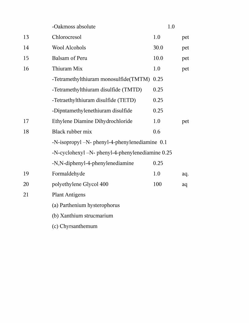
-Oakmoss absolute 1.0
13 Chlorocresol 1.0 pet
14 Wool Alcohols 30.0 pet
15 Balsam of Peru 10.0 pet
16 Thiuram Mix 1.0 pet
-Tetramethylthiuram monosulfide(TMTM) 0.25
-Tetramethylthiuram disulfide (TMTD) 0.25
-Tetraethylthiuram disulfide (TETD) 0.25
-Dipntamethylenethiuram disulfide 0.25
17 Ethylene Diamine Dihydrochloride 1.0 pet
18 Black rubber mix 0.6
-N-isopropyl –N- phenyl-4-phenylenediamine 0.1
-N-cyclohexyl –N- phenyl-4-phenylenediamine 0.25
-N,N-diphenyl-4-phenylenediamine 0.25
19 Formaldehyde 1.0 aq.
20 polyethylene Glycol 400 100 aq
21 Plant Antigens
(a) Parthenium hysterophorus
(b) Xanthium strucmarium
(c) Chyrsanthemum
ALLERGENS AND THEIR OCCURRENCES IN OUR ENVIRONMENT
1. Chromium (Potassium Dichromate)
Chromium is the fourth most common material in the earth’s crust.32 It is
probably more accurate to use the term chromate, because chromium is unique in that
the metal itself does not sensitize, but rather its salts.33 Hexavalent chromate is the most
powerful sensitizing chromate because of its solubility and capacity to penetrate the
skin. Fregert et al pointed out the advantage of converting hexavalent chromium in
cement to an insoluble trivalent form via the addition of ferrous sulphate.34
There are many causes of chromate allergy other than cement, including
chrometanned leather, anti – rust, paint, timber preservative, the wood pulp industry, ash
either from burnt wood in general or matches with chromate in the match head, coolants
and machine oil. Welding, dye industry, chromium plating, bleaches, detergents etc. 33
Chromium compounds have been recognized for their primary irritant as well as for
their potent sensitizing properties. In men the chromates are the most frequent industrial
sensitizer, the commonest source being cement in the building industry. The chrome salt
is an accidental contaminant of cement and is not an `additive’ in the usual sense. The
role of chromium in foods in the production of chromate dermatitis is highly
controversial. 32 Chromium may cause air borne contact dermatitis.
2. Neomycin Sulphate
It was first isolated in 1949 from Streptomyces fradiae. It consists of two active

components, neomycin B (78-88%) and neomycin C (10-16%). The third component
present only in small amounts (2-5%) is the degradation product neamine (neomycin A).
it is still one of the most commonly used topical antibiotics for treating varying
cutaneous infections. It is used either as such or in combination with a corticosteroid
depending upon the pathology of the disease. Hypersensitivity to neomycin is likely to
be missed due to the relatively mild dermatitis it produces and secondly because of its
frequent use in combination with topical steroids which suppresses its allergic action. It
is a known potent sensitizer all over the world. The reported incidence varies from 2.5-
6% and even more 35, 36, 37 In India the reported incidence is much higher due to its testing
in selected group of patients rather than routine testing in all patients. 38,39,40,41 There are
also various reports of cross sensitivity with the neomycin group of antibiotics; it cross
reacts with framycetin, gentamicin, kanamycin, tobramycin, streptomycin and
bacitracin. 42
3. Cobalt Chloride
Cobalt is frequently combined with nickel as a contaminant and the two metals
always occur together. Cobalt is a contaminant of cement, and in cement dermatitis
sensitivity to cobalt as well as chromate may occur. 43, 44 Cobalt dermatitis may occur in
those involved in the manufacture of polyester resins and paints, hard metals used for
cutting and drilling tools, and in the manufacture and use of cement. It may also occur in
produces of pottery, ceramics, metal alloys, glass carbide and pigments.
A combined cobalt and nickel sensitivity is more common in women because they

are sensitized by nickel, which always contains an impurity of cobalt. It is a matter of
debate whether nickel – cobalt combined allergy is due to independent sensitization or
cross sensitization.45 Depending on the source of the contact, the pattern of cobalt
dermatitis is in many cases identical with that of either nickel or chromate dermatitis.
Dental plates and fillings may release sufficient amounts of cobalt to cause stomatitis or
vesicular hand eczema in sensitive patients. A more wide spread disseminated or
nummular eruption may also occur.
4. Benzocaine :
Benzocaine is a p-aminobenzoic acid derivative used as a local anaesthetic. It is a
common and potent sensitizer. It is usually applied in the orifices of the body and to raw
intertriginous areas, which renders sensitization easier. It can cross react with other
compounds. 25% of benzocaine sensitivity patients react to paraphenylene diamine and
paramminobenzoic acid esters used in sunscreening agents. 46 It also cross reacts with
procaine, sulphonamides and certain dyes. 47, 48 In order to detect more patients sensitive
to topical anaesthetics it is necessary to test with other “Caine” anaesthetics. 49,50
5. Formaldehyde :
Formaldehyde is a ubiquitous and potent sensitizer, industrially, domestically and
medically. Formaldehyde exposure is difficult to estimate because the chemical besides
being manufactured, imported and used as such is incorporated into a large variety of
products and reactants in many chemical process, including formaldehyde releasers,

polymerized plastics, working fluids, medicaments, fabrics, cosmetics and detergents. 51
Shampoos may contain formaldehyde. Because they are quickly diluted and
washed off, only exquistively formaldehyde consumers develop dermatitis of the scalp
and face. Formaldehyde dermatitis from textiles is rare today because manufacturers
have improved the fabric finish treatment and reduced the amount of formaldehyde
residues in new clothing. 51 Garments made from 100% acrylic, polyester, linen, silk,
nylon and cotton are generally considered to be formaldehyde free. 52 The frequency of
formaldehyde positive patch tests in eczema patients is around 3% to 4% 53
6. p-Phenlenediamine (PPD):
p-Phenlenediamine (PPD) is a colourless compound that acts as a primary
intermediate in hair dyes. It is oxidized by hydrogen per oxide and then polymerized to a
colour within the hair by a coupler. Most cases of contact allergy to PPD occur due to
contact with hair dyes, in either the client or hair dressers. 54 Once the hair is dyed and
polymerized, it has been said to be nonallergic; however cases are occasionally seen
in which people react to other persons dyed hair. This may be due to the dyeing not
being carried out properly, leaving unploymerized hair dye. 54
Patients with PPD allergy may cross react with benzocaine, procaine,
sulphonamide, PABA sunscreens, azo and aniline dyes, anthraquinone and
antihistamines. 55 Immediate type hypersensitivity to PPD, with extension urticarial
reactions has been reported. 56

7. Parabens:
Parabens are alkyl esters of p-hydroxybenzoic acid. They are quite soluble in fats
and are effective preservatives for cosmetics drugs and foods. In addition, many
parentrally administered medications, especially those in multidose packages, also
contain parabens as preservatives. Parabens are known to produce contact
hypersensitivity but are not strong sensitizers. 57 In India the incidence in a selected
group of patients was over 5%. 58 Paraben sensitive leg ulcer patients can often use
paraben preserved cosmetics on normal skin without adverse effect. 59
8. Nickel Sulphate:
Nickel is ubiquitous in the environment and constitutes about 0.008% of the
earth’s crust. Humans are constantly exposed although in variable amounts. Metal
nickel as well as nickel salts give rise to contact allergy, metallic nickel only after
corrosion. The corrosiveness of sweat, saliva and other body fluids to nickel and nickel
alloys is a primary importance. 60 The commonest cause of sensitization is ear piercing,
61 particularly if there is a history of irritation at the time of piercing. Nickel sensitive
patients with vesicular hand eczema worsen after an oral challenge with a diet naturally
high in nickel. 62
Food with high nickel contents are canned food, acid foods cooked in stainless
steel utensils, instant tea, beans, mushroom, onions, spinach, tomato, peas, tea, cocoa
and chocolate. The nickel content of food is partially determined by the components of
the soil in which it is grown, fungicides used on it and the equipment used in handling
the food. 63 Nickel allergy does not seem to increase the change of developing other
allergies64 with the exception of cobalt, copper and pallidium since these metals are
commonly associated with nickel.
9. Colophony (Rosi):
Colophony (rosin) is a widespread, naturally occurring material, which is the
residue left after distilling off the volatile oil from the Oleoresin obtained from trees of
the family Pinanceae. This yellow resin is used in the production of varnished, printing
inks, paper, soldering fluxes, adhesive, polish, waxes, cosmetics (mascara, eye shadow),
topical medicaments, and is a component of dental impression material and periodontal
packings. Cross reactions between rosin, balsam of peru, oil of turpentine, wood tar,
pine resin and spruce resin may occur. 65 The allergencity of colophony can be reduced
by chemical modification i.e., by hydrogenation of the non aromatic double bonds in the
resin.
10. Epoxy Resin:
Of all epoxy resins 95% consists of a glycidyl ether group formed by the reaction
of bisphenol A with epichlorohydrin. Theoretically there are many different chemical
compositions which can be used to make an epoxy resin. Epichlorohydrin / bisphenol A
epoxy resin can vary in molecular weight from 340 to much larger polymers, the larger
polymers having a much lower sensitizing capacity. 66
Epoxy resin compounds should therefore contain little or no low molecular weight
epoxy resin. The higher the molecular weight, the less sensitizing the compound is. once
epoxy resin becomes hardened, its sensitizing capacity becomes markedly reduced, but
so called cured resins can contain uncured molecules and so have been known to
sensitize. 67 Epoxy resins are used as adhesives and in paints, requiring great hardness
and durability, for instance, in ships, in electrical insulation, as an additive to cement for
quick bonding and strength. A negative patch test to epoxy resin does not necessarily
mean that the patient is not allergic to the epoxy product which they have been using,
for the following reasons.
• There may be some other epoxy resin in the compound
• They may be allergic to some other compound in the resin, for instance, dyes,
filers, plasticizers etc.
• They may be allergic to the hardner 67
Both epoxy resins and hardners can be irritant, as well as sensitizing, and if a
patch test is applied at more than 1%, it may produce an irritant reaction. One of the
commonest sources of sensitization in industry is the use of epoxy resin with fibre glass
to make strong sheeting used for various purposes such as hulls for boats.
11. Fragrance Mix:
Fragrance and flavour substances are strong smelling organic compounds with
characteristic, usually pleasant odors. 68 Fragrances are ubiquitous and used in perfumes
and perfumed products. They are found not only in cosmetics but also in detergents,
fabric softners and other household products. Flavours are used for the flavouring of
toothpastes, food and beverages. Perfume allergy evaluation may be difficult. A
complete perfume compound consists of from 10 to more than 300 basic components
selected from over 5000 raw materials, which can be divided into the following. 68, 69
• 500 natural products isolated from various parts of plants, e.g., blossoms, buds,
fruit, peel, seeds, leaves, bark, wood, roots or resinous exudates ;
• 5 animal products and their extracts (ambergris from the sperm whale, musk
Tonkin from the testes of musk deer, castoreum from breaver glands, beeswax
absolute from beeswax, and civet from glands of civet cat)
• Over 4000 synthetic fragrances
The most common reaction to fragrance materials is allergic contact dermatitis,
but contact urticaria, photodermatitis and irritation may occur. Perfume allergy
evaluation is made more difficult by the fact a that labeling of perfumes with their
ingredients is not required by law and by the secrecy policy of perfume manufacturers.
That certain perfumes are sensitizers and photosensitizers (others are solely
photosensitizers) adds to the investigator’s frustration. 70
Screening with individual fragrances is impractical and time consuming and may
give rise to multiple positive reactions and the excited – skin syndrome. Therefore, a
perfume screening mix for patch testing has been developed to increase the ability to
detect perfume allergy. 71 The current fragrance mix consists of light ingredients, each at
a concentration of 1% : cinnamaldehyde, Cinnamyl alcohol, Eugenol, alpha amyl
cinnamaldehyde, hydroxyl citronellal, geraniol, isoeugenol and oak moss absoluate,
with sorbitan sesquioleate as emulsifier. It has shown to be a valuable screening agent
for perfume dermatitis. 72
12. Mercaptobenzothiazole (MBT):
Although a component of the mercaptomix, it is included in the standard series at
2% w/w on its own since the mix failed to detect 30% of patients who were MBT
allergic when compared to simultaneous testing with 1% MBT, and 12 of 24 individuals
who reacted to 2% MBT did not react to the mix.73 According to Cronin, 74 Women who
react to MBT have probably been sensitized by gloves or shoes, but in men the
sensitization is probably by foot wear. Among numerous, other sources of contact with
MBT are rubbers containing MBT and rubber handles, masks, elastic bands, tubing,
elasticated garments and artificial limits. 75 MBT may be present in a variety of
nonrubber products, including cutting oils, greases, coolants, anti – freezer, fungicides,
adhesives and veterinary medicaments.76

13. PEG – 400
Poly ethylene glycol is a mixture of glycols. The lower molecular weights from
200 to 700 are liquids, while the higher weights 1000 to 6000 are solids. PEG of
varying molecular weights are used extensively as vehicles in topical medicaments,
suppositories, shampoos, detergents, hair dressing, insect repellents, cosmetics, tooth
pastes and contraceptives.
In industry the PEG are used as solvents for nitrocellulose, as plasticizers for glue
and casein, and as wetting agents in epoxy hardners. The low molecular poly ethylenes
from 200 to 400 may cause allergic contact urticaria and eczema. The higher
polyethylenes are not sensitizers. 77
14. Chlorocresol (4- Chloro – m – Cresol):
It is an efficient bactericide used as a preservative. In veterinary medicine it is
used as in pesticides and fungicides. Chlorocresol dermatitis may occur from
corticosteroid creams in which it is used as a preservative. It is also used in topical
antiseptics, pharmaceutical products, protein shampoo, baby cosmetic, cooling fluids,
adhesives and glues, inks, paints etc. it cross reacts with 4-chloro – 3 – xylenol (Dettol).
It has a low sensitizing potential and is an infrequent sensitizer. 78
15. Wool Alcohols (Lanolin)

Lanolin is a natural product from sheep fleece and consists of a complex mixture
of esters and polyesters of high molecular weight alcohols and fatty acids. The
composition varies from time to time and place to place. Wool alcohols are a complex
mixture of alcohols derived from hydrolysis of the oily, wavy fraction of sheep fleece.
The general incidence of lanolin allergy is low. Lanolin allergy is most common among
leg allergy patients. 79The use of lanolin extends from topical preparations to polishes,
anti – corrosives, printing ink and paper constituents. The allergens in lanolin are
unknown but are probably present in its alcoholic fraction. Their allergen city is
increased by the simultaneous presence of detergent. Removal of the free fatty alcohols
and detergents from lanolin reduced the hypersensitivity by 99% in selected lanolin
sensitive patients. 80
16. Balsam of Peru:
Balsam of Peru is the natural resinous balsam which exudes from the trunk of the
Central American tree Myroxylon pereirae after scarification of the bark. It consists of
essential oil and resin, and is thus of the oleoresin type. The composition varies, and
standardization is based on physical characteristics and the identification of some major
chemical constituents. Balsam of peru contains 30-40% of resins of unkown
composition, while the remaining 60% - 70% consists of well known chemicals: benzyl
benzoate, benzyl cinnamate, cinnamic acid, benzoic acid, vanillin, farnesol and
nerolidol.
Many perfumes and flavorings contain components either identical with, or cross
reacting with, materials, contained in balsam of peru and other natural resins. Positive
patch tests with one or more of these substances are often an indication of perfume
allergy. The high incidence of perfume allergy is attributed to the widespread use of
perfumes in cosmetics, topical preparation and household products. Systemic reactions
following ingestion of balsams in eczema patients may result in flare – ups of their
dermatitis. 81 The International Fragrance Association recommends that balsam of peru
should not be used as a fragrance ingredient due to its sensitizing properties. 82 Another
interesting phenomenon regarding perfume allergy is the quenching phenomenon
described by Opdyke. 83 The sensitizing properties of Cinnamaldehyde, Citral and
PHenylacetaldehyde were inhibited by eugenol, limonene and phenyl ethyl alcohol
respectively. The mechanism behind quenching of sensitization is not known. The
quenching effect seems to operate at two levels : Induction and Elicitation.It may exert
its effect through blockade of antigen – presenting cells or by physicochemical
mechanisms. 84,85 A product use test is important in the evaluation of a patient with
suspected perfume allergy because of false positive patch test reactions. Generally, the
composition of perfumes is complex, and the ingredients are not known to the
investigator. The patient may tolerate some and not other perfumed products.
17. Thiuram Mix:
The thiuram mix used in this series contains the following four compounds, each
at a dilution of 0.25%.
• Tetra ethyl thiuram disulphide (TETD ; disulfiram)
• Tetra methyl thiuram disulphide (TMTD)
• Tetra methyl thiuram monosulphide (TMTM)
• Dipenta methylene thiuram disulphide (PTD)
These chemicals are used in the vulcanization of rubber as accelerating agents.
They increase the rate of cross – linking by sulphur between the hydrocarbon chains of
the uncured rubber and may also donate some sulphur to the reaction. In the fully cured
product, unreacted accelerators remain. Some of these may migrate over time on to the
surface of the finished article, together with other chemicals. 86 The use of thiurams is
ubiquitous in the rubber industry. The compound are encountered in rubbers for both
industrial and domestic use.
Different manufacturers have preferences for particular thiurams which they use
for particular applications. This fact probably explains geographical variations in the
incidence of sensitivity to components of the mix. 87 Gloves are the commonest cause
of rubber dermatitis, and the allergen is usually a thiuram. Release of thiuram from
rubber gloves into sweat may vary between brands. 88 In individuals who are sensitive to
thiurams the use of polyvinyl chloride plastic gloves, shoes with leather or polyurethane
soles, and clothing elasticated with lycra (a polyurethane elastomer) may be required
where indicated to reduce personal exposure to the allergens. 86 Thiurmas have found
wide use as fungicides, particularly for agricultural purposes but also for such
applications as wall paper adhesives and paints. They have also been used in animal
repellents. TETD, when administered systemically, causes inhibition of the enzyme
aldehyde, which causes skin irritation, erythema and urticaria. In the form of Antabuse,
TETD is used to treat alcoholism. TETD has been used to treat vesicular hand eczema in
nickel sensitive individuals. 89 A wide spread eczematous reaction may develop after the
systemic administration of TETD to previously sensitized individuals. 90
18. Ethylenediamine dihydrochloride:
It is a colourless strongly alkaline caustic liquid used as a stabilizer in topical
preparations. It has other uses, and dermatitis has been described due to it from the
following sources :Floor polish remover 91 Epoxy hardner 92 and Coolant oil. 93 Its use
has also been described in a number of other industries, rubber, dyes, insecticides, and
synthetic waxes. There is a potential problem with systemic administration in those
sensitized, either with drugs which contain ethylenediamine, for instance aminophylline,
or with drugs chemically related to it, including, various antihistamines, among which
are hydroxyzine hydrochloride, piperazine and cyclizine. Cases have been described
with generalized erythroderma in patients who have become allergic to piperazine in
local applications, who receive piperazine phosphate for thread worms. 94

19. Black Rubber Mix:
• N-isopropyl – N – phenyl – 4 phenylenediamine (IPPD) 0.1%
• N – cyclohexyl – N- phenyl – 4- phenylenediamine (CPPD) 0.25%
• N, N-diphenyl – 4 – phenylenediamine (DPPD) 0.25%
These amines are used as antioxidants and antizonants in the production of rubber.
They prevent rubber from drying or cracking by preventing oxidation by atmospheric
oxygen or by decreasing the effect of ozone.95 These substances are used widely in
polymers (rubber, adhesives, and plastics), gasoline, lubricants and food; cured rubber
accounts for the major consumption.

AIM
1. To determine the causes of hand eczema among the patients attending out
patient department of Dermatology during the period of August 2007-
September2009.
2. To report occupations frequently associated with hand dermatitis.
3. To indicate which substances were the more common allergens among individuals
evaluated by patch testing for hand dermatitis.
4. To determine the predominant age group affected by hand eczema.
5. To estimate the underlying atopy association in patients with hand eczema
6. To determine the common site of involvement.
7. To document the various morphological types of hand eczema.

MATERIALS AND METHODS
One hundred patients presenting with hand eczema were selected from out
patient Department of Dermatology at Madras Medical College, Chennai during the
period of August 2007- September 2009. A detailed history was recorded with particular
emphasis to occupation, types of agents handled during daily activities, and a thorugh
clinical examination was done to document the distribution patterns and types of lesions.
KOH examination of scrapings from the lesions was carried out in all the patients to rule
out dermatophytosis and scabies.
Patch testing was done in all cases utilizing the Indian standard series approved by
CODFI (Contact and Occupational Dermatoses Forum of India) and
manufactured/supplied by Systopic Laboratories, New Delhi. The standard patch testing
technique using aluminium chambers was done and reactions were interpreted as
recommended by International Contact Dermatitis Group (ICRG).
Patch testing with the plant antigens was done in suspected individuals. All the
housewives were patch tested with 8 % solution of the soap used by them and with
onion and garlic paste freshly prepared. Oils and other liquid contactants were used as
such in suspected individuals.
The results were tabulated and analyzed. Ethical committee clearance was taken
from the institute.

Test site:
Chambers were applied upon clinically normal upper back of the patient who had
no active dermatitis anywhere upon the body. The patch tests were applied in strips of 10
units i.e., two vertical columns of 5 units, starting from left scapular region to the right
scapular region avoiding the vertebral column. The number and exact positions of patch
with names of antigens were recorded.
Exposure time:
All the patients were told to return at 48 hrs and advised to avoid bath, exposure
to sunlight and dislodgement of the patches. When the patches were removed, the test
sites were marked with gentian violet. For soaps the contact time was 24 hrs.96
Time of reading:
The readings were taken 30 minutes after the removal of patches at 48 hrs. For
soaps reading was taken at 24 hours.
Interpretation of reactions:
Reactions were graded according to the recommendations of ICDRG
( International Contact Dermatitis Research Group ).
? : doubtful reaction, faintly macular erythema only.
+ : weak ( non-vesicular ) positive reation, erythema, infiltration, possibly
papules.
++ : strong ( vesicular) positive reaction, erythema, infiltration, papules, vesicles.
+++ : extreme positive reaction, bullous reaction
- : negative reaction
IR : irritant reaction.
Exclusion criteria
Care was taken not to patch test those:
With active disease
On steroids
On antimetabolites
False positive patch test reaction:
Excessive concentration
Impure substance
Irritant vehicle
Excess allergen applied
Uneven dispersion
Current/ recent dermatitis in the patch test site
Current dermatitis at a distant site
Pressure effect of hard materials
Adhesive tape reactions
Artifact
Angry back.
False negative patch test reaction:
Insufficient concentration
Insufficient amount applied
Poor occlusion of patches
Patches applied at the wrong sites
Inappropriate vehicles
Readings performed too early
Substance degraded
Pretreatment of patch test site with topical steroid
UV irradiation of patch test site
Systemic treatment with immunosuppresssants

Adverse reactions to patch test:
Flare up of dermatitis
Pigmentary changes ( hypo or hyperpigmentation) or
Keloid at test site
Bacterial infection
Viral infection
Active sensitization
Anaphylaxis
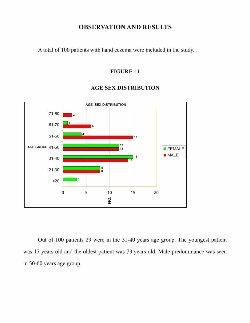
OBSERVATION AND RESULTS
A total of 100 patients with hand eczema were included in the study.
FIGURE - 1
AGE SEX DISTRIBUTION
AGE- SEX DISTRIBUTION
8
14
12
15
6
2
3
8
15
12
4
1
0 5 10 15 20
<20
21-30
31-40
41-50
51-60
61-70
71-80
AGE GROUP
NO
.
FEMALE
MALE
Out of 100 patients 29 were in the 31-40 years age group. The youngest patient
was 17 years old and the oldest patient was 73 years old. Male predominance was seen
in 50-60 years age group.

FIGURE – 2
SEX DISTRIBUTION
SEX DISTRIBUTION
57%
43% MALE
FEMALE
Majority of patients 57 % were males. Male: Female ratio was 1.32:1
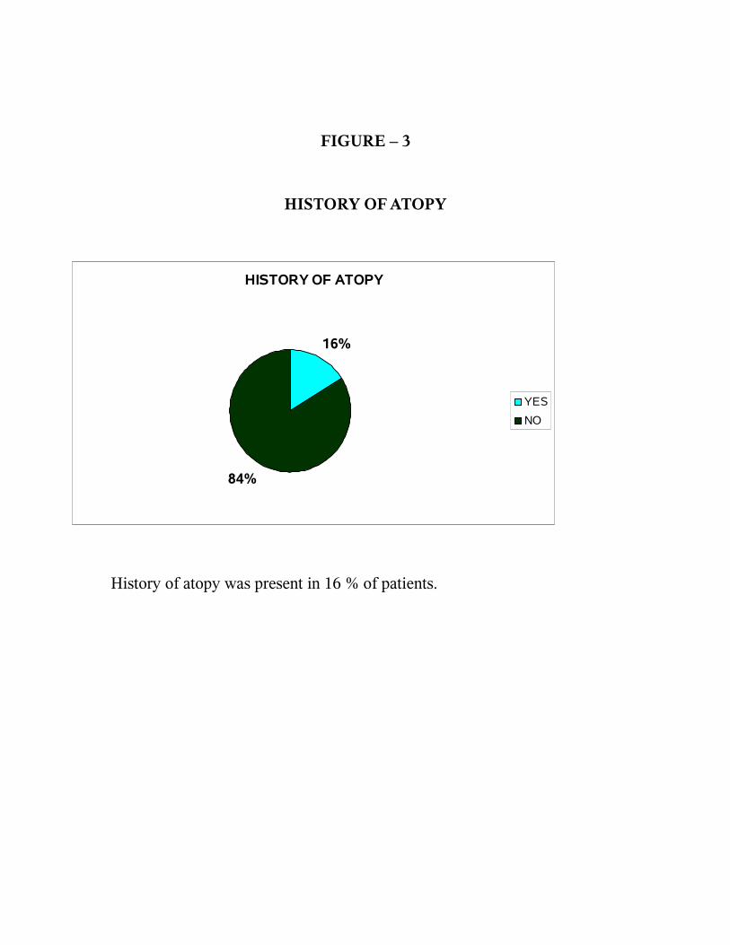
FIGURE – 3
HISTORY OF ATOPY
HISTORY OF ATOPY
16%
84%
YES
NO
History of atopy was present in 16 % of patients.

FIGURE – 4
OCCUPATION
OCCUPATION
31
27
10
6
4 43 3 3
21 1 1 1 1 1 1
0
5
10
15
20
25
30
35
HO
US
E W
IFE
MA
SO
N
FA
RM
ER
PA
INT
ER
HO
TE
L W
OR
KE
R
CL
ER
K
FL
OW
ER
VE
ND
OR
SE
CU
RIT
Y
WE
LD
ER
TA
ILO
R
BA
RB
ER
EN
GIN
EE
R
LE
AT
HE
R W
OR
K
PL
UM
BE
R
PR
ES
S
NU
RS
E
ST
UD
EN
T
NO
.
NO.
Among the various occupational groups, housewives formed the majority and
accounted for 31% of total cases. Masons constituted second major group (27%).

FIGURE - 5
PATCH TEST RESULT
PATCH TEST RESULT
76%
24%
POSITIVE
NEGATIVE
Positive patch test result was seen in 76 % of patients with hand eczema.

FIGURE – 6
ISS AND PLANT ANTIGEN SENSITIVITY
ISS AND PLANT ANTIGENS
34
13
6 5 52 2 1 1 1 1 1
0
5
10
15
20
25
30
35
40
PO
TA
SS
IUM
DIC
HR
OM
AT
E
NIC
KE
L
PA
RT
HE
NIU
M
FO
RM
AL
DE
HY
DE
XA
NT
HIU
M
CO
BA
LT
FR
AG
RE
NC
E M
IX
CH
RY
SA
NT
HE
MU
M
PE
G
EP
OX
Y R
ES
IN
ET
HY
LE
NE
DIA
MIN
E
PP
D
ANTIGENS
The most common sensitizer in our study group was potassium dichromate,
constituting 44.73 % (34) followed by nickel sulphate i.e., 17.10 % (13), parthenium
7.89% (6), formaldehyde 5 (6.57%), xanthium 5 (6.57%), cobalt 2 ( 2.63%), fragrance
mix 2 (2.67%), chrysanthemum 1 (1.31%), polyethylene glycol 1 (1.31%), epoxy resin 1
(1.31%) , ethylene diamine 1 (1.31%) and PPD 1 (1.31%) .
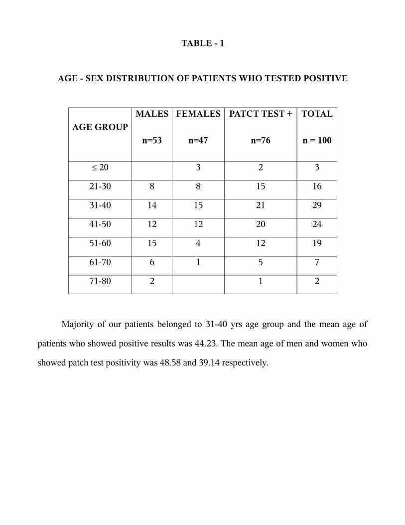
TABLE - 1
AGE - SEX DISTRIBUTION OF PATIENTS WHO TESTED POSITIVE
AGE GROUP
MALES
n=53
FEMALES
n=47
PATCT TEST +
n=76
TOTAL
n = 100
≤ 20 3 2 3
21-30 8 8 15 16
31-40 14 15 21 29
41-50 12 12 20 24
51-60 15 4 12 19
61-70 6 1 5 7
71-80 2 1 2
Majority of our patients belonged to 31-40 yrs age group and the mean age of
patients who showed positive results was 44.23. The mean age of men and women who
showed patch test positivity was 48.58 and 39.14 respectively.

FIGURE – 7
IRRITANT REACTION TO SOAP AMONG HOUSEWIVES
IRRITANT REACTION TO SOAP AMONG HOUSE WIVES
65%
35%
POSITIVE
NEGATIVE
Among the house wives 35 % showed irritant reaction in the patch test done with
soaps used by them.

FIGURE – 8
SENSITIVITY TO ONION AND GARLIC AMONG HOUSEWIVES
SENSITIVITY TO ONION AND GARLIC AMONG HOUSEWIVES
16%
84%
POSITIVE
NEGATIVE
Among the house wives 16 % showed sensitivity to onion and garlic.
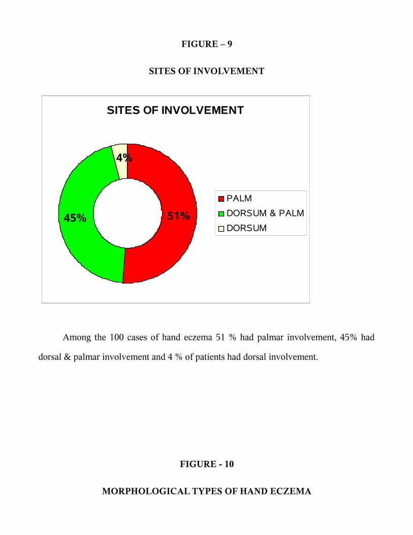
FIGURE – 9
SITES OF INVOLVEMENT
SITES OF INVOLVEMENT
51%45%
4%
PALM
DORSUM & PALM
DORSUM
Among the 100 cases of hand eczema 51 % had palmar involvement, 45% had
dorsal & palmar involvement and 4 % of patients had dorsal involvement.
FIGURE - 10
MORPHOLOGICAL TYPES OF HAND ECZEMA

MORPHOLOGICAL TYPES OF HAND ECZEMA
69
12 115 2 1
01020304050607080
WE
AR
& T
EA
RD
ER
MA
TIT
IS
HY
PE
RK
ER
AT
OT
IC
FIN
GE
R T
IP
PO
MP
HO
LYX
R.F
.F.P
AP
RO
N E
CZ
EM
A
Among the various morphological types of hand eczema wear and tear dermatitis
was the frequent type in our study group constituting 69 % follwed by hyperkeratotic
type (12 %).

DISCUSSION
A total of 100 patients were included in the study, out of which 76% showed
positivity in patch testing in concurrence with the studies done earlier which ranges from
50% to 92.5% . Minocha 97 et al., reported 56.5% contact sensitivity among the patients
with palmar hyperkeratotic dermatitis. Templet 98 et al., reported 54.4 % among patients
with hand eczema. Huda 99 et al., reported 92.5% positivity among patients with hand
dermatitis. Kishore 100 et al., reported 82.5% positivity among hand eczema. Laxmisha
101 et al., reported 52.5% positivity among patients with hand eczema.
Majority of our patients belong to 31-50 yrs age group and the mean age of
patients who showed positive results was 44.23 which is in concurrence with other
studies. 100,102 The mean age of men and women who showed patch test positivity were
48.58 and 39.14 respectively. Women showed positive patch test at an earlier age, this
could be because of the earlier sensitization to allergens like nickel, cobalt and fragrance
mix used in artificial jewelries and cosmetic products.
Among the 100 patients studied 57 (57%) were males and 43 (43%) were females,
the ratio being 1.32:1 which is contrary to most studies where the incidence was higher
in females.97,98,103 This could be because of high number of semiskilled construction
workers in our study group. Kishore 100 et al., and Laxmisha 101 et al., reported male
predominance in their study group.
Atopic diathesis is the most common endogenous cause of hand eczema. In our

study group 16% of patients were atopics. Suman and Reddy reported history of atopy in
36% of their patients with hand eczema. 8104Laxmisha 101 et al., had reported that only
one out of 36 patients had atopy history in their study group.
Occupation has significant bearing on hand eczema because of exposure to
various contactants at workplace. 105.106 In fact, occupational hand eczema comprises
90% to 95% of all occupational skin diseases in Denmark. 105 Among the various
occupational groups, housewives formed the majority and accounted for 31% of total
cases in our study, which is in concordance with other studies.97,100,104 This can be
explained on the basis of their coming in contact with agents of wide variety during day-
to day routines of household work of cooking, washing, cleansing and milking, feeding
of animals particularly by housewives of rural background in India. Masons constituted
second major group (27%) which is higher when compared to Laxmisha 101 et al ., and
Suman and Reddy 104 who has reported 14% masons in their study group. This could be
because of the growth in construction industry in our region. The other occupational
groups encountered in our study were, farmers (10 %), painters (6 %), hotel workers (4
%), clerk (4 %), flower vendors (3 %), security (3 %), welder (3 %), tailor ( 2 %), barber
(1 %), mechanical engineer (1%), leather worker ( 1 %), plumber ( 1 %), press (1 %),
nurse ( 1 % ) and student (1 %). The contact with water, which is hypotonic, and the
dissolution of the surface lipids by detergents or solvents, may be the reason for a higher
incidence of contact allergy in people involved in the above occupations.
The most common sensitizer in our study group was potassium dichromate,
constituting 44.73 % (34) with a male predominance ( 85.29 %).This could be because
of a large number of construction workers in our study group. Chromates are present in
cements, leather, matches, bleaches, yellow paints, varnishes, certain chromates
containing glues, soap, and detergents.107 Chromates are part of earth's crust, and traces
of chromates are present in practically all raw materials. 107 Similar findings were
reported by Laxmisha 101 et al., and Kishore 100et al.,
The next common allergen was nickel sulphate i.e., 17.10 % (13), with a female
predominance ( 84.61 %). Majority of the patients with nickel allergy were house wives
as they are exposed to utensils, door handles, knobs, artificial jewelry etc. Nickel is
ubiquitous in the environment and constitutes about 0.008% of the earths crust. Nickel
in metal and salt form gives rise to contact allergy, metallic nickel only after
corrosion.108 The corrosiveness of sweat, saliva and other body fluids to nickel and
nickel alloys is of primary importance. Similar findings have been reported in various
studies. 103,109,110
Among those positive patients the common sensitizer in males was potassium
dichromate (70.73%) and in the females it was nickel sulphate (31.42 %).
Positive reaction to parthenium antigen was seen in 6 patients (7.89%). Out of 6
patients 5 were farmers and one patient was a house wife. Men predominated in the
parthenium sensitive group with 66.6% of the parthenium-positive patients being males.
This may be due to greater outdoor exposure in men. Sesquiterpene lactones are the
main sensitizers of the Compositae family. Other components, thiophenes and acetylenes
are said to elicit only phytophotodermatitis, but recent studies have demonstrated that
some thiophenes and benzofuran derivates possess not only phototoxic activity, but also
sensitizing properties. Clinical manifestations vary from generalized eczema (20-30%),
eczema of hands and face (24%), hand (36-44%), or facial eczema (11-28%). 110 Bajaj 111
et al., reported that 2 patients out of 71 cases of parthenium dermatitis presented with
hand eczema ( 2.81%). Singhal 112 et al., have found hands and/or feet dermatitis to be
the most frequent sites affected.
Out of 6 patients with parthenium positivty 5 showed positive reaction to
xanthium. This could be because of cross sensitivity. One patient showed positivity to
chrysanthemum , he was a flower vendor and occupational exposure could be the
reason for the eczema. Xanthium strumarium is a weed belonging to compositae family
with sesquiterpene lactone as the sensitizer. Parthenium hysterophorus and X..
strumarium have shown a high rate of cross-sensitivity in Indian patients,113 whereas the
prevalence of cross reaction with chrysanthemum is generally low.114,115
Sensitivity to formaldehyde was seen in 5 (6.57 %) patients. Formaldehyde is a
ubiquitous sensitizer industrially, domestically and medically. The frequency of
formaldehyde positivity in eczema patients is around 3 - 4%. 116 Among the five positive
patients 3 were hotel workers, one was a paramedical personnel and the other one was a
painter. One patient showed epoxy resin sensitivity and two patients showed fragrence
mix sensitivity along with formaldehyde positivity.
Cobalt sensitivity was observed in two ( 2.63%) patients. Among the two one was
a construction worker who also showed chromate sensitivity and the other one was a
painter with PEG sensitivity. Cobalt and nickel are present in cement and allergy to these
metals can occur in the construction workers. However isolated occupationally relevant
allergy to these metals from cement without concomitant allergy to chromate is very
rare. It is because cobalt and nickel are present in insoluble form that has very low
sensitization potential.21 Thus, allergy to these metals is usually secondary in the setting
of damaged skin incurred upon by the existing chromate allergy. Greater the clinical
severity of chromate allergy more is the chance of sensitization from cobalt and nickel.
117
Fragrence mix positivity was seen in 2 ( 2.63%) patients and these patients
showed formaldehyde sensitivity also. In India the use of perfumes and fragrances are
on the rise. We feel that in the near future more people with allergy to fragrence mix
may be detected.
One patient (1.31%) showed sensitivity to Poly ethylene glycol (PEG). The
patient was a painter. PEG is used as a solvent, plasticizer and wetting agent.
Occupational exposure could be the reason for the sensitivity in this patient, who also
showed sensitivity to Cobalt.
One patient (1.31%) showed sensitivity to Epoxy resin, he was a painter by
occupation. Since epoxy resins are used as additive in paint occupational exposure could
be the reason for this patients sensitivity. This patient also showed formaldehyde
sensitivity.

Ethylenediamine dihydrochloride sensitivity was seen in one patient who was a
leather worker. Occupational exposure could be the reason for his sensitivity. This
patient showed positive reaction to potassium dichromate also.
One patient showed sensitivity to PPD. This patient was a barber who handles hair
dyes regularly during his work and this could be the reason for his hand eczema.
One patient was a plumber and he showed sensitivity to the glue used in his daily
work. Patch testing was done with the glue as such and the nature of substances in the
glue could not be determined.
Among the 31 house wives patch tested with soaps and detergents 20 ( 64.51%)
showed irritant reaction, among these 20 patients 9 patients did not show sensitivity to
any other allergens. Chronic irritant dermatitis to the soaps and detergents could be the
reason for the hand eczema in these patients. Detergents as a major cause of hand
dermatitis among house wives has been reported in various studies.108,118
Garlic and onion has been reported as the most common sensitizer among the
vegetables in hand dermatitis seen in house wives by various authors.103,118,119 In our
study 5 out of 31 house wives had sensitivity to onion and garlic. All the patients had
finger tip eczema, which is the pattern seen in hand dermatitis due to vegetables.
The predominant site of involvement were palms (51 %), dorsum & palm (45 %)
and only dorsal involvement in 4%. Dry, scaly skin was the most common

morphological picture, followed by hyperpigmentation, fissuring, and lichenification.
Similar observation has been reported by Suman and Reddy.104 Among the various
morphological types of hand eczema wear and tear dermatitis was the frequent type in
our study group constituting 69 % follwed by hyperkeratotic ( 12 %), finger tip (11%),
pompholyx ( 5%), recurrent focal palmar peeling ( 2%) and apron eczema ( 1%).
SUMMARY
This study was undertaken using Indian standard series, plant antigens in
suspected individuals, soaps, detergents, onion and garlic in housewives for patch testing
patients with hand eczema.
Majority of patients belonged to 31-50 yrs age group. The mean age of patients
who showed patch test postivity was 44.23. Hand eczema was predominantly seen in
men in our study. Male : Female ratio was 1.32:1. Women showed positive patch test at
an earlier age than men.
History of atopy was seen in 16 % of patients with hand eczema. Among the
various occupational groups housewives formed the majority comprising 31 % followed
by masons (27%).
Patch test was positive in 76 % of patients with hand eczema.
The most common sensitizer in our study group was potassium dichromate,
constituting 44.73 % (34), followed by nickel sulphate 17.10 % (13). Among those
positive patients the common sensitizer in males was potassium dichromate (70.73%)
and in the females it was nickel sulphate (31.42 %).
The other antigens that showed positive reactions are, parthenium (7.89 % ),
formaldehyde (6.57% ), xanthium (6.57% ), cobalt (2.63% ), fragrence mix (2.63% ),
chrysanthemum (1.31% ), PEG (1.31% ), epoxy resin (1.31% ), ethylene diamine
(1.31% ), and PPD (1.31% ).
Out of 6 patients with sensitivity to parthenium 5 showed positive reactions to
xanthium. Among the 2 patients with cobalt sensitivity one showed positive reaction to
chromate and the other patient showed sensitivity to PEG. Fragrence mix positivity was
seen in 2 patients and these patients showed formaldehyde sensitivity also.
Among the 31 house wives patch tested with soaps and detergents 20 ( 64.51%)
showed irritant reaction, among these 20 patients 9 patients did not show sensitivity to
any other allergens. Sensitivity to onion and garlic was seen in 5 house wives.
The predominant site of involvement were palms ( 51 %), dorsum & palm ( 45 %)
and only dorsal involvement in 4%. Dry, scaly skin was the most common
morphological picture, followed by hyperpigmentation, fissuring, and lichenification.
Among the various morphological types of hand eczema wear and tear dermatitis
was the frequent type in our study group constituting 69 % follwed by hyperkeratotic
( 12 %), finger tip (11%), pompholyx ( 5%), recurrent focal palmar peeling ( 2%) and
apron eczema ( 1%).

CONCLUSION
We encountered a high degree of patch test positivity in patients with hand
eczema and the Indian standard series proved to be very useful, but lacking in certain
cases like hand eczema in housewives. Housewives formed the bulk of our study group
and a high degree of sensitivity to vegetables has been established in the past. Inclusion
of the extracts of common vegetables and fruits in the series would be of immense value.
Since more than half of hand dermatitis cases may be related to occupation, a thorough
history should be taken by a knowledgeable clinician. Potentially relevant allergens in
the workplace must be identified and tested. These allergens may not be contained in
standard trays. A specific patch test series for the hands as in footwear series or textile
series will be an aid in diagnosis of hand eczema.

BIBLIOGRAPHY
1. Holden CA, Berth-Jones J. Eczema, lichenification, prurigo and erythroderma. In:
Burns T, Breathnach S, Cox N, Griffiths C, editors. Rook's Textbook of
Dermatology, 7 th ed. Oxford: Blackwell Science; 2004. p. 17.1-17.55.
2. Li LF, Wang J. Contact hypersensitivity in hand dermatitis. Contact Dermatitis
2002;47:206-9.
3. Rietschel RL, Fowler JF Jr. Hand dermatitis due to contactants: Special
considerations. Fisher's Contact Dermatitis. 5 th ed. Philadelphia: Lippincott
Williams and Wilkins; 2001. p. 261-78.
4. Hebra F. On Diseases of Skin. New Sydenham Society.London 1868. chap 19.
5. Fox T. Skin diseases. 3rd ed., Henry Renshaw.London 1873.chap 10.
6. Radcliffe – Crocker. Diseases of skin.3 rd ed.,H.K.Lewis London, 1903, 147.
7. Meding B, Järvholm B. Hand eczema in Swedish adults - changes in prevalence
between 1983 and 1996. J Invest Dermatol. 2002 ;118:719-23.
8. Agrup G. Hand eczema and other hand dermatoses in South Sweden. Acta Derm
Venereol. 1969;49:73-84.
9. Montnémery P, Nihlén U, Löfdahl CG, Nyberg P, Svensson A. Prevalence of
hand eczema in an adult Swedish population and the relationship to risk
occupation and smoking. Acta Derm Venereol. 2005;85:429-32.

10. Coenraids PJ, Smit H. dermatoses. In: McDonald JC editor. Epidemiology:work
related diseases. 2nd ed. London:BMJ Books;2000.p.175-94.
11. Perry AD, Trafeli JP. Hand dermatitis: review of etiology, diagnosis, and
treatment. J Am Board Fam Med. 2009 ;22:325-30.
12. Bikowski JB. Hand eczema: diagnosis and management. Cutis. 2008 ;82:9-15.
13. Nanko H. treatment of house wives hand eczema. J Jap Med Ass.2004;47:44-51.
14. Tagami H. stratum corneum as skin barrier. Journal of the Japanese society of
dermatology 1998;108:713-717.
15. Hamanaka S. Skin glycolipids and their function. Journal of the Japanese society
of dermatology. 2002;112:107-115.
16. Meding B. Epidemiology of hand eczema in an industrial city. Acta Derm
Venereol.1990;153:1-43.
17. Rystedt I. Hand eczema inpatients with history of atopic manifestations in
childhood. Acta Derma Venereol.1985;65:305-12.
18. Lee HJ, Ha SS, Ahn WK. Clinical evaluation of atopic hand dermatitis. Paediatr
Dermatol. 2001;18:102-6.
19. Mending B, Swanbeck G. epidemiology of different types of hand eczema in an
industrial city. Acta Derma Vnereol. 1989;69:227-33.
20. Suman M, Reddy BS. Pattern of contact sensitivity in Indian patients with hand
eczema. J Dermatol. 2003 ;30:649-54.
21. Burton J L, Rook A, Wilkinson D S. Eczema, lichen simplex, erythroderma and
prurigo. In : Textbook of Dermatology (Rook A, Wilkinson D S, Ebling F J G, et
al, eds), 4th edn. Bombay : Oxford University Press, 1987; 367 – 418
22. Pasricha J S. Contact dermatitis in India, 2nd ed. New Delhi : Offsetters, 1988.
23. Stuckert J, Nedorost S. Low-cobalt diet for dyshidrotic eczema patients. Contact
Dermatitis. 2008;59:361-5.
24. Thierse HJ, Gamerdinger K, Junkes C, Guerreiro N, Weltzien HU. T cell receptor
(TCR) interaction with haptens: metal ions as non-classical haptens. Toxicology.
2005;209:101-7.
25. Lofgren SM, Warshaw EM. Dyshidrosis: epidemiology, clinical characteristics,
and therapy. Dermatitis. Dec 2006;17:165-81.
26. Guillet MH, Wierzbicka E, Guillet S, Dagregorio G, Guillet G. A 3-year causative
study of pompholyx in 120 patients. Arch Dermatol. Dec 2007;143(12):1504-8.
27. Por A, Ket NS. Investigative techniques in contact dermatitis. In : Ket NS, Goh
CL, editors. The Principles and Practice of Contact and Occupational
Dermatology in the Asia-Pacific Region. World Scientific: New Jersey; 2001. p.
47-57.
28. Ghosh S. Atlas and Synopsis of Contact and Occupational dermatology. 1st ed.
Institute of Allergic and Immunologic Skin Diseases: Kolkata; 2006. p .25-61.
29. Bajaj AK. Patch testing: An overview. In : Ghosh S, editor. Recent Advances in
Dermatology. Jaypee Brothers: Delhi; 2004. p. 136-6.
30. Sulzberger MB, Wise F. The contact or patch test in dermatology. Arch Dermatol
Syph 1931;29:519-31.
31. Colman CD. The use and abuse of patch tests. In : Maibach HI, Gollin GA,
editors. Occupational and Industrial Dermatology. Mosby: Chicago; 1982. p. 131-
2.
32. Fisher AA: Chromate dermatitis and cement burns. In contact deramtits 3rd
edn.,Lea and febiger, Philaadelphia,1986;762-772.
33. Klaus E. Anderson, Desmond Burrows and Ian R White: Allergens from the
standard series. In, text book of contact dermatitis. Eds – R.J.G. Rycroft,
T.Menne, P.J frosch 2 nd edn. Springer – Verlag publication 1995;421-424.
34. Fregert S, Gruyberger B, Sandahl E: Reduction of chromate in cement by iron
sulphate. Contact Dermatits 1979;5:39.
35. Goh Cl. Contact sensitivity in Singapore. Hifu (Skin research) 1986;28 ( suppl)
41-51.
36. Nethercote JR. Results of routine patch testing of 200 patients in Toronto, Canada.
Contact Dermatits 1982;8:309-395.

37. Rudner E, Clendenning WE, Epstein E et al. epidemiology of contact dermatitis in
North America. Arch Dermatol 1973;108:537-540.
38. Fraki JF, Peltonen L Hopsu – Hova VK. Allergy to various components of topical
preparations in stasis dermatitis and leg ulcer. Contact Dermatits 1979;5:97-100.
39. Pasricha JS, Bharathi G. contact hypersensitivity due to local antibacterial agents.
Ind J Dermatol Venereol Leprol 1981;47:27-30.
40. Surrinder K, Sharma VK. Indigenous patch test unit resembling Finn chamber.
Ind J Dermatol Venereol Leprol 1986;52:332-336.
41. George N.D, Srinivas CR, Balachandran C, Shenoi SD. Sensitivity to va rious
ingredients of topical preparations following prolonged use. Contact Dermatitis
1990;23:367-368.
42. Forstrom L, Pirila V. Cross sensitivity within the neomycin group of antibiotics.
Contact Dermatits;1978:4:312.
43. Goh CL, Gan SL, Ngni SJ. Occupational dermatitis in a prefabrication
construction factory. Contact dermatitis 1986;15:235-240.
44. Kiel – swier Czynska M. occupational dermatitis and allergy to metals in polish
construction workers manufacturing prefabricated building units. Contact
Dermatitis 1990;23:27-32
45. Cavelier C, Fousserean J, Gille P, Zissne D. allergy to nickel or cobalt; tolerance
to nickel and cobalt samples in man and in the guinea pig allergic or sensitized to
these metals. Contact Dermatitis 1989;21:72-78.
46. Hjorth N, Willkinson D, Magnusson B et al., G lyceryl – p- aminobenzoate patch
testing in benzocaine sensitive subjects. Contact dermatitis 1978;4:46-48.
47. Adriani J, Dalili H. penetration of local anaesthetics through epithelial barier.
Anesth Analg 1971;50:834-841.
48. Fisher AA. Local anaesthetics in contact dermatitis. 3rd edn. Lea and Febiger,
Philadelphia:220-227.
49. Wilkinson JD. Preliminary patch testing with 25% and 15% “ Caine mixes”.
Contact dermatitis 1990;22:244-245.
50. Beck MH, Holden A. benzocaine an unsatisfactory indicator of topical local
anaesthetic sensitization for the UK. Br J Dermatol 1998;118:91-94.
51. Klaus E, Andersen, Desmond Burrows and Ian R. White. In, text book of contact
dermatitis. Eds: R.J.G Rycroft, T.Menne, P.J.Frosch; Allergens from the standard
series. 2nd edn, Springer – Veralg publication 1995; 438-439.
52. Adams RM, Fisher AA. Contact allergen alternatives : J Am Acad Dermatol 1986;
14:951-969.
53. Chirstoffersen J. Clinical patch test data evaluated by multivariate analysis.
Contact Dermatits 1989;21:291-299.
54. Klaus E Andersen, Desmond Burrows and Ian R. White. In, text book of contact
dermatitis. Eds: R.J.G Rycroft, T.Menne, P.J.Frosch; Allergens from the standard

series. 2nd edn, Springer – Veralg publication 1995; 444-445.
55. Mackie BS, Mackie LE. Cross sensitization in dermatitis due to hair dyes. Aust J
Dermatol 1964;7:189.
56. Calnan CD. Reaction to artificial colouring materials. J Soc Cosmet Chem
1967;18:215.
57. Seidenari S, Manzini BM, Danere P. Patch and prick test studyof 593 healthy
subjects. Contact Dermatitis 1990;23:162- 167
58. Bajaj AK, Chaterjee AK. Paraben contact hyper sensitivity. Ind J Dermatol
Venerol Leprol 1985;1:319-21.
59. Fisher AA. The paraben paradox. Cutis 1973;12:830-32.
60. Morgan LG, Flint GN. Nickel alloys and coatings: release of nickel. In:Maibach
Hi, Menne T (eds) Nickel and the skin : Immunology and toxicaology CRC, Boca
Raton. 45-54.
61. Larsson – Stynne B, Windstrom L. ear pirecing – a cause of nickel allergy in
school girls. Contact Drmatitis 1985;13:289-93.
62. Nielsen GD et al. Nickel sensitive patients with vesicular hand eczema: oral
challenge with a diet naturally high in nickel. Br J Dermatol 1990;122:299-308.
63. Fisher AA. Nickel- the ubiquitous contact allergen : textbook of contact
dermatitis, 3 rd edn. Lea and Febiger 1986:745-761.
64. Cammintausta K, Kalimo K. Do positive nickel reactions increase non specific
patch test reactivity?. Contact Dermatitis 1987;16:160-163.
65. Fisher AA. Contact dermatitis, 3 rd edn., Lea and febiger, Philadelphia, 665-674.
66. Fregert S, Thorgeirsson A, Patch testing with lower molecular oligomers of epoxy
resin in humans. Contact Dermatits 1977;3:301-303.
67. Klaus E, Andersen, Desmond Burrows and Ian R. White. In, text book of contact
dermatitis. Eds: R.J.G Rycroft, T.Menne, P.J.Frosch; Allergens from the standard
series. 2nd edn, Springer – Veralg publication 1995; p 452.
68. Bauer K. Flavours and fragrances. In: Von Apre Hj ( eds) Ullmans encyclopedia
of industrial chemistry, vol. AIi.VCH, Weinheins, p.144-246.
69. Larsen WG. Perfume Dermatits. In Fisher AA (ed). Contact dermatitis, 3rd edn.
Lea and Febiger. Philadelphia,pp.394-404.
70. Klaus E, Andersen, Desmond Burrows and Ian R. White. In, text book of contact
dermatitis. Eds: R.J.G Rycroft, T.Menne, P.J.Frosch; Allergens from the standard
series. 2nd edn, Springer – Veralg publication 1995; 416-460.
71. Larsen WG. Perfume Dermatitis. A study of 20 patients. Arch Dermatol
1977;113:623-627.
72. De Groot Ac. Adverse reations to cosmetics. Thesis. State university of
Groningen. 1988.

73. Andersen KE, Burrows D, Cronin E, Dooms GA. Recommended changes to
standard series. Contact Dermatitis 1988; 5: 389-390.
74. Cronin E. Contact Dermatitis. Churchill Livingstone, Edinburg, pp 734-735.
75. Conde – Salazar L, Volpe MG, Guimaranes D. allergic contact dermatitis from a
suction socket prosthesis. Contact Dermatits 19:305-306.
76. Taylor JS. Rubber. In: Fisher AA(ed), 3rd edn, Contact Dmatitis, Lea and Febiger,
Philadelphia,p.623
77. Fisher AA. Dermatitis to preservative and other additives in cosmetics and
medications; In contact dermatitis, 3rd edn, 1986. Lea and Febiger 238-257.
78. Burry Tn, Kirk J, Reid JG, Turner T. Chlorocresol sensitivity. Contact Dermatitis
1975;1;41.
79. Klaus E, Andersen, Desmond Burrows and Ian R. White. In, text book of contact
dermatitis. Eds: R.J.G Rycroft, T.Menne, P.J.Frosch; Allergens from the standard
series. 2nd edn, Springer – Veralg publication 1995; 460.
80. Clark EW, Blondeel A, Cronin E, Olefe JA, Wilkinson DS. Lanolin of reduced
sensitizing potential. Contact Dermatitis 19891;7:80-83.
81. Veien N. Systemically induced eczema in adult. Acta Derm Venereal ( suppl)
(stock) 1989 147.
82. Fisher AA, Dooms – Goossens A. the effect of perfume ageing on the allergenicity
of individual perfume ingredients. Contact Dermatitis !(&^;2:155-159.
83. Opdyke DLK. Inhibition od sensitization reactions induced by certain aldehydes.
Food cosmet Toxicol 1976;14:197-198.
84. Hanau D. The influence of Limonene on induced delayed hypersensitivity to citral
in guinea pigs I. Histological study. Acta Derm Venereal ( stock) 63:1-7.
85. Barbier P, Benezra C. The influence of Limonene on induced delayed
hypersensitivity to citral in guinea pigs II.label distribution in the skin of 14 c –
labeled citral. Acta Derm Venereal ( stock) 63:93-96.
86. Klaus E, Andersen, Desmond Burrows and Ian R. White. In, text book of contact
dermatitis. Eds: R.J.G Rycroft, T.Menne, P.J.Frosch; Allergens from the standard
series. 2nd edn, Springer – Veralg publication 1995; 446-447.
87. Cronin E .contact Dermatitis. Churchil Livingstone, Edinburg, p 729.
88. Knudsen BB, Larsen E, Egsgaard H, Menne T. Release of thiuram and carbamates
from rubber gloves. Contact Dermatits 1993;28:63-69.
89. Kaaber K, Menne T, Veien N, Hongaard P. Treatment of dermatitis with antabuse;
a double blind study. Contact Dermatits 1983;9:297-299.
90. Vn Hecke E, Vermaner F. allergic contact dermatitis by oral disulfurams. Contact
Dermatits 1984;10:254.
91. Agner T, Menne T. Sensitivity to clioquinol and chlorquinaldol in the quinoline
mix. Conatact Dermatits 1993; 29:163.

92. Groot Ac, Weyland JW, Kathon CG. A review. J Am Acad Dermatol 1988;18:350-
358.
93. Hasson A, Guimaraens D, conde-salazar C. Patch testing sensitivity to the
preservative Kathon CG in Spain. Contact Dermatitis 1990;22:257-261.
94. Klaus E, Andersen, Desmond Burrows and Ian R. White. In, text book of contact
dermatitis. Eds: R.J.G Rycroft, T.Menne, P.J.Frosch; Allergens from the standard
series. 2nd edn, Springer – Veralg publication 1995; 433-434.
95. Klaus E, Andersen, Desmond Burrows and Ian R. White. In, text book of contact
dermatitis. Eds: R.J.G Rycroft, T.Menne, P.J.Frosch; Allergens from the standard
series. 2nd edn, Springer – Veralg publication 1995; 448-449.
96. Lakshmi C, Srinivas CR, Anand CV, Mathew AC. Irritancy ranking of 31
cleansers in the Indian market in a 24-h patch test. Int J Cosmet Sci
2008;30:277-283.
97. YC Minocha, A Dogra, VK Sood. Contact sensitivity in palmar hyperkeratotic
dermatitis. Ind J Dermatol Venerol Leprol. 1993; 59: 60-63.
98. Templet JT, Hall S, Belsito DV. Etiology of hand dermatitis among patients
referred for patch testing. Dermatitis. 2004 ;15:25-32.
99. MM Huda, UK Paul. Patch testing in contact dermatitis of hands and
feet.1996;62: 361-362.
100. Nanda B Kishore, AD Belliappa, Narendra J Shetty, D Sukumar, S Ravi. Hand
eczema-Clinical patterns and role of patch testing. Ind J DermatolVenerol Leprol.
2005;71: 207—208.
101. Laxmisha C, Kumar S, Nath AK, Thappa DM. Patch testing in hand eczema at a
tertiary care center. 2008; 74 : 498-499.
102. Lantinga H, Nater JP, Coenraades PJ. Prevalence, incidence and course of
eczema on the hand and forearms in a sample for the general population. Contact
Dermatitis 1984;10:35-40.
103. Bajaj A K. Contact dermatitis of hands. Ind J Dermatol Venereol Leprol 1983; 49:
195-9.
104. Suman M, Reddy BS. Pattern of contact sensitivity in Indian patients with hand
eczema. J Dermatol. 2003;30:649-54.
105. Skoet R, Olsen J, Mathiesen B, Iversen L, Johansen JD, Agner T. A survey of
occupational hand eczema in Denmark. Contact Dermatitis 2004;51:159-66.
106. Templet JT, Hall S, Belsito DV. Etiology of hand dermatitis among patients
referred for patch testing. Dermatitis 2004;15:25-32.
107. Rietschel RL, Fowler JF Jr. Hand dermatitis due to contactants: Special
considerations. Fisher's Contact Dermatitis. 5th ed. Philadelphia: Lippincott
Williams and Wilkins; 2001. p. 261-78
108. Singh G, Singh K K. Contact dermatitis of hands. Ind J Dermatol Venereol
Leprol 1986; 52 : 152 - 4.
109. Sharma N L, Sharma R C. Contact dermatitis to metals in Simla (HP). Ind J
Dermatol Venereol Leprol 1985; 51 148-50.
110. Jovanović M, Poljacki M. Compositae dermatitis. Med Pregl. 2003;56:43-9.
111. Bajaj AK , Saraswat A, Mukhija G, Rastogi S, Yadav S. Patch testing experience
with 1000 patients. Ind J Dermatol Venereol Leprol 2007; 73: 313-318.
112. Singhal V, Reddy BS. Common contact sensitizers in Delhi. J Dermatol
2000;27:440-5.
113. Pasricha JS, Bhaumik P, Agarwal A. Contact Dermatitis due to Xanthium
strumarium. Indian J Dermatol Venereol Leprol 1990;56:319-21.
114. Pasricha JS, Nandakishore T. Air borne contact dermatitis due to chrysanthemum
with true cross sensitivity to Parthenium hysterophorus and Xanthium strumarium
. Indian J Dermatol Venereol Leprol 1992;58:268-71.
115. Sharma SC, Tanwar RC, Kau S. Contact dermatitis from chrysanthemums in
India. Contact Dermatitis 1989;21:69-71.
116. Christoffersen J. Clinical patch test data evaluated by multivariate analysis.
Contact dermatitis 1989;21:291-299.
117. Fregert S, Gruvberger B. Solubility of cobalt in cement. Contact dermatitis 1978;
4:14-8.
118. Pasricha J S, Kanwar A J. Substances causing contact dermatitis. Ind J Dermatol

Venereol Leprol 1978; 44 : 264 – 8.
119. Sinha S M, Pasricha J S, Sharma R C, et al. Vegetables responsible for contact
dermatitis of hands. Arch Dermatol 1977; 113: 776-9.
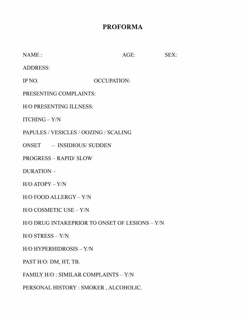
PROFORMA
NAME : AGE: SEX:
ADDRESS:
IP NO. OCCUPATION:
PRESENTING COMPLAINTS:
H/O PRESENTING ILLNESS:
ITCHING – Y/N
PAPULES / VESICLES / OOZING / SCALING
ONSET – INSIDIOUS/ SUDDEN
PROGRESS – RAPID/ SLOW
DURATION –
H/O ATOPY – Y/N
H/O FOOD ALLERGY – Y/N
H/O COSMETIC USE – Y/N
H/O DRUG INTAKEPRIOR TO ONSET OF LESIONS – Y/N
H/O STRESS – Y/N
H/O HYPERHIDROSIS – Y/N
PAST H/O: DM, HT, TB.
FAMILY H/O : SIMILAR COMPLAINTS – Y/N
PERSONAL HISTORY : SMOKER , ALCOHOLIC.
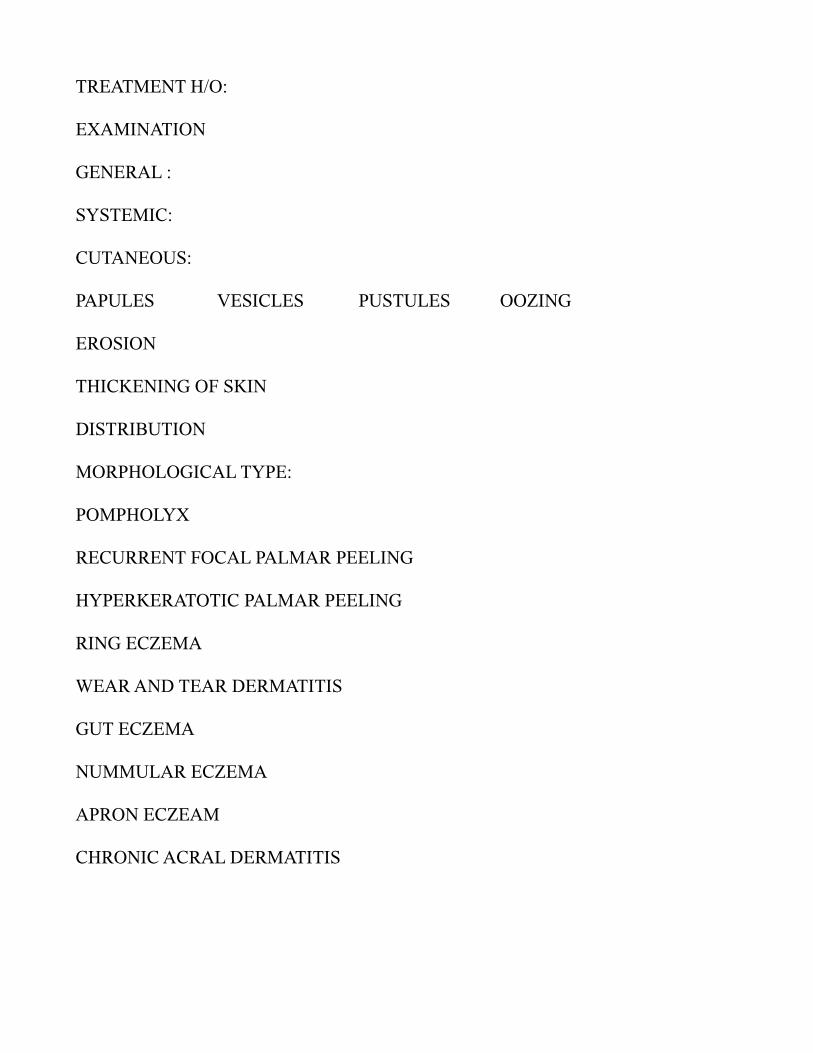
TREATMENT H/O:
EXAMINATION
GENERAL :
SYSTEMIC:
CUTANEOUS:
PAPULES VESICLES PUSTULES OOZING
EROSION
THICKENING OF SKIN
DISTRIBUTION
MORPHOLOGICAL TYPE:
POMPHOLYX
RECURRENT FOCAL PALMAR PEELING
HYPERKERATOTIC PALMAR PEELING
RING ECZEMA
WEAR AND TEAR DERMATITIS
GUT ECZEMA
NUMMULAR ECZEMA
APRON ECZEAM
CHRONIC ACRAL DERMATITIS

NAIL:
PITTING RIDGING DYSTROPHY
THICKENING DISCOLOURATION
HAIR:
MUCOSA:
ORAL
GENITAL
INVESTIGATION:
PATCH TEST :
BIOPSY :
DIAGNOSIS :