Sankara Nethralaya Clinical Practice Patterns in OPHTHALMOLOGY

18 Stephenson Way, London, NW1 2HD T. 020 7935 0702 [email protected] rcophth.ac.uk @RCOphth © The Royal College of Ophthalmologists 2016 All rights reserved For permission to reproduce and of the content contained herein please contact [email protected]
Ophthalmic Services Guidance:
Clinical Audit and Clinical Effectiveness in Ophthalmology
October 2016
2016/PROF/344 2
Date of review: October 2019
Contents
1. Introduction 3
2. Clinical audit 3
3. Why audit? 4
4. Key bodies 6
5. The National Ophthalmology Database (NOD) Audit and Ophthalmic Datasets7
6. Who should do audit? 8
7. Undertaking an audit 8
8. Identify a topic or a problem for audit 9
9. The team 9
10. Identify standards 10
11. What exactly to measure and methodology 11
12. Sampling 12
13. Data collection 12
14. Always do a pilot! 13
15. Data analysis 13
16. Identify changes required – action planning 14
17. Presentation of audit 14
18. Re-audit 15
19. Clinical guidelines and national guidance 15
20. Key bodies 16
National Institute for Health and Care Excellence (NICE) 16
The Royal College of Ophthalmologists 18
The National Guideline Centre 19
National Guideline Clearinghouse 19
Scottish Intercollegiate Guidelines Network (SIGN) 19
The Guidelines and Audit Implementation Network (GAIN) 19
Cochrane 19
21. Conclusions 20
22. Authors for 2016 revision 20
23. Previous Versions 20
24. Resources 20
25. Papers for possible audit standards 21
Age-related Macular Degeneration 21
Cataract Surgery 22
Cataract Surgery – Biometry 23
Diabetic Retinopathy Laser Treatment 23
Diabetic Retinopathy 24
Local Anaesthesia for Ocular Surgery 24
Glaucoma Surgery 24
Glaucoma Visual Loss 25
Postoperative Endophthalmitis 25
Ocular Trauma 25
Vitreoretinal Surgery 26
26. Guideline resources for possible audit standards 27

2016/PROF/344 3
1. Introduction
All doctors have a duty to understand, and participate in delivering quality, safety and clinical governance in modern healthcare as described in the General Medical Council’s (GMC’s) Good Medical Practice Domain 2.
This document aims to provide a simple overview of the principles and practice of clinical effectiveness and clinical audit for ophthalmologists, and should be read in conjunction with the College publication Quality, safety and clinical governance in ophthalmology: an overview.
Clinical effectiveness is one of the three arms of healthcare quality defined by Lord Darzi in 2008 and can be defined as “Care which provides good outcomes, that is good results or success of care for patients. This can be assessed both through clinical outcome measures, such as mortality/survival rates, complication rates, and through patient reported outcome measures (PROMs) such as a patient’s assessment of their own symptoms and quality of life measures”. Clinical audit and the use of evidence based guidelines fall within this quality domain.
2. Clinical audit
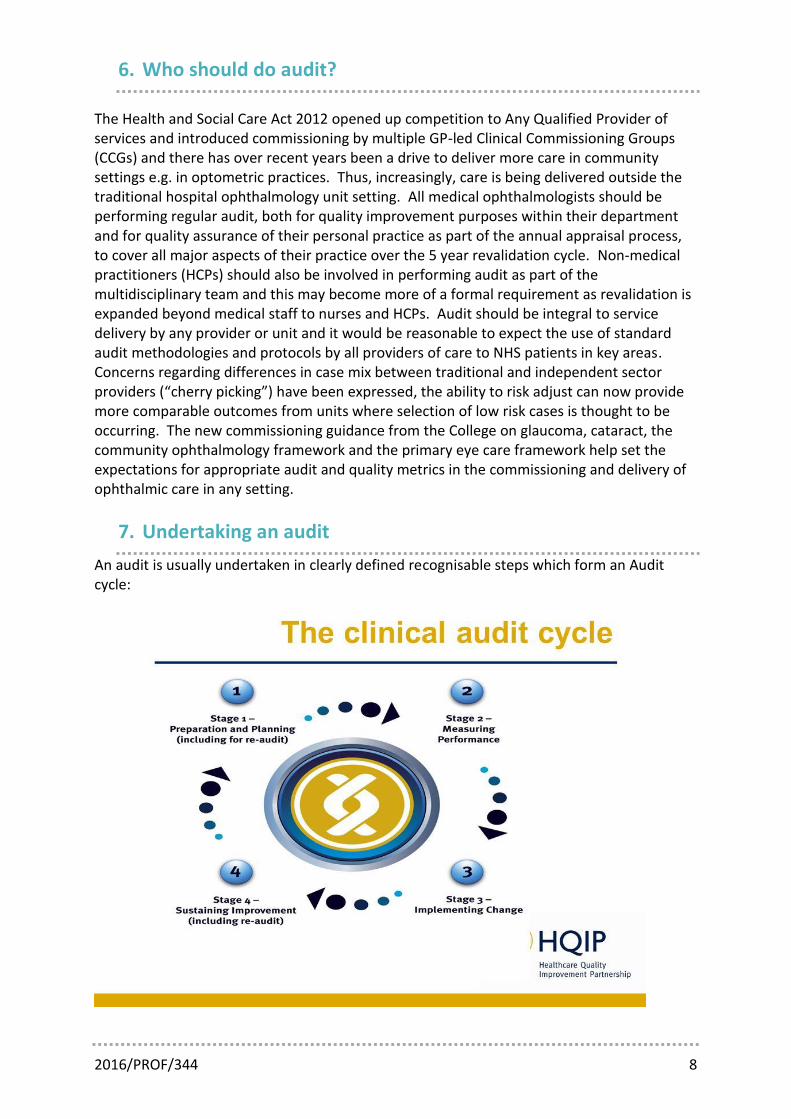
Clinical audit is defined by the National Institute for Health and Care Excellence (NICE) as “a quality improvement process that seeks to improve patient care and outcomes through systematic review of care against explicit criteria and the implementation of change.’ It differs from research in that, whereas research defines what is best practice where there is uncertainty and advances our knowledge, audit measures whether the already known best practice is actually occurring in clinic care and nearly always it should lead to change and reassessment later. The audit cycle describes this continuous process of review and implementation of change and involves the following steps:
1. Identify the topic and the question 2. Select standards – the audit criteria (audit criteria are simply the explicit standards
against which care will be measured)
3. Measure performance/practice
4. Compare performance with standards and see where the standards are not achieved – these are the areas for improvement
5. Make improvements 6. Re-audit to ensure improvement has been achieved
For an audit to be worthwhile it requires careful planning. The choice of topic must be relevant to both the local situation and the patients and professionals involved. The aims and standards must be both realistic and valid. Data collection must be well thought out and undertaken accurately so that when data are analysed, a true and representative impression of practice is formed. The results must be carefully interpreted and any changes to practice implemented with full agreement of those involved or affected. Re-audit at a later time completes the audit cycle and should affirm adjustments to practice implemented in the earlier cycle(s) have had their desired effect and have not created other issues which need addressing. Most clinical audits that, fail do so because they are undertaken as a tick box
2016/PROF/344 4
exercise to fulfil a perceived duty and/or due to the failure to devote some thought and time to what is being done, and why and how it might help care and patients.
3. Why audit?
Apart from the fact that it is a professional and regulatory duty for providers and individual ophthalmologists to participate in clinical audit, it can, if done well, be very beneficial for the auditor, the unit and patients who can be reassured that good care is being provided. Audits are broadly undertaken for two purposes:
1. Simple and brief quality assurance (QA) audits to ensure key areas reach a minimum standard e.g. annual audit of personal posterior capsular rupture rate in cataract surgery for annual appraisal.
2. Quality improvement (QI) audit. Quality assurance audits allow you to:
Demonstrate current best practice is in place
High quality
Low harm
Provide evidence that there is good quality of care to
Patients
Regulators such as the Care Quality Commissioning
Peers
Professional bodies e.g. General Medical Council, The Royal College of Ophthalmologists
Commissioners
Share your outcomes
Benchmarking for other units
Present at conferences and publish in journals
National ophthalmology audit
Fulfil appraisal / revalidation
It is worth bearing in mind that these sorts of audits are likely to become more frequently required as the introduction of outcome based commissioning and specialised commissioning by the new Health and Social Care Act (2012) means that at least some of any provider’s income will be based on the achievement of key standards of care which require an audit to assess.

2016/PROF/344 5
There are certain areas where you would reasonably expect any provider or professional providing this care to perform regular QA audits:
Table 1 Quality assurance audits
Audit Guidance adherence Outcomes
Cataract surgery College guidelines on cataract and cataract anaesthesia NICE (once published). College Quality Standards for Cataract Adherence to protocol if any extended roles for Health Care Practitioners (HCPs)
Posterior capsular rupture rate Visual Acuity (VA) results Ideally via National Ophthalmalgia Database audit Actual vs planned postop refraction Endophthalmitis rates Number wrong Intraocular Lenses
Medical retina Adherence to NICE guidance on intravitreal injections College AMD guidelines College RVO guidelines College DRS guidelines College QS for MR Adherence to protocol if any extended roles HCPs
Injection endophthalmitis rates Other injection complications % eligible Certificate of Vision Impairment registered Number never events (wrong drug, wrong eye) injections Delays in follow up VA loss and gain after intravitreal injections in Age-related Macular Degeneration (AMD)
Vitreoretinal College Quality Standards for vitreo retinal surgery
% reattachment primary retinal detachment surgery % closure macular hole surgery Macular hole VA outcomes Complications Ideally via national VR dataset
Adnexal College Quality Standards for adnexal
Paediatric & strabismus College Quality Standards for Paediatric Ophthalmology
College Retinopathy of Prematurity guidelines

2016/PROF/344 6
Neuro-ophthalmology College Quality Standards for neuro-ophthalmology
NICE TIA guidance
Glaucoma Adherence to NICE guidance on glaucoma care Adherence to protocol if any extended roles HCPs
% eligible CVI registered Delays in follow-up Success trabeculectomy and tube surgery
Cornea College Quality Standards for cornea
% submission yellow forms corneal national audit % success/failure at 1 year for PK and lamellar grafts
Theatres Use ophthalmic WHO checklist audit
Unexpected return to theatres Never events
Quality Improvement audits allow you to:
Improve quality of care
Reduce risk, harm and clinical mistakes
Increase efficiency and effectiveness
Direct resources or provide evidence for requirement of resources (e.g. in a business case) for staff, equipment, training
4. Key bodies
Healthcare Quality Improvement Partnership (HQIP) is an independent organisation led by
the Academy of Medical Royal Colleges, The Royal College of Nursing and National Voices,
established in 2008 to increase the impact of clinical audit on quality improvement. HQIP
commissions, manages, supports and promotes national and local quality improvement and
audit. This includes the national and local clinical audit programmes, the Clinical Outcome
Review Programmes, the National Joint Registry and the Royal College of Ophthalmologists’
National Ophthalmology Audit.
The Royal College of Ophthalmologists (RCOphth) provides advice and guidance on clinical
audit in ophthalmology and also promotes audit via: its scientific publications; publication of
various national audits or surveys; national ophthalmology datasets; the National
Ophthalmology Database Audit; and presentations at the Annual Congress. At Congress, high
quality audits are accepted as poster and oral presentations.

2016/PROF/344 7
5. The National Ophthalmology Database (NOD) Audit and Ophthalmic Datasets
Previous efforts to establish a national electronic care record solution (Connecting for Health and the National Programme for IT) disappointingly failed to deliver, but the collection and analysis of comparable data as a by-product of routine clinical work across many ophthalmic units remains an achievable ambition through intraoperable specialty systems deployed at local level. Adherence to agreed data forms and formats are key to valid ‘cross boundary’ and ‘through time’ comparisons. The Cataract National Data set has paved the way for this in ophthalmology and generated multiple publications including a multicentre analysis of over 180,000 cataract operations. Such large volumes of data not only provide precise benchmarking estimates but also permit statistically powerful analyses of risk indicators which can then be used to risk adjust for the case complexity of individual surgeons, thus making comparisons against benchmarks and their confidence limits more meaningful. Of potentially immediate benefit to patients is the ability to routinely quantify risk strata preoperatively in order to ensure that higher risk operations are undertaken by the most experienced surgeons and consent discussions can be based on individualised risk rates.
The RCOphth has been commissioned by HQIP to provide the National Ophthalmology Database (NOD) Audit as part of the National Clinical Audit and Patient Outcomes Programme (NCAPOP). The NOD Audit will prospectively collect, collate and analyse the cataract surgery national dataset from all centres providing NHS cataract surgery in England and Wales to update benchmark standards of care and provide a powerful quality improvement tool. In addition to cataract surgery, electronic ophthalmology feasibility audits will be undertaken for glaucoma, retinal detachment surgery and age related macular degeneration (AMD).
The national cataract audit will utilise validated and risk (case complexity) adjusted measures of quality which discriminate between centres and surgeons. Initially these will be based on legacy (historic) EMR data up to the end of the 2014-15 NHS year. All EMR databases with national dataset compliant data will be eligible for data submission. The first prospective phase of the audit is due to report on surgery undertaken between 1 September 2015 and 31 August 2016. To facilitate participation centres will be provided with EMR data collection tools where needed, allowing both EMR and paper based centres to be included. In addition, there is a web based tool for optometrists to return visual acuity and refractive data directly back into the patient’s EMR record. Following appropriate local information governance permissions data from electronically enabled units are remotely extracted to a secure server within the NHS firewall. Data are pseudonymised, checked for errors as far as possible and descriptive analyses produced. Summarised data are presented on the audit website, www.nodaudit.org.uk such that contributors are able to view data from their own centre in the context of data from all participating centres. Contributors are also able to use these data for personal audit with benefits in terms of appraisal and revalidation. Relevant aspects of the audit will be submitted for peer reviewed publication in medical journals and prospective results will in due course be publically available via annual reports and the NOD Audit website.
A number of non-cataract national datasets are available (e.g. retinal detachment, macular hole, strabismus, corneal cross linking, cosmetic surgery) and more details of these can be found on the College website.
2016/PROF/344 8
6. Who should do audit?
The Health and Social Care Act 2012 opened up competition to Any Qualified Provider of services and introduced commissioning by multiple GP-led Clinical Commissioning Groups (CCGs) and there has over recent years been a drive to deliver more care in community settings e.g. in optometric practices. Thus, increasingly, care is being delivered outside the traditional hospital ophthalmology unit setting. All medical ophthalmologists should be performing regular audit, both for quality improvement purposes within their department and for quality assurance of their personal practice as part of the annual appraisal process, to cover all major aspects of their practice over the 5 year revalidation cycle. Non-medical practitioners (HCPs) should also be involved in performing audit as part of the multidisciplinary team and this may become more of a formal requirement as revalidation is expanded beyond medical staff to nurses and HCPs. Audit should be integral to service delivery by any provider or unit and it would be reasonable to expect the use of standard audit methodologies and protocols by all providers of care to NHS patients in key areas. Concerns regarding differences in case mix between traditional and independent sector providers (“cherry picking”) have been expressed, the ability to risk adjust can now provide more comparable outcomes from units where selection of low risk cases is thought to be occurring. The new commissioning guidance from the College on glaucoma, cataract, the community ophthalmology framework and the primary eye care framework help set the expectations for appropriate audit and quality metrics in the commissioning and delivery of ophthalmic care in any setting.
7. Undertaking an audit
An audit is usually undertaken in clearly defined recognisable steps which form an Audit cycle:
2016/PROF/344 9
8. Identify a topic or a problem for audit
The first step is to identify the problem or topic that needs to be audited. At this stage it is
important to define the exact focus and scope of the audit and detail the aims of the project.
Decide whether this is a short quality assurance audit or one to drive improvement.
Remember there is little to be gained from undertaking a time-consuming audit on a topic
that does examine an area which requires change to ultimately benefit patients and improve
care. Certain factors should be considered when selecting a topic to ensure that the
investment of time and effort lead to worthwhile improvement, either directly or indirectly,
in the care provided for patients.
Is the condition or event common? Does it affect a reasonable number of patients? Or are many patients at risk? What is the level of severity of the risk?
Does the problem affect morbidity or important aspects of service organisation?
Is there evidence that care could be improved (e.g. complaints or serious incidents)?
Is the topic a national or regional priority? (e.g. indicated by the presence of NICE or other national guidance, national confidential enquiry (NCEPOD) issues, national safety alerts, national audits etc.)
Is it an area of concern for clinical governance?
Are local professionals (both clinical and other) interested in the topic and do they share the perception that a problem exists?
Is the potential for benefit worth the effort and costs required for the audit?
Is this a significant issue for the whole unit or the commissioners?
Is it a second cycle?
Is it a new type of care or service with uncertainties about success?
Is it on your unit’s or organisation’s annual audit plan?
9. The team
It is important to put together a suitable audit group which should be multidisciplinary,
include representatives of everyone involved in the service or who might have an insight
into issues or the area, or whose work might need to change. For instance, if the audit is
likely to show there is a problem with booking patients into clinic, it is crucial to involve the
administrative or reception staff who may understand what the issues are and ensure you
collect the data which will allow you to decide what to change. Many in the group will be
purely advisory or simply to be kept informed, others will have key roles to perform and a
smaller number with decide the exact methodology and perform the analysis.
It is also worth thinking about the involvement of patients and carers where relevant and
this is encouraged at a national level. It is absolutely crucial to involve patients in some way
where you are developing a patient questionnaire or directly assessing the patient
2016/PROF/344 10
experience as part of the audit. Do not forget to involve and use staff from your own clinical
audit department.
Ophthalmologists in training often move posts or attachments and this is one of the
commonest reasons why audits are not finished. Ensure there is a permanent member of
staff on the team or someone to hand over the project to if the audit lead is likely to move
on.
In planning an audit, it is necessary to identify the resources required and to check that
these are available. Considerations for this should include:
Clinicians time
Support staff requirements
IT requirements
Data handling and analysis support
10. Identify standards
The standards to be reached for the audit must be set prior to data collection and it is vital
that all those involved in the project agree with them. They need to be explicit (that is
clearly defined including exclusions), objective and measurable, and the achievement level
must be stated and be realistic – it may not always be possible to achieve 100% success or
the success achieved in a clinical trial, even in best practice in real life clinical settings and
benchmark standards may be required. All standards should be considered carefully to
ensure their applicability to your local population, case mix, and practice.
Standards for audit should be based on the best available evidence or guidelines, and agreed
and widely accepted. Good sources of standards include:
The Royal College of Ophthalmologists and other national audits and national databases
The Royal College of Ophthalmologists Clinical and Ophthalmic Service Guidelines
NICE guidance
National service frameworks (e.g. diabetes, paediatrics)
National Confidential Enquiries
CQC and NHSLA
American Academy of Ophthalmology and similar respected international professional and specialty body guidance
Major published treatment trials
Cochrane Library – Cochrane Eyes and Vision Group
Effectiveness Bulletins Centre for Reviews and Dissemination, University of York
The references section has a list of relevant guidelines, quality standards and journal
publications which may be used to identify audit standards but many more are available and
these will change as new publications emerge.
2016/PROF/344 11
Prior to undertaking an audit, it is good practice to research the published literature using:
NHS Evidence (http://www.nice.org.uk/#tab1)
Cochrane Eyes and Vision Group www.cochrane.org
Scottish Inter-Collegiate Guideline Network (SIGN) www.sign.ac.uk
Pubmed / Medline (http://www.ncbi.nlm.nih.gov/sites/entrez?db=pubmed)
Embase; Healthstar
Remember – clinical guidelines and relevant literature may need to be adapted so that they
are applicable to the local patient population and local service provision.
11. What exactly to measure and methodology
It is important to define what exactly needs to be measured to assess whether the agreed
standards are being achieved. The choices are broadly categorised as:
Structure – the establishment in which you are working e.g.
staff available / staff training
equipment
physical environment
time allowed
Process – does what you do comply with guidelines and protocols to deliver the right things to the right patient at the right time e.g.
Record keeping
History and examination
Interventions
Investigations
Drugs / surgery, procedures
Outcome – what is the result of your care for the patient e.g.
better health/vision, quality of life, longer life, reduced symptoms
fewer adverse events such as loss of vision, complications, failure of surgery
It is often much easier to measure structure and process as it is quick and simple and the
assumption is that doing the right thing (adhering to good practice guidance) with the right
staff, equipment and facilities will inevitably lead to good results for patients (outcomes).
Measuring outcomes, that is what results the care achieved for patients, can be harder as it
can be difficult to measure, or the results may take time to be apparent. For instance, it is
much easier to measure whether a glaucoma patient received appropriate examination and
drugs at the right time that to measure whether your care meant they retained their visual
field so that they could continue to drive. However, outcomes are what really matter to
patients.
2016/PROF/344 12
Methods of data collection and analysis must then be designed. Consider whether data
should be collected about events that are about to happen (prospective audit) or events that
have already taken place (retrospective audit). Prospective audit has the advantage of
allowing all the relevant data to be collected, the disadvantage being that the knowledge
that data are being collected can influence people's usual behaviour and it is likely to take
longer to finish. Retrospective audit is the most common form of clinical audit, and usually
examines what has been recorded in patient notes. This has the advantage that the data
collected are not influenced by a knowledge that the audit is taking place, but a limitation is
that only routinely collected data are available for analysis. Any missing data are usually
unobtainable and can lead in bias in the results.
12. Sampling
In the absence of full electronic working it is rarely practical to collect data on all patients or
events for an audit. Thus it is important to ensure that those included in the audit are
representative of the underlying population.
The two key issues associated with obtaining a representative sample are:
Patient selection
Sample size
Selecting a sample can be complicated and if necessary it may be advisable to obtain some
help in deciding how to do this and how many patients to include. The principles of random
selection and sufficient sample size must be respected and decided upon before data
collection begins. Choosing the next or last ten patients rarely provides a true
representation of the underlying patient population or routine clinical practice. Sample sizes
of around 100 are often sufficient for audits of common conditions or events. Where
specific precision for estimates is required a formal power calculation can be performed with
the help of a statistician.
13. Data collection
Data collection tools should be as brief, simple and user friendly as possible. When
designing a proforma or questionnaire it is useful to consider the following points:
Are all variables of interest identified?
Are all variables necessary?
Are the sections / questions in logical order
Are all the questions appropriate?
Are there question which tell you not only whether or not the standard is achieved but why this might be the case if not?
2016/PROF/344 13
Are all the questions worded in a clear, concise and unambiguous way
Is the layout attractive? Is it compact without being crammed and is there enough space to write? Can you use tick boxes or Yes/No answers for easier data collection?
If using a questionnaire for patients, ensure the language is suitable and test it out with
patient input and with your patient experience lead. Ensure whoever collects the data and
fills in the form understands it and has the knowledge to understand and enter the data. If
there is more than one data collector, ensure that they will be consistent in their approach.
14. Always do a pilot!
Test out your audit tool and do a mini-analysis on a few cases to see if it the form works, is
usable and if it is providing you with the data and answers you need. It is much better to
undertake a pilot, make any necessary changes, and then commence data collection, than to
collect a lot of data and then to realise that you did not get the information required.
15. Data analysis
This should be kept simple, and should be appropriate to answering the audit questions.
Clinical audits seldom require complicated statistical analysis, but if in doubt ask for advice if
required before you start the audit. More often than not simply identifying proportions of
events or patients will be sufficient to inform the majority of audits. Standards are
frequently expressed as percentages, for example success rates or complication rates. It is
good practice to provide confidence intervals (95%) for percentages and averages as this
aids understanding of the precision of the estimate under consideration. Always include in
your audit documents and reports the numbers as well as the percentages which is
particularly helpful when someone else comes to re-audit. Explain your calculation
methods, e.g. what are you going to do with missing data or ‘non-responses’?
Data for 67 cases: 32 met the criterion, 16 did not and 19 did not provide a response
Calculation of success might be
32/67 = 48%
32/(67-19) = 67%
Compliance with standards is:
Number of people whose care is consistent with the criterion / Number of people to whom
the measure applies (total – exceptions) X100
Remember – data collection forms or spreadsheets which have patient identifiable
information are confidential and will need to be stored in line with information governance

2016/PROF/344 14
(IG) requirements and such data must remain within the trust firewall at all times. Avoid
patient details being included in the data analysis file. A decoding file with the relationship
between audit ID number and patient ID can be held securely and separately from the data
analysis file. This minimises IG risk but provides a mechanism for returning back to the
patient in the event of a serious issue arising which needs to be addressed.
16. Identify changes required – action planning
If practice does not meet the standards it will usually be necessary to make some changes.
Discussion between all those affected by proposed changes in practice is required,
presenting the findings, discussion of issues and possible causes, and deciding what actions
are needed. This may involve presenting and discussion in more than one meeting.
Resource implications should be taken into account. Unanimous agreement with the
changes from key players is essential, otherwise improvement is unlikely, and things could
carry on being done in the same unsatisfactory way as before. Changes do not have to be
complicated, but should be achievable. If the standards have been met this indicates that
performance is at a satisfactory level. The opportunity should still be taken to consider if
there are possibilities for improvements.
The implementation of agreed changes may involve other people (e.g. other professionals,
managers or commissioners) and it is important to inform and involve all those affected.
A clear timetable should be agreed to facilitate a successful implementation. Ideally an
action plan should include several actions presented in the following way:
Issue Action needed By whom (action lead) By when (timescale
17. Presentation of audit
Presentation of an audit should cover the steps described above with the following headings in an audit report and/or an audit PowerPoint presentation. Where presentation is in written report form a structured abstract or summary is helpful:
Title
Audit team
Background
Aim/objectives
Setting or service
Evidence base
Standards
Methods (copy of data collection form in appendix)
Results
Interpretation of findings/conclusions
Action plan
Date for re-audit
2016/PROF/344 15
HQIP have lots of resources on their website including detailed guidance to undertaking
audits and template audit reports: http://www.hqip.org.uk/resources/.
18. Re-audit
The re-assessment of quality is the final stage of the audit and is often referred to as 'closing
the loop'. This entails returning to the topic after an appropriate length of time to re-
measure the quality of practice to ensure that the actions have been implemented and that
standards have improved as a result of the changes in practice; and that the changes have
not thrown up other issues which need addressing. Occasionally initial audit will have
confirmed excellent practice with no adjustments necessary. In this instance, a re-audit may
be considered unnecessary.
19. Clinical guidelines and national guidance
Clinical guidelines are defined as systematically derived statements that help practitioners to
make decisions about care in specific clinical circumstances and should be research or
evidence based. The use of evidence-based medicine is essential in maintaining a high
standard of care and guidelines are often the most effective way of ensuring practice is
consistent, evidence based and supported by a consensus of experts. Keeping up-to-date
with recent clinical guidelines is a requirement of the GMC “Duties of a Doctor” and
guidelines provide standards against which current clinical practice can be audited.
Guidelines do allow deviation from the prescribed pathway according to individual
circumstances and where reasons can be clearly demonstrated and documented. In other
words, they do not have to be slavishly followed where there is good reason to do
otherwise. The NICE approach is that their clinical guidelines are intended to apply to 80%
of people on 80% of occasions. A number of other guides exist which may influence clinical
practice.
Policy documents take the form of formal written statements detailing particular actions to
be taken in particular situations and are contractually binding. A policy assists management
and staff to make correct decisions; deal effectively with and comply with relevant
legislation, organisational rules and good practice. A policy document should be regarded as
mandatory, with deviation only in exceptional circumstances. Policies in hospitals are
primarily operational tools for example a sick leave policy, or a ‘did not attend’ policy.
Procedure documents take the form of a set of detailed step by step instructions that
describe the appropriate method for carrying out simple tasks or activities to maintain high
quality and to ensure efficiency, consistency and safety, e.g. a biometry procedure, or a
venepuncture procedure.

2016/PROF/344 16
Protocols are rigid statements which set out a precise sequence of activities to be adhered
to in the management of a specific clinical condition. These allow little or no flexibility or
variation, e.g. protocol for technicians seeing patients in a glaucoma clinic, diabetic
retinopathy screening imaging protocol.
20. Key bodies
National Institute for Health and Care Excellence (NICE)
The role of NICE is to improve outcomes for people using the NHS and other public health
and social care services by producing evidence based guidance and advice, developing
quality standards and metrics for services and providing a range of information services for
commissioners, practitioners and managers across the spectrum of health and social care.
NICE issues guidance in five categories:
NICE guidelines
Technology appraisals guidance (mostly drugs)
Interventional procedures guidance
Medical technologies and diagnostics guidance
They produce the following performance standards:
Quality standards
Quality outcomes frameworks
Clinical commissioning group outcomes indicator
They also run NICE evidence, an online search engine for guidance, medicines and
prescribing support and the BNF.
NICE guidelines include clinical guidelines, and guidelines for public health, social care, safe
staffing and medicines practice. There are a number of NICE clinical guidelines that focus
primarily on ophthalmic practice and these are:
Glaucoma (CG85) [Limited update in development – publication expected mid 2017]
Cataracts in Adults (Guideline in development – publication expected mid 2017)
Macular Degeneration (Guideline in development – publication expected mid 2017)
However, there are many other guidelines which have relevance for the ophthalmologist
such as those concerned with diabetic care.
Technology appraisals guidance assess the clinical and cost effectiveness of health
technologies, such as new pharmaceutical and biopharmaceutical products, but also include
procedures, devices and diagnostic agents, hoping to ensure equitable access to the most
2016/PROF/344 17
clinically and cost-effective treatments that are available. There are a number of TAs which
are directly relevant to ophthalmology for example:
Choroidal neovascularisation (pathological myopic) – Ranibizumab (TA298)
Dry eye disease – Ciclosporin (TA369)
Macular degeneration (age-related) – Aflibercept (TA294)
Macular oedema (retinal vein occlusion) – Dexamethasone (TA229)
Vitreomacular traction – Ocriplasmin (TA 297)
Interventional procedures guidance cover procedures used for diagnosis or for treatments
that involve gaining access into the body (e.g. surgery, endoscopy), or using electromagnetic
radiation (e.g. X-rays, lasers) and examine safety, whether efficacy is sufficient for routine
use and whether special consenting is needed. They essentially cover new innovative
therapies allowing them to be safely and cautiously introduced into routine care.
Ophthalmology is a rapidly changing highly technical surgical field so, not surprisingly, there
are many IPGs which are relevant for example:
Adnexal and Lacrimal – Endoscopic dacryocystorhinostomy (IPG113)
Corneal and Refractive – Accommodating intraocular lenses for cataract (IPG209)
Corneal and Refractive – Collagen cross‑linkage using riboflavin and ultraviolet A for keratoconus and keratectasia (IPG466)
Glaucoma – Trabecular stent bypass micro-surgery (IPG396)
Macular Degeneration (age-related) – Macular translocation with 360° retinotomy (IPG340)
Macular Degeneration (age-related) – Miniature lens systems (IPG272)
Medical Retina – Insertion of a subretinal prosthesis system for retinitis pigmentosa (IPG537)
Strabismus – Tenotomy of horizontal eye muscles for nystagmus (IPG299)
NICE medical technologies and diagnostics guidance addresses specific technologies and
diagnostic tests notified to NICE by manufacturers and developers who examine the ‘case
for adoption’ based on the claimed advantages of introducing the innovation compared with
current management of the condition aiming to ensure the NHS is able to adopt clinically
and cost effective technologies and diagnostics rapidly and consistently.
Quality standards are concise sets of statements, with accompanying metrics, to measure
priority quality improvements within a particular area of care and are usually derived from
NICE's guidance. These are very useful tools to decide whether or not care is compliant with
the guidance and to use for clear audit standards.

2016/PROF/344 18
The Royal College of Ophthalmologists
The Royal College of Ophthalmologists maintains its own guideline development programme
and over the past decade has produced guideline statements on many major
ophthalmological conditions and areas which are available on the college website.
Clinical Guidelines Where NICE guidance is available it is College policy to adopt such
guidance and avoid duplication. Occasionally supplementary College guidance may be
necessary to clarify specific issues, the College is however mindful to not contradict guidance
issued by NICE. For more recent clinical guidelines, the College has attempted to adopt the
methodology and rigour of the NICE approach, with more robust though fewer guidelines
produced and concentration on major areas of care. Although the College does not possess
the infrastructure and resource needed to work to the same standards as NICE, more recent
guidelines have been widely consulted on and are better evidence based.
Examples of currently available College clinical guidelines include:
Cataract Surgery Guidelines 2010 (will be superseded by NICE guideline)
Age-Related Macular Degeneration: Guidelines for Management 2013 (will be superseded by NICE guideline)
Diabetic Retinopathy Guidelines 2012
Retinal Vein Occlusion 2015
Local Anaesthesia in Ophthalmic Surgery 2012
Abusive Head Trauma and the Eye in Infancy 2013
Retinopathy of Prematurity Guideline 2008
Ophthalmic services guidance is intended to inform both ophthalmologists and those
managing eye services what the expected standards of practice are and how these should be
achieved through adequate staffing levels, proper facilities and appropriate managerial
support as well as clinical input. These are based on published evidence as far much as is
possible but, in the absence of high quality published evidence for some areas of practice,
expert consensus is also used to make recommendations.
Examples of these include:
Ophthalmic day care and inpatient facilities 2012
Managing an outbreak of postoperative endophthalmitis 2016
Ophthalmic imaging 2016
Ophthalmic services for children 2012
Ophthalmic pathology 2016
Quality Standards are a set of simple self-assessment tools for a number of clinical services
including subspecialty care (cataract, glaucoma, adnexal, medical retina (including age-
related macular degeneration [AMD]), diabetic retinopathy, vitreoretinal surgery, neuro-
ophthalmology) and care for specific groups (children and young adults and adults with
2016/PROF/344 19
learning difficulties, adults with sight loss and dementia). The tools focus on service
provision not outcomes, and do not attempt to assess every aspect of service but try to
focus on a small number of key areas. It is not expected that most clinical services will
achieve a perfect score, and the results should be used in conjunction with other methods of
quality assessment to support learning and improvement.
Commissioning Guidance The College process for development of commissioning guidelines
has gained NICE approval and the College has issued NICE accredited commissioning
guidance for services in the areas of glaucoma, cataract, as well as frameworks for
community ophthalmology and primary eye care.
The National Guideline Centre Previously the National Clinical Guideline Centre (NCGC), the National Guideline Centre was
formed in 2009 and is one of the largest clinical guideline development organisations in the
world. The work of the NGC is overseen by a governance partnership between the Royal
Colleges of General Practitioners, Nursing, Physicians and Surgeons. Each college is
represented on the NGC management board, alongside representatives from the Royal
College of Physicians Patient and Carer Network, the UK Cochrane Centre, and NHS England.
It is a multi-disciplinary health service research team which produces and publishes
guidelines on behalf of NICE for use in England and Wales.
National Guideline Clearinghouse www.guideline.gov
This is the largest database of appraised guidelines in the world. It is biased towards US
guidance but it does include guidelines from other countries.
Scottish Intercollegiate Guidelines Network (SIGN) www.sign.ac.uk
SIGN develops and publishes clinical guidelines for use in Scotland.
The Guidelines and Audit Implementation Network (GAIN)
This is the clinical and social care regional audit and guidelines body for Northern Ireland,
formed after the amalgamation of CREST (Clinical Resource Efficiency Support Team with the
Regional Multi-professional Audit Group (RMAG) and the Northern Ireland Audit Advisory
Committee (NIRAAC).
Cochrane Cochrane is a global independent network of researchers, clinical professionals, patients and
carers with 37,000 contributors from more than 130 countries producing the Cochrane
Reviews, a database of systematic reviews and meta-analyses which summarise and
interpret the results of medical research, particularly well-conducted controlled trials
2016/PROF/344 20
considered free from commercial sponsorship and other conflicts of interest. This is a huge and
important resource for gold standard detailed evidence based information.
21. Conclusions
There is a wealth of evidence, guidance and standards available for ophthalmologists on
practice and on how to measure whether care is safe and of a high quality. There is a huge
amount of material available particularly from the College and from NICE websites. All
ophthalmologists have a duty to be involved in providing clinically effective care both
personally and within their teams and units and need to make use of guidelines and clinical
audit, which are powerful and useful tools if used well.
22. Authors for 2016 revision
Ian Rodrigues, Informatics and Audit Sub-committee Member, Royal College of
Ophthalmologists
Melanie Hingorani, Chair of Quality and Safety Group, Royal College of Ophthalmologists
John Sparrow, Chair Informatics and Audit Sub-committee, Clinical lead for National
Ophthalmology Audit, Royal College of Ophthalmologists
23. Previous Versions
Original Paper 2002
John Sparrow, Audit and Clinical Effectiveness Lead, Royal College of Ophthalmologists
Richard Wormald, Consultant Ophthalmologist, Moorfields Eye Hospital
John Thompson, Epidemiologist, Leicester Royal Infirmary
Barny Foot, Audit Fellow, Royal College of Ophthalmologists
Revisions 2005, 2008, 2012
John Sparrow, Audit and Clinical Effectiveness Lead, Royal College of Ophthalmologists
24. Resources
Good Medical Practice. General Medical Council 2016. http://www.gmc-uk.org/guidance/index.asp
Quality, safety and clinical governance in ophthalmology: an overview. Royal College of Ophthalmologists 2016. https://www.rcophth.ac.uk/standards-publications-research/
2016/PROF/344 21
High Quality Care for All: the next stage review final report. “The Darzi report” Crown Copyright 2008 https://www.gov.uk/government/uploads/system/uploads/attachment_data/file/228836/7432.pdf
Principles for Best Practice in Clinical Audit, NICE/CHI (2002)
HQIP website. http://www.hqip.org.uk/
National Ophthalmology Database Audit https://www.nodaudit.org.uk/
The Health and Social Care Act 2012. http://services.parliament.uk/bills/2010-12/healthandsocialcare.html
Commissioning guide: Cataract surgery. Royal College of Ophthalmologists Clinical Council for Eye Health Commissioning 2016. https://www.rcophth.ac.uk/standards-publications-research/commissioning-in-ophthalmology/
Commissioning guide: Glaucoma. RCOphth, Clinical Council for Eye Health Commissioning, 2016. https://www.rcophth.ac.uk/standards-publications-research/commissioning-in-ophthalmology/
Community Ophthalmology Framework. RCO Clinical Council for Eye Health Commissioning 2015. https://www.rcophth.ac.uk/wp-content/uploads/2015/07/Community-Ophthalmology-Framework.pdf
Primary Eye Care Framework 2016. RCOphth, Clinical Council for Eye Health. https://www.rcophth.ac.uk/2016/07/ccehc-publish-primary-eye-care-framework/
NICE http://www.nice.org.uk
National Clinical Guideline Centre (NCGC) www.ncng.ac.uk
Cochrane http://www.cochranelibrary.com/
25. Papers for possible audit standards
Age-related Macular Degeneration Madhusudhana KC, Lee AY, Keane PA, Chakravarthy U, Johnston RL, Egan CA, Sim
D, Zarranz-Ventura J, Tufail A, McKibbin M; UK AMD EMR Study Group. UK Neovascular Age-Related Macular Degeneration Database. Report 6: time to retreatment after a pause in therapy. Outcomes from 92 976 intravitreal ranibizumab injections. Br J Ophthalmol. 2016 Mar 30. pii: bjophthalmol-2015-308077.
Lee AY, Lee CS, Butt T, Xing W, Johnston RL, Chakravarthy U, Egan C, Akerele T, McKibbin M, Downey L, Natha S, Bailey C, Khan R, Antcliff R, Varma A, Kumar V, Tsaloumas M, Mandal K, Liew G, Keane PA, Sim D, Bunce C, Tufail A; UK AMD EMR Users Group. UK AMD EMR USERS GROUP REPORT V: benefits of initiating ranibizumab therapy for neovascular AMD in eyes with vision better than 6/12. Br J Ophthalmol. 2015 Aug;99(8):1045-50.
Liew G, Lee AY, Zarranz-Ventura J, Stratton I, Bunce C, Chakravarthy U, Lee CS, Keane PA, Sim DA, Akerele T, McKibbin M, Downey L, Natha S, Bailey C, Khan R, Antcliff R, Armstrong S, Varma A, Kumar V, Tsaloumas M, Mandal K, Egan C, Johnston RL, Tufail A. The UK Neovascular AMD Database Report 3: inter-centre variation in visual acuity outcomes and establishing real-world measures of care. Eye (Lond). 2016 Jul 15. doi: 10.1038/Eye.2016.149.
2016/PROF/344 22
Zarranz-Ventura J, Liew G, Johnston RL, Xing W, Akerele T, McKibbin M, Downey L, Natha S, Chakravarthy U, Bailey C, Khan R, Antcliff R, Armstrong S, Varma A, Kumar V, Tsaloumas M, Mandal K, Bunce C, Tufail A; United Kingdom Age-Related Macular Degeneration Electronic Medical Records Users Group. The neovascular age-related macular degeneration database: report 2: incidence, management, and visual outcomes of second treated eyes. Ophthalmology. 2014 Oct;121(10):1966-75.
Writing Committee for the UK Age-Related Macular Degeneration EMR Users Group. The neovascular age-related macular degeneration database: multicenter study of 92 976 ranibizumab injections: report 1: visual acuity. Ophthalmology. 2014 May;121(5):1092-101.
Ranibizumab and bevacizumab for neovascular age-related macular degeneration. CATT Research Group, Martin DF, Maguire MG, Ying GS, Grunwald JE, Fine SL, Jaffe GJ. N Engl J Med. 2011 May 19;364(20):1897-908.
Regillo CD, Brown DM, Abraham P, Yue H, Ianchulev T, Schneider S, Shams N; The PIER Study Group. Randomized, Double-Masked, Sham-Controlled Trial of ranibizumab for Neovascular Age-related Macular Degeneration: PIER Study Year 1. Am J Ophthalmol. 2008 Feb;145(2):239-248.
Ghanchi FD, Fullarton J, Blake J, Harding SP; PDTUG(UK). The introduction of verteporfin photodynamic therapy in the UK: PDT users group (PDTUG) surveillance programme report 1. Eye (Lond). 2008 May;22(5):671-7.
Rosenfeld PJ, Brown DM, Heier JS et al Boyer DS, for the MARINA Study Group. Ranibizumab for neovascular age related macular degeneration. N Engl J Med. 355(14),1419-1431 (2006).
Brown DM, Kaiser PK, Michels M et al; ANCHOR Study Group. Ranibizumab versus verteporfin for neovascular age-related macular degeneration. N Engl J Med. 355(14),1432-44 (2006).
Cataract Surgery Day AC, Donachie PH, Sparrow JM, Johnston RL; Royal College of
Ophthalmologists' National Ophthalmology Database. United Kingdom National Ophthalmology Database Study of Cataract Surgery: Report 3: Pseudophakic Retinal Detachment. Ophthalmology. 2016 Aug;123(8):1711-5.
Day AC, Donachie PH, Sparrow JM and Johnston RL. The Royal College of Ophthalmologists' National Ophthalmology Database study of cataract surgery: report 2, relationships of axial length with ocular copathology, preoperative visual acuity, and posterior capsule rupture. Eye (Lond). 2015 Dec 29(12): 1528-1537.
Day AC, Donachie PH, Sparrow JM, Johnston RL. The Royal College of Ophthalmologists' National Ophthalmology Database study of cataract surgery: report 1, visual outcomes and complications. Eye (Lond). 2015 Apr;29(4):552-60. doi: 10.1038/eye.2015.3. Epub 2015 Feb 13.
Sparrow JM, Taylor H, Qureshi K, Smith R, Birnie K, Johnston RL. The Cataract National Dataset Electronic Multi-centre Audit of 55,567 Operations: Risk Indicators for Monocular Visual Acuity Outcomes. Eye 2012;26:821–826
Sparrow JM, Taylor H, Qureshi K, Smith R, Johnston RL. The cataract national data set electronic multi-centre audit of 55,567 operations: case-mix adjusted surgeon's outcomes for posterior capsule rupture. Eye 2011; 25:1010-5.
2016/PROF/344 23
Johnston RL, Taylor H, Smith R, Sparrow JM. The Cataract National Dataset electronic multi-centre audit of 55,567 operations: variation in posterior capsule rupture rates between surgeons. Eye 2010; 24:888-93.
Narendran N, Jaycock P, Johnston RL, Taylor H, Adams M, Tole DM, Asaria RH, Galloway P, Sparrow JM. The Cataract National Dataset electronic multicentre audit of 55,567 operations: risk stratification for posterior capsule rupture and vitreous loss. Eye 2009; 23:31-7.
Jaycock P, Johnston RL, Taylor H, Adams M, Tole DM, Galloway P, Canning C, Sparrow JM. The Cataract National Dataset electronic multi-centre audit of 55,567 operations: updating benchmark standards of care in the United Kingdom and internationally. Eye 2009; 23:38-49.
Benzimra JD, Johnston RL, Jaycock P, Galloway PH, Lambert G, Chung AK, Eke T, Sparrow JM. The Cataract National Dataset electronic multicentre audit of 55,567 operations: antiplatelet and anticoagulant medications. Eye 2009; 23:10-6.
Johnston RL, Sparrow JM, Canning C, Tole D, Price NC, UK Cataract EPR users group. Pilot National Electronic Cataract Surgery Survey: I. method, descriptive and process features. Eye 2005 Jul;19;788-794.
Cataract Surgery – Biometry Aristodemou P, Knox Cartwright NE, Sparrow JM, Johnston RL. First eye
prediction error improves second eye refractive outcome results in 2129 patients after bilateral sequential cataract surgery. Ophthalmology 2011; 118:1701-9.
Aristodemou P, Knox Cartwright NE, Sparrow JM, Johnston RL. Formula choice: Hoffer Q, Holladay 1, or SRK/T and refractive outcomes in 8108 eyes after cataract surgery with biometry by partial coherence interferometry. J Cataract Refract Surg 2011; 37:63-71.
Aristodemou P, Knox Cartwright NE, Sparrow JM, Johnston RL. Intraocular lens formula constant optimization and partial coherence interferometry biometry: Refractive outcomes in 8108 eyes after cataract surgery. J Cataract Refract Surg 2011; 37:50-62.
Aristodemou P, Knox Cartwright NE, Sparrow JM, Johnston RL. Letter to the Editor: Biometry formula choice and cataract refractive outcomes. Clinical & Experiment Ophthalmology 2010; 38:536–7.
Knox Cartwright NE, Johnston RL, Jaycock PD, Tole DM, Sparrow JM. The Cataract National Dataset electronic multicentre audit of 55,567 operations: when should IOLMaster biometric measurements be rechecked? Eye 2010; 24:894-900.
Kugelberg M, Lundstrom M. Factors related to the degree of success in achieving target refraction in cataract surgery: Swedish National Cataract Register study. J Cataract Refract Surg 2008; 34:1935-9.
Diabetic Retinopathy Laser Treatment Bailey CC, Sparrow JM, Grey RHB, Cheng H Diabetic Retinopathy Laser
Treatment: 1 Maculopathy Eye 1998:12:69-76
Bailey CC, Sparrow JM, Grey RHB, Cheng H Diabetic Retinopathy Laser Treatment: II Proliferative retinopathy Eye 1998:12:77-84
2016/PROF/344 24
Bailey CC, Sparrow JM, Grey RHB, Cheng H Diabetic Retinopathy Laser Treatment: III Clinical outcomes Eye 1999:13:151-159
Diabetic Retinopathy Keenan TD, Johnston RL, Donachie PH, Sparrow JM, Stratton IM, Scanlon P.
United Kingdom National Ophthalmology Database Study: Diabetic Retinopathy; Report 1: prevalence of centre - involving diabetic macular oedema and other grades of maculopathy and retinopathy in hospital eye services. Eye (Lond). 2013 Dec;27(12):1397-404.
UK National Screening Committee 2011 Key Performance Indicators for Screening, Version 1.7 (accessed 22.01.12) http://www.screening.nhs.uk/kpi
DESP Quality Assurance Standards Release 7 Version 1.3, 23 January 2012 (accessed 23.01.12) http://diabeticeye.screening.nhs.uk
Local Anaesthesia for Ocular Surgery Lee RM, Thompson JR, Eke T. Severe adverse events associated with local
anaesthesia in cataract surgery: 1 year national survey of practice and complications in the UK. Br J Ophthalmol. 2016 Jun;100(6):772-6.
El-Hindy N, Johnston RL, Jaycock P, Eke T, Braga AJ, Tole DM, Galloway P, Sparrow JM. The Cataract National Dataset Electronic Multi-centre Audit of 55,567 operations: anaesthetic techniques and complications. Eye 2009; 23:50-5.
Eke T, Thompson JR. Serious complications of local anaesthesia for cataract surgery: a 1 year national survey in the United Kingdom. Br J Ophthalmol 2007; 91:470-5.
Eke T, Thompson JR The National Survey of Local Anaesthesia for Ocular Surgery: I Survey methodology and current practice Eye 1999:13:189-195
Eke T, Thompson JR The National Survey of Local Anaesthesia for Ocular Surgery: II Safety profiles of local anaesthesia techniques Eye 1999:13:196-204
Glaucoma Surgery Ke M, Guo J, Qian Z. Meta analysis of non-penetrating trabecular surgery versus
trabeculectomy for the treatment of open angle glaucoma. J Huazhong Univ Sci Technolog Med Sci 2011; 31:264-70.
Gedde SJ, Schiffman JC, Feuer WJ, Herndon LW, Brandt JD, Budenz DL. Three-year follow-up of the tube versus trabeculectomy study. Am J Ophthalmol 2009 ;148:670-84.
King AJ, Rotchford AP, Alwitry A, Moodie J. Frequency of bleb manipulations after trabeculectomy surgery. Br J Ophthalmol 2007; 91:873-7.
Edmunds B, Thompson JR, Salmon JF, Wormald RP The National Survey of Trabeculectomy: I Sample and methods Eye 1999:13:524-530
Edmunds B, Thompson JR, Salmon JF, Wormald RP The National Survey of Trabeculectomy: II Variations in surgical technique and outcome Eye 1999:13:524-530
Edmunds B, Thompson JR, Salmon JF, Wormald RP. The national survey of trabeculectomy. III. Early and late complications. Eye 2002:16;297-303
Molteno AC, Bosma NJ, Kittelson JM. Otago glaucoma surgery outcome study: long-term results of trabeculectomy--1976 to 1995. Ophthalmology 1999 ;106:1742-50.

2016/PROF/344 25
NICE Glaucoma Clinical Guideline appendix for Evidence Summaries and Meta-analyses https://www.nice.org.uk/guidance/cg85/evidence/full-guideline-243555949
Glaucoma Visual Loss Boodhna T, Saunders LJ, Crabb DP. Are rates of vision loss in patients in English
glaucoma clinics slowing down over time? Trends from a decade of data. Eye Lond Engl. 2015 Dec;29(12):1613-9.
Boodhna T, Crabb DP. Disease severity in newly diagnosed glaucoma patients with visual field loss: trends from more than a decade of data. Ophthalmic Physiol Opt J Br Coll Ophthalmic Opt Optom. 2015 Mar;35(2):225-30.
Hu S, Smith ND, Saunders LJ, Crabb DP. Patterns of Binocular Visual Field Loss Derived from Large-Scale Patient Data from Glaucoma Clinics. Ophthalmology. 2015 Dec;122(12):2399-406.
Saunders LJ, Russell RA, Crabb DP. Measurement precision in a series of visual fields acquired by the standard and fast versions of the Swedish interactive thresholding algorithm: analysis of large-scale data from clinics. JAMA Ophthalmol. 2015 Jan;133(1):74-80.
Saunders LJ, Russell RA, Kirwan JF, McNaught AI, Crabb DP. Examining visual field loss in patients in glaucoma clinics during their predicted remaining lifetime. Invest Ophthalmol Vis Sci. 2014 Jan;55(1):102-9.
Saunders LJ, Russell RA, Crabb DP. Practical landmarks for visual field disability in glaucoma. Br J Ophthalmol. 2012 Sep;96(9):1185 9.
Fung SSM, Lemer C, Russell RA, Malik R, Crabb DP. Are practical recommendations practiced? A national multi-centre cross-sectional study on frequency of visual field testing in glaucoma. Br J Ophthalmol. 2013 Jul;97(7):843-7.
Postoperative Endophthalmitis Arshinoff SA, Bastianelli PA. Incidence of postoperative endophthalmitis after
immediate sequential bilateral cataract surgery. J Cataract Refract Surg 2011; 37:2105-14.
Sparrow JM. Monte-Carlo simulation of random clustering of endophthalmitis following cataract surgery. Eye 2007; 21:209-13.
Barry P, Seal DV, Gettinby G, Lees F, Peterson M, Revie CW. ESCRS study of prophylaxis of postoperative endophthalmitis after cataract surgery: Preliminary report of principal results from a European multicenter study. J Cataract Refract Surg 2006; 32:407-10.
Kamalarajah S, Silvestri G, Sharma N, Khan A, Foot B, Ling R, Cran G, Best R. Surveillance of endophthalmitis following cataract surgery in the UK. Eye 2004; 18:580-7.
Ocular Trauma Man CY, Steel D. Visual outcome after open globe injury: a comparison of two
prognostic models--the Ocular Trauma Score and the Classification and Regression Tree. Eye 2010; 24:84-9.
Desai P, MacEwen CJ, Baines P, Minassian DC Incidence of cases of ocular trauma admitted to hospital and incidence of blinding outcome.Br J Ophthalmol. 1996 Jul;80(7):592-6.

2016/PROF/344 26
Desai P, MacEwen CJ, Baines P, Minassian DC Epidemiology and implications of ocular trauma admitted to hospital in Scotland. J Epidemiol Community Health. 1996 Aug;50(4):436-41.
MacEwen J, Baines P, Desai P Eye injuries in children: the currents picture Br J Ophthalmology 1999:83:933-936
Vitreoretinal Surgery Jackson TL, Johnston RL, Donachie PH, Williamson TH, Sparrow JM, Steel DH. The
Royal College of Ophthalmologists' National Ophthalmology Database Study of Vitreoretinal Surgery: Report 6, Diabetic Vitrectomy. JAMA Ophthalmol. 2016 Jan;134(1):79-85.
Sallam AA, Donachie PH, Williamson TH, Sparrow JM, Johnston RL. The Royal College of Ophthalmologists' National Ophthalmology Database Study of vitreoretinal surgery: report 5, anaesthetic techniques. Br J Ophthalmol. 2016 Feb;100(2):246-52.
Jackson TL, Donachie PH, Williamson TH, Sparrow JM, Johnston RL. The royal college of ophthalmologists' national ophthalmology database study of vitreoretinal surgery: Report 4, Epiretinal Membrane. Retina. 2015 Aug;35(8):1615-21.
Keenan TD, Johnston RL, Donachie PH, Sparrow JM, Stratton IM, Scanlon P. United Kingdom National Ophthalmology Database Study: Diabetic Retinopathy; Report 1: prevalence of centre - involving diabetic macular oedema and other grades of maculopathy and retinopathy in hospital eye services. Eye (Lond). 2013 Dec;27(12):1397-404.
Jackson TL, Donachie PH, Sparrow JM, Johnston RL. United Kingdom National Ophthalmology Database study of vitreoretinal surgery: report 2, macular hole. Ophthalmology. 2013 Mar;120(3):629-34.
Jackson TL, Donachie PH, Sparrow JM, Johnston RL. United Kingdom National Ophthalmology Database Study of Vitreoretinal Surgery: report 1; case mix, complications, and cataract. Eye (Lond). 2013 May;27(5):644-51.
Mitry D, Awan MA, Borooah S, Siddiqui MA, Brogan K, Fleck BW, Wright A, Campbell H, Singh J, Charteris DG, Yorston D. Surgical outcome and risk stratification for primary retinal detachment repair: results from the Scottish Retinal Detachment study. Br J Ophthalmol. 2012 May;96(5):730-4.
Johnson Z, Ramsay A, Cottrell D, Mitchell K, Stannard K. Triple cycle audit of primary retinal detachment surgery. Eye 2002 Sep;16(5):513-8.
Thompson JA, Snead MP, Billington BM, Barrie T, Thompson JR, Sparrow JM. National audit of the outcome of primary surgery for rhegmatogenous retinal detachment. I. Sample and Methods. Eye 2002:16;766-770.
Thompson JA, Snead MP, Billington BM, Barrie T, Thompson JR, Sparrow JM. National audit of the outcome of primary surgery for rhegmatogenous retinal detachment. II. Clinical Outcomes. Eye 2002:16;771-777.

2016/PROF/344 27
26. Guideline resources for possible audit standards
The Royal College of Ophthalmologists Clinical Guidelines and Ophthalmic Services Guidance
Cataract Surgery
Cataract Surgery Guidelines 2010
Cataract Surgery Commissioning Guide 2015
Glaucoma
Glaucoma Commissioning Guide
Medical Retina
Age-Related Macular Degeneration: Guidelines for Management 2013
Age-related macular degeneration: Commissioning better eye care
Delivery of Diabetic Eye Care 2009
Diabetic Retinopathy Guidelines 2012
Diabetic Retinopathy Screening (DRS) and the Ophthalmology Clinic set up in England 2010
Maximising Capacity in AMD Services
Retinal Vein Occlusion 2015
Ophthalmic Surgery
Local Anaesthesia in Ophthalmic Surgery 2012
Intra-ocular injections by non-medical health care professionals 2013
Managing an outbreak of postoperative endophthalmitis
Prevention of transmission of blood-borne viruses in ophthalmic surgery
Standards for the Retrieval of Human Ocular Tissue used in Transplantation, Research and Training 2013
Theatres
Paediatric Ophthalmology
Abusive Head Trauma and the Eye in Infancy 2013
Adalimumab for children with uveitis
Management of Strabismus in Childhood 2012
Ophthalmic Services for Children
Recording Ophth features of suspected paediatric head trauma
Retinopathy of Prematurity Guideline
Vision screening for children
2016/PROF/344 28
Vitreo Retinal
Management of Retinal Detachment
Other Clinical Guidelines
Emergency Eye Care
Eye Care Services for Adults with Learning Disabilities 2015
Management of visual problems in people with learning disabilities
Management of patients with Grave’s orbitopathy: initial assessment, management outside specialised centres and referral pathways
Ocular Pathology
Primary Care Ophthalmology
Referral guidelines for adult ocular tumours including choroidal naevi 2009
Review of the Ocular side effects of Topiramate 2010
The Ocular Side-Effects of Vigabatrin (Sabril) Information and Guidance for Screening 2008
UK guidelines for the management of Stevens-Johnson syndrome/toxic epidermal necrolysis 2016
Vision Standards for Driving
Ophthalmic Service Guidelines
Audit & Clinical Effectiveness Information for Ophthalmologists
Informatics (update due 2017)
Ophthalmic Day care and Inpatient Facilities
Ophthalmic Imaging
Ophthalmic Instrument Decontamination
Ophthalmic Outpatient Department
Patient Safety in Ophthalmology
Quality safety and clinical governance in ophthalmology: an overview
Sustainability in Ophthalmology May 2013
Quality Standards:
Quality Standards for Services to Patients with Learning Disabilities
Quality Standards & Quality Indicators for Ophthalmic Care and Services for Children and Young People
Quality Standards for Oculoplastic Surgery Services
Quality standards for diabetic retinopathy services in NHS Scotland
Quality standards for diabetic retinopathy services for England, Wales and Northern Ireland
Quality Assurance Self Test for AMD Services
Quality standard for people with sight loss and dementia in an ophthalmology department
Quality Standard for Adnexal Services
Quality Standard for Cataract Services
Quality Standards for Cornea Services
Quality Standard for Glaucoma Services
Quality Standard for Medical Retina Disease Services
Quality Standard for Neuro-ophthalmology Services
2016/PROF/344 29
Quality Standard for Vitreoretinal Services
Refractive Surgery Standards
National Institute for Health and Care Excellence Clinical Guidelines
Glaucoma (CG85) [Limited update in development - publication expected mid 2017]
Cataracts in Adults (Guideline in development - publication expected mid 2017)
Macular Degeneration (Guideline in development - publication expected mid 2017)
Technology Appraisals
Choroidal neovascularisation (pathological myopic) - Ranibizumab (TA298)
Dry eye disease - Ciclosporin (TA369)
Macular degeneration (age-related) - Aflibercept (TA294)
Macular degeneration (age-related) - Photodynamic Therapy (TA68)
Macular degeneration (age-related) - Ranibizumab and Pegaptanib (TA155)
Macular oedema (retinal vein occlusion) - Dexamethasone (TA229)
Macular oedema (retinal vein occlusion) - Aflibercept (TA305)
Macular oedema (retinal vein occlusion) - Ranibizumab (TA283)
Macular oedema (diabetic) - Aflibercept (TA346)
Macular oedema (diabetic) - Dexamethasone (TA349)
Macular oedema (diabetic) – Fluocinolone (TA301)
Macular oedema (diabetic) - Ranibizumab (TA274)
Vitreomacular traction - Ocriplasmin (TA297)
Interventional Procedures
Adnexal & Lacrimal - Endoscopic dacryocystorhinostomy (IPG113)
Adnexal & Lacrimal - Retrobulbar irradiation for thyroid eye disease (IPG148)
Corneal & Refractive - Accommodating intraocular lenses for cataract (IPG209)
Corneal & Refractive - Collagen cross‑linkage using riboflavin and ultraviolet A for keratoconus and keratectasia (IPG466)
Corneal & Refractive - Corneal graft–keratoprosthesis for severe corneal opacity in wet blinking eyes (IPG534)
Corneal & Refractive - Corneal implants for keratoconus (IPG227)
Corneal & Refractive - Corneal implants for refractive error (IPG225)
Corneal & Refractive - Corneal inlay implantation for presbyopia (IPG455)
Corneal & Refractive - Endothelial transplantation (IPG304)
Corneal & Refractive - Hydrogel keratoprosthesis (IPG69)
Corneal & Refractive - Intraocular lens insertion for refractive error, with preservation of the natural lens (IPG289)
Corneal & Refractive - Laser correction of refractive error following non-refractive ophthalmic surgery (IPG385)

2016/PROF/344 30
Corneal & Refractive - Multifocal (non-accommodative) intraocular lenses during cataract surgery (IPG264)
Corneal & Refractive - Photorefractive (laser) (IPG164)
Corneal & Refractive - Phototherapeutic laser keratectomy for corneal surface irregularities (IPG358)
Corneal & Refractive - Scleral expansion surgery for presbyopia (IPG70)
Corneal & Refractive - Tissue-cultured limbal stem cell allograft transplantation for regrowth of corneal epithelium (IPG216)
Glaucoma - Canaloplasty (IPG260)
Glaucoma - Trabecular stent bypass micro-surgery (IPG396)
Glaucoma - Trabeculotomy ab interno (IPG397)
Macular Degeneration (age-related) - Epiretinal brachytherapy (IPG415)
Macular Degeneration (age-related) - Limited macular translocation (IPG339)
Macular Degeneration (age-related) - Macular translocation with 360° retinotomy (IPG340)
Macular Degeneration (age-related) - Miniature lens system implantation for advanced age-related macular degeneration (IPG565)
Macular Degeneration (age-related) - Radiotherapy (IPG49)
Macular Degeneration (age-related) - Transpupillary thermotherapy (IPG58)
Medical Retina - Arteriovenous crossing sheathotomy for branch retinal vein occlusion (IPG334)
Medical Retina - Insertion of a subretinal prosthesis system for retinitis pigmentosa (IPG537)
Medical Retina - Insertion of an epiretinal prosthesis for retinitis pigmentosa (IPG519)
Strabismus - Opaque intraocular lens for intractable double vision (IPG293)
Strabismus - Tenotomy of horizontal eye muscles for nystagmus (IPG299)
NICE Quality Standards
Diabetes in adults (QS6)
Glaucoma (QS7)