Clinical Approach to Acute Arthritis Azam amini Rheumatologist Boushehr university of medical...
75
Clinical Approach to Acute Arthritis Azam amini Rheumatologist Boushehr university of medical science
-
Upload
gilbert-scott -
Category
Documents
-
view
219 -
download
2
Transcript of Clinical Approach to Acute Arthritis Azam amini Rheumatologist Boushehr university of medical...
Clinical Approach to Acute Arthritis
Azam amini
Rheumatologist
Boushehr university of medical science

Acute Arthritis
The sudden onset of inflammation of the joint, causing severe pain, swelling, and redness.
Structural changes in the joint itself may result from persistence of this condition.

Signs of Inflammation
Swelling
Warmth
Erythema
Tenderness
Loss of function

Key Points
Distinguish arthritis from soft tissue non articular syndromes
If the problem is articular distinguish single joint from multiple joint involvement
Inflammatory or non-inflammatory disease
Always consider septic arthritis!

Articular Vs. Periarticular
Clinical feature Articular Periarticular
Anatomic structure
Painful site
Pain on movement
Swelling
Synovium, cartilage, capsule
Diffuse, deep
Active/passive, all planes
Common
Tendon, bursa, ligament, muscle, bone
Focal “point”
Active, in few planes
Uncommon

Inflammatory Vs. Noninflammatory
Feature Inflammatory Noninflammatory
Pain (when?)
Swelling
Erythema
Warmth
AM stiffness
Systemic features
î ESR, CRP
Synovial fluid WBC
Examples
Yes (AM)
Soft tissue
Sometimes
Sometimes
Prominent
Sometimes
Frequent
WBC >2000
Septic, RA, SLE, Gout
Yes (PM)
Bony
Absent
Absent
Minor (< 30 ‘)
Absent
Uncommon
WBC < 2000
OA, AVN
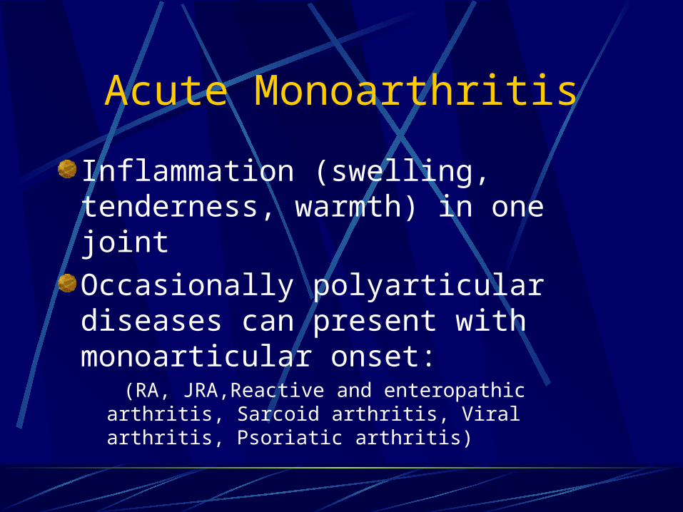
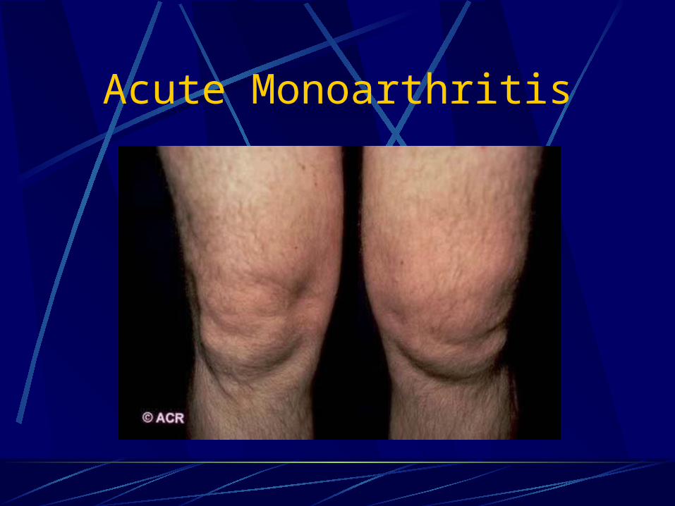
Acute Monoarthritis
Inflammation (swelling, tenderness, warmth) in one joint
Occasionally polyarticular diseases can present with monoarticular onset: (RA, JRA,Reactive and enteropathic arthritis,
Sarcoid arthritis, Viral arthritis, Psoriatic arthritis)

Acute Monoarthritis - Etiology
THE MOST CRITICAL DIAGNOSIS TO CONSIDER: INFECTION !SepticCrystal deposition (gout, pseudogout)Traumatic (fracture, internal derangement)Other (hemarthrosis, osteonecrosis, presentation of polyarticular disorders)

Questions to Ask – History Helps in DD
Pain come suddenly, minutes? – fracture.0ver several hours or 1-2 days? –infectious, crystals, inflammatory arthropathy.History of IV drug abuse or a recent infection? – septic joint.Previous similar attacks? – crystals or inflammatory arthritis.Prolonged courses of steroids? – infection or osteonecrosis of the bone.
Acute Monoarthritis
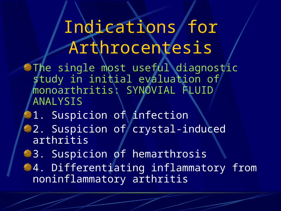
Indications for Arthrocentesis
The single most useful diagnostic study in initial evaluation of monoarthritis: SYNOVIAL FLUID ANALYSIS1. Suspicion of infection2. Suspicion of crystal-induced arthritis3. Suspicion of hemarthrosis4. Differentiating inflammatory from noninflammatory arthritis

Tests to Perform on Synovial Fluid
Low threshold for doing Gram stain and cultures .
Total leukocyte count/differential: inflammatory vs. non-inflammatory.
Polarized microscopy to look for crystals.
Not necessary routinely: Chemistry (glucose, total protein, LDH) unlikely to yield helpful information beyond the previous tests.
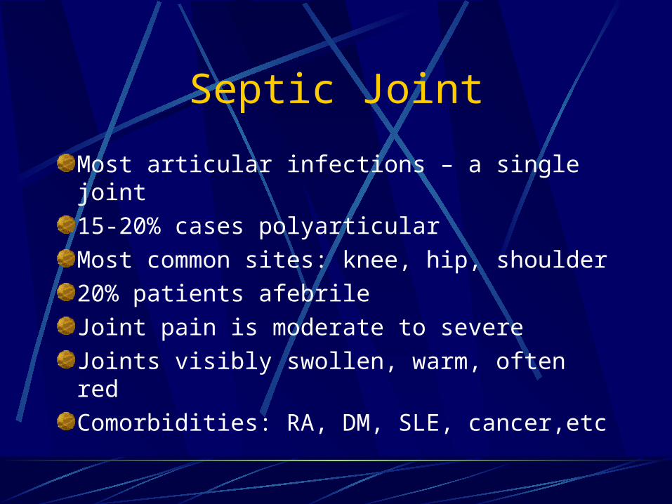
Septic Joint
Most articular infections – a single joint
15-20% cases polyarticular
Most common sites: knee, hip, shoulder
20% patients afebrile
Joint pain is moderate to severe
Joints visibly swollen, warm, often red
Comorbidities: RA, DM, SLE, cancer,etc

Septic Joint - Nongonococcal
80-90% monoarticular
Most develop from hematogenous spread
Most common:Gram positive aerobes (80%)Majority with Staph aureus (60%)Gram negative 18%

Septic Joint - Gonococcal
Most common cause of septic arthritis
Often preceded by disseminated gonococcemia
Sexually active individual, 5-7 days h/o fever, chills, skin lesions, migratory arthralgias and tenosynovitis persistent monoarthritis
Women often menstruating or pregnant
Genitourinary disease often asymptomatic

Disseminated Gonococcemia – Pustules
Gout
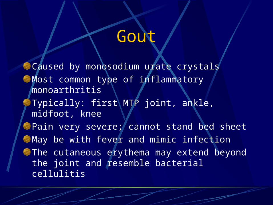
Caused by monosodium urate crystals
Most common type of inflammatory monoarthritis
Typically: first MTP joint, ankle, midfoot, knee
Pain very severe; cannot stand bed sheet
May be with fever and mimic infection
The cutaneous erythema may extend beyond the joint and resemble bacterial cellulitis
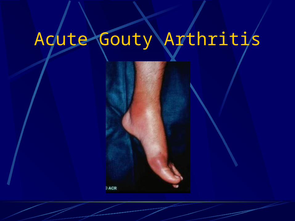
Acute Gouty Arthritis

Risk Factors
Primary gout: Obesity, hyperlipidemia, diabetes mellitus, hypertension, and atherosclerosis.
Secondary gout: alcoholism, drug therapy (diuretics, cytotoxics), myeloproliferative disorders, chronic renal failure.

Urate Crystals
Needle-shaped
Strongly negative birefringent

CPPD Crystals Deposition Disease
Can cause monoarthritis clinically indistinguishable from gout – Pseudogout.
Often precipitated by illness or surgery.
Pseudogout is most common in the knee (50%) and wrist.
Reported in any joint (Including MTP).
CPPD disease may be asymptomatic (deposition of CPP in cartilage).

Associated Conditions
Hyperparathyroidism
Hypercalcemia
Hypocalciuria
Hemochromatosis
Hypothyroidism
Gout
Aging

CPPD Crystals
Rod or rhomboid-shaped
Weakly positive birefringent
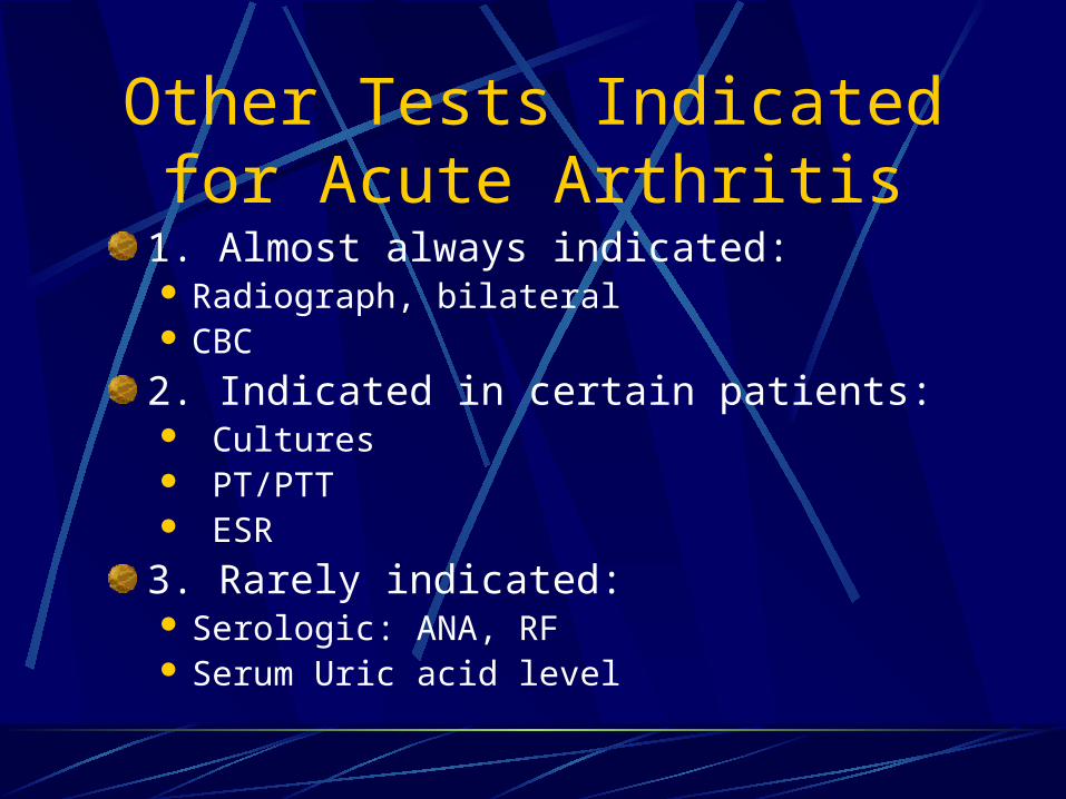
Other Tests Indicated for Acute Arthritis
1. Almost always indicated: Radiograph, bilateral CBC
2. Indicated in certain patients: Cultures PT/PTT ESR
3. Rarely indicated: Serologic: ANA, RF Serum Uric acid level

Polyarthritis
Definite inflammation (swelling, tenderness, warmth of > 5 jointsA patient with 2-4 joints is said to have pauci- or oligoarticular arthritis

Acute Polyarthritis
InfectionGonococcalMeningococcalLyme diseaseRheumatic feverBacterial endocarditisViral (rubella, parvovirus, Hep. B)
Inflammatory
RA
JRA
SLE
Reactive arthritis
Psoriatic arthritis
Polyarticular gout
Sarcoid arthritis

Inflammatory Vs. Noninflammatory
Feature Inflammatory Mechanical
Morning stiffness
Fatigue
Activity
Rest
Systemic
Corticosteroid
>1 h
Profound
Improves
Worsens
Yes
Yes
< 30 min
Minimal
Worsens
Improves
No
No
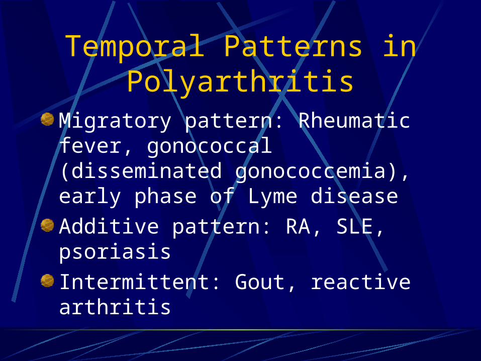
Temporal Patterns in Polyarthritis
Migratory pattern: Rheumatic fever, gonococcal (disseminated gonococcemia), early phase of Lyme disease
Additive pattern: RA, SLE, psoriasis
Intermittent: Gout, reactive arthritis

Patterns of Joint Involvement
Symmetric polyarthritis involving small and large joints: viral, RA, SLE, one type of psoriatic (the RA-like).
Asymmetric, oligo- and polyarthritis involving mainly large joints, preferably lower extremities, especially knee and ankle : reactive arthritis, one type of psoriatic, enteropathic arthritis.
DIP joints: Psoriatic.

Viral Arthritis
Younger patientsUsually presents with prodrome, rashHistory of sick contactPolyarthritis similar to acute RAPrognosis good; self-limitedExamples: Parvovirus B-19, Rubella, Hepatitis B and C, Acute HIV infection, Epstein-Barr virus, mumps
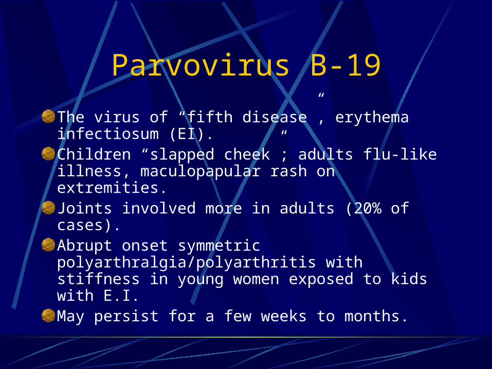
Parvovirus B-19
The virus of “fifth disease”, erythema infectiosum (EI).Children “slapped cheek”; adults flu-like illness, maculopapular rash on extremities.Joints involved more in adults (20% of cases).Abrupt onset symmetric polyarthralgia/polyarthritis with stiffness in young women exposed to kids with E.I.May persist for a few weeks to months.

Viral Arthritides - Parvovirus

Rubella Arthritis
German measles.
Young women exposed to school-aged children.
Arthritis in 1/3 of natural infections; also following vaccination.
Morbilliform rash, constitutional symptoms.
Symmetric inflammatory arthritis (small and large joints).

Rheumatoid ArthritisSymmetric, inflammatory polyarthritis, involving large and small jointsAcute, severe onset 10-15 %; subacute 20%Hand characteristically involvedAcute hand deformity: fusiform swelling of fingers due to synovitis of PIPsRF may be negative at onset and may remain negative in 15-20%! RA is a clinical diagnosis, no laboratory test is diagnostic, just supportive!
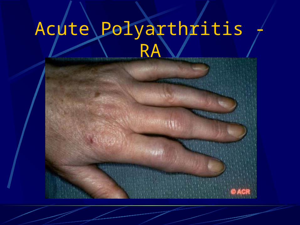
Acute Polyarthritis - RA
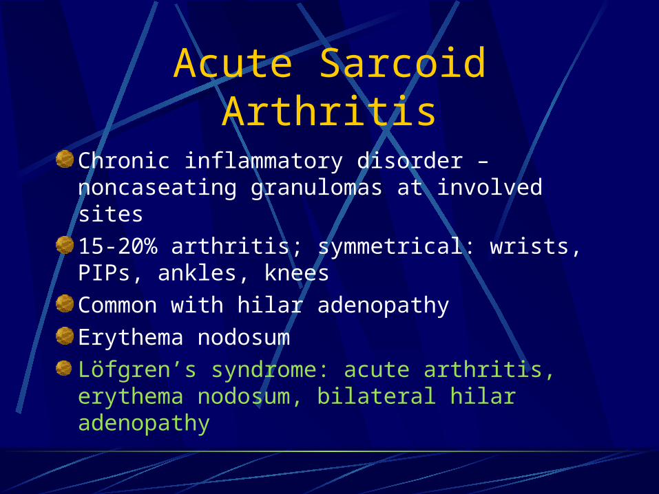
Acute Sarcoid Arthritis
Chronic inflammatory disorder – noncaseating granulomas at involved sites
15-20% arthritis; symmetrical: wrists, PIPs, ankles, knees
Common with hilar adenopathy
Erythema nodosum
Löfgren’s syndrome: acute arthritis, erythema nodosum, bilateral hilar adenopathy

Acute Polyarthritis in Sarcoidosis

Reactive Arthritis
Infection-induced systemic disease with inflammatory synovitis from which viable organisms cannot be cultured Association with HLA B 27 Asymmetric, oligoarticular, knees, ankles, feet40% have axial disease (spondylarthropathy)Enthesitis: inflammation of tendon-bone junction (Achilles tendon, dactylitis)Extraarticular: rashes, nails, eye involvement

Asymmetric, Inflammatory Oligoarthritis

Enthesitis in Reactive Arthritis

Keratoderma Blenorrhagica – Reactive
Arthritis

Reactive Arthritis - Conjunctivitis

Reactive Arthritis – Palate Erosions

Psoriatic Arthritis
Prevalence of arthritis in Psoriasis 5-7%Dactilytis (“sausage fingers”), nail changesSubtypes: Asymmetric, oligoarticular- associated dactylitis Predominant DIP involvement – nail changes Polyarthritis “RA-like” – lacks RF or nodules Arthritis mutilans – destructive erosive hands/feet Axial involvement –spondylitis – 50% HLAB27 (+) HIV-associated – more severe

Acute Polyarthritis - Psoriatic

Dactylitis “Sausage Toes” – Psoriasis

Psoriasis
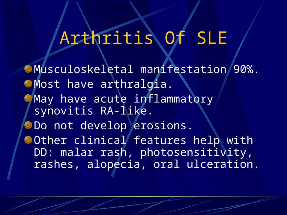
Arthritis Of SLE
Musculoskeletal manifestation 90%.Most have arthralgia.May have acute inflammatory synovitis RA-like.Do not develop erosions.Other clinical features help with DD: malar rash, photosensitivity, rashes, alopecia, oral ulceration.

Butterfly Rash – SLE
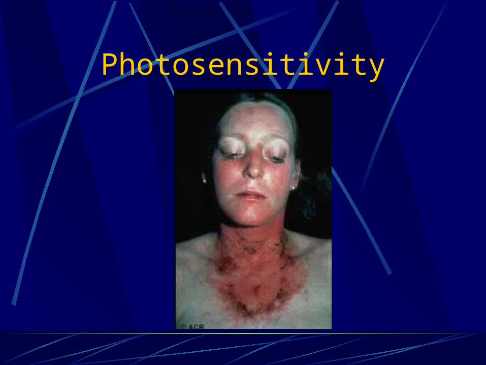
Photosensitivity
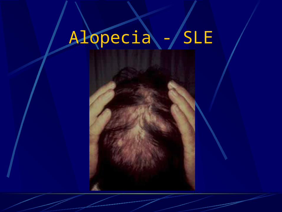
Alopecia - SLE
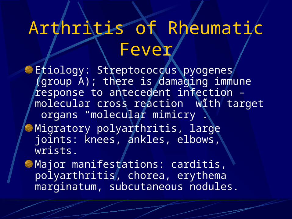
Arthritis of Rheumatic Fever
Etiology: Streptococcus pyogenes (group A); there is damaging immune response to antecedent infection – molecular cross reaction with target organs “molecular mimicry”.Migratory polyarthritis, large joints: knees, ankles, elbows, wrists.Major manifestations: carditis, polyarthritis, chorea, erythema marginatum, subcutaneous nodules.

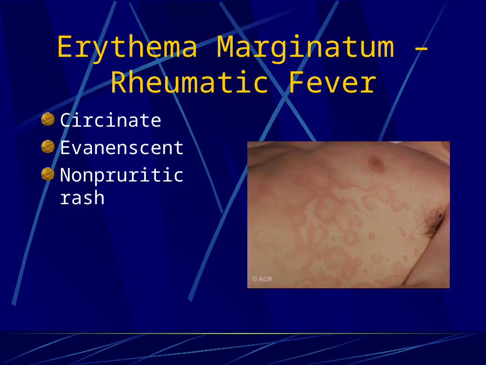
Erythema Marginatum – Rheumatic Fever
Circinate
Evanenscent
Nonpruritic rash
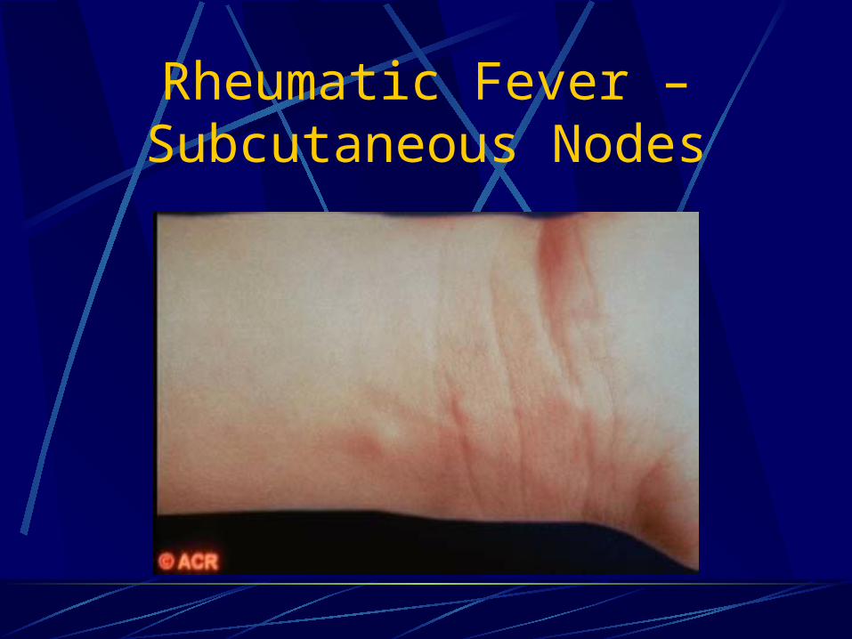
Rheumatic Fever – Subcutaneous Nodes

Gouty Arthritis
Skin Lesions Useful in Diagnosis
Psoriatic plaquesKeratoderma Blenorrhagicum (reactive arthritis)Butterfly rash (SLE)Salmon-colored rash of JRA, adult Still’sErythema marginatum (Rheumatic Fever)Vesicopustular lesions (gonococcal arthritis)Erythema nodosum (acute sarcoid, enteropathic arthritis)

Disseminated Gonococcemia – Pustules

Keratoderma Blenorrhagica – Reactive
Arthritis
Erythema Marginatum – Rheumatic Fever
Circinate
Evanenscent
Nonpruritic rash

Adult Still’s Disease and JRA Rash
Salmon or pale-pink BlanchingMacules or maculopapulesTransient (minutes or hours)Most common on trunkFever related

SLE – Face Rash

SLE – Interarticular Rash Hands

Keratoderma Blenorrhagicum

Erythema Nodosum
Sarcoidosis
Inflammatory Bowel Disease – related arthritis
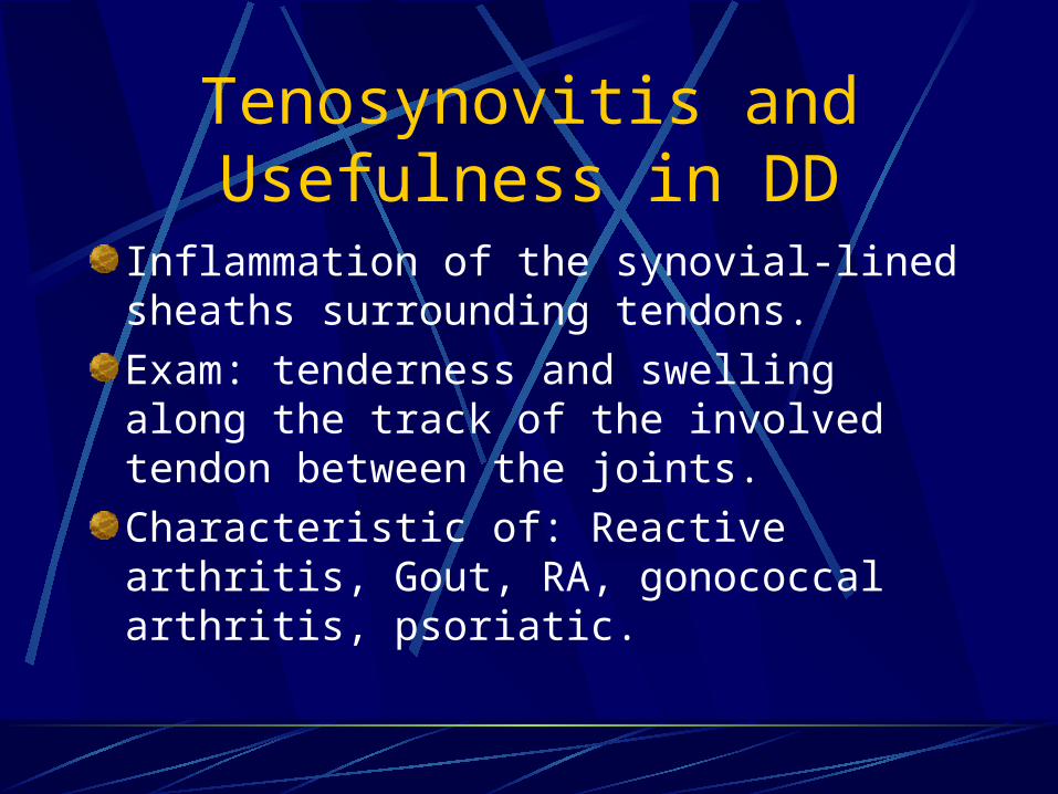
Tenosynovitis and Usefulness in DD
Inflammation of the synovial-lined sheaths surrounding tendons.
Exam: tenderness and swelling along the track of the involved tendon between the joints.
Characteristic of: Reactive arthritis, Gout, RA, gonococcal arthritis, psoriatic.
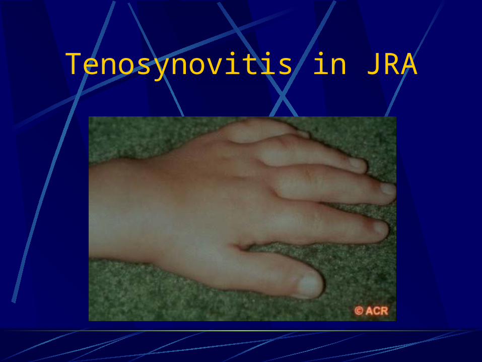
Tenosynovitis in JRA

Dactylitis “Sausage Toes” – Psoriasis, Reactive,
Enteropathic
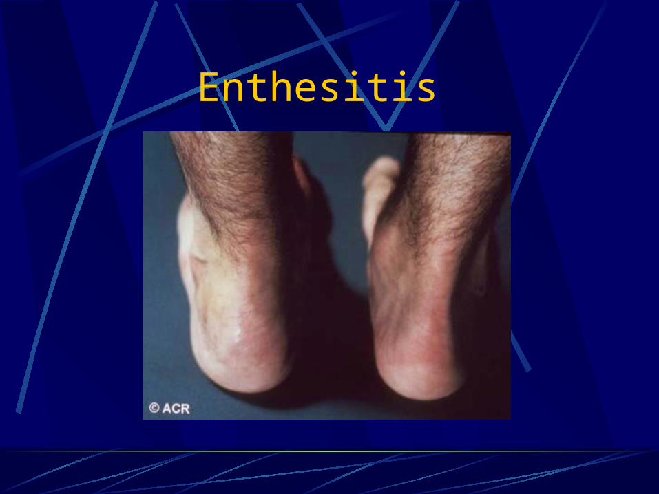
Enthesitis

Extraarticular Features Helpful in DD
Eye involvement: conjunctivitis in reactive arthritis, uveitis in enteropathic and sarcoidosis, episcleritis in RA
Oral ulcerations: painful in reactive arthritis and enteropathic, not painful in SLE
Nail lesions: pitting (psoriasis), onycholysis (reactive arthritis)
Alopecia (SLE)

Reactive Arthritis - Conjunctivitis
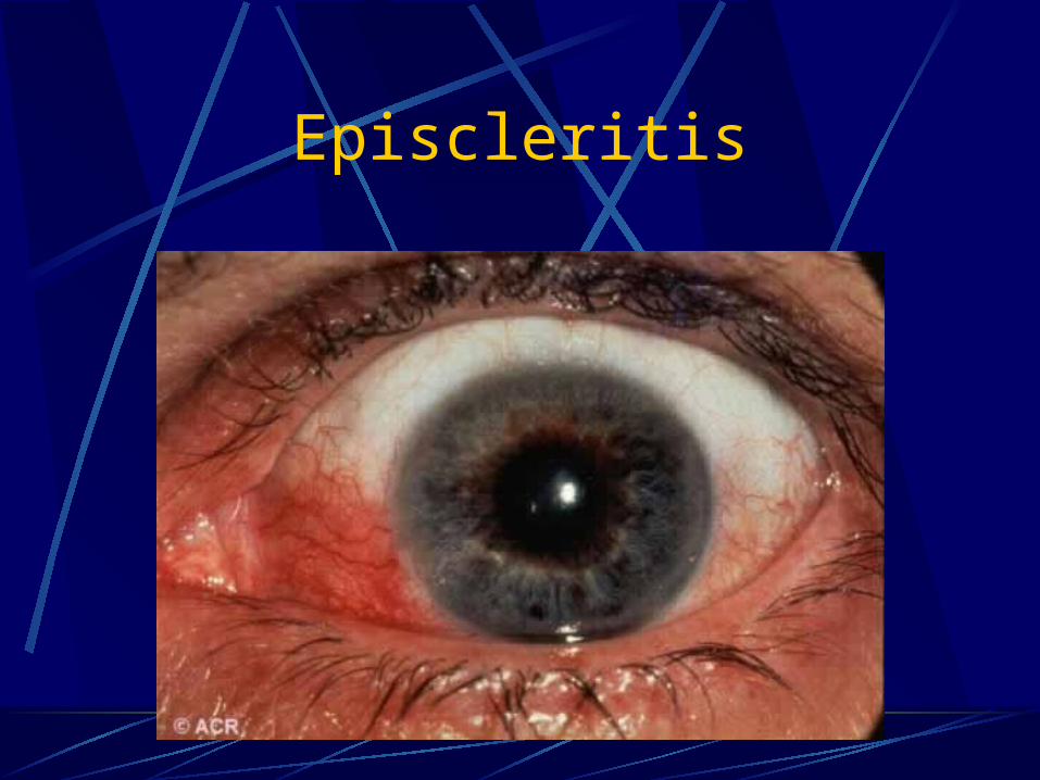
Episcleritis
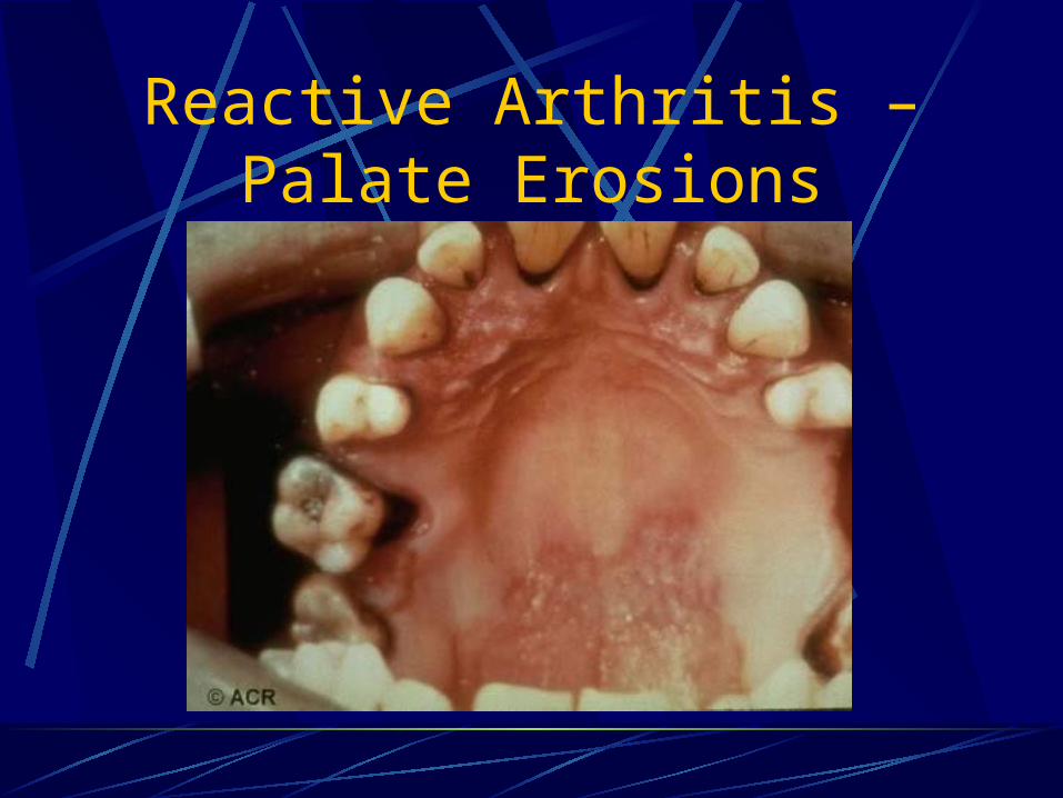
Reactive Arthritis – Palate Erosions

Alopecia - SLE
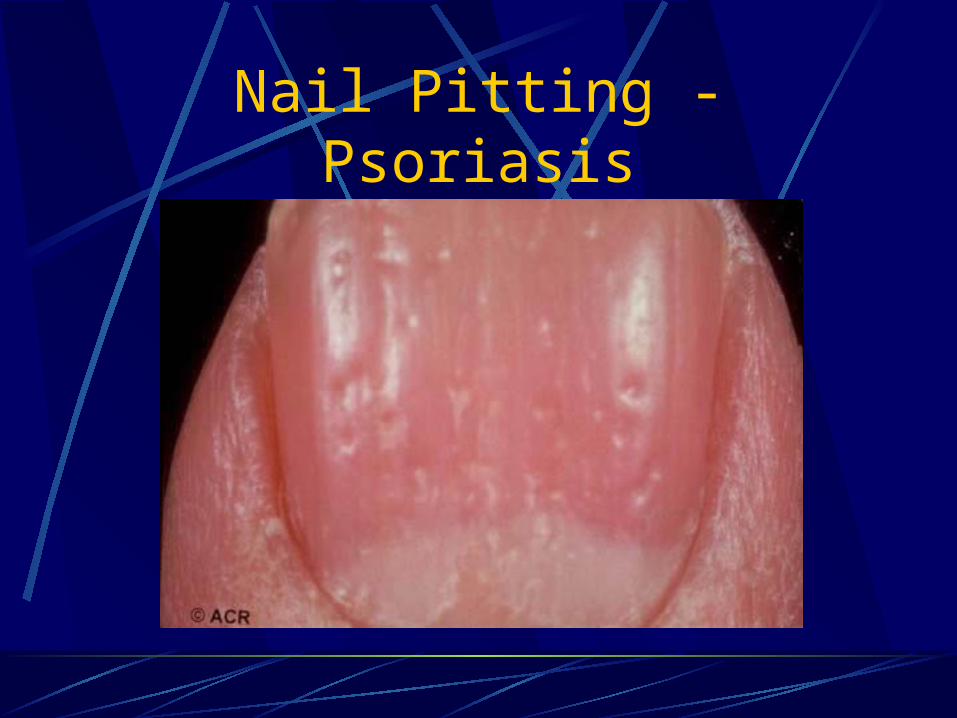
Nail Pitting - Psoriasis
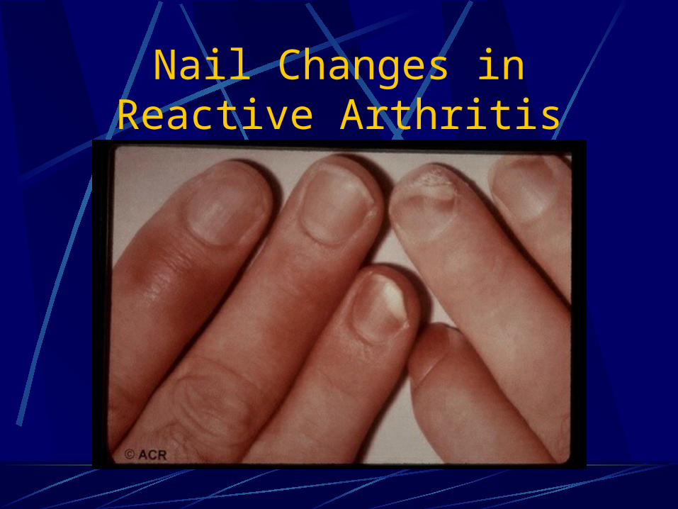
Nail Changes in Reactive Arthritis