Clinical applications of circulating tumor cells in pharmacotherapy...
40
MOL#108142 1 Clinical applications of circulating tumor cells in pharmacotherapy: challenges and perspectives Tong Wu, Bin Cheng and Liwu Fu State Key Laboratory of Oncology in South China, Collaborative Innovation Center for Cancer Medicine, Guangdong Esophageal Cancer Institute, Sun Yat-Sen University Cancer Center, Guangzhou, China (T.W., L.F.); Department of Oral Medicine, Guanghua School of Stomatology, Guangdong Provincial Key Laboratory of Stomatology, Sun Yat-sen University, Guangzhou, China (T.W., B.C.) This article has not been copyedited and formatted. The final version may differ from this version. Molecular Pharmacology Fast Forward. Published on March 29, 2017 as DOI: 10.1124/mol.116.108142 at ASPET Journals on February 17, 2019 molpharm.aspetjournals.org Downloaded from
Transcript of Clinical applications of circulating tumor cells in pharmacotherapy...
MOL#108142
1
Clinical applications of circulating tumor cells in
pharmacotherapy: challenges and perspectives
Tong Wu, Bin Cheng and Liwu Fu
State Key Laboratory of Oncology in South China, Collaborative Innovation Center for
Cancer Medicine, Guangdong Esophageal Cancer Institute, Sun Yat-Sen University
Cancer Center, Guangzhou, China (T.W., L.F.); Department of Oral Medicine,
Guanghua School of Stomatology, Guangdong Provincial Key Laboratory of
Stomatology, Sun Yat-sen University, Guangzhou, China (T.W., B.C.)
This article has not been copyedited and formatted. The final version may differ from this version.Molecular Pharmacology Fast Forward. Published on March 29, 2017 as DOI: 10.1124/mol.116.108142
at ASPE
T Journals on February 17, 2019
molpharm
.aspetjournals.orgD
ownloaded from
MOL#108142
2
Running title: Clinical applications of CTCs in pharmacotherapy
Correspondence authority:
Liwu Fu, Professor and Director, Cancer Institute, Cancer Center, Sun Yat-sen
University, Guangzhou, 510060, China. Email: [email protected]. Tel: +86-(20)-
8734-3163; Fax: +86-(20)-8734-3170
Number of text pages: 41
Number of tables: 1
Number of figures: 3
Number of references: 113
Number of words in Abstract: 117
Number of words in introduction: 460
Number of words in main text: 2450
Abbreviations: ALK, anaplastic lymphoma kinase; AR-V7, androgen receptor splice
variant 7; CK, cytokeratins; CRPC, castration-resistant prostate cancer; CTCs,
circulating tumor cells; CTM, circulating tumor microemboli; DFS, disease-free
survival; EGFR, epidermal growth factor receptor; EMT, epithelial to mesenchymal
transition; ER, estrogen receptor; ERCC1, excision repair cross-complementation
group 1; ERK, extracellular signal-regulated kinases; HER2, human epidermal growth
factor receptor 2; MET, mesenchymal to epithelial transition; MAPK, mitogen-
activated protein kinases; PI3K, phosphatidylinositide 3-kinases; mTOR, mammalian
target of rapamycin; PD-L1, programmed death-ligand 1
This article has not been copyedited and formatted. The final version may differ from this version.Molecular Pharmacology Fast Forward. Published on March 29, 2017 as DOI: 10.1124/mol.116.108142
at ASPE
T Journals on February 17, 2019
molpharm
.aspetjournals.orgD
ownloaded from
MOL#108142
3
Abstract
Circulating tumor cells (CTCs) have been identified as one approach to ultrasensitive
liquid biopsy for real-time monitoring of cancer patients. The detection of CTCs in
peripheral blood from cancer patients is promising, but its application still faces serious
challenges with respect to specificity and sensitivity. Here, we review the significant
roles of CTCs in metastasis, and the strengths and weaknesses of currently available
methods for CTC detection and characterization. Moreover, we discuss the clinical
application of CTCs as markers for patient prognosis, and we specifically focus on the
application of CTCs as indicators in cancer pharmacotherapy. Characterization of the
detected CTCs will provide new biological perspectives and clinical applications for
the treatment of cancer patients with metastasis.
This article has not been copyedited and formatted. The final version may differ from this version.Molecular Pharmacology Fast Forward. Published on March 29, 2017 as DOI: 10.1124/mol.116.108142
at ASPE
T Journals on February 17, 2019
molpharm
.aspetjournals.orgD
ownloaded from
MOL#108142
4
1. Introduction
A fraction of the cancer cells that disseminate from primary tumors and migrate to
distant sites will result in the formation of a lethal metastatic tumor (Hanahan and
Weinberg, 2011). Migrating tumor cells found in the peripheral bloodstream are
called circulating tumor cells (CTCs), while theor counterparts found in bone marrow
are called disseminated tumor cells (DTCs) (Alix-Panabieres and Pantel, 2016).
CTCs have been regarded as a critical stage in the development of metastasis, in that
they contain genetic and molecular information about the cancer as well as its
evolutionary adaptation to prior therapies (Alix-Panabieres and Pantel, 2016).
However, CTCs have also been detected in the blood circulation of healthy volunteers,
and in patients with benign diseases of the lung, colon, pancreas, and breast (Alva et
al., 2015; Cauley et al., 2015; Franken et al., 2012; Ilie et al., 2014; Liu et al., 2015;
Markou et al., 2014; Pantel et al., 2012).
CTCs have paved new diagnostic avenues in liquid biopsy diagnostics, especially for
tumors that are not easy to biopsy and for metastatic lesions (Azarin et al., 2015).
Early detection of cancer metastasis is always difficult, not to mention its prevention or
cure. With its non-invasive nature and real-time advantage, cancer screening for
CTCs can be applied to populations at higher risk. Therefore, oncologists place high
hopes on CTC-based screening methods which have been found to be more sensitive
than current imaging methods such as PET scan (Hegemann et al., 2016). The
consistency between CTCs and their primary tumors is encouraging, and may provide
This article has not been copyedited and formatted. The final version may differ from this version.Molecular Pharmacology Fast Forward. Published on March 29, 2017 as DOI: 10.1124/mol.116.108142
at ASPE
T Journals on February 17, 2019
molpharm
.aspetjournals.orgD
ownloaded from
MOL#108142
5
an excellent opportunity for clinicians to examine mutations of key genes that are not
detected through traditional blood-based assays (Nagrath et al., 2016). In the era of
precision medicine in cancer therapy, systemic monitoring of response to anti-cancer
therapies is a key step toward providing personalized care. An increasing body of
evidence indicates that, in the course of the treatment, assessment of the molecular
characteristics of the progressive disease is more significant than depending on the
primary tumor samples, which do not reflect the evolution of the tumor (Alix-
Panabieres and Pantel, 2016). For their comprehensive information regarding the
whole disease, studies of CTCs not only reveal the underlying mechanism of
tumorigenesis and metastasis, but also provide a non-invasive method for cancer
diagnosis, prognosis, and pharmacotherapy monitoring (Masuda et al., 2016).
Recent research has demonstrated that CTCs, an integral part of the “liquid biopsy”,
have great potential to change the status quo of anticancer therapy; however the
approach remains technically challenging. Following a short discussion of the
significant roles of CTCs in cancer metastasis and currently available methods for the
CTCs detection and molecular characterization,this review will focus on the clinical
applications of CTCs as markers for prognosis prediction in cancer patients and as
indicators in cancer pharmacotherapy (Figure 1).
2. CTCs and cancer metastasis
The malignant form of cancer and the cause for more than 90% of cancer-associated
This article has not been copyedited and formatted. The final version may differ from this version.Molecular Pharmacology Fast Forward. Published on March 29, 2017 as DOI: 10.1124/mol.116.108142
at ASPE
T Journals on February 17, 2019
molpharm
.aspetjournals.orgD
ownloaded from
MOL#108142
6
mortality is metastasis, which os characterized by the ability of cancer cells to invade
into the surrounding tissue and disseminate throughout the body to establish secondary
tumors in distant organs (Gupta and Massague, 2006). As shown in Figure 2, the
sequential metastasis processes first initiates with a loss of adhesion of tumor cells in
the primary site and their migration out of the primary tumor. Secondly, the tumor
cells attach to blood vessels and invade into the blood or lymphatic circulation in a
process called intravasation (Fidler, 2003). As the tumor cells circulate to the
secondary site, the tumor cells intrude blood vessels to adhere to the target organ
endothelium and migrate into the parenchyma; this is called extravasation (Chaffer and
Weinberg, 2011; Klein, 2009; Sosa et al., 2014). Therefore, the existence of CTCs
has been recognized as an important “intermediate step” in cancer metastasis. CTCs
represent a stem-like sub-population of cells that are capable of immigration and tumor
initiation (Al-Hajj et al., 2003; Ricci-Vitiani et al., 2007; Stewart et al., 2011). During
the metastasis process, many cell surface markers of CTCs undergo change. The
activation of epithelial to mesenchymal transition (EMT) facilitates tumor cell invasion
and dissemination during intravasation, while its reverse process, a mesenchymal to
epithelial transition (MET), is believed to support extravasation once cancer cells have
arrived in distant organs (Acloque et al., 2009; Nieto, 2013; Thiery et al., 2009). It
has been speculated that the entire process in which CTCs seed metastasis occurs with
extremely low efficiency; only 0.01% of all CTCs can survive and form
micrometastases in distant organs (Luzzi et al., 1998). Even if CTCs succeed in
intravasation, most of them cannot survive the adverse environment in the bloodstream
This article has not been copyedited and formatted. The final version may differ from this version.Molecular Pharmacology Fast Forward. Published on March 29, 2017 as DOI: 10.1124/mol.116.108142
at ASPE
T Journals on February 17, 2019
molpharm
.aspetjournals.orgD
ownloaded from
MOL#108142
7
and eventually die from anoikis. Therefore, CTCs sometimes aggregate to form
microemboli (circulating tumor microemboli, CTM), which may endow tumor cells
advantages in survival and enhabces CTCs viability and motility (Krebs et al., 2014).
For several decades, the lack of relevant models for metastasis research extensively
limited further investigation. CTCs are the true link between primary and metastatic
tumors, and thus create a new opportunity for investigators to explore valuable features
of both primary and metastatic sites, as well as specific details of the processes of
intravasation, migration, and extravasation.
3. CTC detection
As mentioned above, after release from the main and/or metastatic tumor site into the
blood circulation, the conditions in the blood are harsh for epithelial tumor cells, and
the survival time of CTCs is extremely short (half-life: 1–2.4 hours) (Meng et al., 2004).
Apoptotic and fragmented CTCs are frequently detected in the peripheral bloodstream
of cancer patients (Larson et al., 2004). Therefore, a pivotal challenge for the clinical
application of CTCs is the capability of the current CTC technology to efficiently
capture the extremely rare CTC population from patient blood samples for subsequent
processing (Nelson, 2010).
In the past decade, one of the most widely used strategies to detect CTCs has been the
use of epithelial markers such as cytokeratins (CK) and EpCAM, which are not detected
This article has not been copyedited and formatted. The final version may differ from this version.Molecular Pharmacology Fast Forward. Published on March 29, 2017 as DOI: 10.1124/mol.116.108142
at ASPE
T Journals on February 17, 2019
molpharm
.aspetjournals.orgD
ownloaded from
MOL#108142
8
on the surrounding mesenchymal blood cells. CellSearch, the only U.S. Food and
Drug Administration (FDA) approved CTC technology to monitor metastatic breast
cancer patients, is a case in point (Ferreira et al., 2016). The CellSearch system uses
ferrofluid functionalized nanoparticles containing EpCAM antibody for magnetic
separation of EpCAM positive cells after blood centrifugation for the enrichment.
The cells are then selected by immunostaining for the expression of CKs 8, 18 and 19,
and as well as for negative staining for CD45 (Riethdorf et al., 2007).
However, further research has demonstrated that epithelial tumor cells are likely to
undergo EMT, which results in decreased expression of epithelial markers and
increased plasticity, migration, and invasiveness (Mani et al., 2008). These partial
EMT tumor cells, also called the “intermediate phenotype,” have the highest versatility
to adapt to the microenvironment in secondary sites (Tam and Weinberg, 2013).
Therefore, in recent years a variety of devices have been developed for the enrichment
and detection of CTCs undergoing EMT, in addition to the approaches selecting for
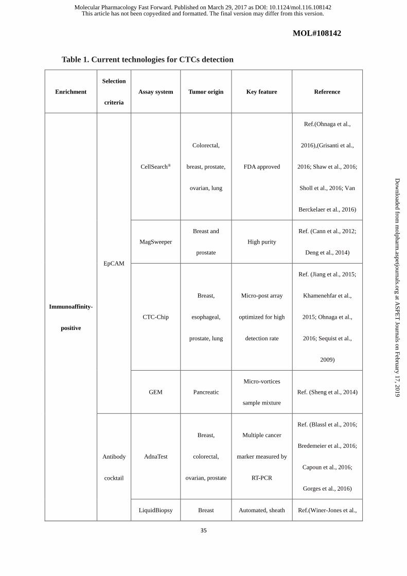
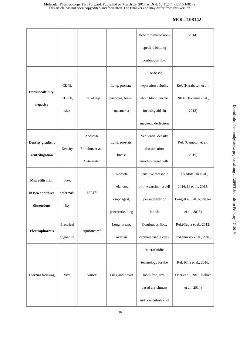
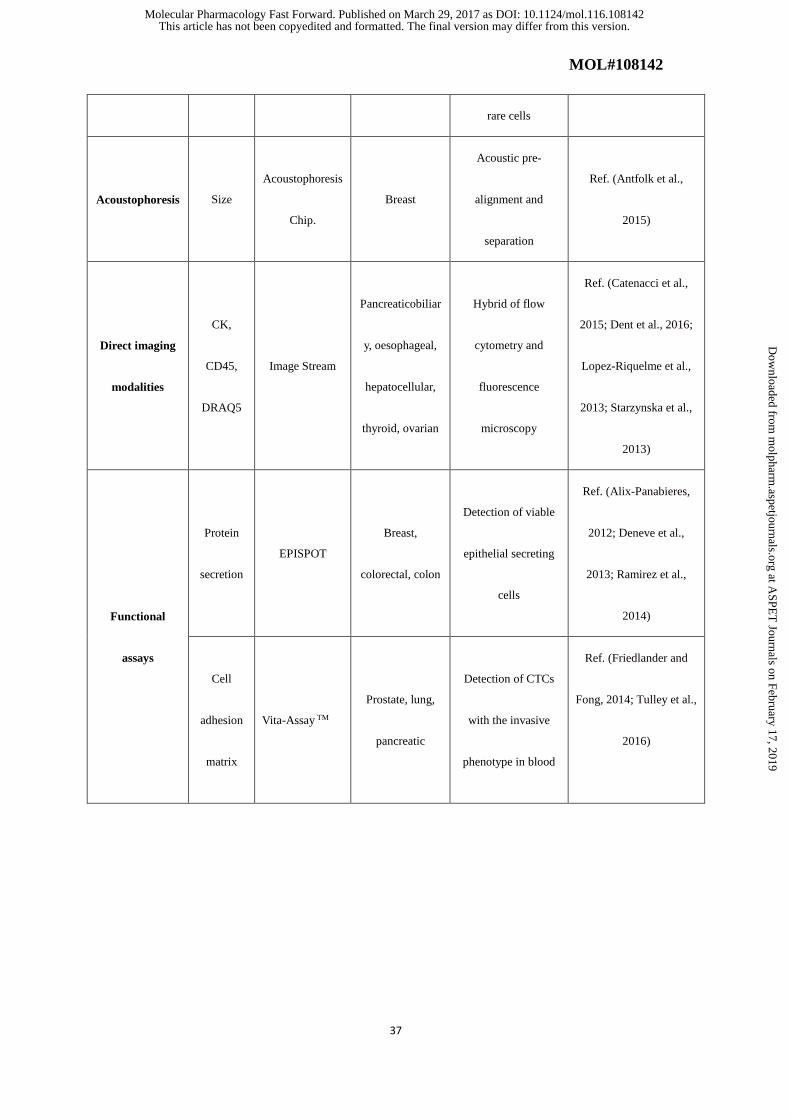
epithelial markers (Alix-Panabieres and Pantel, 2014). CTC assays usually involves
two steps: first, an enrichment step increases the percentage of CTCs, making it easier
ti detect single tumor cells. Specifically, CTCs can be enriched by their biologic
characteristics (e.g., protein markers) or on the basis of their physical properties (e.g.,
size, density, deformability, or electric charges). Second, in the detection step, CTCs
can be selected using different criteria such as immunologic, molecular, or functional
assays (Table 1) (Ferreira et al., 2016). Nowadays, although CTC technologies have
This article has not been copyedited and formatted. The final version may differ from this version.Molecular Pharmacology Fast Forward. Published on March 29, 2017 as DOI: 10.1124/mol.116.108142
at ASPE
T Journals on February 17, 2019
molpharm
.aspetjournals.orgD
ownloaded from
MOL#108142
9
developed rapidly, sensitivity and specificity are still problems that hinder the clinical
utilization of CTCs for guiding personalized treatment of cancer patients (Hardingham
et al., 2015).
4. CTCs as prognostic markers in cancer
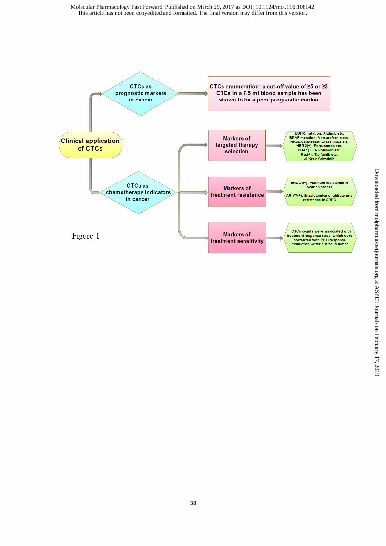
To date, CTCs enumeration has been widely used as a prognostic index for patient
overall survival rate. A cut-off value of ≥5 or ≥3 CTCs in 7.5 ml blood has been
proved to be a poor prognostic index in several cancers, including breast cancer (Zhang
et al., 2012), colorectal cancer (Cohen et al., 2008), prostate cancer (de Bono et al.,
2008) , lung cancer (Krebs et al., 2011) , bladder cancer (Gazzaniga et al., 2014) , liver
cancer (Schulze et al., 2013), esophageal cancer (Vashist et al., 2012), melanoma (Rao
et al., 2011) , head and neck carcinoma (Nichols et al., 2012), and pancreatic cancer
(Han et al., 2014).
The association between detection of CTCs and clinical outcome has been most widely
studied in breast cancer. For example, CTCs were analyzed in a pool of 2026 patients
with early stage breast cancer before pharmacotherapy and 1492 patients after
pharmacotherapy using the CellSearch System (Rack et al., 2014). In the pre-
pharmacotherapy group, CTCs were found in 21.5% of patients, in which 19.6% were
lymph node-negative and 22.4% were node-positive. No correlation was found between
CTCs and tumor size, grading, or hormone receptor status. CTCs were detected in 22.1%
of 1492 patients after chemotherapy. The presence of CTCs was associated with poor
This article has not been copyedited and formatted. The final version may differ from this version.Molecular Pharmacology Fast Forward. Published on March 29, 2017 as DOI: 10.1124/mol.116.108142
at ASPE
T Journals on February 17, 2019
molpharm
.aspetjournals.orgD
ownloaded from

MOL#108142
10
rates for disease-free survival (DFS), distant DFS, breast cancer-specific survival, and
overall survival. CTCs were identified as an independent prognostic index for DFS
in multivariable analysis. Patients with more than five CTCs per 30 mL blood had the
worst prognosis. These results from a large-scale trial of patients with breast cancer
suggested that CTCs have prognostic value (Rack et al., 2014).
Of note, in these reports indicating that CTCs can be used as a reliable early index of
disease progression and survival as compared to traditional methods, a significant
proportion of patients with obvious distant metastases were negative for CTCs. This
result implied that CTCs undergoing EMT transformation can be missed by epithelial
marker based detection methods, such as the CellSearch system. Therefore, large-scale
multicenter trials with improved CTC detection techniques and well-defined endpoints
are needed to support the clinical utility of CTC detection in cancer patients.
5. CTC as indicators in pharmacotherapy
CTCs may be disseminated from the primary tumor or from a number of metastatic
sites. Therefore, CTCs offer a wealth of genetic and molecular information cincerning
the cancer at the protein, RNA, and genome levels (Meng et al., 2004). Bin addition
to CTC detection, significant effort has been made towards CTC characterization. In
the era of precision medicine of cancer therapy, identification of CTCs expressing
certain markers can be used to specifically monitor cancer therapy.
This article has not been copyedited and formatted. The final version may differ from this version.Molecular Pharmacology Fast Forward. Published on March 29, 2017 as DOI: 10.1124/mol.116.108142
at ASPE
T Journals on February 17, 2019
molpharm
.aspetjournals.orgD
ownloaded from
MOL#108142
11
5.1.CTCs as markers for targeted therapy selection
Molecular alterations in CTCs have proved to be highly consisted with the primary
tumor, which provides robust evidence for the clinical application of targeted therapy
in cancer. Several studies have suggested CTCs as an index of therapy selection and,
furthermore, as a real-time biopsy to reflect the effect of a particular therapy.
For example, BRAF mutation between primary tumors and metastases have been
described within a patient; these mutations mediate tumor proliferation through
activation of the RAF–MEK–ERK pathway (Lin et al., 2011). Therefore, the BRAF
mutation status in CTCs collected from patients with metastatic melanoma is a pivotal
index for selecting targeted therapies such as Vemurafenib and Dabrafenib (Jang and
Atkins, 2014; Reid et al., 2015). Another case in point are EGFR mutations in lung
cancer. A group of pulmonary adenocarcinoma that have activating EGFR mutations
are exclusively sensitive to EGFR tyrosine kinase inhibitors (Mok et al., 2009).
Therefore, EGFR mutations in CTCs are clinical biomarkers for categorization of
pharmacotherapies target in metastatic lung cancer with respect to treatment with
Erlotinib, Afatinib, and Osimertinib (Breitenbuecher et al., 2014; Kuwano et al., 2016).
In addition to melanoma and lung cancer, therapeutic targets were identified in breast
cancer. The PI3K/AKT/mTOR pathway is frequently altered in cancer. PI3K is a cell
membrane signal transduction molecule that supports cell survival and growth, making
it a popular therapeutic target (Akinleye et al., 2013; Wong et al., 2010). PIK3CA
mutations were identified in CTCs from metastasis breast cancer patients by CellSearch
This article has not been copyedited and formatted. The final version may differ from this version.Molecular Pharmacology Fast Forward. Published on March 29, 2017 as DOI: 10.1124/mol.116.108142
at ASPE
T Journals on February 17, 2019
molpharm
.aspetjournals.orgD
ownloaded from

MOL#108142
12
enrichment, DNA extraction, and whole genome amplification (Schneck et al., 2013).
Agents targeting this pathway, such as Everolimus and Temsirolimus, are promising
(Johnston, 2015).
Another application of CTCs is for the detection of various biomarkers expressed in
advanced disease that reflect the progression of the cancer. Hormone receptor status
is one of the most well-established predictors for endocrine adjuvant or palliative
therapy of primary and metastatic breast cancer. However, hormone receptor status
changes during the course of disease progression. Variations in the expression of ER
and HER2 can occur in advanced breast cancer, and has been readily detected in CTCs.
Monitoring of these changes is helpful in selecting chemotherapies, especially those
targeting HER2 receptor such as Trastuzumab, Lapatinib, Pertuzumab and T-DM1
(Aktas et al., 2011; Hernández-Blanquisett et al., 2016; Thompson et al., 2010; Turner
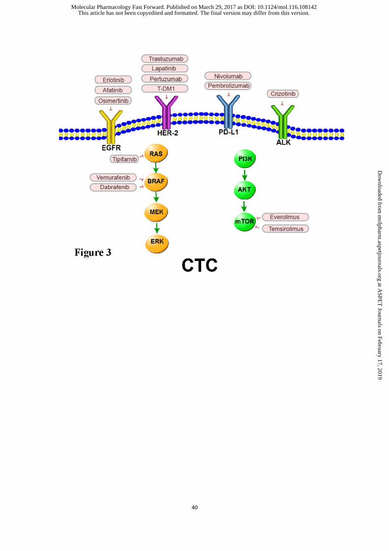
and Di Leo, 2013) . In addition, several therapeutic targets such as ALK (Ilie et al., 2012;
Pailler et al., 2013; Pailler et al., 2013), PD-L1 (Jing et al., 2016), and RAS (Karandish
and Mallik, 2016), were also detected in CTCs collected from breast, colorectal,
prostate and ovarian cancer patients (Figure 3).
5.2. CTCs as markers of treatment resistance
It has been reported that, in both early and metastatic cancers, the presence of CTCs
following treatment can act as a predictive index of the possibility of disease recurrence
(Alix-Panabieres and Pantel, 2013; Xenidis et al., 2007). These persisting CTCs are
This article has not been copyedited and formatted. The final version may differ from this version.Molecular Pharmacology Fast Forward. Published on March 29, 2017 as DOI: 10.1124/mol.116.108142
at ASPE
T Journals on February 17, 2019
molpharm
.aspetjournals.orgD
ownloaded from

MOL#108142
13
resistant to treatment and can thus be involved in cancer progression. Therefore, there
is an urgent need to identify effective therapies in patients with “therapy-resistant”
CTCs. Several studies have been reported based on this strategy. For example,
platinum resistance is one of the most recognized clinical challenges for ovarian cancer
pharmacotherapy. While detection of excision repair cross-complementation group 1
(ERCC1) protein in the primary tumor by immunohistochemistry is inaccurate for the
prediction of platinum resistance, the presence of ERCC1(+) CTCs in blood can be used
as a diagnosis biomarker in ovarian cancer to predict platinum resistance (Kuhlmann et
al., 2014). In metastatic castration-resistant prostate cancer (CRPC), the presence of
androgen receptor splice variant 7 (AR-V7) in CTCs is associated with resistance to
enzalutamide or abiraterone, but not to taxanes (Antonarakis et al., 2015; Antonarakis
et al., 2014) . In AR-V7-positive patients, Taxanes are more efficacious than
enzalutamide or abiraterone therapy in AR-V7-positive tumors, while in AR-V7-
negative, taxanes , enzalutamide, and abiraterone have quite similar efficacies
(Antonarakis et al., 2015). AR-V7 expressed in CTCs may therefore serve as a
biomarker for CRPC treatment selection (Onstenk et al., 2015). These results add to
existing evidence that CTCs are a valuable tool to optimize personalized cancer
treatments and improve the prognosis for therapy-resistant patients.
5.3.CTC as a biomarker for treatment sensitivity
Increasing evidence points out the significance of evaluating molecular features of the
advancing disease during therapy, instead of depending on the primary tumor sample
This article has not been copyedited and formatted. The final version may differ from this version.Molecular Pharmacology Fast Forward. Published on March 29, 2017 as DOI: 10.1124/mol.116.108142
at ASPE
T Journals on February 17, 2019
molpharm
.aspetjournals.orgD
ownloaded from
MOL#108142
14
that are unable to reflect the progression of the tumor and target associated features
(Alix-Panabieres and Pantel, 2016). Considering the easy availability of blood, it has
been suggested that CTCs can serve as a “real-time liquid biopsy” to provide
information of the current disease without invasive biopsy (Lianidou and Markou,
2011).
CTC enumeration is one of the most widely-used criteria to monitor systemic anticancer
therapy. The significance of CTC enumeration in monitoring anticancer therapy has
been demonstrated in metastatic breast cancer patients receiving first-line
chemotherapy. In the SWOG 0500 (NCT00382018) clinical trial, patients with
metastatic breast cancer had CTC enumeration before cycles 1 and 2 with or without
targeted therapy in combination with first-line chemotherapy. Patients with sustained
increasea in CTC number (≥5 CTCs/7.5 mL) after one cycle treatment were regarded
as at higher risk group for early cancer progression. These patients were randomly
designated into a continued first-line pharmacotherapy group or another treatment
group before radiologic evaluation of progression (Bidard et al., 2016). For patients
with continued increases in CTC numbers after first-line chemotherapy, a more
effective treatment than standard chemotherapy is needed (Smerage et al., 2014).
Several multicenter clinical trials testing anticancer therapy monitoring based on CTCs
are still in progress, including the STIC CTC METABREAST clinical trial in France
(NCT01710605). In this trial, breast cancer patients with more than 5 CTC counts in
7.5 mL blood received chemotherapy, while patients with no more than 5 CTCs in 7.5
This article has not been copyedited and formatted. The final version may differ from this version.Molecular Pharmacology Fast Forward. Published on March 29, 2017 as DOI: 10.1124/mol.116.108142
at ASPE
T Journals on February 17, 2019
molpharm
.aspetjournals.orgD
ownloaded from
MOL#108142
15
mL blood received endocrine therapy as the first-line treatment (Lianidou and Markou,
2011). In a phase II trial of Erlotinib and Pertuzumab in advanced non-small cell lung
cancer, CTC counts were associated with treatment response rates, which were
correlated with fludeoxyglucose-positron emission tomography (Punnoose et al., 2012) .
6. Future prospect and challenges
There is no doubt that innovative approached utilizing CTCs have paved new diagnostic
avenues for the next generation of liquid biopsy diagnostics, especially in tumors that
are not easy to biopsy and in metastatic lesions. Furthermore, based on their non-
invasive and real-time advantages, CTCs can be applied for cancer screening of
populations at higher risk. Identification and characterization of CTCs have been
applied in several key clinical areas, such as prognosis prediction, systemic
pharmacotherapy selection, and monitoring. However, although some promising
results have been reported, detection of CTCs still faces serious challenges including
sensitivity and specificity. In the future, more efficient capture systems and larger
panels of detection markers will be explored to avoid losing assay specificity while
increasing sensitivity. In conclusion, the detection and characterization of CTCs will
provide new biological perspectives and clinical implications for cancer patients,
especially during pharmacotherapy.
Authorship contribution
Wrote or contributed to the writing of manuscript: Wu, Cheng, and Fu.
This article has not been copyedited and formatted. The final version may differ from this version.Molecular Pharmacology Fast Forward. Published on March 29, 2017 as DOI: 10.1124/mol.116.108142
at ASPE
T Journals on February 17, 2019
molpharm
.aspetjournals.orgD
ownloaded from
MOL#108142
16
References:
Abdallah EA, Fanelli MF, Souza ESV, Machado NM, Gasparini JJ, Araujo DV, Ocea LM, Buim ME,
Tariki MS, Alves VS, Piana DAV, Dettino AL, Abdon LDMC and Chinen LT (2016) MRP1
expression in CTCs confers resistance to irinotecan-based chemotherapy in metastatic colorectal
cancer. INT J CANCER 139(4):890-898.
Acloque H, Adams MS, Fishwick K, Bronner-Fraser M and Nieto MA (2009) Epithelial-mesenchymal
transitions: the importance of changing cell state in development and disease. J CLIN INVEST
119(6):1438-1449.
Akinleye A, Avvaru P, Furqan M, Song Y and Liu D (2013) Phosphatidylinositol 3-kinase (PI3K)
inhibitors as cancer therapeutics. J HEMATOL ONCOL 6(1):88.
Aktas B, Muller V, Tewes M, Zeitz J, Kasimir-Bauer S, Loehberg CR, Rack B, Schneeweiss A and Fehm
T (2011) Comparison of estrogen and progesterone receptor status of circulating tumor cells and the
primary tumor in metastatic breast cancer patients. GYNECOL ONCOL 122(2):356-360.
Al-Hajj M, Wicha MS, Benito-Hernandez A, Morrison SJ and Clarke MF (2003) Prospective
identification of tumorigenic breast cancer cells. Proc Natl Acad Sci U S A 100(7):3983-3988.
Alix-Panabieres C (2012) EPISPOT assay: detection of viable DTCs/CTCs in solid tumor patients.
Recent Results Cancer Res 195:69-76.
Alix-Panabieres C and Pantel K (2013) Circulating tumor cells: liquid biopsy of cancer. CLIN CHEM
59(1):110-118.
Alix-Panabieres C and Pantel K (2014) Challenges in circulating tumour cell research. NAT REV
CANCER 14(9):623-631.
This article has not been copyedited and formatted. The final version may differ from this version.Molecular Pharmacology Fast Forward. Published on March 29, 2017 as DOI: 10.1124/mol.116.108142
at ASPE
T Journals on February 17, 2019
molpharm
.aspetjournals.orgD
ownloaded from
MOL#108142
17
Alix-Panabieres C and Pantel K (2016) Clinical Applications of Circulating Tumor Cells and Circulating
Tumor DNA as Liquid Biopsy. CANCER DISCOV 6(5):479-491.
Alva A, Friedlander T, Clark M, Huebner T, Daignault S, Hussain M, Lee C, Hafez K, Hollenbeck B,
Weizer A, Premasekharan G, Tran T, Fu C, Ionescu-Zanetti C, Schwartz M, Fan A and Paris P
(2015) Circulating Tumor Cells as Potential Biomarkers in Bladder Cancer. J Urol 194(3):790-798.
Antfolk M, Magnusson C, Augustsson P, Lilja H and Laurell T (2015) Acoustofluidic, label-free
separation and simultaneous concentration of rare tumor cells from white blood cells. ANAL CHEM
87(18):9322-9328.
Antonarakis ES, Lu C, Luber B, Wang H, Chen Y, Nakazawa M, Nadal R, Paller CJ, Denmeade SR,
Carducci MA, Eisenberger MA and Luo J (2015) Androgen Receptor Splice Variant 7 and Efficacy
of Taxane Chemotherapy in Patients With Metastatic Castration-Resistant Prostate Cancer. JAMA
Oncol 1(5):582-591.
Antonarakis ES, Lu C, Wang H, Luber B, Nakazawa M, Roeser JC, Chen Y, Mohammad TA, Chen Y,
Fedor HL, Lotan TL, Zheng Q, De Marzo AM, Isaacs JT, Isaacs WB, Nadal R, Paller CJ, Denmeade
SR, Carducci MA, Eisenberger MA and Luo J (2014) AR-V7 and resistance to enzalutamide and
abiraterone in prostate cancer. N Engl J Med 371(11):1028-1038.
Azarin SM, Yi J, Gower RM, Aguado BA, Sullivan ME, Goodman AG, Jiang EJ, Rao SS, Ren Y, Tucker
SL, Backman V, Jeruss JS and Shea LD (2015) In vivo capture and label-free detection of early
metastatic cells. NAT COMMUN 6:8094.
Bidard FC, Proudhon C and Pierga JY (2016) Circulating tumor cells in breast cancer. MOL ONCOL
10(3):418-430.
Blassl C, Kuhlmann JD, Webers A, Wimberger P, Fehm T and Neubauer H (2016) Gene expression
This article has not been copyedited and formatted. The final version may differ from this version.Molecular Pharmacology Fast Forward. Published on March 29, 2017 as DOI: 10.1124/mol.116.108142
at ASPE
T Journals on February 17, 2019
molpharm
.aspetjournals.orgD
ownloaded from

MOL#108142
18
profiling of single circulating tumor cells in ovarian cancer - Establishment of a multi-marker gene
panel. MOL ONCOL 10(7):1030-1042.
Bredemeier M, Edimiris P, Tewes M, Mach P, Aktas B, Schellbach D, Wagner J, Kimmig R and Kasimir-
Bauer S (2016) Establishment of a multimarker qPCR panel for the molecular characterization of
circulating tumor cells in blood samples of metastatic breast cancer patients during the course of
palliative treatment. ONCOTARGET 7(27):41677-41690.
Breitenbuecher F, Hoffarth S, Worm K, Cortes-Incio D, Gauler TC, Kohler J, Herold T, Schmid KW,
Freitag L, Kasper S and Schuler M (2014) Development of a highly sensitive and specific method
for detection of circulating tumor cells harboring somatic mutations in non-small-cell lung cancer
patients. PLOS ONE 9(1):e85350.
Campton DE, Ramirez AB, Nordberg JJ, Drovetto N, Clein AC, Varshavskaya P, Friemel BH, Quarre
S, Breman A, Dorschner M, Blau S, Blau CA, Sabath DE, Stilwell JL and Kaldjian EP (2015) High-
recovery visual identification and single-cell retrieval of circulating tumor cells for genomic
analysis using a dual-technology platform integrated with automated immunofluorescence staining.
BMC CANCER 15(1).
Cann GM, Gulzar ZG, Cooper S, Li R, Luo S, Tat M, Stuart S, Schroth G, Srinivas S, Ronaghi M, Brooks
JD and Talasaz AH (2012) mRNA-Seq of single prostate cancer circulating tumor cells reveals
recapitulation of gene expression and pathways found in prostate cancer. PLOS ONE 7(11):e49144.
Capoun O, Mikulova V, Jancikova M, Honova H, Kolostova K, Sobotka R, Michael P, Zima T, Hanus
T and Soukup V (2016) Prognosis of Castration-resistant Prostate Cancer Patients - Use of the
AdnaTest(R) System for Detection of Circulating Tumor Cells. ANTICANCER RES 36(4):2019-
2026.
This article has not been copyedited and formatted. The final version may differ from this version.Molecular Pharmacology Fast Forward. Published on March 29, 2017 as DOI: 10.1124/mol.116.108142
at ASPE
T Journals on February 17, 2019
molpharm
.aspetjournals.orgD
ownloaded from
MOL#108142
19
Catenacci DV, Chapman CG, Xu P, Koons A, Konda VJ, Siddiqui UD and Waxman I (2015) Acquisition
of Portal Venous Circulating Tumor Cells From Patients With Pancreaticobiliary Cancers by
Endoscopic Ultrasound. GASTROENTEROLOGY 149(7):1794-1803.
Cauley CE, Pitman MB, Zhou J, Perkins J, Kuleman B, Liss AS, Fernandez-Del CC, Warshaw AL,
Lillemoe KD and Thayer SP (2015) Circulating Epithelial Cells in Patients with Pancreatic Lesions:
Clinical and Pathologic Findings. J Am Coll Surg 221(3):699-707.
Chaffer CL and Weinberg RA (2011) A perspective on cancer cell metastasis. SCIENCE
331(6024):1559-1564.
Che J, Yu V, Dhar M, Renier C, Matsumoto M, Heirich K, Garon EB, Goldman J, Rao J, Sledge GW,
Pegram MD, Sheth S, Jeffrey SS, Kulkarni RP, Sollier E and Di Carlo D (2016) Classification of
large circulating tumor cells isolated with ultra-high throughput microfluidic Vortex technology.
ONCOTARGET 7(11):12748-12760.
Cohen SJ, Punt CJ, Iannotti N, Saidman BH, Sabbath KD, Gabrail NY, Picus J, Morse M, Mitchell E,
Miller MC, Doyle GV, Tissing H, Terstappen LW and Meropol NJ (2008) Relationship of
circulating tumor cells to tumor response, progression-free survival, and overall survival in patients
with metastatic colorectal cancer. J CLIN ONCOL 26(19):3213-3221.
de Bono JS, Scher HI, Montgomery RB, Parker C, Miller MC, Tissing H, Doyle GV, Terstappen LW,
Pienta KJ and Raghavan D (2008) Circulating tumor cells predict survival benefit from treatment
in metastatic castration-resistant prostate cancer. CLIN CANCER RES 14(19):6302-6309.
Deneve E, Riethdorf S, Ramos J, Nocca D, Coffy A, Daures JP, Maudelonde T, Fabre JM, Pantel K and
Alix-Panabieres C (2013) Capture of viable circulating tumor cells in the liver of colorectal cancer
patients. CLIN CHEM 59(9):1384-1392.
This article has not been copyedited and formatted. The final version may differ from this version.Molecular Pharmacology Fast Forward. Published on March 29, 2017 as DOI: 10.1124/mol.116.108142
at ASPE
T Journals on February 17, 2019
molpharm
.aspetjournals.orgD
ownloaded from
MOL#108142
20
Deng Y, Zhang Y, Sun S, Wang Z, Wang M, Yu B, Czajkowsky DM, Liu B, Li Y, Wei W and Shi Q
(2014) An integrated microfluidic chip system for single-cell secretion profiling of rare circulating
tumor cells. Sci Rep 4:7499.
Dent BM, Ogle LF, O'Donnell RL, Hayes N, Malik U, Curtin NJ, Boddy AV, Plummer ER, Edmondson
RJ, Reeves HL, May FE and Jamieson D (2016) High-resolution imaging for the detection and
characterisation of circulating tumour cells from patients with oesophageal, hepatocellular, thyroid
and ovarian cancers. INT J CANCER 138(1):206-216.
Dhar M, Wong J, Karimi A, Che J, Renier C, Matsumoto M, Triboulet M, Garon EB, Goldman JW,
Rettig MB, Jeffrey SS, Kulkarni RP, Sollier E and Di Carlo D (2015) High efficiency vortex
trapping of circulating tumor cells. BIOMICROFLUIDICS 9(6):64116.
Ferreira MM, Ramani VC and Jeffrey SS (2016) Circulating tumor cell technologies. MOL ONCOL
10(3):374-394.
Fidler IJ (2003) The pathogenesis of cancer metastasis: the 'seed and soil' hypothesis revisited. NAT REV
CANCER 3(6):453-458.
Franken B, de Groot MR, Mastboom WJ, Vermes I, van der Palen J, Tibbe AG and Terstappen LW
(2012) Circulating tumor cells, disease recurrence and survival in newly diagnosed breast cancer.
BREAST CANCER RES 14(5):R133.
Friedlander TW and Fong L (2014) The end of the beginning: circulating tumor cells as a biomarker in
castration-resistant prostate cancer. J CLIN ONCOL 32(11):1104-1106.
Gazzaniga P, de Berardinis E, Raimondi C, Gradilone A, Busetto GM, De Falco E, Nicolazzo C,
Giovannone R, Gentile V, Cortesi E and Pantel K (2014) Circulating tumor cells detection has
independent prognostic impact in high-risk non-muscle invasive bladder cancer. INT J CANCER
This article has not been copyedited and formatted. The final version may differ from this version.Molecular Pharmacology Fast Forward. Published on March 29, 2017 as DOI: 10.1124/mol.116.108142
at ASPE
T Journals on February 17, 2019
molpharm
.aspetjournals.orgD
ownloaded from
MOL#108142
21
135(8):1978-1982.
Gorges TM, Stein A, Quidde J, Hauch S, Rock K, Riethdorf S, Joosse SA and Pantel K (2016) Improved
Detection of Circulating Tumor Cells in Metastatic Colorectal Cancer by the Combination of the
CellSearch(R) System and the AdnaTest(R). PLOS ONE 11(5):e155126.
Grisanti S, Antonelli A, Buglione M, Almici C, Foroni C, Sodano M, Triggiani L, Greco D, Palumbo C,
Marini M, Magrini SM, Berruti A and Simeone C (2016) Analysis of Circulating Tumor Cells in
Prostate Cancer Patients at PSA Recurrence and Review of the Literature. ANTICANCER RES
36(6):2975-2981.
Gupta GP and Massague J (2006) Cancer metastasis: building a framework. CELL 127(4):679-695.
Gupta V, Jafferji I, Garza M, Melnikova VO, Hasegawa DK, Pethig R and Davis DW (2012)
ApoStream(), a new dielectrophoretic device for antibody independent isolation and recovery of
viable cancer cells from blood. BIOMICROFLUIDICS 6(2):24133.
Han L, Chen W and Zhao Q (2014) Prognostic value of circulating tumor cells in patients with pancreatic
cancer: a meta-analysis. Tumour Biol 35(3):2473-2480.
Hanahan D and Weinberg RA (2011) Hallmarks of cancer: the next generation. CELL 144(5):646-674.
Hardingham JE, Grover P, Winter M, Hewett PJ, Price TJ and Thierry B (2015) Detection and Clinical
Significance of Circulating Tumor Cells in Colorectal Cancer--20 Years of Progress. MOL MED 21
Suppl 1:S25-S31.
Hegemann M, Stenzl A, Bedke J, Chi KN, Black PC and Todenhofer T (2016) Liquid biopsy: ready to
guide therapy in advanced prostate cancer? BJU INT.
Hernández-Blanquisett A, Touya D, Strasser-Weippl K, Ruiz R, St LJ and Goss P (2016) Current and
emerging therapies of HER2-positive metastatic breast cancer. Breast (Edinburgh, Scotland)
This article has not been copyedited and formatted. The final version may differ from this version.Molecular Pharmacology Fast Forward. Published on March 29, 2017 as DOI: 10.1124/mol.116.108142
at ASPE
T Journals on February 17, 2019
molpharm
.aspetjournals.orgD
ownloaded from
MOL#108142
22
29:170-177.
Ilie M, Hofman V, Long-Mira E, Selva E, Vignaud JM, Padovani B, Mouroux J, Marquette CH and
Hofman P (2014) "Sentinel" circulating tumor cells allow early diagnosis of lung cancer in patients
with chronic obstructive pulmonary disease. PLOS ONE 9(10):e111597.
Ilie M, Long E, Butori C, Hofman V, Coelle C, Mauro V, Zahaf K, Marquette CH, Mouroux J, Paterlini-
Brechot P and Hofman P (2012) ALK-gene rearrangement: a comparative analysis on circulating
tumour cells and tumour tissue from patients with lung adenocarcinoma. ANN ONCOL
23(11):2907-2913.
Jang S and Atkins MB (2014) Treatment of BRAF-Mutant Melanoma: The Role of Vemurafenib and
Other Therapies. Clinical Pharmacology & Therapeutics 95(1):24-31.
Jiang R, Lu YT, Ho H, Li B, Chen JF, Lin M, Li F, Wu K, Wu H, Lichterman J, Wan H, Lu CL, OuYang
W, Ni M, Wang L, Li G, Lee T, Zhang X, Yang J, Rettig M, Chung LW, Yang H, Li KC, Hou Y,
Tseng HR, Hou S, Xu X, Wang J and Posadas EM (2015) A comparison of isolated circulating
tumor cells and tissue biopsies using whole-genome sequencing in prostate cancer. ONCOTARGET
6(42):44781-44793.
Jing W, Li M, Zhang Y, Teng F, Han A, Kong L and Zhu H (2016) PD-1/PD-L1 blockades in non-small-
cell lung cancer therapy. Oncotargets & Therapy 9.
Johnston SR (2015) Enhancing Endocrine Therapy for Hormone Receptor-Positive Advanced Breast
Cancer: Cotargeting Signaling Pathways. Cancerspectrum Knowledge Environment 107(10).
Karabacak NM, Spuhler PS, Fachin F, Lim EJ, Pai V, Ozkumur E, Martel JM, Kojic N, Smith K, Chen
PI, Yang J, Hwang H, Morgan B, Trautwein J, Barber TA, Stott SL, Maheswaran S, Kapur R, Haber
DA and Toner M (2014) Microfluidic, marker-free isolation of circulating tumor cells from blood
This article has not been copyedited and formatted. The final version may differ from this version.Molecular Pharmacology Fast Forward. Published on March 29, 2017 as DOI: 10.1124/mol.116.108142
at ASPE
T Journals on February 17, 2019
molpharm
.aspetjournals.orgD
ownloaded from
MOL#108142
23
samples. NAT PROTOC 9(3):694-710.
Karandish F and Mallik S (2016) Biomarkers and Targeted Therapy in Pancreatic Cancer. Biomarkers
in Cancer 8(Suppl 1):27-35.
Khamenehfar A, Beischlag TV, Russell PJ, Ling MT, Nelson C and Li PC (2015) Label-free isolation of
a prostate cancer cell among blood cells and the single-cell measurement of drug accumulation using
an integrated microfluidic chip. BIOMICROFLUIDICS 9(6):64104.
Klein CA (2009) Parallel progression of primary tumours and metastases. NAT REV CANCER 9(4):302-
312.
Krebs MG, Metcalf RL, Carter L, Brady G, Blackhall FH and Dive C (2014) Molecular analysis of
circulating tumour cells-biology and biomarkers. NAT REV CLIN ONCOL 11(3):129-144.
Krebs MG, Sloane R, Priest L, Lancashire L, Hou JM, Greystoke A, Ward TH, Ferraldeschi R, Hughes
A, Clack G, Ranson M, Dive C and Blackhall FH (2011) Evaluation and prognostic significance of
circulating tumor cells in patients with non-small-cell lung cancer. J CLIN ONCOL 29(12):1556-
1563.
Kuhlmann JD, Wimberger P, Bankfalvi A, Keller T, Scholer S, Aktas B, Buderath P, Hauch S, Otterbach
F, Kimmig R and Kasimir-Bauer S (2014) ERCC1-positive circulating tumor cells in the blood of
ovarian cancer patients as a predictive biomarker for platinum resistance. CLIN CHEM
60(10):1282-1289.
Kuwano M, Sonoda K, Murakami Y, Watari K and Ono M (2016) Overcoming drug resistance to
receptor tyrosine kinase inhibitors: Learning from lung cancer. PHARMACOL THERAPEUT
161:97-110.
Larson CJ, Moreno JG, Pienta KJ, Gross S, Repollet M, O'Hara SM, Russell T and Terstappen LW (2004)
This article has not been copyedited and formatted. The final version may differ from this version.Molecular Pharmacology Fast Forward. Published on March 29, 2017 as DOI: 10.1124/mol.116.108142
at ASPE
T Journals on February 17, 2019
molpharm
.aspetjournals.orgD
ownloaded from
MOL#108142
24
Apoptosis of circulating tumor cells in prostate cancer patients. Cytometry A 62(1):46-53.
Li H, Song P, Zou B, Liu M, Cui K, Zhou P, Li S and Zhang B (2015) Circulating Tumor Cell Analyses
in Patients With Esophageal Squamous Cell Carcinoma Using Epithelial Marker-Dependent and -
Independent Approaches. MEDICINE 94(38):e1565.
Lianidou ES and Markou A (2011) Circulating tumor cells in breast cancer: detection systems, molecular
characterization, and future challenges. CLIN CHEM 57(9):1242-1255.
Lin J, Goto Y, Murata H, Sakaizawa K, Uchiyama A, Saida T and Takata M (2011) Polyclonality of
BRAF mutations in primary melanoma and the selection of mutant alleles during progression. Br J
Cancer 104(3):464-468.
Liu HY, Qian HH, Zhang XF, Li J, Yang X, Sun B, Ma JY, Chen L and Yin ZF (2015) Improved method
increases sensitivity for circulating hepatocellular carcinoma cells. World J Gastroenterol
21(10):2918-2925.
Long E, Ilie M, Bence C, Butori C, Selva E, Lalvée S, Bonnetaud C, Poissonnet G, Lacour J, Bahadoran
P, Brest P, Gilson E, Ballotti R, Hofman V and Hofman P (2016) High expression of TRF2, SOX10,
and CD10 in circulating tumor microemboli detected in metastatic melanoma patients. A potential
impact for the assessment of disease aggressiveness. Cancer Medicine 5(6):1022-1030.
Lopez-Riquelme N, Minguela A, Villar-Permuy F, Ciprian D, Castillejo A, Alvarez-Lopez MR and Soto
JL (2013) Imaging cytometry for counting circulating tumor cells: comparative analysis of the
CellSearch vs ImageStream systems. APMIS 121(12):1139-1143.
Luzzi KJ, MacDonald IC, Schmidt EE, Kerkvliet N, Morris VL, Chambers AF and Groom AC (1998)
Multistep nature of metastatic inefficiency: dormancy of solitary cells after successful extravasation
and limited survival of early micrometastases. AM J PATHOL 153(3):865-873.
This article has not been copyedited and formatted. The final version may differ from this version.Molecular Pharmacology Fast Forward. Published on March 29, 2017 as DOI: 10.1124/mol.116.108142
at ASPE
T Journals on February 17, 2019
molpharm
.aspetjournals.orgD
ownloaded from
MOL#108142
25
Mani SA, Guo W, Liao MJ, Eaton EN, Ayyanan A, Zhou AY, Brooks M, Reinhard F, Zhang CC,
Shipitsin M, Campbell LL, Polyak K, Brisken C, Yang J and Weinberg RA (2008) The epithelial-
mesenchymal transition generates cells with properties of stem cells. CELL 133(4):704-715.
Markou A, Farkona S, Schiza C, Efstathiou T, Kounelis S, Malamos N, Georgoulias V and Lianidou E
(2014) PIK3CA mutational status in circulating tumor cells can change during disease recurrence
or progression in patients with breast cancer. CLIN CANCER RES 20(22):5823-5834.
Masuda T, Hayashi N, Iguchi T, Ito S, Eguchi H and Mimori K (2016) Clinical and biological
significance of circulating tumor cells in cancer. MOL ONCOL 10(3):408-417.
Meng S, Tripathy D, Frenkel EP, Shete S, Naftalis EZ, Huth JF, Beitsch PD, Leitch M, Hoover S, Euhus
D, Haley B, Morrison L, Fleming TP, Herlyn D, Terstappen LW, Fehm T, Tucker TF, Lane N,
Wang J and Uhr JW (2004) Circulating tumor cells in patients with breast cancer dormancy. CLIN
CANCER RES 10(24):8152-8162.
Mok TS, Wu YL, Thongprasert S, Yang CH, Chu DT, Saijo N, Sunpaweravong P, Han B, Margono B,
Ichinose Y, Nishiwaki Y, Ohe Y, Yang JJ, Chewaskulyong B, Jiang H, Duffield EL, Watkins CL,
Armour AA and Fukuoka M (2009) Gefitinib or carboplatin-paclitaxel in pulmonary
adenocarcinoma. N Engl J Med 361(10):947-957.
Nagrath S, Jack RM, Sahai V and Simeone DM (2016) Opportunities and Challenges for Circulating
Pancreatic Tumor Cells. GASTROENTEROLOGY.
Nelson NJ (2010) Circulating tumor cells: will they be clinically useful? J Natl Cancer Inst 102(3):146-
148.
Nichols AC, Lowes LE, Szeto CC, Basmaji J, Dhaliwal S, Chapeskie C, Todorovic B, Read N,
Venkatesan V, Hammond A, Palma DA, Winquist E, Ernst S, Fung K, Franklin JH, Yoo J,
This article has not been copyedited and formatted. The final version may differ from this version.Molecular Pharmacology Fast Forward. Published on March 29, 2017 as DOI: 10.1124/mol.116.108142
at ASPE
T Journals on February 17, 2019
molpharm
.aspetjournals.orgD
ownloaded from
MOL#108142
26
Koropatnick J, Mymryk JS, Barrett JW and Allan AL (2012) Detection of circulating tumor cells in
advanced head and neck cancer using the CellSearch system. Head Neck 34(10):1440-1444.
Nieto MA (2013) Epithelial plasticity: a common theme in embryonic and cancer cells. SCIENCE
342(6159):1234850.
Ohnaga T, Shimada Y, Takata K, Obata T, Okumura T, Nagata T, Kishi H, Muraguchi A and Tsukada
K (2016) Capture of esophageal and breast cancer cells with polymeric microfluidic devices for
CTC isolation. Mol Clin Oncol 4(4):599-602.
Onstenk W, Sieuwerts AM, Kraan J, Van M, Nieuweboer AJ, Mathijssen RH, Hamberg P, Meulenbeld
HJ, De Laere B, Dirix LY, van Soest RJ, Lolkema MP, Martens JW, van Weerden WM, Jenster
GW, Foekens JA, de Wit R and Sleijfer S (2015) Efficacy of Cabazitaxel in Castration-resistant
Prostate Cancer Is Independent of the Presence of AR-V7 in Circulating Tumor Cells. EUR UROL
68(6):939-945.
O'Shannessy DJ, Davis DW, Anderes K and Somers EB (2016) Isolation of Circulating Tumor Cells
from Multiple Epithelial Cancers with ApoStream((R)) for Detecting (or Monitoring) the
Expression of Folate Receptor Alpha. Biomark Insights 11:7-18.
Ozkumur E, Shah AM, Ciciliano JC, Emmink BL, Miyamoto DT, Brachtel E, Yu M, Chen PI, Morgan
B, Trautwein J, Kimura A, Sengupta S, Stott SL, Karabacak NM, Barber TA, Walsh JR, Smith K,
Spuhler PS, Sullivan JP, Lee RJ, Ting DT, Luo X, Shaw AT, Bardia A, Sequist LV, Louis DN,
Maheswaran S, Kapur R, Haber DA and Toner M (2013) Inertial focusing for tumor antigen-
dependent and -independent sorting of rare circulating tumor cells. SCI TRANSL MED 5(179):147r-
179r.
Pailler E, Adam J, Barthélémy A, Oulhen M, Auger N, Valent A, Borget I, Planchard D, Taylor M and
This article has not been copyedited and formatted. The final version may differ from this version.Molecular Pharmacology Fast Forward. Published on March 29, 2017 as DOI: 10.1124/mol.116.108142
at ASPE
T Journals on February 17, 2019
molpharm
.aspetjournals.orgD
ownloaded from
MOL#108142
27
André F (2013) Detection of circulating tumor cells harboring a unique ALK rearrangement in
ALK-positive non-small-cell lung cancer. Journal of Clinical Oncology Official Journal of the
American Society of Clinical Oncology 31(18):2273-2281.
Pailler E, Adam J, Barthelemy A, Oulhen M, Auger N, Valent A, Borget I, Planchard D, Taylor M, Andre
F, Soria JC, Vielh P, Besse B and Farace F (2013) Detection of circulating tumor cells harboring a
unique ALK rearrangement in ALK-positive non-small-cell lung cancer. J CLIN ONCOL
31(18):2273-2281.
Pailler E, Auger N, Lindsay CR, Vielh P, Islas-Morris-Hernandez A, Borget I, Ngo-Camus M, Planchard
D, Soria JC, Besse B and Farace F (2015) High level of chromosomal instability in circulating tumor
cells of ROS1-rearranged non-small-cell lung cancer. ANN ONCOL 26(7):1408-1415.
Pantel K, Deneve E, Nocca D, Coffy A, Vendrell JP, Maudelonde T, Riethdorf S and Alix-Panabieres C
(2012) Circulating epithelial cells in patients with benign colon diseases. CLIN CHEM 58(5):936-
940.
Punnoose EA, Atwal S, Liu W, Raja R, Fine BM, Hughes BG, Hicks RJ, Hampton GM, Amler LC,
Pirzkall A and Lackner MR (2012) Evaluation of circulating tumor cells and circulating tumor DNA
in non-small cell lung cancer: association with clinical endpoints in a phase II clinical trial of
pertuzumab and erlotinib. CLIN CANCER RES 18(8):2391-2401.
Rack B, Schindlbeck C, Juckstock J, Andergassen U, Hepp P, Zwingers T, Friedl TW, Lorenz R, Tesch
H, Fasching PA, Fehm T, Schneeweiss A, Lichtenegger W, Beckmann MW, Friese K, Pantel K and
Janni W (2014) Circulating tumor cells predict survival in early average-to-high risk breast cancer
patients. J Natl Cancer Inst 106(5).
Ramirez JM, Fehm T, Orsini M, Cayrefourcq L, Maudelonde T, Pantel K and Alix-Panabieres C (2014)
This article has not been copyedited and formatted. The final version may differ from this version.Molecular Pharmacology Fast Forward. Published on March 29, 2017 as DOI: 10.1124/mol.116.108142
at ASPE
T Journals on February 17, 2019
molpharm
.aspetjournals.orgD
ownloaded from
MOL#108142
28
Prognostic relevance of viable circulating tumor cells detected by EPISPOT in metastatic breast
cancer patients. CLIN CHEM 60(1):214-221.
Rao C, Bui T, Connelly M, Doyle G, Karydis I, Middleton MR, Clack G, Malone M, Coumans FA and
Terstappen LW (2011) Circulating melanoma cells and survival in metastatic melanoma. INT J
ONCOL 38(3):755-760.
Reid AL, Freeman JB, Millward M, Ziman M and Gray ES (2015) Detection of BRAF-V600E and
V600K in melanoma circulating tumour cells by droplet digital PCR. CLIN BIOCHEM 48(15):999-
1002.
Ricci-Vitiani L, Lombardi DG, Pilozzi E, Biffoni M, Todaro M, Peschle C and De Maria R (2007)
Identification and expansion of human colon-cancer-initiating cells. NATURE 445(7123):111-115.
Riethdorf S, Fritsche H, Muller V, Rau T, Schindlbeck C, Rack B, Janni W, Coith C, Beck K, Janicke F,
Jackson S, Gornet T, Cristofanilli M and Pantel K (2007) Detection of circulating tumor cells in
peripheral blood of patients with metastatic breast cancer: a validation study of the CellSearch
system. CLIN CANCER RES 13(3):920-928.
Schneck H, Blassl C, Meier-Stiegen F, Neves RP, Janni W, Fehm T and Neubauer H (2013) Analysing
the mutational status of PIK3CA in circulating tumor cells from metastatic breast cancer patients.
MOL ONCOL 7(5):976-986.
Schulze K, Gasch C, Staufer K, Nashan B, Lohse AW, Pantel K, Riethdorf S and Wege H (2013)
Presence of EpCAM-positive circulating tumor cells as biomarker for systemic disease strongly
correlates to survival in patients with hepatocellular carcinoma. INT J CANCER 133(9):2165-2171.
Sequist LV, Nagrath S, Toner M, Haber DA and Lynch TJ (2009) The CTC-chip: an exciting new tool
to detect circulating tumor cells in lung cancer patients. J THORAC ONCOL 4(3):281-283.
This article has not been copyedited and formatted. The final version may differ from this version.Molecular Pharmacology Fast Forward. Published on March 29, 2017 as DOI: 10.1124/mol.116.108142
at ASPE
T Journals on February 17, 2019
molpharm
.aspetjournals.orgD
ownloaded from
MOL#108142
29
Shaw JA, Guttery DS, Hills A, Fernandez-Garcia D, Page K, Rosales BM, Goddard KS, Hastings RK,
Luo J, Ogle O, Woodley L, Ali S, Stebbing J and Coombes RC (2016) Mutation Analysis of Cell-
Free DNA and Single Circulating Tumor Cells in Metastatic Breast Cancer Patients with High
Circulating Tumor Cell Counts. CLIN CANCER RES.
Sheng W, Ogunwobi OO, Chen T, Zhang J, George TJ, Liu C and Fan ZH (2014) Capture, release and
culture of circulating tumor cells from pancreatic cancer patients using an enhanced mixing chip.
LAB CHIP 14(1):89-98.
Sholl LM, Aisner DL, Allen TC, Beasley MB, Cagle PT, Capelozzi VL, Dacic S, Hariri LP, Kerr KM,
Lantuejoul S, Mino-Kenudson M, Raparia K, Rekhtman N, Roy-Chowdhuri S, Thunnissen E, Tsao
M, Vivero M and Yatabe Y (2016) Liquid Biopsy in Lung Cancer: A Perspective From Members
of the Pulmonary Pathology Society. ARCH PATHOL LAB MED 140(8):825-829.
Smerage JB, Barlow WE, Hortobagyi GN, Winer EP, Leyland-Jones B, Srkalovic G, Tejwani S, Schott
AF, O'Rourke MA, Lew DL, Doyle GV, Gralow JR, Livingston RB and Hayes DF (2014)
Circulating tumor cells and response to chemotherapy in metastatic breast cancer: SWOG S0500. J
CLIN ONCOL 32(31):3483-3489.
Sollier E, Go DE, Che J, Gossett DR, O'Byrne S, Weaver WM, Kummer N, Rettig M, Goldman J, Nickols
N, McCloskey S, Kulkarni RP and Di Carlo D (2014) Size-selective collection of circulating tumor
cells using Vortex technology. LAB CHIP 14(1):63-77.
Sosa MS, Bragado P and Aguirre-Ghiso JA (2014) Mechanisms of disseminated cancer cell dormancy:
an awakening field. NAT REV CANCER 14(9):611-622.
Starzynska T, Dabkowski K, Blogowski W, Zuba-Surma E, Budkowska M, Salata D, Dolegowska B,
Marlicz W, Lubikowski J and Ratajczak MZ (2013) An intensified systemic trafficking of bone
This article has not been copyedited and formatted. The final version may differ from this version.Molecular Pharmacology Fast Forward. Published on March 29, 2017 as DOI: 10.1124/mol.116.108142
at ASPE
T Journals on February 17, 2019
molpharm
.aspetjournals.orgD
ownloaded from
MOL#108142
30
marrow-derived stem/progenitor cells in patients with pancreatic cancer. J CELL MOL MED
17(6):792-799.
Stewart JM, Shaw PA, Gedye C, Bernardini MQ, Neel BG and Ailles LE (2011) Phenotypic
heterogeneity and instability of human ovarian tumor-initiating cells. Proc Natl Acad Sci U S A
108(16):6468-6473.
Tam WL and Weinberg RA (2013) The epigenetics of epithelial-mesenchymal plasticity in cancer. NAT
MED 19(11):1438-1449.
Thiery JP, Acloque H, Huang RY and Nieto MA (2009) Epithelial-mesenchymal transitions in
development and disease. CELL 139(5):871-890.
Thompson AM, Jordan LB, Quinlan P, Anderson E, Skene A, Dewar JA and Purdie CA (2010)
Prospective comparison of switches in biomarker status between primary and recurrent breast
cancer: the Breast Recurrence In Tissues Study (BRITS). BREAST CANCER RES 12(6):R92.
Tulley S, Zhao Q, Dong H, Pearl ML and Chen WT (2016) Vita-Assay Method of Enrichment and
Identification of Circulating Cancer Cells/Circulating Tumor Cells (CTCs). Methods Mol Biol
1406:107-119.
Turner NH and Di Leo A (2013) HER2 discordance between primary and metastatic breast cancer:
assessing the clinical impact. CANCER TREAT REV 39(8):947-957.
Van Berckelaer C, Brouwers AJ, Peeters DJ, Tjalma W, Trinh XB and van Dam PA (2016) Current and
future role of circulating tumor cells in patients with epithelial ovarian cancer. Eur J Surg Oncol
42(12):1772-1779.
Vashist YK, Effenberger KE, Vettorazzi E, Riethdorf S, Yekebas EF, Izbicki JR and Pantel K (2012)
Disseminated tumor cells in bone marrow and the natural course of resected esophageal cancer.
This article has not been copyedited and formatted. The final version may differ from this version.Molecular Pharmacology Fast Forward. Published on March 29, 2017 as DOI: 10.1124/mol.116.108142
at ASPE
T Journals on February 17, 2019
molpharm
.aspetjournals.orgD
ownloaded from
MOL#108142
31
ANN SURG 255(6):1105-1112.
Winer-Jones JP, Vahidi B, Arquilevich N, Fang C, Ferguson S, Harkins D, Hill C, Klem E, Pagano PC,
Peasley C, Romero J, Shartle R, Vasko RC, Strauss WM and Dempsey PW (2014) Circulating
tumor cells: clinically relevant molecular access based on a novel CTC flow cell. PLOS ONE
9(1):e86717.
Wong KK, Engelman JA and Cantley LC (2010) Targeting the PI3K signaling pathway in cancer. CURR
OPIN GENET DEV 20(1):87-90.
Xenidis N, Markos V, Apostolaki S, Perraki M, Pallis A, Sfakiotaki G, Papadatos-Pastos D, Kalmanti L,
Kafousi M, Stathopoulos E, Kakolyris S, Mavroudis D and Georgoulias V (2007) Clinical relevance
of circulating CK-19 mRNA-positive cells detected during the adjuvant tamoxifen treatment in
patients with early breast cancer. ANN ONCOL 18(10):1623-1631.
Zhang L, Riethdorf S, Wu G, Wang T, Yang K, Peng G, Liu J and Pantel K (2012) Meta-analysis of the
prognostic value of circulating tumor cells in breast cancer. CLIN CANCER RES 18(20):5701-5710.
This article has not been copyedited and formatted. The final version may differ from this version.Molecular Pharmacology Fast Forward. Published on March 29, 2017 as DOI: 10.1124/mol.116.108142
at ASPE
T Journals on February 17, 2019
molpharm
.aspetjournals.orgD
ownloaded from

MOL#108142
32
Footnotes
This work was supported by grants from National Nature Scientific Foundation [No.
81473233, 81600878], International Collaboration Science Research Foundation of
Guangdong Province [No. 2013B051000046], Medical Science and Technology
Research Fund of Guangdong Province [No. B2013145], Natural Science Foundation
of Guangdong Province [No.2016A030310217] and Medical Scientific Research
Foundation of Guangdong Province [No.A2016096].
This article has not been copyedited and formatted. The final version may differ from this version.Molecular Pharmacology Fast Forward. Published on March 29, 2017 as DOI: 10.1124/mol.116.108142
at ASPE
T Journals on February 17, 2019
molpharm
.aspetjournals.orgD
ownloaded from
MOL#108142
33
Figure legends
Figure 1. Clinical application of CTCs as liquid biopsy. Recent research has
demonstrated that CTCs, an integral part of the “liquid biopsy”, have great potential to
change the status quo of cancer therapy. One of the most commonly used clinical
applications of CTCs is as markers for cancer patient prognosis prediction based on
CTC enumeration. Other categories of CTC clinical application are as indicators in
cancer pharmacotherapy such as markers of targeted therapy selection, treatment
resistance and sensitivity.
Figure 2.Schematic representation of the participation of circulating tumor cells
(CTCs) in multiple stages of metastasis. The sequential metastasis process initiates
with a loss of adhesion of tumor cells in the primary site and their migration out of the
primary tumor. Next, the tumor cells attach to the blood vessels and invade into the
blood or lymphatic circulation, which is called intravasation. The activation of
epithelial to mesenchymal transition (EMT) facilitates tumor cell invasion and
dissemination during intravasation. CTCs sometimes aggregate to form microemboli
(circulating tumor microemboli, CTM), which may endow tumor cells with advantages
in survival and enhances their viability and motility. As the tumor cells circulate to the
secondary site, they intrude blood vessels to adhere to the target organ endothelium and
migrate into the parenchyma, which is called extravasation. Activation of mesenchymal
to epithelial transition (MET) is believed to support this extravasation once cancer cells
have arrived in distant organs.
This article has not been copyedited and formatted. The final version may differ from this version.Molecular Pharmacology Fast Forward. Published on March 29, 2017 as DOI: 10.1124/mol.116.108142
at ASPE
T Journals on February 17, 2019
molpharm
.aspetjournals.orgD
ownloaded from

MOL#108142
34
Figure 3. Chemotherapeutic targets identified in circulating tumor cells (CTCs)
and theor representative target agents. Several therapeutic targets such as EGFR,
HER-2, ALK, PD-L1, RAS were detected in CTCs collected from lung, breast,
colorectal, prostate and ovarian cancer. These proteins are clinical biomarkers for target
therapy selection.
This article has not been copyedited and formatted. The final version may differ from this version.Molecular Pharmacology Fast Forward. Published on March 29, 2017 as DOI: 10.1124/mol.116.108142
at ASPE
T Journals on February 17, 2019
molpharm
.aspetjournals.orgD
ownloaded from
MOL#108142
35
Table 1. Current technologies for CTCs detection
Enrichment
Selection
criteria
Assay system Tumor origin Key feature Reference
Immunoaffinity-
positive
EpCAM
CellSearch®
Colorectal,
breast, prostate,
ovarian, lung
FDA approved
Ref.(Ohnaga et al.,
2016),(Grisanti et al.,
2016; Shaw et al., 2016;
Sholl et al., 2016; Van
Berckelaer et al., 2016)
MagSweeper
Breast and
prostate
High purity
Ref. (Cann et al., 2012;
Deng et al., 2014)
CTC-Chip
Breast,
esophageal,
prostate, lung
Micro-post array
optimized for high
detection rate
Ref. (Jiang et al., 2015;
Khamenehfar et al.,
2015; Ohnaga et al.,
2016; Sequist et al.,
2009)
GEM Pancreatic
Micro-vortices
sample mixture
Ref. (Sheng et al., 2014)
Antibody
cocktail
AdnaTest
Breast,
colorectal,
ovarian, prostate
Multiple cancer
marker measured by
RT-PCR
Ref. (Blassl et al., 2016;
Bredemeier et al., 2016;
Capoun et al., 2016;
Gorges et al., 2016)
LiquidBiopsy Breast Automated, sheath Ref.(Winer-Jones et al.,
This article has not been copyedited and formatted. The final version may differ from this version.Molecular Pharmacology Fast Forward. Published on March 29, 2017 as DOI: 10.1124/mol.116.108142
at ASPE
T Journals on February 17, 2019
molpharm
.aspetjournals.orgD
ownloaded from
MOL#108142
36
flow minimized non-
specific binding
continuous flow
2014)
Immunoaffinity-
negative
CD45,
CD66b,
size
CTC-iChip
Lung, prostate,
pancreas, breast,
melanoma
Size-based
separation debulks
whole blood, inertial
focusing aids in
magnetic deflection
Ref. (Karabacak et al.,
2014; Ozkumur et al.,
2013)
Density gradient
centrifugation
Density
Accucyte
Enrichment and
CyteSealer
Lung, prostate,
breast
Sequential density
fractionation
enriches target cells.
Ref. (Campton et al.,
2015)
Microfiltration
in two and three
dimensions
Size,
deformabi
lity
ISET®
Colorectal,
melanoma,
esophageal,
pancreatic, lung
Sensitive threshold
of one carcinoma cell
per milliliter of
blood.
Ref.(Abdallah et al.,
2016; Li et al., 2015;
Long et al., 2016; Pailler
et al., 2015)
Electrophoresis
Electrical
Signature
ApoStream®
Lung, breast,
ovarian
Continuous flow,
captures viable cells.
Ref.(Gupta et al., 2012;
O'Shannessy et al., 2016)
Inertial focusing Size Vortex Lung and breast
Microfluidic
technology for the
label-free, size-
based enrichment
and concentration of
Ref. (Che et al., 2016;
Dhar et al., 2015; Sollier
et al., 2014)
This article has not been copyedited and formatted. The final version may differ from this version.Molecular Pharmacology Fast Forward. Published on March 29, 2017 as DOI: 10.1124/mol.116.108142
at ASPE
T Journals on February 17, 2019
molpharm
.aspetjournals.orgD
ownloaded from
MOL#108142
37
rare cells
Acoustophoresis Size
Acoustophoresis
Chip.
Breast
Acoustic pre-
alignment and
separation
Ref. (Antfolk et al.,
2015)
Direct imaging
modalities
CK,
CD45,
DRAQ5
Image Stream
Pancreaticobiliar
y, oesophageal,
hepatocellular,
thyroid, ovarian
Hybrid of flow
cytometry and
fluorescence
microscopy
Ref. (Catenacci et al.,
2015; Dent et al., 2016;
Lopez-Riquelme et al.,
2013; Starzynska et al.,
2013)
Functional
assays
Protein
secretion
EPISPOT
Breast,
colorectal, colon
Detection of viable
epithelial secreting
cells
Ref. (Alix-Panabieres,
2012; Deneve et al.,
2013; Ramirez et al.,
2014)
Cell
adhesion
matrix
Vita-Assay TM
Prostate, lung,
pancreatic
Detection of CTCs
with the invasive
phenotype in blood
Ref. (Friedlander and
Fong, 2014; Tulley et al.,
2016)
This article has not been copyedited and formatted. The final version may differ from this version.Molecular Pharmacology Fast Forward. Published on March 29, 2017 as DOI: 10.1124/mol.116.108142
at ASPE
T Journals on February 17, 2019
molpharm
.aspetjournals.orgD
ownloaded from
38
This article has not been copyedited and formatted. The final version may differ from this version.Molecular Pharmacology Fast Forward. Published on March 29, 2017 as DOI: 10.1124/mol.116.108142
at ASPE
T Journals on February 17, 2019
molpharm
.aspetjournals.orgD
ownloaded from

39
This article has not been copyedited and formatted. The final version may differ from this version.Molecular Pharmacology Fast Forward. Published on March 29, 2017 as DOI: 10.1124/mol.116.108142
at ASPE
T Journals on February 17, 2019
molpharm
.aspetjournals.orgD
ownloaded from
40
This article has not been copyedited and formatted. The final version may differ from this version.Molecular Pharmacology Fast Forward. Published on March 29, 2017 as DOI: 10.1124/mol.116.108142
at ASPE
T Journals on February 17, 2019
molpharm
.aspetjournals.orgD
ownloaded from