CLINICAL AND HISTOPATHOLOGICAL EXAMINATION … Dec06/Avon.dec06.pdf · The degree to which this ......
10
The Journal of Forensic Odonto-Stomatology, Vol.24 No.2, December 2006 SL Avon, JT Mayhall, RE Wood University of Toronto, Laval University and Office of the Chief Coroner of Ontario, Canada ABSTRA ABSTRA ABSTRA ABSTRA ABSTRACT CT CT CT CT Under rigorously controlled laboratory conditions, mechanically induced simulated human bite marks were made on pig skin to enable the clinical and histopatholgical study of experimental bite marks in-vivo. A series of bite marks were created on the abdomen and thorax of live anaesthetized juvenile pigs at specific times just prior to and after death. Following the release of the biting force clinical observations of antemortem wounds revealed slow diminishment of the bite indentations presumably due to dermal elastic recovery. Minutes after euthanasia of the animals, the indentations of the teeth from the postmortem bite marks faded rapidly. After the biting process the animals were placed on either the right or left side and this side was maintained until necropsy to examine for dependant and non-dependent side differences. All bite mark injuries located on the non-dependent side revealed specific pattern characteristics. However, on the dependent side whether the bite mark was antemortem or postmortem in areas of livor mortis, no clear pattern was visible. Histologically, the observations for each bite mark specimen were categorised by the presence or absence of extravasated red blood cells in the fatty or muscle layers. The histopathological findings correlate with the clinical observations of antemortem and postmortem bite marks located on the non-dependent side in regard to muscular erythema and extravasated red blood cells. It is clinically difficult to comment on temporal relationship of a bite mark in relation to time of death in areas affected by blood-pooling seen on the dependent side. In these situations, histopathological studies could be a reliable alternative to provide information regarding antemortem or postmortem injuries. (J Forensic Odontostomatol 2006;24:53-62) Key words: bite mark, skin, histopathology INTRODUCTION Bite marks comprise a physical alteration in a medium caused by forceful contact of teeth either alone or in combination with other parts of the mouth. 1,2 Human bite marks vary in location, appearance, and severity. They are usually identified by their clinical appearance, a circular or oval injury consisting of one arch or two arches. 3 Along the periphery of the arches are a series of individual abrasions, contusions and/or lacerations reflecting the size, shape, arrangement and distribution of the features of the contacting surfaces of teeth. 4 Several factors contribute to the appearance of a bite mark. These include resiliency of the material bitten; anatomic location; force applied during the bite; tongue pressure; suction during biting; dragging movements from relative movement of the biter and victim; whether the person is living or deceased at the time of the bite mark; and the time lapse between when the bite is produced and the examination. 1,5 Table 1 presents an overview of some of the factors in relation to the biter and the bitten that might influence the quality of bite marks on human tissue. 6 Of these factors, both the mechanism and forces influence the appearance of bite marks. There is certainly a biting force produced by the teeth but there may also be a combined sucking and tongue thrusting force which has been defined in the past as the suckling force. 7-9 In general, as bite pressure increases the forms of the cutting edges, especially from the lower incisors will leave an impression on the skin. In wide biting, the palatal surfaces of upper incisors can leave an impression on the skin that can play a major role in determining the appearance of a wound. Skin is a poor medium for registration of patterned injuries left by various tools, weapons or teeth. 2 The thickness of the epidermis, composition of the underlying dermis, musculature, adipose tissue, CLINICAL CLINICAL CLINICAL CLINICAL CLINICAL AND HIST AND HIST AND HIST AND HIST AND HISTOP OP OP OP OPATHOL THOL THOL THOL THOLOGICAL EXAMIN OGICAL EXAMIN OGICAL EXAMIN OGICAL EXAMIN OGICAL EXAMINATION TION TION TION TION OF EXPERIMENT OF EXPERIMENT OF EXPERIMENT OF EXPERIMENT OF EXPERIMENTAL BITE MARKS AL BITE MARKS AL BITE MARKS AL BITE MARKS AL BITE MARKS IN-VIV IN-VIV IN-VIV IN-VIV IN-VIVO Avon, Mayhall, Wood 53
Transcript of CLINICAL AND HISTOPATHOLOGICAL EXAMINATION … Dec06/Avon.dec06.pdf · The degree to which this ......
The Journal of Forensic Odonto-Stomatology, Vol.24 No.2, December 2006
SL Avon, JT Mayhall, RE Wood
University of Toronto, Laval University and Office of the Chief Coroner of Ontario, Canada
ABSTRAABSTRAABSTRAABSTRAABSTRACTCTCTCTCTUnder rigorously controlled laboratory conditions,mechanically induced simulated human bite markswere made on pig skin to enable the clinical andhistopatholgical study of experimental bite marksin-vivo.
A series of bite marks were created on theabdomen and thorax of live anaesthetized juvenilepigs at specific times just prior to and after death.Following the release of the biting force clinicalobservations of antemortem wounds revealedslow diminishment of the bite indentationspresumably due to dermal elastic recovery.Minutes after euthanasia of the animals, theindentations of the teeth from the postmortembite marks faded rapidly. After the biting processthe animals were placed on either the right or leftside and this side was maintained until necropsyto examine for dependant and non-dependentside differences. All bite mark injuries located onthe non-dependent side revealed specific patterncharacteristics. However, on the dependent sidewhether the bite mark was antemortem orpostmortem in areas of livor mortis, no clearpattern was visible. Histologically, theobservations for each bite mark specimen werecategorised by the presence or absence ofextravasated red blood cells in the fatty or musclelayers.
The histopathological findings correlate with theclinical observations of antemortem and postmortembite marks located on the non-dependent side inregard to muscular erythema and extravasated redblood cells. It is clinically difficult to comment ontemporal relationship of a bite mark in relation totime of death in areas affected by blood-poolingseen on the dependent side. In these situations,histopathological studies could be a reliablealternative to provide information regardingantemortem or postmortem injuries.(J Forensic Odontostomatol 2006;24:53-62)
Key words: bite mark, skin, histopathology
INTRODUCTIONBite marks comprise a physical alteration in amedium caused by forceful contact of teeth eitheralone or in combination with other parts of themouth.1,2 Human bite marks vary in location,appearance, and severity. They are usually identifiedby their clinical appearance, a circular or oval injuryconsisting of one arch or two arches.3 Along theperiphery of the arches are a series of individualabrasions, contusions and/or lacerations reflectingthe size, shape, arrangement and distribution of thefeatures of the contacting surfaces of teeth.4
Several factors contribute to the appearance of abite mark. These include resiliency of the materialbitten; anatomic location; force applied during thebite; tongue pressure; suction during biting; draggingmovements from relative movement of the biter andvictim; whether the person is living or deceased atthe time of the bite mark; and the time lapse betweenwhen the bite is produced and the examination.1,5
Table 1 presents an overview of some of the factorsin relation to the biter and the bitten that mightinfluence the quality of bite marks on human tissue.6
Of these factors, both the mechanism and forcesinfluence the appearance of bite marks. There iscertainly a biting force produced by the teeth butthere may also be a combined sucking and tonguethrusting force which has been defined in the pastas the suckling force.7-9 In general, as bite pressureincreases the forms of the cutting edges, especiallyfrom the lower incisors will leave an impression onthe skin. In wide biting, the palatal surfaces of upperincisors can leave an impression on the skin thatcan play a major role in determining the appearanceof a wound.
Skin is a poor medium for registration of patternedinjuries left by various tools, weapons or teeth.2 Thethickness of the epidermis, composition of theunderlying dermis, musculature, adipose tissue,
CLINICAL CLINICAL CLINICAL CLINICAL CLINICAL AND HISTAND HISTAND HISTAND HISTAND HISTOPOPOPOPOPAAAAATHOLTHOLTHOLTHOLTHOLOGICAL EXAMINOGICAL EXAMINOGICAL EXAMINOGICAL EXAMINOGICAL EXAMINAAAAATIONTIONTIONTIONTIONOF EXPERIMENTOF EXPERIMENTOF EXPERIMENTOF EXPERIMENTOF EXPERIMENTAL BITE MARKS AL BITE MARKS AL BITE MARKS AL BITE MARKS AL BITE MARKS IN-VIVIN-VIVIN-VIVIN-VIVIN-VIVOOOOO
Avon, Mayhall, Wood 53
The Journal of Forensic Odonto-Stomatology, Vol.24 No.2, December 2006
curvature, looseness or adherence to underlyingtissues will vary with anatomical location and withinand between individuals10. From the time the injuryis made until the information is obtained, skin maycontinue to change. The presence of elastic fibresallows stretching of the skin during the biting processor when the evidence is collected. The degree towhich this phenomenon occurs depends on differentfactors and properties of the skin.11 The non-linearnature of skin forms pre-existing tension lines similarto Langer’s Lines.9 These directional variations alterwith movements and changes in body position.Distortions in bite marks, which are produced by suchdirectional variations, will therefore be dependent onthe position of the subject during biting as well as thelocation of the bite.8,9 Due to the elastic fibres in thedermis, skin tension varies greatly with the locationof the bitten area. The resulting pattern depends onfactors such as whether the bite was made into looseor firm skin, on a flat or curved surface or whether thebody part was flexed or extended.2,12
If the victim is alive, bruising may appear, ifdeceased, then postmortem changes will occur.13
Many variables can affect the complex biologicevents occurring within an injury. Table 2 presentsa list of variables affecting the appearance ofbruises.14
Histologically, leukocyte infiltration is the first sign ofvital reaction at a damaged site. However, the degreeof intensity of leukocyte infiltration indicative of a vitalresponse is controversial.15 In a living individual duringthe first few hours of wound healing,polymorphonuclear leukocytes accumulate in thewound periphery, but they do not yet constitute awell-defined zone around the wound. For some,cellular infiltration becomes well marked within 1-2hours,16,17 2-6 hours,18 4-8 hours,19 and can extendup to 24 hours after wounding.20
Presently there are no textbooks or papers thatconvincingly age bite mark contusions in relation totime of death although a few studies have describedcolor changes in the healing bruises in live humanskin.12,17,18 It is the capillaries of the subcutaneoustissues, which make the greatest contribution tobruising. Extravasated blood will spread along anyline of cleavage in tissue producing a discolouredarea. In the live subject, further colour changes takeplace as haemoglobin breaks down.14 The shape of abruise may or may not therefore reflect the shape ofthe object causing it. All of this will further vary withthe physical properties of the tissues: the localvascularity, the time elapsed since injury and thedegree of injury.14
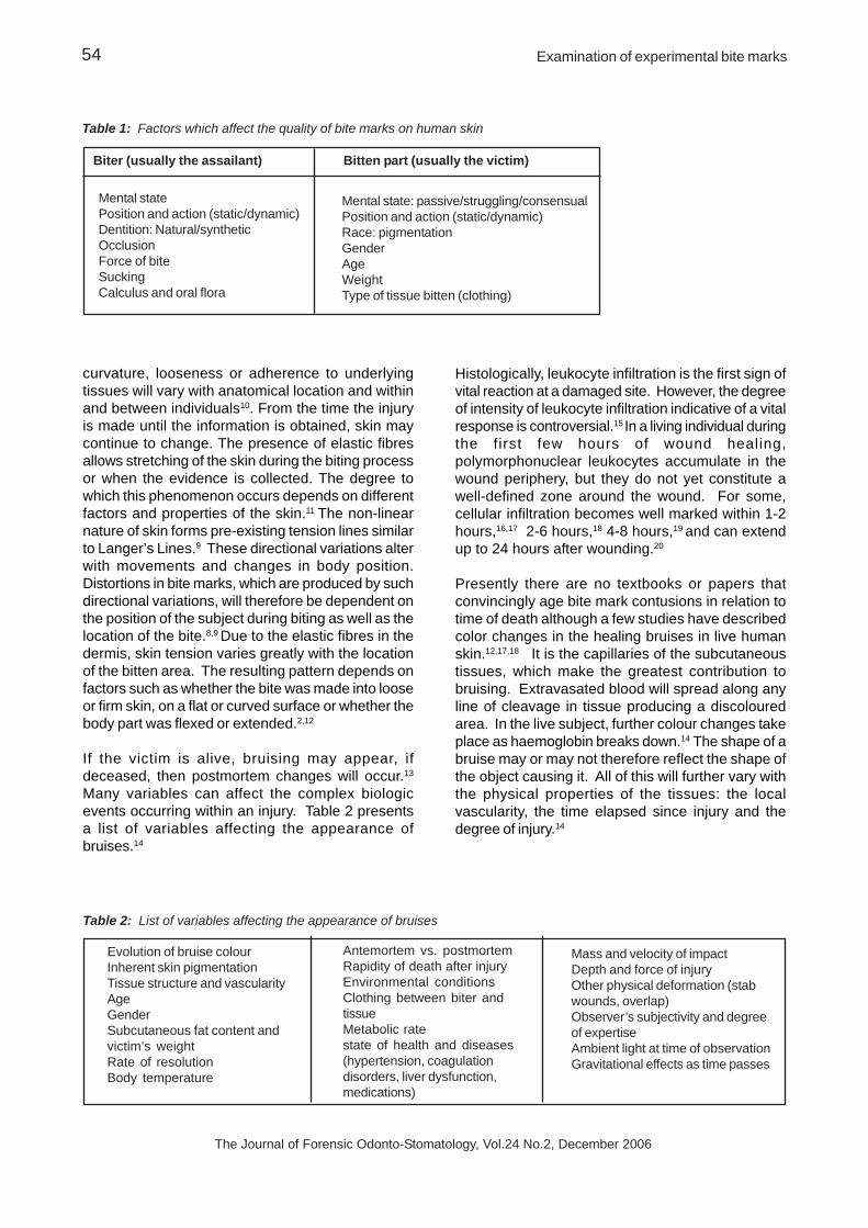
Table 1: Factors which affect the quality of bite marks on human skin
Biter (usually the assailant) Bitten part (usually the victim)
Mental statePosition and action (static/dynamic)Dentition: Natural/syntheticOcclusionForce of biteSuckingCalculus and oral flora
Mental state: passive/struggling/consensualPosition and action (static/dynamic)Race: pigmentationGenderAgeWeightType of tissue bitten (clothing)
Table 2: List of variables affecting the appearance of bruises
Evolution of bruise colourInherent skin pigmentationTissue structure and vascularityAgeGenderSubcutaneous fat content andvictim’s weightRate of resolutionBody temperature
Antemortem vs. postmortemRapidity of death after injuryEnvironmental conditionsClothing between biter andtissueMetabolic ratestate of health and diseases(hypertension, coagulationdisorders, liver dysfunction,medications)
Mass and velocity of impactDepth and force of injuryOther physical deformation (stabwounds, overlap)Observer’s subjectivity and degreeof expertiseAmbient light at time of observationGravitational effects as time passes
Examination of experimental bite marks54

The Journal of Forensic Odonto-Stomatology, Vol.24 No.2, December 2006
The mechanical properties affecting the quality of abite mark on human skin may be applicable to pigskin. Just how the pattern varies and how it is relatedto changes in the epidermal-dermal tissues remainsunknown. A recent study provided informationshowing that clearly demarcated bite marks occurat or around the time of death.21
Animal skin differs morphologically from that ofhumans, making extrapolation to human histologyproblematic. Species-to-species variation isobserved in terms of epidermal and dermalthickness, types and arrangements of hair follicles,and adnexal structures. Variation is also present inthe morphology between regions of the body in anindividual animal.22 While similarities between pigand human skin are numerous in regard to structural,functional and biochemical characteristics, there aredifferences with respect to structure,immunohistochemistry and function.23 Nevertheless,pig skin possess an epidermis and dermiscomparable to human skin making it the mostsuitable animal that can be used as a model for thestudy of pattern injuries.24,25
PURPOSE OF THE STUDYThe purpose of this study was to examineexperimental bite mark wounds inflicted at knownintervals before and after death on an in-vivo porcinemodel. The study included clinical andhistopathological observations to evaluate theusefulness of microscopic versus macroscopicexaminations of bite marks as a method of decidingif such injuries were inflicted before or after death.
MATERIALS AND METHODSBiting deviceAn instrument was constructed to mechanicallyproduce simulated human bite marks on skin. Thisdevice was previously used for studies of ageing ofbite marks.21 The biting device consists of removablechrome-cobalt upper and lower dentitions fixed to alocking C-clamp #11 vice-grip.* A pressure-sensitiveload cell and a pre-configured indicator** were addedto the device to display live loads for pressureconsistency at a pre-selected incisor tooth. Studieshave shown that the pressure exerted by humanincisor teeth with range from 6.0 to 23.5 kg with amean of 8.9 to 11.4 kg.26,27 Pressure consistency wasselected at 23 kg to represent the “maximum” forceapplied by human incisor teeth.
Biting procedure on porcine skinEthics and Animal Care Committee approval wasobtained from the Division of Comparative Medicine(DCM) of the University of Toronto.
Three young female Yorkshire pigs were used asrecipients for antemortem and postmortem bite markinjuries. The mean age of the three pigs was 12 weekswith a mean weight of 36.2 kg. The pigs werepurchased by the DCM 5 days prior to the procedure.The pigs were acclimated in a temperature (22°C),light-dark (12h/12h) regulated facility and receivedcomplete examination and blood tests to rule outany systemic diseases or haematological disorders.
On the day of the experiment, all the results from theblood tests were normal. Sedation of the pigs wasachieved with an intramuscular injection of 16.0 cc ofketamine (100 mg/ml) in the right thigh. The abdominalregion of the pig represents the widest surface andthe thinnest epidermis and cornified layer.28,29 Thecleavage lines are mostly transverse in arrangement30
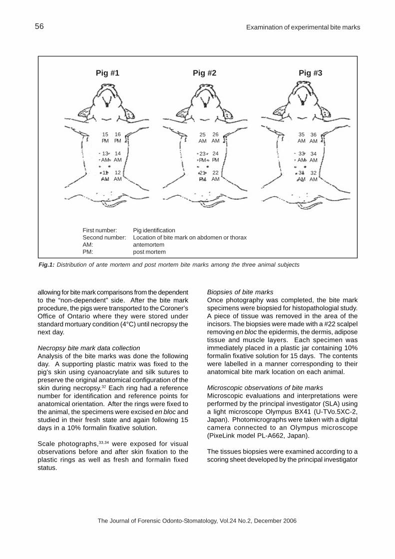
and are similar in orientation to the tension lines ofhuman skin. The bite marks were made at specificanatomical regions on the pig’s abdomen and thorax.Comparison was possible because the antemortemand postmortem locations were reproduced from pigto pig, but did not overlap with each other (Fig 1).Therefore, anatomical antemortem and postmortemlocations could be compared. Both sides of the pig’sabdomen and thorax were bitten to study the effect ofpostmortem lividity since after biting, the animals werelaid on one side until the time of the necropsy thenext day.
A series of simulated bite marks were created onthe abdomen and thorax with the biting device usinga pressure consistency of approximately 23 kg heldfor 60 seconds around the time of death. Each pigreceived 6 bite mark injuries (4 antemortem and 2postmortem) located at specific sites on theabdominal and thoracic surfaces. With the animalsunder general anaesthesia, four antemortem bitemarks were made one after the other with the timeranging from 10 minutes to 2 minutes before clinicaldeath. Approximately 2 minutes after the fourthantemortem bite mark, the pigs were sacrificed withintravenous Tanax®*** Four minutes after clinicaldeath, postmortem bite marks were made with thetimes of bite application ranging between 4 minutesand 9 minutes after death.
After the procedure, the pig was positioned on oneside to allow settling of blood by gravitationalforces.31 This side was termed the “dependent” side
* MasterCraft® Canadian Tire Corporation Toronto, ON,Canada
** A-Tech Instruments Ltd., Scarborough, ON,Canada
*** Intervet Canada Ltd, Whitby, ON, Canada
Avon, Mayhall, Wood 55
The Journal of Forensic Odonto-Stomatology, Vol.24 No.2, December 2006
allowing for bite mark comparisons from the dependentto the “non-dependent” side. After the bite markprocedure, the pigs were transported to the Coroner’sOffice of Ontario where they were stored understandard mortuary condition (4°C) until necropsy thenext day.
Necropsy bite mark data collectionAnalysis of the bite marks was done the followingday. A supporting plastic matrix was fixed to thepig’s skin using cyanoacrylate and silk sutures topreserve the original anatomical configuration of theskin during necropsy.32 Each ring had a referencenumber for identification and reference points foranatomical orientation. After the rings were fixed tothe animal, the specimens were excised en bloc andstudied in their fresh state and again following 15days in a 10% formalin fixative solution.
Scale photographs,33,34 were exposed for visualobservations before and after skin fixation to theplastic rings as well as fresh and formalin fixedstatus.
Biopsies of bite marksOnce photography was completed, the bite markspecimens were biopsied for histopathologial study.A piece of tissue was removed in the area of theincisors. The biopsies were made with a #22 scalpelremoving en bloc the epidermis, the dermis, adiposetissue and muscle layers. Each specimen wasimmediately placed in a plastic jar containing 10%formalin fixative solution for 15 days. The contentswere labelled in a manner corresponding to theiranatomical bite mark location on each animal.
Microscopic observations of bite marksMicroscopic evaluations and interpretations wereperformed by the principal investigator (SLA) usinga light microscope Olympus BX41 (U-TVo.5XC-2,Japan). Photomicrographs were taken with a digitalcamera connected to an Olympus microscope(PixeLink model PL-A662, Japan).
The tissues biopsies were examined according to ascoring sheet developed by the principal investigator
Fig.1: Distribution of ante mortem and post mortem bite marks among the three animal subjects
First number: Pig identificationSecond number: Location of bite mark on abdomen or thoraxAM: antemortemPM: post mortem
Pig #1 Pig #2 Pig #3
15PM
13AM
11AM
16PM
14AM
12AM
25AM
23PM
21PM
26AM
24PM
22AM
36AM
34AM
32AM
35AM
33AM
31AM
Examination of experimental bite marks56

The Journal of Forensic Odonto-Stomatology, Vol.24 No.2, December 2006
to evaluate the correlation between antemortem andpostmortem tissues as well as the effect of livormortis (see Table 3 for scoring sheet). Evidence ofblood vessels, blood vessel dilatation and congestionalong with extravasated red blood cells in the varioustissue layers were all evaluated. The evaluation wassemi-quantitative using a score of plus (+) and minus(–) signs according to the presence or absence of thecharacteristic studied. The blood vessels andvasodilatation were evaluated at 20X magnification.Extravasated red blood cells were confirmed in thetissue type in which they were found whether it wasconnective tissue, fat or muscle tissue. The observedsamples were graded “–” if there was noextravasated red blood cells, “+” if extravasated redblood cells were seen in at least one area, “++” fortwo to three areas, and “+++” if more than threeareas were seen in the tissue. This scoring sheetallows a better depiction of the histopathologicalsituation and facilitates correlation with clinicalcharacteristics observed on antemortem andpostmortem tissue reaction.
RESULTSClinical observations of bite marksOn the day the injuries were inflicted the markingswere clearly evident and viewable as distinctive ovalpatterns. On some specimens, the cutting edges ofthe incisors left an impression on the skin while onother specimens, the palatal surfaces of upperincisors could be appreciated. Antemortem clinical
Figs 2 A and B: Antemortem bite mark present on the non-dependent side. A. Skin surface side showing white indentationwith peripheral erythema. B. Deep tissue aspect (muscular side) showing intramuscular erythema caused by the bite(arrows)
A
13 A
B13 A
observations after release of the biting force revealedslow but progressing fading of the impressions due toelastic recovery of the skin. After clinical death, bitemark indentations faded rapidly. Approximately 18hours after the biting procedure, clinical observationsrevealed rigor mortis and livor mortis on the dependentside on the three pigs.
All bite marks on the non-dependent side revealeddetailed pattern characteristics. The most stable anddetailed bite mark injuries were those made fiveminutes prior to death followed by those made fiveminutes after death. Antemortem bite marks on thenon-dependent side showed pale centralindentations surrounded by red outlines (Figs 2 A andB). The class characteristics represented by linear ortriangle shapes and individual characteristics withteeth angulations or rotations were recognizable evenwhen the tissue was excised. The postmortem bitemarks from the non-dependent side were homogenous,pale and less defined. They lacked central whiteindentations and showed less class and individualcharacteristics than the antemortem ones (Figures 3A and B). Only antemortem bite marks from the non-dependent side exhibited areas of erythema.
The anatomical location of the antemortem andpostmortem bite marks did not influence the patterncharacteristics on the abdomen or the thorax of theanimals.
Avon, Mayhall, Wood 57

The Journal of Forensic Odonto-Stomatology, Vol.24 No.2, December 2006
The pattern injury of the bite marks was influenced bythe presence of livor mortis. Bite marks located onthe dependent side, where areas were discolouredby the settling of blood by gravitational forces withinvessels, showed clear white indentations on a purplishblue background. Whether the bite mark was madeantemortem or postmortem did not show any particularpattern characteristics of the tissues in a livor mortisarea (Figs 4 A and B).
Figs 3 A and B: Postmortem bite mark present on the non-dependent side. A. Skin surface side showing pale, homogenousredness at the side of indentation. B. Deep tissue aspect (muscular side) showing no intramuscular erythema
.
Figs 4 A and B: Postmortem bite mark present on the dependent side. A. Skin surface side showing white indentation.B. Deep tissue aspect (muscular side) showing redness caused by livor mortis
15 A
A
15 A
B
16 A
A
16 A
B
Histopathological observations of bite marksMicroscopic observations of all the examined tissuesshowed orthokeratinised stratified squamousepithelium with underlying connective tissue,adipose tissue and muscle. The observations foreach specimen depended upon whether the biopsiedtissue was antemortem, postmortem or if the tissuewas affected by livor mortis. Table 3 shows a semi-quantitative evaluation for each specimen accordingto blood vessel configuration and the presence ofextravasated red blood cells.
Examination of experimental bite marks58
The Journal of Forensic Odonto-Stomatology, Vol.24 No.2, December 2006
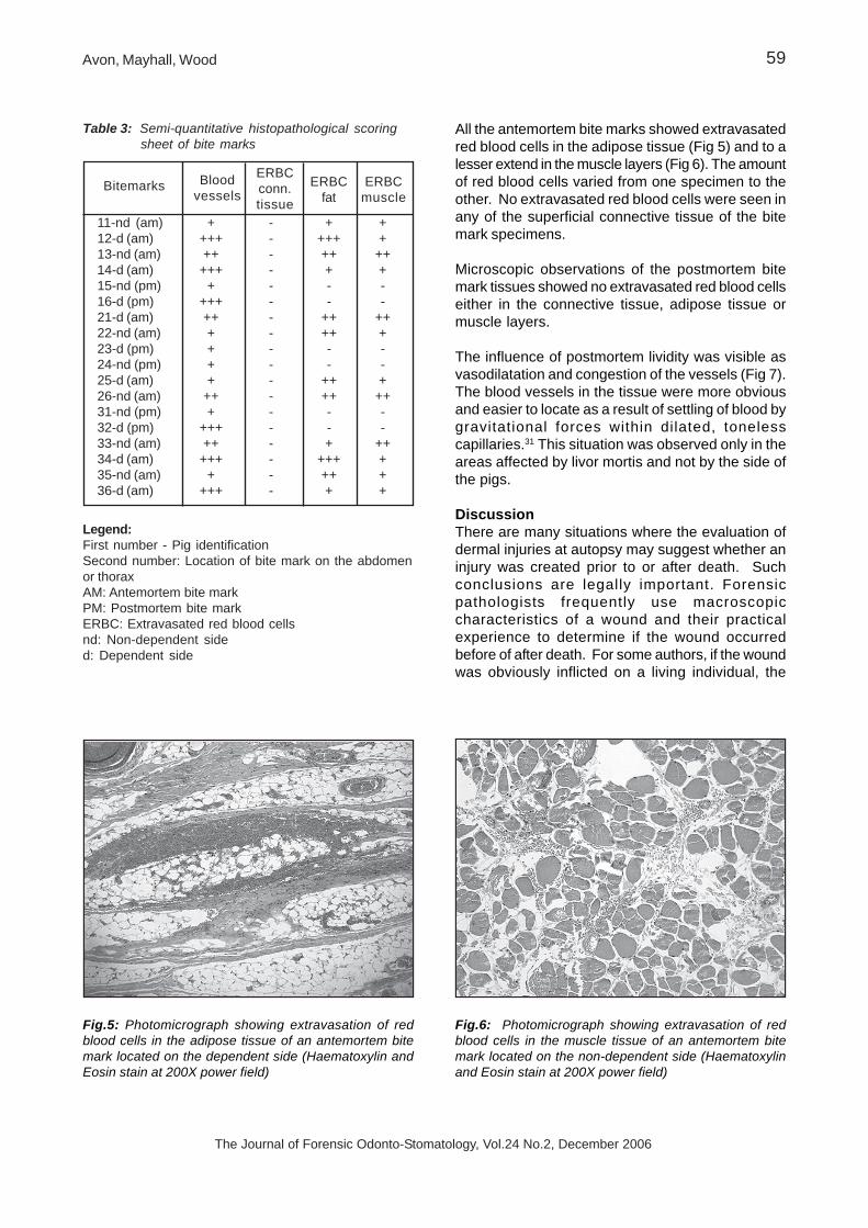
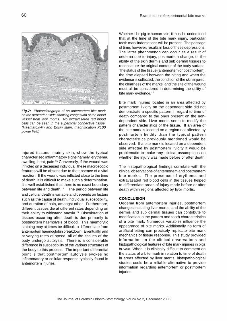
All the antemortem bite marks showed extravasatedred blood cells in the adipose tissue (Fig 5) and to alesser extend in the muscle layers (Fig 6). The amountof red blood cells varied from one specimen to theother. No extravasated red blood cells were seen inany of the superficial connective tissue of the bitemark specimens.
Microscopic observations of the postmortem bitemark tissues showed no extravasated red blood cellseither in the connective tissue, adipose tissue ormuscle layers.
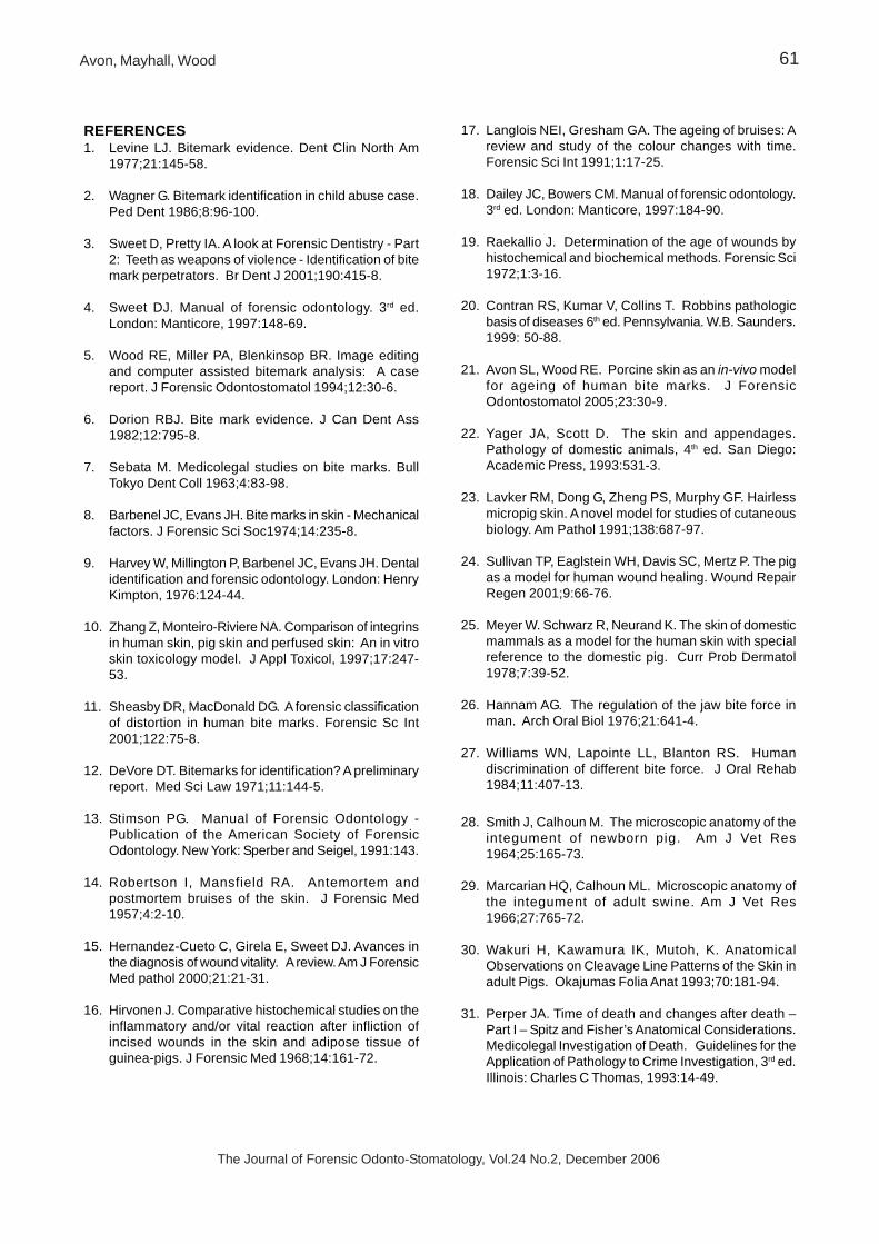
The influence of postmortem lividity was visible asvasodilatation and congestion of the vessels (Fig 7).The blood vessels in the tissue were more obviousand easier to locate as a result of settling of blood bygravitational forces within dilated, tonelesscapillaries.31 This situation was observed only in theareas affected by livor mortis and not by the side ofthe pigs.
DiscussionThere are many situations where the evaluation ofdermal injuries at autopsy may suggest whether aninjury was created prior to or after death. Suchconclusions are legally important. Forensicpathologists frequently use macroscopiccharacteristics of a wound and their practicalexperience to determine if the wound occurredbefore of after death. For some authors, if the woundwas obviously inflicted on a living individual, the
Fig.5: Photomicrograph showing extravasation of redblood cells in the adipose tissue of an antemortem bitemark located on the dependent side (Haematoxylin andEosin stain at 200X power field)
Fig.6: Photomicrograph showing extravasation of redblood cells in the muscle tissue of an antemortem bitemark located on the non-dependent side (Haematoxylinand Eosin stain at 200X power field)
Table 3: Semi-quantitative histopathological scoringsheet of bite marks
11-nd (am) + - + +12-d (am) +++ - +++ +13-nd (am) ++ - ++ ++14-d (am) +++ - + +15-nd (pm) + - - -16-d (pm) +++ - - -21-d (am) ++ - ++ ++22-nd (am) + - ++ +23-d (pm) + - - -24-nd (pm) + - - -25-d (am) + - ++ +26-nd (am) ++ - ++ ++31-nd (pm) + - - -32-d (pm) +++ - - -33-nd (am) ++ - + ++34-d (am) +++ - +++ +35-nd (am) + - ++ +36-d (am) +++ - + +
Bitemarks Bloodvessels
ERBCconn.tissue
ERBCfat
ERBCmuscle
Legend:First number - Pig identificationSecond number: Location of bite mark on the abdomenor thoraxAM: Antemortem bite markPM: Postmortem bite markERBC: Extravasated red blood cellsnd: Non-dependent sided: Dependent side
59Avon, Mayhall, Wood
The Journal of Forensic Odonto-Stomatology, Vol.24 No.2, December 2006
injured tissues, mainly skin, show the typicalcharacterised inflammatory signs namely, erythema,swelling, heat, pain.15 Conversely, if the wound wasinflicted on a deceased individual, these macroscopicfeatures will be absent due to the absence of a vitalreaction. If the wound was inflicted close to the timeof death, it is difficult to make such a determination.It is well established that there is no exact boundarybetween life and death.15 The period between lifeand cellular death is variable and depends on factorssuch as the cause of death, individual susceptibility,and duration of pain, amongst other. Furthermore,different tissues die at different rates depending ontheir ability to withstand anoxia.15 Discoloration oftissues occurring after death is due primarily topostmortem haemolysis of blood. This haemolyticstaining may at times be difficult to differentiate fromantemortem haemoglobin breakdown. Eventually, andat varying rates of speed, all of the tissues of thebody undergo autolysis. There is a considerabledifference in susceptibility of the various structures ofthe body to this process. The important differentialpoint is that postmortem autolysis evokes noinflammatory or cellular response typically found inantemortem injuries.
Whether it be pig or human skin, it must be understoodthat at the time of the bite mark injury, particulartooth mark indentations will be present. The passageof time, however, results in loss of these depressions.The latter phenomenon can occur as a result ofoedema due to injury, postmortem change, or theability of the skin dermis and sub dermal tissues toreconstitute the original contour of the body surface.The status of the tissue (antemortem or postmortem),the time elapsed between the biting and when theevidence is collected, the condition of the skin injured,the clearness of the marks, and the site of the woundmust all be considered in determining the utility ofbite mark evidence.1,7
Bite mark injuries located in an area affected bypostmortem lividity on the dependent side did notdemonstrate a specific pattern in regard to time ofdeath compared to the ones present on the non-dependent side. Livor mortis seem to modify thepattern characteristics of the tissue. If an area ofthe bite mark is located on a region not affected bypostmortem lividity than the typical patterncharacteristics previously mentioned would beobserved. If a bite mark is located on a dependentside affected by postmortem lividity it would beproblematic to make any clinical assumptions onwhether the injury was made before or after death.
The histopathological findings correlate with theclinical observations of antemortem and postmortembite marks. The presence of erythema andextravasated red blood cells in the tissues helpedto differentiate areas of injury made before or afterdeath within regions affected by livor mortis.
CONCLUSIONOedema from antemortem injuries, postmortemchanges including livor mortis, and the ability of thedermis and sub dermal tissues can contribute tomodification in the pattern and tooth characteristicsof a bite mark. Numerous variables influence theappearance of bite marks. Additionally no form ofartificial biting can precisely replicate bite markmechanics or tissue response. This study providedinformation on the clinical observations andhistopathological features of bite mark injuries in pigsin-vivo. When it is clinically difficult to comment onthe status of a bite mark in relation to time of deathin areas affected by livor mortis, histopathologicalstudies could be a reliable alternative to provideinformation regarding antemortem or postmorteminjuries.
Fig.7: Photomicrograph of an antemortem bite markon the dependent side showing congestion of the bloodvessel from livor mortis. No extravasated red bloodcells can be seen in the superficial connective tissue.(Haematoxylin and Eosin stain, magnification X100power field)
Examination of experimental bite marks60
The Journal of Forensic Odonto-Stomatology, Vol.24 No.2, December 2006
REFERENCES1. Levine LJ. Bitemark evidence. Dent Clin North Am
1977;21:145-58.
2. Wagner G. Bitemark identification in child abuse case.Ped Dent 1986;8:96-100.
3. Sweet D, Pretty IA. A look at Forensic Dentistry - Part2: Teeth as weapons of violence - Identification of bitemark perpetrators. Br Dent J 2001;190:415-8.
4. Sweet DJ. Manual of forensic odontology. 3rd ed.London: Manticore, 1997:148-69.
5. Wood RE, Miller PA, Blenkinsop BR. Image editingand computer assisted bitemark analysis: A casereport. J Forensic Odontostomatol 1994;12:30-6.
6. Dorion RBJ. Bite mark evidence. J Can Dent Ass1982;12:795-8.
7. Sebata M. Medicolegal studies on bite marks. BullTokyo Dent Coll 1963;4:83-98.
8. Barbenel JC, Evans JH. Bite marks in skin - Mechanicalfactors. J Forensic Sci Soc1974;14:235-8.
9. Harvey W, Millington P, Barbenel JC, Evans JH. Dentalidentification and forensic odontology. London: HenryKimpton, 1976:124-44.
10. Zhang Z, Monteiro-Riviere NA. Comparison of integrinsin human skin, pig skin and perfused skin: An in vitroskin toxicology model. J Appl Toxicol, 1997;17:247-53.
11. Sheasby DR, MacDonald DG. A forensic classificationof distortion in human bite marks. Forensic Sc Int2001;122:75-8.
12. DeVore DT. Bitemarks for identification? A preliminaryreport. Med Sci Law 1971;11:144-5.
13. Stimson PG. Manual of Forensic Odontology -Publication of the American Society of ForensicOdontology. New York: Sperber and Seigel, 1991:143.
14. Robertson I, Mansfield RA. Antemortem andpostmortem bruises of the skin. J Forensic Med1957;4:2-10.
15. Hernandez-Cueto C, Girela E, Sweet DJ. Avances inthe diagnosis of wound vitality. A review. Am J ForensicMed pathol 2000;21:21-31.
16. Hirvonen J. Comparative histochemical studies on theinflammatory and/or vital reaction after infliction ofincised wounds in the skin and adipose tissue ofguinea-pigs. J Forensic Med 1968;14:161-72.
17. Langlois NEI, Gresham GA. The ageing of bruises: Areview and study of the colour changes with time.Forensic Sci Int 1991;1:17-25.
18. Dailey JC, Bowers CM. Manual of forensic odontology.3rd ed. London: Manticore, 1997:184-90.
19. Raekallio J. Determination of the age of wounds byhistochemical and biochemical methods. Forensic Sci1972;1:3-16.
20. Contran RS, Kumar V, Collins T. Robbins pathologicbasis of diseases 6th ed. Pennsylvania. W.B. Saunders.1999: 50-88.
21. Avon SL, Wood RE. Porcine skin as an in-vivo modelfor ageing of human bite marks. J ForensicOdontostomatol 2005;23:30-9.
22. Yager JA, Scott D. The skin and appendages.Pathology of domestic animals, 4th ed. San Diego:Academic Press, 1993:531-3.
23. Lavker RM, Dong G, Zheng PS, Murphy GF. Hairlessmicropig skin. A novel model for studies of cutaneousbiology. Am Pathol 1991;138:687-97.
24. Sullivan TP, Eaglstein WH, Davis SC, Mertz P. The pigas a model for human wound healing. Wound RepairRegen 2001;9:66-76.
25. Meyer W. Schwarz R, Neurand K. The skin of domesticmammals as a model for the human skin with specialreference to the domestic pig. Curr Prob Dermatol1978;7:39-52.
26. Hannam AG. The regulation of the jaw bite force inman. Arch Oral Biol 1976;21:641-4.
27. Williams WN, Lapointe LL, Blanton RS. Humandiscrimination of different bite force. J Oral Rehab1984;11:407-13.
28. Smith J, Calhoun M. The microscopic anatomy of theintegument of newborn pig. Am J Vet Res1964;25:165-73.
29. Marcarian HQ, Calhoun ML. Microscopic anatomy ofthe integument of adult swine. Am J Vet Res1966;27:765-72.
30. Wakuri H, Kawamura IK, Mutoh, K. AnatomicalObservations on Cleavage Line Patterns of the Skin inadult Pigs. Okajumas Folia Anat 1993;70:181-94.
31. Perper JA. Time of death and changes after death –Part I – Spitz and Fisher’s Anatomical Considerations.Medicolegal Investigation of Death. Guidelines for theApplication of Pathology to Crime Investigation, 3rd ed.Illinois: Charles C Thomas, 1993:14-49.
Avon, Mayhall, Wood 61
The Journal of Forensic Odonto-Stomatology, Vol.24 No.2, December 2006
32. Sweet DJ, Bastien RB. Use of an Acrylonitrile-Butadiene-Styrene (ABS) plastic ring as a matrix in therecovery of bite mark evidence. J Forens Sci1991;36:1565-7.
33. Krauss T. Photographic techniques of concern in metricbite mark analyses. J Forens Sci 1984;29:633-8.
34. Hyzer W, Thomas P, Krauss T. The bite mark standardreference scale - ABFO No.2. J Forens Sci1988;33:498-506.
Address for correspondence:Sylvie Louise AvonFaculté de médecine dentaireUniversité LavalCite universitaireSte-Foy, QuébecCANADA G1K 7P4
Examination of experimental bite marks62