Clinical and electrophysiological effects of intravenous ... · CaseNo. Cardiacdisease CHF...
8
British Heart Journal, 1977, 39, 309-316 Clinical and electrophysiological effects of intravenous quinidine in man1 DAVID S. HIRSCHFELD, CLARENCE T. UEDA, MALCOLM ROWLAND, AND MELVIN M. SCHEINMAN2 From the Medical Service, San Francisco General Hospital, and the Department of Medicine and the Cardiovascular Research Institute, University of California, San Francisco, U.S.A. Quinidine gluconate (total dose 4-4 to 9-1 mg/kg) was infused intravenously over 22 minutes in 20 patients with either frequent premature ventricular contractions or supraventricular arrhythmias, 16 of whom had bundle-branch block. Therapeutic plasma quinidine levels (3 to 7mg/l) were achieved in 15. Heart rate, atrioventricular nodal, and infranodal conduction times did not change significantly. The QRS duration increased significantly from 128 ±30 to 134 ±29 ms at peak plasma quinidine levels (P < 0.01). Mild hypo- tension occurred during infusion in most patients. Two patients had a severe but transient toxic response characterised by hypotension, nausea, vomiting, and diaphoresis. Atrioventricular dissociation with escape His bundle or fascicular rhythm occurred in 1 patient with sinus bradycardia. Bundle-branch block does not contraindicate administration of quinidine. Quinidine gluconate administered intravenously (0.3 to 0 4 mg/kg per min) is frequently associated with hypotension and should be used only in an intensive care setting and with careful monitoring of blood pressure. Oral administration of quinidine has proved to be effective and safe for the control of atrial and ventricular arrhythmias provided that plasma levels are kept within the therapeutic range (Sokolow and Ball, 1956), whereas the intravenous use of quini- dine has been associated with serious untoward re- actions (Armbrust and Levine, 1950; Chapman, 1945; Hepburn and Rykert, 1937; Riseman and Linenthal, 1941; Strong and Munroe, 1940) and is generally considered to be contraindicated (Selzer, 1972). Quinidine delays conduction and increases refractoriness of the His-Purkinje system and slows conduction in ventricular muscle. Because of these actions, quinidine is believed by some to be rela- tively contraindicated in patients with bundle- branch block (Sokolow and Perloff, 1961). In the study reported here, we attempted to correlate the electrophysiological effects of quinidine on atrio- ventricular and intraventricular conduction with plasma drug levels in patients with bundle-branch block in an effort to define a rate of intravenous administration of this drug in man which yields "Supported in part by funds from the Bay Area Heart Research Committee and in part by research grants HL 06285 and GM 16496 from the National Institutes of Health, Bethesda, Maryland. 'This study was performed during tenure of the American Heart Association Teaching Scholar in Cardiology. Received for publication 23 April 1976 therapeutic blood levels within a relatively short period of time but without undue toxic effects. Subjects and methods Twenty patients with either frequent premature ventricular contractions or supraventricular ar- rhythmias in whom prolonged therapy with oral quinidine was anticipated were studied. Informed consent was obtained and the study conducted in accordance with a protocol approved by the Com- mittee on Human Experimentation of the University of California, San Francisco. No patient was re- ceiving other antiarrhythmic agents at the time of the study. A hexapolar electrode catheter was intro- duced percutaneously via the right femoral vein, and His bundle recordings were obtained by pre- viously described techniques (Damato et al., 1969; Scheinman et al., 1973). External scalar leads X, Y, and inverse Z of the Frank orthogonal system were recorded simultaneously with the His bundle electrogram at a paper speed of 100 mm/s. The atrioventricular nodal conduction time (AH in- terval) was measured from the earliest rapid de- flection of the atrial electrogram to the initial de- flection of the His bundle depolarisation. The con- duction time from bundle to ventricle (HQ interval) 309 on 5 March 2019 by guest. Protected by copyright. http://heart.bmj.com/ Br Heart J: first published as 10.1136/hrt.39.3.309 on 1 March 1977. Downloaded from
-
Upload
nguyendang -
Category
Documents
-
view
219 -
download
0
Transcript of Clinical and electrophysiological effects of intravenous ... · CaseNo. Cardiacdisease CHF...

British Heart Journal, 1977, 39, 309-316
Clinical and electrophysiological effects ofintravenous quinidine in man1DAVID S. HIRSCHFELD, CLARENCE T. UEDA, MALCOLM ROWLAND,AND MELVIN M. SCHEINMAN2
From the Medical Service, San Francisco General Hospital, and the Department of Medicine and theCardiovascular Research Institute, University of California, San Francisco, U.S.A.
Quinidine gluconate (total dose 4-4 to 9-1 mg/kg) was infused intravenously over 22 minutes in 20 patientswith either frequent premature ventricular contractions or supraventricular arrhythmias, 16 of whom hadbundle-branch block. Therapeutic plasma quinidine levels (3 to 7mg/l) were achieved in 15. Heart rate,atrioventricular nodal, and infranodal conduction times did not change significantly. The QRS durationincreased significantly from 128 ±30 to 134±29 ms at peak plasma quinidine levels (P < 0.01). Mild hypo-tension occurred during infusion in most patients. Two patients had a severe but transient toxic responsecharacterised by hypotension, nausea, vomiting, and diaphoresis. Atrioventricular dissociation with escapeHis bundle or fascicular rhythm occurred in 1 patient with sinus bradycardia. Bundle-branch block does notcontraindicate administration of quinidine. Quinidine gluconate administered intravenously (0.3 to 0 4mg/kg per min) is frequently associated with hypotension and should be used only in an intensive care settingand with careful monitoring of blood pressure.
Oral administration of quinidine has proved to beeffective and safe for the control of atrial andventricular arrhythmias provided that plasma levelsare kept within the therapeutic range (Sokolow andBall, 1956), whereas the intravenous use of quini-dine has been associated with serious untoward re-actions (Armbrust and Levine, 1950; Chapman,1945; Hepburn and Rykert, 1937; Riseman andLinenthal, 1941; Strong and Munroe, 1940) and isgenerally considered to be contraindicated (Selzer,1972). Quinidine delays conduction and increasesrefractoriness of the His-Purkinje system and slowsconduction in ventricular muscle. Because of theseactions, quinidine is believed by some to be rela-tively contraindicated in patients with bundle-branch block (Sokolow and Perloff, 1961). In thestudy reported here, we attempted to correlate theelectrophysiological effects of quinidine on atrio-ventricular and intraventricular conduction withplasma drug levels in patients with bundle-branchblock in an effort to define a rate of intravenousadministration of this drug in man which yields"Supported in part by funds from the Bay Area Heart ResearchCommittee and in part by research grants HL 06285 and GM 16496from the National Institutes of Health, Bethesda, Maryland.'This study was performed during tenure of the American HeartAssociation Teaching Scholar in Cardiology.Received for publication 23 April 1976
therapeutic blood levels within a relatively shortperiod of time but without undue toxic effects.
Subjects and methods
Twenty patients with either frequent prematureventricular contractions or supraventricular ar-rhythmias in whom prolonged therapy with oralquinidine was anticipated were studied. Informedconsent was obtained and the study conducted inaccordance with a protocol approved by the Com-mittee on Human Experimentation ofthe Universityof California, San Francisco. No patient was re-ceiving other antiarrhythmic agents at the time ofthe study. A hexapolar electrode catheter was intro-duced percutaneously via the right femoral vein,and His bundle recordings were obtained by pre-viously described techniques (Damato et al., 1969;Scheinman et al., 1973). External scalar leads X, Y,and inverse Z of the Frank orthogonal system wererecorded simultaneously with the His bundleelectrogram at a paper speed of 100 mm/s. Theatrioventricular nodal conduction time (AH in-terval) was measured from the earliest rapid de-flection of the atrial electrogram to the initial de-flection of the His bundle depolarisation. The con-duction time from bundle to ventricle (HQ interval)
309
on 5 March 2019 by guest. P
rotected by copyright.http://heart.bm
j.com/
Br H
eart J: first published as 10.1136/hrt.39.3.309 on 1 March 1977. D
ownloaded from

Hirschfeld, Ueda, Rowland, and Scheinman
was measured from the initial His deflection to theearliest onset of ventricular depolarisation recordedon the external leads; the onset of the V deflectionin the His bundle recordings was not used.
Preliminary studies showed that infusion of lessthan 3 mg/kg body weight over 22 minutes failed toproduce therapeutic blood levels of quinidine(3 to 7 mg/l). In the present study, quinidinegluconate was diluted in 50 ml 5 per cent dextrosein water and administered over 22 minutes by a
Harvard constant infusion pump to provide a totaldose of from 4'4 to 9-1 mg/kg quinidine gluconate(average 6.9) at infusion rates of 0-20 to 045 mg/kgper min (average 0 34). Blood specimens for quini-dine assay were withdrawn every 5 minutes duringinfusion and for 20 minutes afterwards from a poly-ethylene catheter inserted into a vein in the op-posite arm. Simultaneously with withdrawal ofbloodspecimens, the systemic blood pressure was
measured by the cuff method, the His bundleelectrogram recorded, and 30 second electrocardio-graphic recordings were obtained in order to countpremature beats. Patients were observed closely andasked to report any symptoms during the study.Plasma quinidine levels were measured by two
techniques: (1) a standard colorimetric method in-volving alkaline extraction into ethylene chloride(Brodie et al., 1946), and (2) a more specific extrac-tion procedure coupled to a thin layer chromato-graphy separation (Ueda et al., 1976). Plasma
quididine levels are expressed as mg/l of quinidinebase. Where applicable, the data were analysedusing Student's paired t test.
Results
Pertinent clinical information, peak plasma quini-dine levels, and electrophysiological data for eachpatient are summarised in Tables 1 and 2. Twelvepatients manifested either left bundle-branch block,right bundle-branch block with left anterior fasci-cular block, or right bundle-branch block with leftposterior fascicular block; of these, 8 had prolongedHQ intervals (>55 ms). Four patients had rightbundle-branch block alone or isolated left anteriorfascicular block; of these, one had a prolonged HQinterval. Four had no intraventricular conductionabnormality; of these, one had a slightly prolongedHQ interval (58 ms). Of the 20 patients, 13 had pre-mature beats at the time of the study.
Plasma quinidine levels tended to increase pro-gressively during infusion, and in all but one patientthe peak quinidine level was achieved at the end ofthe infusion (Fig. 1). Therapeutic plasma quinidinelevels were achieved (3 to 7 mg/ml) when the in-fusion was stopped in 15 patients. There was no
significant difference in plasma quinidine levelsdetermined by the colorimetric assay and the morespecific chromatographic technique. Values referred
Table 1 Pertinent clinical and electrocardiographic data and amount of quinidine administered
Case No. Cardiac disease CHF Conduction disturbance Arrhythmia Amount quinidine infused Peak quinidine plasma level(mg/kg) (mg/i)
-1 CAD Yes LAHB PVC 7-5 3-12 CAD Yes LAHB, RBBB PVC 7-1 4 93 CAD Yes LBBB PAC 7-2 3-54 CAD Yes LAHB PVC 91 4-35 CAD Yes RBBB PVC 5-4 3-36 CAD No None PVC 9 0 8-47 Cardiomyopathy Yes LPHB, RBBB PVC 6-7 0-88 RHD Yes LBBB, SB PAT* 7-3 5-89 Unknown No None PVC 4-6 1-410 Unknown No LAHB, RBBB PAC 5-8 4-611 Cardiomyopathy Yes LAHB, SB PJC 6-6 4-112 Unknown No LBBB PAC 7-4 3-013 CAD No None PAT* 9-0 4-014 HHD Yes None Atrial flutter 8-8 5-815 CAD No LPHB, RBBB PVC 9 0 2-916 CAD Yes LBBB PVC 5-4 5-317 HHD Yes LBBB PVC 7-2 2-818 Unknown Yes LAHB, RBBB PVC 5-2 3-719 Cardiomyopathy No LAHB, RBBB PVC, PAC 6-0 1-620 Unknown Yes LBBB PVC, PAC 4-4 3-2Mean ESD 6-9 +1-5 3-8 ±1-7
CHP, congestive heart failure; CAD, coronary artery disease; RHD, rheumatic heart disease; HHD, hypertensive heart disease; LAHB,left anterior hemiblock; RBBB, right bundle-branch block; LBBB, left bundle-branch block; LPHB, left posterior hemiblock; SB, sinusbradycardia; PAC, premature atrial contractions; PVC, premature ventricular contractions; PAT, paroxysmal atrial tachycardia; PJC,premature junctional contractions.*Not at time of study.
310
on 5 March 2019 by guest. P
rotected by copyright.http://heart.bm
j.com/
Br H
eart J: first published as 10.1136/hrt.39.3.309 on 1 March 1977. D
ownloaded from
Effects of intravenous quinidine in man
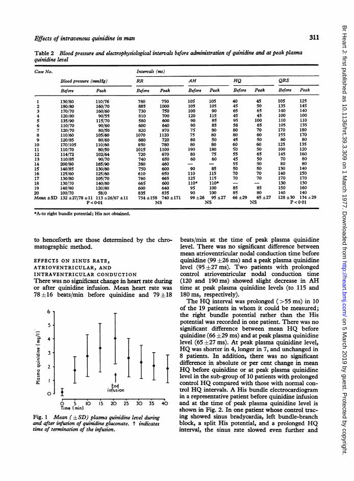
Table 2 Blood pressure and electrophysiological intervals before administration of quinidine and at peak plasmaquinidine level
Case No. Intervals (ms)
Blood pressure (mmHg) RR AH HQ QRS
Before Peak Before Peak Before Peak Before Peak Before Peak
1 130/80 110/76 780 750 105 105 40 45 105 1252 180/80 160/70 885 1000 105 105 45 50 135 1453 170/70 160/60 730 750 100 90 65 65 140 1404 120/80 90/55 810 700 120 115 45 45 100 1005 135/90 115/70 580 600 90 85 95 100 110 1106 110/70 90/60 600 640 90 85 58 65 105 1357 120/70 80/50 820 870 75 80 80 70 170 1808 110/60 105/60 1070 1120 75 80 80 60 155 1709 120/85 80/60 880 720 80 50 45 50 80 8010 170/105 110/60 850 780 80 80 60 60 125 13511 110/70 80/50 1015 1100 190 180 50 50 100 12012 114/72 102/64 720 670 80 75 55 65 145 16013 110/85 90/70 740 650 60 60 45 50 70 8014 200/90 165/90 380 460 - - 55 50 80 8015 140/85 130/80 750 600 90 85 50 50 130 14016 125/60 125/60 610 650 110 115 70 70 140 15017 130/80 105/70 760 665 125 115 70 70 170 17018 130/70 140/80 665 600 110* 110* - - 130 13019 140/90 120/80 600 640 95 100 85 85 150 16020 100/70 58/0 835 835 90 100 85 80 140 140Mean +SD 132 +27/78 ±11 113 ±26/67 +11 754 +158 740±171 99 +26 95 +27 66 +29 65 +27 128 ±30 134 ±29
P < 0-01 NS NS NS P < 0-01
*A-to right bundle potential; His not obtained.
to henceforth are those determinedmatographic method.
by the chro-
EFFECTS ON SINUS RATE,ATRIOVENTRICULAR, ANDINTRAVENTRICULAR CONDUCTIONThere was no significant change in heart rate duringor after quinidine infusion. Mean heart rate was78 ±16 beats/min before quinidine and 79 ±18
0 l
I~~~~~End
infusion
.
10Time (min)
I15 20 25 30 35
Fig. 1 Mean ( LSD) plasma quinidine level duringand after infiusion of quinidine gluconate. t indicatestime of termination of the infusion.
beats/min at the time of peak plasma quinidinelevel. There was no significant difference betweenmean atrioventricular nodal conduction time beforequinidine (99 ±26 ms) and a peak plasma quinidinelevel (95 +27 ms). Two patients with prolongedcontrol atrioventricular nodal conduction time(120 and 190ms) showed slight decrease in AHtime at peak plasma quinidine levels (to 115 and180 ms, respectively).The HQ interval was prolonged (>55 ms) in 10
of the 19 patients in whom it could be measured;the right bundle potential rather than the Hispotential was recorded in one patient. There was nosignificant difference between mean HQ beforequinidine (66 ±29 ms) and at peak plasma quinidinelevel (65 +27 ms). At peak plasma quinidine level,HQ was shorter in 4, longer in 7, and unchanged in8 patients. In addition, there was no significantdifference in absolute or per cent change in meanHQ before quinidine or at peak plasma quinidinelevel in the sub-group of 10 patients with prolongedcontrol HQ compared with those with normal con-trol HQ intervals. A His bundle electrocardiogramin a representative patient before quinidine infusionand at the time of peak plasma quinidine level isshown in Fig. 2. In one patient whose control trac-ing showed sinus bradycardia, left bundle-branchblock, a split His potential, and a prolonged HQinterval, the sinus rate slowed even further and
6
5
4
3
2
.E
m-a0E0
311
on 5 March 2019 by guest. P
rotected by copyright.http://heart.bm
j.com/
Br H
eart J: first published as 10.1136/hrt.39.3.309 on 1 March 1977. D
ownloaded from

Hirschfeld, Ueda, Rowland, and Scheinman
Fig. 2 His bundle electrograms(HBE) of Case 17. (a) Beforeinfusion of quinidine gluconate.A= atrial potential, H=Hisspike, V=ventricular potential.(b) HBE at peak plasmaquinidine level. AH is 10 msshorter than in (a) and HQ andQRS are unchanged.
atrioventricular dissociation developed (Fig. 3),with the block localised within the His bundleduring drug infusion. Second degree or completeatrioventricular block did not develop in any otherpatient.Mean QRS duration increased significantly from
128+30 before quinidine to 134 +29ms at peakplasma quinidine level (P < 0-01), but there was nosignificant difference in the absolute or per centchange in 7 patients with either normal or leftanterior fascicular block compared with the 13patients with bundle-branch block. In no patient
did the QRS duration increase by greater than 28per cent.
EFFECTS OF CONGESTIVE HEART FAILUREOf the 20 patients, 13 were in congestive heartfailure with varying degrees of compensation at thetime of the study. The group in heart failure showedno significant difference from those not in heartfailure, in heart rate or atrioventricular or intra-ventricular conduction times either before or afterquinidine infusion. Furthermore, there was nodifference in the peak plasma level of quinidine
312
on 5 March 2019 by guest. P
rotected by copyright.http://heart.bm
j.com/
Br H
eart J: first published as 10.1136/hrt.39.3.309 on 1 March 1977. D
ownloaded from

Effects of intravenous quinidine in man
x
Iy.
AH=75z HH'=60HQ=80
BE1
BE2
A
x
f =RR1250.1280 msAA^=1200-1i30
V
I
HBE1
HBE2
'V,v
O
H H'
- L. ..
H H'IA
-s 1- 4I -mw1 -/l-- A t oU F.JA V
Fig. 3 His bundle electrogramsof Case 18. (a) Control tracing
~Y'.~'Y showing two spikes (HH')H '1 falling between A and V
representing split His potential1 e-'*~~'~'~' with spontaneous atrial cycleA length of 1100 ms. (b) 5 minutes
after termination of infusion,atrioventricular dissociation ispresent. (The atrial cycle lengthis 1230; the ventricular cycle
yfi length is 1280 ms.) Because ofproximity of the atrial andventricular electrogram, Hisspikes are not seen in the firsttwo complexes. (c) Atrio-ventricular dissociationwithin the His-Purkinje system.Note that HH' has lengthened. H'is missing in alternate beats,which is probably because of blockwithin the His. The ventricularpacemaker is probably in thedistal His bundle, but a focus in
_4,'p_,, the right fascicle cannot beexcluded.
v
-IMlO i
A
x_
HBE1 I
V ANH'
NE21ft-
RR a100 mSAHNSOHH090
313
\ /0000
I.
m
'F vT
---11 I~r
IIN
on 5 March 2019 by guest. P
rotected by copyright.http://heart.bm
j.com/
Br H
eart J: first published as 10.1136/hrt.39.3.309 on 1 March 1977. D
ownloaded from

Hirschfeld, Ueda, Rowland, and Scheinman
achieved, when normalised for height and weight,in the patients in heart failure compared with thepatients not in heart failure.
ARRHYTHMIA CONTROLFive patients were having premature supraven-tricular beats and 8 were experiencing prematureventricular beats during the study. One of thesubjects had both premature atrial contractions andpremature ventricular contractions. Ectopic beatswere decreased in frequency (<75% of control)at the time of peak plasma quinidine level in all 9patients in whom therapeutic levels were achieved.The effect on frequency of premature ventricularcontractions in a representative patient is shown inFig. 4.
TOXIC SIDE-EFFECTSThe mean of the systemic blood pressures droppedsignificantly (P < 0-01) from control values (132 ±27/78 ± 11 mmHg) by 5 minutes after starting in-fusion and was lowest (113 ±26/67 ± 11 mmHg)at the end of infusion when plasma quinidine levelswere highest. Five patients had a drop in systolicblood pressure to < 100 mnmHg but remainedasymptomatic. In 2 other patients, diaphoresis andnausea occurred concomitantly with a distinct fallin blood pressure and small changes in heart rate;both responded favourably to raising the legs andstopping the infusion. Their plasma quinidinelevels were not excessive (1.4 and 3-2 mg/l) at thetime of hypotension and the rates of infusion (0 35and 0-20 mg/kg per min) were no more rapid thanin the remaining 18 patients (mean 034 mg/kg permin). No other toxic responses to quinidine in-fusion were observed, and no patients experiencedtinnitus, vertigo, or diarrhoea.
32 -
24
8E
C- b
8-1
0
0
0
II:.-end infusion
0 10 20 30 40
Fig. 4 Frequency of premature ventricular contractionsduring quinidine infusion in Case 6 illustrating suppressionof ectopic beats at termination of infusion and return ofpremature ventricular contractions afterwards.
Discussion
EFFECT OF QUINIDINEQuinidine is considered by many to be the mainstayof chronic oral antiarrhythmic therapy. It is ofobvious practical importance for the clinician to beaware of the relative risks of quinidine therapy forpatients with atrioventricular or intraventricularconduction delay. Experimental studies in animalsshowed that administration of quinidine in amountsthat produce blood levels considered to be withinthe therapeutic range in man result in little changein atrioventricular nodal conduction time (Wallaceet al., 1966) but definite increases in infranodal andintraventricular conduction times (Wallace et al.,1966; Sokolow, 1951; Vaughan Williams, 1958;Swain and Weidner, 1957). For this reason, somebelieve that the presence of bundle-branch blockconstitutes a relative contraindication to the use ofquinidine (Sokolow and Perloff, 1961). The electro-cardiograms in 16 of our patients had either uni-fascicular or bifascicular block and 10 had pro-longed infranodal conduction times. Therapeuticblood levels of quinidine resulted in no significantchange in atrioventricular conduction times and insmall increases in intraventricular conduction times.Though the QRS duration increased significantly(P<0.01) after quinidine, the QRS axis did notchange nor did a new bundle-branch block developin any patient. In one patient with sinus bradycardiaand a prolonged sinus node recovery time, sinusslowing, and atrioventricular dissociation associatedwith an escape His bundle or fascicular focus de-veloped during quinidine infusion. The degree ofatrioventricular block did not increase in any otherpatient.The only other study in which His bundle re-
cordings were used to assess the effects of quinidinegluconate in man is that of Josephson et al. (1974),in which the drug was administered intramuscularly.Their results are similar to ours with regard to theeffect of quinidine on heart rate and atrioventricularnodal and intraventricular conduction times butdiffer in that a small but significant increase in infra-nodal conduction time (mean 6 ms) was observedin their study. The reason for the difference is un-certain, though in the study of Josephson et al.(1974), larger doses (600 to 800 mg quinidinegluconate) were administered and His bundle re-cordings were made at higher plasma quinidinelevels (4-6 versus 3-8 mg/l). It is also possible thatthe hypotension caused by intravenous administra-tion of the drug in our patients provoked catechol-amine stimulation or increased sympathetic tonewhich tended to diminish the direct effect of quini-dine on the infranodal conduction system.
314
%.O
on 5 March 2019 by guest. P
rotected by copyright.http://heart.bm
j.com/
Br H
eart J: first published as 10.1136/hrt.39.3.309 on 1 March 1977. D
ownloaded from
Effects of intravenous quinidine in man
Reports on the safety of intravenous administra-tion of quinidine have yielded conflicting conclu-sions. Severe toxic reactions (Chapman, 1945;Hepburn and Rykert, 1937; Riseman and Linen-thal, 1941; Strong and Munroe, 1940) and death(Armbrust and Levine, 1950; Acierno and Gubner,1951) were attributed to the intravenous route andsome clinicians have expressed the view that theintravenous route is never (Selzer, 1972) or almostnever (Goldberg, 1974; Friedberg, 1966) acceptablemedical practice. In these reports either large dosesof quinidine were administered (intravenously)rapidly or the patients appeared to be moribund atthe time of infusion. However, Clagett (1950)administered quinidine lactate intravenously indoses up to 3-25 g but at infusion rates comparableto ours, and the only side-effects observed werenausea and vomiting in 3 of the 13 patients studied.In his study, as in ours, quinidine administrationwas associated with a decrease or abolition of supra-ventricular or ventricular arrhythmias and did notproduce bundle-branch or atrioventricular block orincreased ventricular ectopia. Asymptomatic mildto moderate decreases in systemic blood pressureoccurred in 17 of our patients, including reductionof systolic pressure to < 100 mmHg in 5, and a toxicreaction characterised by the abrupt onset ofnausea, intense diaphoresis, and severe hypotensionoccurred in 2 patients during infusion. Both of thesepatients actually received smaller amounts ofquinidine because infusion was terminated pre-maturely, and plasma quinidine levels were in thesubtherapeutic or low therapeutic range (1-4 and3-2 mg/i). In all patients who had a fall in bloodpressure, the hypotension persisted for 5 to 10minutes after termination of infusion. Our datasuggest that careful monitoring of the systemicblood pressure is mandatory when quinidine isadministered intravenously. Moreover, an alpha-sympathetic agonist should be readily available be-cause the hypotensive effect of intravenous quini-dine is most likely the result of reversible alpha-receptor blockade (Ferrer et al., 1948; Luchi et al.,1963) rather than of a direct myocardial action.
PLASMA QUINIDINE LEVELSAlthough there was a progressive rise in plasmaquinidine level in each patient during infusion ofquinidine, the levels ranged widely even after cor-rection for rates of infusion and body weight. Thisfinding is probably related to the rate and extent ofdrug distribution because only trivial amounts ofthe drug would be expected to be metabolised orexcreted over the relatively short period of thestudy. Regardless of differences in rate and extentof drug distribution, we found that doses of 7 to
9 mg/kg quinidine gluconate regularly resulted inpeak plasma quinidine levels within the therapeuticrange. Of further interest was the observation thatmean peak plasma quinidine levels did not differsignificantly between patients with (10) or without(10) heart failure. These findings are similar to thosedescribed by Bellet et al. (1971), who found nosignificant difference in mean plasma quinidinelevels between normal subjects and patients withcongestive heart failure 2 hours after an oral dose of600 mg quinidine; however, the levels were signi-ficantly higher in patients with heart failure than innormal subjects 4, 6, and 24 hours after the oraldose. These findings are consistent with our ownunpublished observations that the higher plasmaquinidine levels found in patients with heart failureare related to decreased drug clearance rather thanto diminished volume of distribution.
CLINICAL IMPLICATIONS
In this study, the haemodynamic and electro-physiological effects of intravenous administrationof quinidine gluconate which resulted in a relativelyrapid achievement of therapeutic plasma quinidinelevels were assessed. All the patients but one whoreceived 7 to 9 mg/kg achieved therapeutic plasmaquinidine levels within 22 minutes. In a recent studyby Wellens and Durrer (1974) 4 mg/kg of quinidinewas infused intravenously at a rate of 0-8 mg/kg permin in patients with the Wolff-Parkinson-Whitesyndrome and 5 of the 6 patients became hypo-tensive, with sweating, restlessness, and tachycardia.In our study, the average total dose was about twiceas great, but the slower rates of infusion (mean rateof 0-34 mg/kg) probably accounted for the lowerincidence of toxicity. Nevertheless, the slower ratesof infusion used in the present study were still as-sociated with an unacceptably high incidence ofhypotensive reactions. This study has also shownthat therapeutic levels of quinidine were effective indecreasing or abolishing cardiac arrhythmias andwere safe for patients with atrioventricular or intra-ventricular conduction abnormalities in that nohigher degrees of atrioventricular block or increasedventricular ectopic activity were encountered. Onepatient with sinus bradycardia, however, developedtransient atrioventricular dissociation owing tofurther slowing of the sinus rate. For patients re-quiring parenteral quinidine therapy, intravenousadministration appears to have several advantagesover the intramuscular route: there is a more rapidand uniform achievement of therapeutic plasmalevels; drug administration may be readily ter-minated should untoward effects occur; and theintravenous route is painless.
Nevertheless, the relatively high incidence of
315
on 5 March 2019 by guest. P
rotected by copyright.http://heart.bm
j.com/
Br H
eart J: first published as 10.1136/hrt.39.3.309 on 1 March 1977. D
ownloaded from

Hirschfeld, Ueda, Rowland, and Scheinman
hypotensive reactions encountered suggests thatintravenous quinidine administration should bereserved for critical situations where first line drugsare ineffective and only used with ready availabilityof an alpha-sympathetic agonist. Though untowardeffects disappeared shortly after termination of theinfusion and no long-term sequelae were observed,this regimen should rarely, if ever, be used incritically ill patients with hypoxia and/or electrolyteabnormalities (Watanabe et al., 1963; Watanabeand Dreifus, 1967; Holland, 1957), in patients withsinus bradycardia, or in patients receiving drugsthat may potentiate quinidine effects.
Addendum
Since completion of this manuscript, 7 additionalpatients received 8 mg/kg of quinidine intravenouslyand no untoward effects were observed.
The authors wish to thank Gunnard Modin forstatistical analyses.
References
Acierno, L. J., and Gubner, R. (1951). Utility and limitationsof intravenous quinidine in arrhythmias. American HeartJ'ournal, 41, 733-741.
Armbrust, C. A., Jr., and Levine, S. A. (1950). Paroxysmalventricular tachycardia: study of 107 cases. Circulation,1, 28-40.
Bellet, S., Roman, L. R., and Boza, A. (1971). Relationbetween serum quinidine levels and renal function. Studiesin normal subjects and patients with congestive failure andrenal insufficiency. American Journal of Cardiology, 27,368-371.
Brodie, B. B., Udenfriend, S., and Baer, J. E. (1946). Analysisof basic organic compounds in biological tissues: I. Isolationprior to estimation. Federation Proceedings, 5, 124-125.
Chapman, D. W. (1945). Observations on 2 patients withparoxysmal ventricular tachycardia treated by intra-venous administration of quinidine lactate. American HeartJ'ournal, 30, 276-283.
Clagett, A. H., Jr. (1950). Intravenous use of quinidine, withparticular reference to ventricular tachycardia. AmericanJ7ournal of the Medical Sciences, 220, 381-387.
Damato, A. N., Lau, S. H., Helfant, R. H., Stein, E.,Berkowitz, W. D., and Cohen, S. I. (1969). Study of atrio-ventricular conduction in man using electrode catheterrecordings of His bundle activity. Circulation, 39, 287-296.
Ferrer, M. I., Harvey, R. M., Werko, L., Dresdale, D. T.,Cournand, A., and Richards, D. W., Jr. (1948). Someeffects of quinidine sulfate on the heart and circulation inman. American Heart Journal, 36, 816-837.
Friedberg, C. K. (1966). Diseases of the Heart, 3rd ed., p. 553.W. B. Saunders, Philadelphia.
Goldberg, L. I. (1974). Pharmacology of cardiovascular drugs.In The Heart, Arteries and Veins, 3rd ed., p. 1704. Ed. byJ. W. Hurst, R. B. Logue, R. C. Schlant, and N. K.Wenger. McGraw-Hill, New York.
Hepburn, J., and Rykert, H. E. (1937). Use of quinidine sul-fate intravenously in ventricular tachycardia. AmericanHeart Journal, 14, 620-623.
Holland, W. C. (1957). A possible mechanism of action ofquinidine. American Journal of Physiology, 190, 492-494.
Josephson, M. E., Seides, S. F., Batsford, W. P., Weisfogel,G. M., Akhtar, M., Caracta, A. R., Lau, S. H., and Damato,A. N. (1974). The electrophysiological effects of intra-muscular quinidine on the atrioventricular conductingsystem in man. American Heart Journal, 87, 55-64.
Luchi, R. J., Helwig, J., Jr., and Conn, H. L., Jr. (1963).Quinidine toxicity and its treatment. An experimentalstudy. American Heart Journal, 65, 340-348.
Riseman, J. E. F., and Linenthal, H. (1941). Paroxysmal ven-tricular tachycardia; its favorable prognosis in absence ofacute cardiac damage and its treatment with parenterallyadministered quinidine dihydrochloride. American HeartJournal, 22, 219-229.
Scheinman, M. M., Weiss, A., and Kunkel, F. (1973). Hisbundle recordings in patients with bundle branch block andtransient neurologic symptoms. Circulation, 48, 322-330.
Selzer, A. (1972). The use and abuse of quinidine. HeartLung, 1, 755.
Sokolow, M. (1951). The present status of therapy of thecardiac arrhythmias with quinidine. American HeartJournal, 42, 771-797.
Sokolow, M., and Ball, R. E. (1956). Factors influencing con-version of chronic atrial fibrillation with special referenceto serum quinidine concentration. Circulation, 14, 568-583.
Sokolow, M., and Perloff, D. B. (1961). The clinical pharma-cology and use of quinidine in heart disease. Progress inCardiovascular Diseases, 3, 316-330.
Strong, G. F., and Munroe, D. S. (1940). Paroxysmal ven-tricular tachycardia, with report of unusual case. AmericanHeart Journal, 19, 486-491.
Swain, H. H., and Weidner, C. L. (1957). A study of sub-stances which alter intraventricular conduction in theisolated dog heart. Journal of Pharmacology and Experi-mental Therapeutics, 120, 137-146.
Ueda, C. T., Ballard, B. E., and Rowland, M. (1976). Con-centration-time effects in quinidine disposition kinetics inRhesus monkeys. Journal of Pharmacokinetics and Bio-pharmaceutics. In the press.
Vaughan Williams, E. M. (1958). The mode of action ofquinidine on isolated rabbit atria interpreted from intra-cellular potential records. British Journal of Pharmacologyand Chemotherapy, 13, 276-287.
Wallace, A. G., Cline, R. E., Sealy, W. C., Young, W. G., Jr.,and Troyer, W. G., Jr. (1966). Electrophysiologic effects ofquinidine. Studies using chronically implanted electrodes inawake dogs with and without cardiac denervation. Circula-tion Research, 19, 960-969.
Watanabe, Y., and Dreifus, L. S. (1967). Interactions ofquinidine and potassium on atrioventricular transmission.Circulation Research, 20, 434-446.
Watanabe, Y., Dreifus, L. S., and Likoff, W. (1963). Electro-physiologic antagonism and synergism of potassium andantiarrhythmic agents. American J7ournal of Cardiology, 12,702-710.
Wellens, H. G., and Durrer, D. (1974). Effect of procaineamide, quinidine, and ajmaline in the Wolff-Parkinson-White syndrome. Circulation, 50, 114-120.
Requests for reprints to Editorial Office, Room4101, San Francisco General Hospital, 1001Potrero Avenue, San Francisco, California 94110,U.S.A.
316
on 5 March 2019 by guest. P
rotected by copyright.http://heart.bm
j.com/
Br H
eart J: first published as 10.1136/hrt.39.3.309 on 1 March 1977. D
ownloaded from


![[2021]EWHC (QB) CaseNo: G90MA404 …](https://static.fdocuments.us/doc/165x107/618bcc54dc416d05cc58d0a6/2021ewhc-qb-caseno-g90ma404-.jpg)