Clinical and cytopathological aspects in phyllodes tumors ... tumors, with 0.3–0.9% from all of...
7
Romanian Journal of Morphology and Embryology 2009, 50(4):605–611 ORIGINAL PAPER Clinical and cytopathological aspects in phyllodes tumors of the breast ANCA PĂTRAŞCU 1) , CARMEN FLORINA POPESCU 2) , I. E. PLEŞEA 3) , ADRIANA BĂDULESCU 4) , FLORENTINA TĂNASE 1) , GAROFIŢA MATEESCU 5) 1) Department of Obstetrics and Gynecology, University of Medicine and Pharmacy of Craiova 2) Department of Pathology and Cytology, Emergency County Hospital, Craiova 3) Department of Pathology, University of Medicine and Pharmacy of Craiova 4) Department of Surgery, “Titu Maiorescu” University, Bucharest 5) Department of Histology, University of Medicine and Pharmacy of Craiova Abstract The frequency of mesenchymal breast tumors is very low, being represented mostly by tumors with biphasic proliferation (phyllodes tumors) and less by other types of non-epithelial tumors. From clinical point of view, phyllodes tumors (PT) can mimic a breast carcinoma. Therefore, the preoperative diagnosis by cytological examination on material obtained by fine needle aspiration (FNA) is very important for adequate treatment of these tumors. In current study, we assessed clinical aspects of 79 phyllodes tumors regarding patient’s age and localization of the tumors. In 17 out of 79 cases, it has been performed FNA within the tumors with further cytological examination on the smears obtained. The median age of the patients was 46.07-year-old, being progressively higher with grade of the tumors with significant values between benign and borderline tumors (p=0.04954) and between benign and malignant ones (p=0.02890). The distinguish on the smears of stromal fragments and naked stromal nuclei with variable grade of atypia regarding the tumoral type, in detriment of epithelial elements have been conclusive for fibroepithelial lesion as cytopathological diagnosis. The preoperative differentiation between a breast phyllodes tumor and a breast carcinoma is extremely important for avoiding of a useless radical surgery for the patient. If the fine needle aspiration was correctly performed, the accuracy of the cytodiagnosis has been 82% in current study. Keywords: phyllodes tumor (PT), breast, fine needle aspiration (FNA), fibroepithelial lesion (FEL). Introduction Breast cancer involves one of nine women and is the second main cause of death by cancer in women, after cervix cancer. Most of tumors appear because of mammary gland epithelial tissue malignization, ductal carcinoma being the most frequent histopathological form. Lymphomas, sarcomas and malignant melanoma are the most frequent non-epithelial malignant tumors, and phyllodes tumors, with 0.3–0.9% from all of breast tumors and 2–3% from fibroepithelial neoplasms are the most rarely seen. Phyllodes tumor was described for the first time by Johannes Müller, in 1838 [1], who initially named it cystosarcoma phyllodes, choosing the name of sarcoma because of tumor’s macroscopic appearance. Subsequently, lesions with certain malignant evolution have been described, the first case of metastasis phyllodes tumor being described only in 1931 by Lee B and Pack G. Since then the name of “sarcoma” has gained a malignant connotation [2]. Although its frequency is rare, this tumor has an unpredictable evolution, meaning that not all of malignant forms are metastasizing and some of the benign ones can metastasize. The purpose of this study was to distinguish some of the clinical and cytological aspects of phyllodes tumors, the latter being very important for a correct therapeutic approach after cytopathological exam. Material and Methods The study included 79 patients with breast tumors diagnosed as phyllodes tumors. The group was splitted into three subgroups according to histopathological classification, i.e.: benign, borderline and malignant tumors. Preoperative tumoral fine needle aspiration (FNA) was performed by a surgeon in 17 cases for cyto- diagnosis. Obtained smears were fixed by air-drying for 5 minutes and then stained by May–Grünwald–Giemsa (MGG) method. After FNA, all patients underwent surgical excision and the excisional lumps were processed by classical method of formalin fixing and paraffin embedding with subsequent Hematoxylin– Eosin staining. The assessed parameters were divided in two main categories: clinical and cytological.
Transcript of Clinical and cytopathological aspects in phyllodes tumors ... tumors, with 0.3–0.9% from all of...

Romanian Journal of Morphology and Embryology 2009, 50(4):605–611
OORRIIGGIINNAALL PPAAPPEERR
Clinical and cytopathological aspects in phyllodes tumors of the breast
ANCA PĂTRAŞCU1), CARMEN FLORINA POPESCU2), I. E. PLEŞEA3), ADRIANA BĂDULESCU4), FLORENTINA TĂNASE1), GAROFIŢA MATEESCU5)
1)Department of Obstetrics and Gynecology, University of Medicine and Pharmacy of Craiova
2)Department of Pathology and Cytology, Emergency County Hospital, Craiova
3)Department of Pathology, University of Medicine and Pharmacy of Craiova
4)Department of Surgery, “Titu Maiorescu” University, Bucharest
5)Department of Histology, University of Medicine and Pharmacy of Craiova
Abstract The frequency of mesenchymal breast tumors is very low, being represented mostly by tumors with biphasic proliferation (phyllodes tumors) and less by other types of non-epithelial tumors. From clinical point of view, phyllodes tumors (PT) can mimic a breast carcinoma. Therefore, the preoperative diagnosis by cytological examination on material obtained by fine needle aspiration (FNA) is very important for adequate treatment of these tumors. In current study, we assessed clinical aspects of 79 phyllodes tumors regarding patient’s age and localization of the tumors. In 17 out of 79 cases, it has been performed FNA within the tumors with further cytological examination on the smears obtained. The median age of the patients was 46.07-year-old, being progressively higher with grade of the tumors with significant values between benign and borderline tumors (p=0.04954) and between benign and malignant ones (p=0.02890). The distinguish on the smears of stromal fragments and naked stromal nuclei with variable grade of atypia regarding the tumoral type, in detriment of epithelial elements have been conclusive for fibroepithelial lesion as cytopathological diagnosis. The preoperative differentiation between a breast phyllodes tumor and a breast carcinoma is extremely important for avoiding of a useless radical surgery for the patient. If the fine needle aspiration was correctly performed, the accuracy of the cytodiagnosis has been 82% in current study. Keywords: phyllodes tumor (PT), breast, fine needle aspiration (FNA), fibroepithelial lesion (FEL).
Introduction
Breast cancer involves one of nine women and is the second main cause of death by cancer in women, after cervix cancer.
Most of tumors appear because of mammary gland epithelial tissue malignization, ductal carcinoma being the most frequent histopathological form.
Lymphomas, sarcomas and malignant melanoma are the most frequent non-epithelial malignant tumors, and phyllodes tumors, with 0.3–0.9% from all of breast tumors and 2–3% from fibroepithelial neoplasms are the most rarely seen.
Phyllodes tumor was described for the first time by Johannes Müller, in 1838 [1], who initially named it cystosarcoma phyllodes, choosing the name of sarcoma because of tumor’s macroscopic appearance.
Subsequently, lesions with certain malignant evolution have been described, the first case of metastasis phyllodes tumor being described only in 1931 by Lee B and Pack G. Since then the name of “sarcoma” has gained a malignant connotation [2].
Although its frequency is rare, this tumor has an unpredictable evolution, meaning that not all of
malignant forms are metastasizing and some of the benign ones can metastasize.
The purpose of this study was to distinguish some of the clinical and cytological aspects of phyllodes tumors, the latter being very important for a correct therapeutic approach after cytopathological exam.
Material and Methods
The study included 79 patients with breast tumors diagnosed as phyllodes tumors. The group was splitted into three subgroups according to histopathological classification, i.e.: benign, borderline and malignant tumors. Preoperative tumoral fine needle aspiration (FNA) was performed by a surgeon in 17 cases for cyto-diagnosis. Obtained smears were fixed by air-drying for 5 minutes and then stained by May–Grünwald–Giemsa (MGG) method. After FNA, all patients underwent surgical excision and the excisional lumps were processed by classical method of formalin fixing and paraffin embedding with subsequent Hematoxylin–Eosin staining.
The assessed parameters were divided in two main categories: clinical and cytological.

Anca Pătraşcu et al.
606
Clinical parameters
Clinical parameters were: sex and age of patients and tumoral site. They were assessed for the whole group of 79 cases.
Cytological parameters Cytological parameters, consisting in stromal com-
ponents characteristics (stromal fragments and naked stromal nuclei) were assessed only for the 17 cases with FNA. They were:
▪ number and size of stromal fragments; ▪ number and size of naked stromal nuclei; ▪ cellularity; ▪ cells’ aspect and atypia. In order to gather the data, database files containing
all the parameters were created in the computer. The cytopathological study included detailed
assessment of the cytological aspects of stromal and epithelial compounds and also which of them prevailed.
Special attention was given to the stromal com-pound, which was analyzed regarding the two elements: stromal fragments and isolated stromal cells.
Stromal fragments
The following parameters of stromal fragments were assessed:
▪ number: quantified as frequent (over 5) and rare (under 5);
▪ size: small, medium and large; ▪ cellularity: graded using a semiquantitative scale
from 1+ to 3+ (1+, low cellularity; 2+, medium cellu-larity; 3+, high cellularity);
▪ cellular features: thin and flattened cells; big and prominent cells;
▪ atypia: quantified as 0 – absent, 1+ – light, 2+ – medium, 3+ – severe.
Isolated stromal cells and naked stromal nuclei
Isolated stromal cells and naked stromal nuclei have been quantified using a semiquantitative scale as:
▪ 1+: rare; ▪ 2+: moderate number; ▪ 3+: frequent.
The epithelial component
The study of the epithelial component pursued: ▪ the number of cellular groups: considered as
frequent when they were more than 5 and rare when they were less than 5;
▪ the architecture of cellular groups: normal or hyperplastic;
▪ atypia. The features used for defining the ratio between
epithelial and stromal components were: ▪ predominance of the stromal component; ▪ equal ratio between the two of them; ▪ predominance of the epithelial component.
Kruskal–Wallis’ method for comparing median values was used because the groups were small; the results were considered significant if they were lower than 5% (0.05).
The cytopathological study was retrospective and it was correlated with histopathological diagnosis.
Results
Clinical study
Age of patients
Patients’ age ranged between 13 and 82-year-old, the mean age being 46-year-old. Only one patient was 13-year-old and had a benign tumor and two patients were 82-year-old, one having a benign tumor and the other, a malignant one. Four patients were under 20-year-old (one having 13 and three having 18-year-old, respectively); all these patients had benign tumors. The lowest age increased from benign PT-group to malignant PT-group (13 to 38-year-old respectively). The highest age for patients with borderline PT was 69-year-old. The mean age of patients increased also from benign PT-group (42.71-year-old) to malignant PT-group (51.80-year-old), borderline PT-group having a mean age in between (49.53-year-old). Standard deviation was highest in benign PT-group because this group had the widest range of ages (between 13 and 82-year-old) and the highest number of cases. In contrast, borderline PT-group had the smallest standard deviation because this group had the narrowest range of ages and also a reduced number of cases. The malignant group had a standard deviation between the other groups because it had a wide range of ages but a small number of cases (Table 1).
Table 1 – Age distribution of PTs Age groups
[years] Benign
PT Borderline
PT Malignant
PT Total
10–19 4 – – 4 20–29 8 – – 8 30–39 6 3 1 10 40–49 12 6 8 26 50–59 8 8 3 19 60–69 4 2 1 7 70–79 2 – 1 3 >80 1 – 1 2 Total 45 19 15 79
The lowest age 13 32 38 13 The highest age 82 69 82 82 The mean age 42.71 49.53 51.80 46.07
Std. dev. 16.57 9.56 12.85 Legend: PT – phyllodes tumor; Std. dev. – standard deviation.
Sex of patients
All patients included in our study were women.
Tumor site
TPs were more frequently situated in left breast than in the right one (51.9% vs. 45.6%). In only two cases, the tumors were found in both breasts (Figure 1).
Clinical and cytopathological aspects in phyllodes tumors of the breast
60736; 45.6%
2; 2.5%41; 51.9%
Left Breast Right Breast Bilateral Figure 1 – Site distribution of PTs.
Cytopathological study
The cytopathological examination revealed in most of cases (9/17) aspects suggesting a benign fibro-epithelial lesion. High cellularity fields were found in only three cases and definite malignant cellular features, suggesting a sarcomatous lesion were observed in two cases. In one case, although the tumor was clinically obvious, the cytopathological aspect was dominated by an inflammatory background. Finally, in two cases, the cytopathological aspect suggested a fibroadenoma (FA) (Table 2).
Histopathological examination revealed that 11 out of the 17 cases with preoperative cytopatological exami-nation were benign phyllodes tumors, four were border-line and two were malignant (Table 2).
In nine out of the 11 cases with benign phyllodes tumors, the cytopatological diagnosis was in accordance with the histopathological one. In the other two cases, the cytopathology suggested, as mentioned above, a FA.
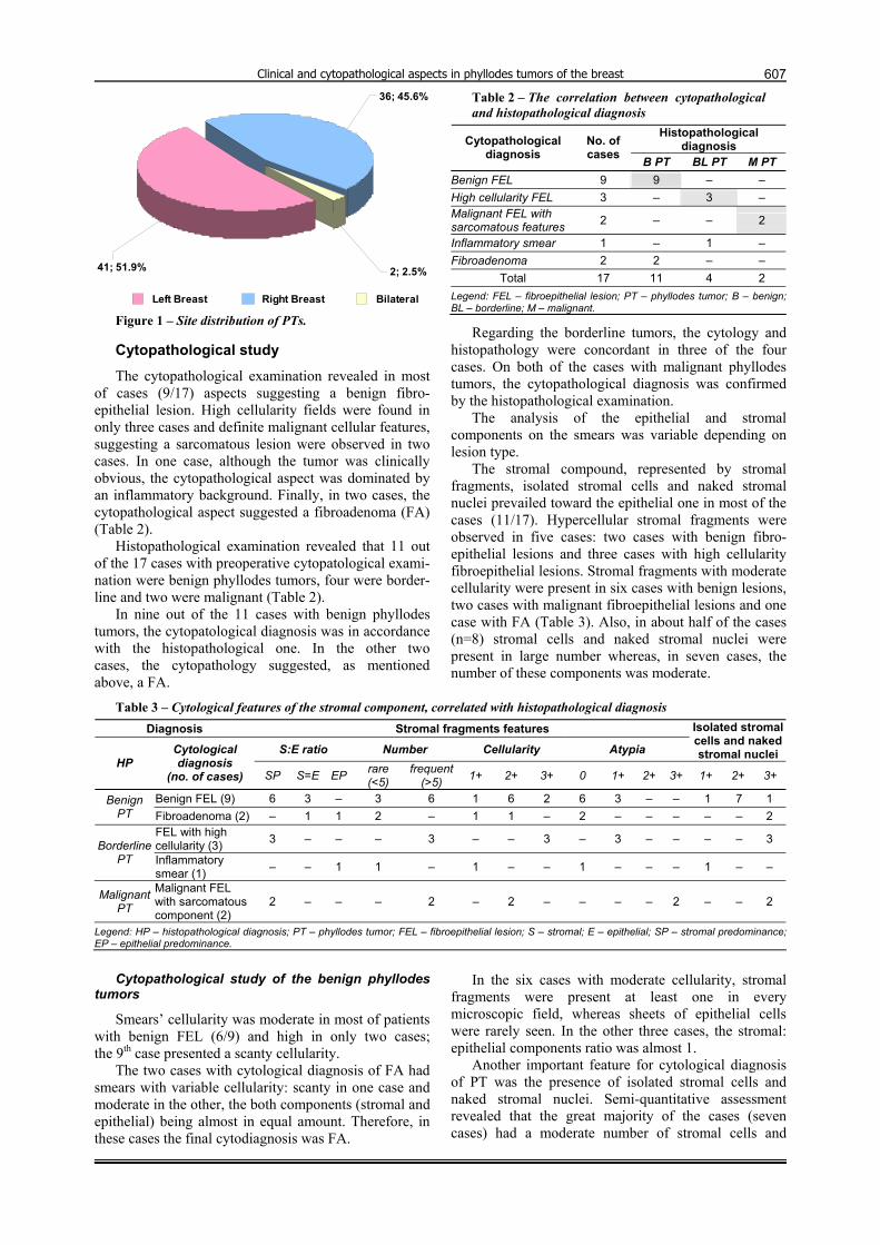
Table 2 – The correlation between cytopathological and histopathological diagnosis
Histopathological diagnosis Cytopathological
diagnosis No. of cases B PT BL PT M PT
Benign FEL 9 9 – – High cellularity FEL 3 – 3 – Malignant FEL with sarcomatous features 2 – – 2
Inflammatory smear 1 – 1 – Fibroadenoma 2 2 – –
Total 17 11 4 2 Legend: FEL – fibroepithelial lesion; PT – phyllodes tumor; B – benign; BL – borderline; M – malignant.
Regarding the borderline tumors, the cytology and histopathology were concordant in three of the four cases. On both of the cases with malignant phyllodes tumors, the cytopathological diagnosis was confirmed by the histopathological examination.
The analysis of the epithelial and stromal components on the smears was variable depending on lesion type.
The stromal compound, represented by stromal fragments, isolated stromal cells and naked stromal nuclei prevailed toward the epithelial one in most of the cases (11/17). Hypercellular stromal fragments were observed in five cases: two cases with benign fibro-epithelial lesions and three cases with high cellularity fibroepithelial lesions. Stromal fragments with moderate cellularity were present in six cases with benign lesions, two cases with malignant fibroepithelial lesions and one case with FA (Table 3). Also, in about half of the cases (n=8) stromal cells and naked stromal nuclei were present in large number whereas, in seven cases, the number of these components was moderate.
Table 3 – Cytological features of the stromal component, correlated with histopathological diagnosis Diagnosis Stromal fragments features
S:E ratio Number Cellularity Atypia
Isolated stromal cells and naked stromal nuclei
HP Cytological diagnosis
(no. of cases) SP S=E EP rare (<5)
frequent (>5) 1+ 2+ 3+ 0 1+ 2+ 3+ 1+ 2+ 3+
Benign FEL (9) 6 3 – 3 6 1 6 2 6 3 – – 1 7 1 Benign PT Fibroadenoma (2) – 1 1 2 – 1 1 – 2 – – – – – 2
FEL with high cellularity (3) 3 – – – 3 – – 3 – 3 – – – – 3 Borderline
PT Inflammatory smear (1) – – 1 1 – 1 – – 1 – – – 1 – –
Malignant PT
Malignant FEL with sarcomatous component (2)
2 – – – 2 – 2 – – – – 2 – – 2
Legend: HP – histopathological diagnosis; PT – phyllodes tumor; FEL – fibroepithelial lesion; S – stromal; E – epithelial; SP – stromal predominance; EP – epithelial predominance.
Cytopathological study of the benign phyllodes
tumors
Smears’ cellularity was moderate in most of patients with benign FEL (6/9) and high in only two cases; the 9th case presented a scanty cellularity.
The two cases with cytological diagnosis of FA had smears with variable cellularity: scanty in one case and moderate in the other, the both components (stromal and epithelial) being almost in equal amount. Therefore, in these cases the final cytodiagnosis was FA.
In the six cases with moderate cellularity, stromal fragments were present at least one in every microscopic field, whereas sheets of epithelial cells were rarely seen. In the other three cases, the stromal: epithelial components ratio was almost 1.
Another important feature for cytological diagnosis of PT was the presence of isolated stromal cells and naked stromal nuclei. Semi-quantitative assessment revealed that the great majority of the cases (seven cases) had a moderate number of stromal cells and

Anca Pătraşcu et al.
608
naked stromal nuclei (2+). In one case, these elements were frequent and, in the other, they were rarely seen. The cells on the smears had monomorphic nuclei, without atypia (Figures 2 and 3).
The cytopathological study of borderline phyllo-des tumors
The main aspects observed on smears obtained from patients with borderline PT, cytologically diagnosed as FEL with high cellularity, were:
▪ the predominance of the stromal component as compared to the epithelial one;
▪ frequent hypercellular stromal fragments, an ave-rage of 2 in each microscopically field (score 3+);
▪ frequent large spindle cells and monomorphic naked stromal nuclei (score 3+), with light anisonucleo-sis (atypia 1+) (Figure 4).
The presence in variable proportions of sheets with epithelial cells with round edges and honeycomb
features and also the presence of naked round monomorphic nuclei (epithelial). In one case, the final cytodiagnosis was not concluding for phyllodes tumor because the material FNA was not representative. The smears showed frequent granulocytes, rare naked nuclei, some of them round shaped, others spindle shaped, rare lymphocytes and erythrocytes.
The cytopathological study of malignant phyllo-des tumors
The smears obtained from the two patients with malignant PT, were cytologically diagnosed as malignant fibroepithelial lesions with sarcomatous componence because they had high cellularity made of stromal fragments and frequent isolated cells with atypia.
Stromal fragments were of variable dimensions, with moderate cellularity (2+), made of dyscohesive spindle cells, with atypical nuclei (Figure 5). We noticed no epithelial elements on the smears.
Figure 2 – Benign phyllodes tumor. Stromal fragment, round and biphasic naked nuclei, FNA (MGG stain, 200×).
Figure 3 – Benign phyllodes tumor. Isolated stromal cells, naked stromal nuclei, FNA (MGG stain, 200×).
Figure 4 – Borderline phyllodes tumor. Smear with frequenttly naked stromal nuclei and stromal fragments, FNA (MGG stain, 200×).
Figure 5 – Malignant phyllodes tumor. Stromal frag-ment, FNA (MGG stain, 400×).
In one case, besides the elements described above,
frequent multinucleated atypical giant cells and naked atypical nuclei were also present (Figure 6).
Because the origin of these cells was uncertain,
FNA was followed by a needle aspiration biopsy, obtaining a tiny fragment, which was embedded in paraffin. The same multinucleated atypical giant cells within a sarcomatous stroma, with high mitotic activity,

Clinical and cytopathological aspects in phyllodes tumors of the breast
609
could be noticed on the histopathological sections too, thus the histopathological diagnosis being malignant PT
with fibrosarcomatous stroma and multinucleated giant cells (Figure 7).
Figure 6 – Malignant phyllodes tumor. Atypical multi-nucleated giant cells, FNA (MGG stain, 100×).
Figure 7 – Malignant phyllodes tumor with fibro-sarcomatous stroma and multinucleated giant cells (HE stain, 40×).
Discussion
Phyllodes tumor remains a challenge for both pathologist and clinician. The most important problem regarding these tumors is that their clinical outcome is unpredictable and is not always related to histological parameters. All tumors are more or less locally aggressive and some of them have the ability to metastasize. Malignant and borderline phyllodes tumors can metastasize more frequently, but metastases of benign phyllodes tumors were also reported [3–5].
In our study, PT had a slight predilection for the right breast. There is little data in the literature concerning the tumor site. In some studies, the frequency of phyllodes tumors was higher in the left breast without any expla-nation of this fact or any correlation between the tumor type and site and no cases with bilateral tumors were found [6, 7]. McDonald JR and Harrington SW reported four cases with bilateral tumors in a study of 13 cases, two of which being adolescents [8].
According to the literature, there is a wide range regarding the age of patients with phyllodes tumors with a mean age of around 45-year-old. Nevertheless, cases of tumors in young women and adolescents were repor-ted [9–13]. Our data were in accordance with those from the literature, our group having age limits between 13 and 82-year-old, with a mean of 46-year-old and also four of the cases with benign tumors appearing in adolescents.
The percentage distribution of the three types of PT depending on age showed that, although the borderline ones appeared at older ages than the benign ones (with significant statistical correlation, p=0.04954) and the malignant appeared at older ages than the benign tumors (with significant statistical correlation, p=0.02890), benign tumors at the age of 82-year-old and malignant ones at the age of 38-year-old were also encountered.
Using the FNA method before surgical excision of the PT is a challenge for the pathologist because false either positive or false negative results could appear.
The cytological diagnosis of PT is mainly suggested by the presence of the stromal elements on the smears which are more numerous and larger than the epithelial ones.
The cells on the smears were classified by Deen SA et al., in 1999 [14], and Jayaram G and Sthaneshwar P in 2002 [15], by comparison with small lymphocytes, in:
▪ short, round/oval cells, two-size smaller than the size of a lymphocyte: considered to be epithelial cells;
▪ long, spindle cells, three-size larger than the size of a lymphocyte: considered to be stromal cells.
Some authors [14, 16–18] considered that the follo-wing aspects should also be taken into consideration in the case of cytological diagnosis of PT:
▪ the presence of hypercellular stromal fragments; ▪ the cellular composition of the stromal fragments; ▪ the amount of naked nuclei on the background of
the smears; ▪ the morphology of the naked nuclei (especially the
bipolar ones). They stated that the presence of the hypercellular
stromal fragments, which they called “phyllodes fragments”, represent the most important aspect for cytodiagnosis, considering that these fragments appear only on phyllodes tumors smears.
Stromal fragments were present most of the smears (94.12%), but they had a variable aspect concerning the size and cellularity, depending, on PT-type, on the one hand, and on quantity of FNA material, on the other hand.
In 82.35% of the cases, these fragments presented high or moderate cellular density (29.41% and 52.94%, respectively). Both their number and cellularity were higher in the borderline PTs as compared to benign ones.
In the malignant tumors, although the smears were hypercellular, the phyllodes fragments were rarely seen and had smaller sizes, the isolated stromal cells and naked stromal nuclei being predominant, both with
Anca Pătraşcu et al.
610
severe nuclei atypia. The epithelial component, represented by sheets of epithelial cells and small, round/oval nuclei, was benign in all of the cases. These results confirm the ones obtained by other authors who have seen the presence of hypercellular stromal fragments in 56% of cases with PT and also in 30% cases with FAs [19]. Nevertheless, those authors explained that, although these fragments can appear on the FA smears, they could not be used as a proof for the diagnosis of PT on the smears.
In a study realized by Veneti S and Manek S in 2001 regarding the cytological differentiation between PT and FAs on FNA fragments it has been shown that stromal fragments in PT have been larger but not more frequent and not with higher cellularity than those in FAs. The same authors reported the fact that, on the PT smears, there were numerous mesenchymal cells and naked stromal nuclei and the ratio between these nuclei and the epithelial ones was higher than 1 [20].
Regarding the cellular compound of the phyllodes fragments, we observed that, in most of them, there were partially dyscohesive, elongated, spindle cells. Some authors considered that this aspect has limited utility because it is not always possible to assess the cells morphology because of the distortion or the artifacts created by the method of obtaining the smears [18, 19].
In the present study, in almost half of the cases (47%) frequent isolated naked stromal cells with scanty cytoplasm and elongated vesicular nuclei and also naked stromal nuclei which were significantly scored with 3+ have also been noticed. According to some data in the literature, the amount of the isolated stromal cells and naked nuclei present on the FNA smears is high in the PT [21]. Krishnamurthy S et al. have obtained the same results in 2000, but they stated that their presence is not an important feature for a certain diagnosis of PT [19].
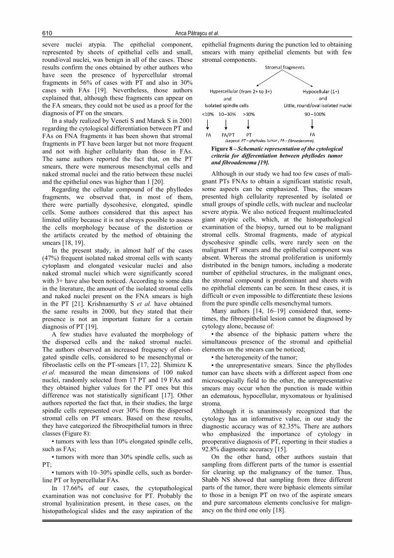
A few studies have evaluated the morphology of the dispersed cells and the naked stromal nuclei. The authors observed an increased frequency of elon-gated spindle cells, considered to be mesenchymal or fibroelastic cells on the PT-smears [17, 22]. Shimizu K et al. measured the mean dimensions of 100 naked nuclei, randomly selected from 17 PT and 19 FAs and they obtained higher values for the PT ones but this difference was not statistically significant [17]. Other authors reported the fact that, in their studies, the large spindle cells represented over 30% from the dispersed stromal cells on PT smears. Based on these results, they have categorized the fibroepithelial tumors in three classes (Figure 8):
▪ tumors with less than 10% elongated spindle cells, such as FAs;
▪ tumors with more than 30% spindle cells, such as PT;
▪ tumors with 10–30% spindle cells, such as border-line PT or hypercellular FAs.
In 17.66% of our cases, the cytopathological examination was not conclusive for PT. Probably the stromal hyalinization present, in these cases, on the histopathological slides and the easy aspiration of the
epithelial fragments during the punction led to obtaining smears with many epithelial elements but with few stromal components.
Figure 8 – Schematic representation of the cytological criteria for differentiation between phyllodes tumor and fibroadenoma [19].
Although in our study we had too few cases of mali-gnant PTs FNAs to obtain a significant statistic result, some aspects can be emphasized. Thus, the smears presented high cellularity represented by isolated or small groups of spindle cells, with nuclear and nucleolar severe atypia. We also noticed frequent multinucleated giant atyipic cells, which, at the histopathological examination of the biopsy, turned out to be malignant stromal cells. Stromal fragments, made of atypical dyscohesive spindle cells, were rarely seen on the malignant PT smears and the epithelial component was absent. Whereas the stromal proliferation is uniformly distributed in the benign tumors, including a moderate number of epithelial structures, in the malignant ones, the stromal compound is predominant and sheets with no epithelial elements can be seen. In these cases, it is difficult or even impossible to differentiate these lesions from the pure spindle cells mesenchymal tumors.
Many authors [14, 16–19] considered that, some-times, the fibroepithelial lesion cannot be diagnosed by cytology alone, because of:
▪ the absence of the biphasic pattern where the simultaneous presence of the stromal and epithelial elements on the smears can be noticed;
▪ the heterogeneity of the tumor; ▪ the unrepresentative smears. Since the phyllodes
tumor can have sheets with a different aspect from one microscopically field to the other, the unrepresentative smears may occur when the punction is made within an edematous, hypocellular, myxomatous or hyalinised stroma.
Although it is unanimously recognized that the cytology has an informative value, in our study the diagnostic accuracy was of 82.35%. There are authors who emphasized the importance of cytology in preoperative diagnosis of PT, reporting in their studies a 92.8% diagnostic accuracy [15].
On the other hand, other authors sustain that sampling from different parts of the tumor is essential for clearing up the malignancy of the tumor. Thus, Shabb NS showed that sampling from three different parts of the tumor, there were biphasic elements similar to those in a benign PT on two of the aspirate smears and pure sarcomatous elements conclusive for malign-ancy on the third one only [18].

Clinical and cytopathological aspects in phyllodes tumors of the breast
611
Therefore, the majority of the data from the litera-ture emphasized the importance of the morphological features of the stromal component on PT FNA smears, including:
▪ the number of stromal fragments (must be frequent); ▪ their size and cellularity (large, hypercellular); ▪ the presence of naked stromal nuclei, larger and
vesicular than those of epithelial cells. The assessment of the cellular and nuclear morphology should be first realized by optic microscopy and confirmed further by computerized cytometry and immunocytochemistry, in difficult cases.
▪ the prevalence of stromal nuclei unto epithelial ones [9, 14, 16–18, 23].
Our aim was to assess the cytological aspects nece-ssary for the diagnosis of PTs on FNA smears. This is very important for future optimal treatment of the tumor, consisting in tumor excision with a limit of at least one centimeter in healthy tissue for avoiding the recurrences, which are seen very often.
Conclusions
In our study, benign forms of phyllodes tumors prevailed, with a mean age of patients around 46-year-old, data similar with those of other authors. The corre-lation between age and PT appearance showed that the older is the patient (more than 45-year-old is) the higher is the probability of the tumor to be malignant. The diagnosis of phyllodes tumor could be established also preoperatively by intratumoral FNA cytology with a high accuracy (82% in our study), by assessing the characteristic features of stromal components (stromal fragments and naked stromal nuclei) on the smears.
References [1] MÜLLER J., Ueber den feinern Bau und die Formen der
krankhaften Geschwülste, G. Reimer, Berlin, 1838, 54–60. [2] LEE B., PACK G., Giant intracanalicular fibroadenomyxoma
of the breast, Am J Cancer, 1931, 15:2583–2609. [3] TURALBA C. I. C., EL-MAHDI A. M., LADAGA L., Fatal metastatic
cystosarcoma phyllodes in an adolescent female: case report and review of treatment approaches, J Surg Oncol, 1986, 33(3):176–181.
[4] ZURRIDA S., BARTOLI C., GALIMBERTI V., SQUICCIARINI P., DELLEDONNE V., VERONESI P., BONO A., DE PALO G., SALVADORI B., Which therapy for unexpected phyllodes tumour of the breast?, Eur J Cancer, 1992, 28(2–3):654–657.
[5] ROWELL M. D., PERRY R. R., HSIU J. G., BARRANCO S. C., Phyllodes tumors, Am J Surg, 1993, 165(3):376–379.
[6] TREVES N., SUNDERLAND D. A., Cystosarcoma phyllodes of the breast: a malignant and a benign tumor; a clinico-pathological study of seventy-seven cases, Cancer, 1951, 4(6):1286–1332.
[7] CHANEY A. W., POLLACK A., MCNEESE M. D., ZAGARS G. K., PISTERS P. W., POLLOCK R. E., HUNT K. K., Primary treat-ment of cystosarcoma phyllodes of the breast, Cancer, 2000, 89(7):1502–1511.
[8] MCDONALD J. R., HARRINGTON S. W., Giant fibro-adenoma of the breast, cystosarcoma phyllodes, Ann Surg, 1950, 131(2):243–251.
[9] STEBBING J. F., NASH A. G., Diagnosis and management of phyllodes tumour of the breast: experience of 33 cases at a specialist centre, Ann R Coll Surg Engl, 1995, 77(3):181–184.
[10] RAJAN P. B., CRANOR M. L., ROSEN P. P., Cystosarcoma phyllodes in adolescent girls and young women: a study of 45 patients, Am J Surg Pathol, 1998, 22(1):64–69.
[11] ANDERSSON A., BERGDAHL L., Cystosarcoma phyllodes in young women, Arch Surg, 1978, 113(6):742–744.
[12] DRAGHI F., SABOLLA L., CAMPANI R., MELONI G., Diagnostic imaging of phyllodes tumors: preliminary observations, Radiol Med, 1996, 91(5):585–589.
[13] TAVASSOLI F. A., Pathology of the breast, 2nd edition, Appleton & Lange, Stanford, Connecticut, 1999, 1–25, 571–631, 633–673, 675–729, 763–827.
[14] DEEN S. A., MCKEE G. T., KISSIN M. W., Differential cytologic features of fibroepithelial lesion of the breast, Diagn Cytopathol, 1999, 20(2):53–56.
[15] JAYARAM G., STHANESHWAR P., Fine-needle aspiration cytology of phyllodes tumors, Diagn Cytopathol, 2002, 26(4):222–227.
[16] DUSENBERY D., FRABLE W. J., Fine needle aspiration cytology of phyllodes tumor. Potential diagnostic pitfalls, Acta Cytol, 1992, 36(2):215–221.
[17] SHIMIZU K., MASAWA N., YAMADA T., OKAMOTO K., KANDA K., Cytologic evaluation of phyllodes tumors as compared to fibroadenomas of the breast, Acta Cytol, 1994, 38(6):891–897.
[18] SHABB N. S., Phyllodes tumor. Fine needle aspiration cytology of eight cases, Acta Cytol, 1997, 41(2):321–326.
[19] KRISHNAMURTHY S., ASHFAQ R., SHIN H. J., SNEIGE N., Distinction of phyllodes tumor from fibroadenoma: a rea-ppraisal of an old problem, Cancer, 2000, 90(6):342–349.
[20] VENETI S., MANEK S., Benign phyllodes tumour vs. fibro-adenoma: FNA cytological differentiation, Cytopathology, 2001, 12(5):321–328.
[21] SILVERMAN J. F., GEISINGER K. R., FRABLE W. J., Fine-needle aspiration cytology of mesenchymal tumors of the breast, Diagn Cytopathol, 1988, 4(1):50–58.
[22] RAO C. R., NARASIMHAMURTHY N. K., JAGANATHAN K., MUKHERJEE G., HAZARIKA D., Cystosarcoma phyllodes. Diagnosis by fine needle aspiration cytology, Acta Cytol, 1992, 36(2):203–207.
[23] BHATTARAI S., KAPILA K., VERMA K., Phyllodes tumor of the breast. A cytohistologic study of 80 cases, Acta Cytol, 2000, 44(5):790–796.
Corresponding author Carmen Florina Popescu, Department of Pathology and Cytology, Emergency County Hospital of Craiova, 1st Tabaci Street, 200642 Craiova, Romania; Phone +40722–775 254, e-mail: [email protected] Received: August 15th, 2009
Accepted: October 25th, 2009